Home Journals Chinese Journal of Alzheimer's Disease and Related Disorders
Chinese Journal of Alzheimer's Disease and Related Disorders

Abbreviation (ISO4): Chinese Journal of Alzheimer's Disease and Related Disorders      Editor in chief: Jun WANG

About  /  Aim & scope  /  Editorial board  /  Indexed  /  Contact  / 

Clinical characteristicsof cerebral amyloid angiopathy complicated by dementia in a series of 7 cases

  • JIN Hong 1 ,
  • GE Fangfang 2 ,
  • ZHANG Zhenxin 3 ,
  • LI Yanfeng , 3
Expand
  • 1 Department of Neurology, Beijing Nuclear Industry Hospital
  • 2 Department of Neurology, Xi’an International Medical Center Hospital
  • 3 Department of Neurology, Peking Union Medical College Hospital, CAMS & PUMC

Received date: 2019-11-19

  Revised date: 2019-11-28

  Online published: 2020-03-25

Abstract

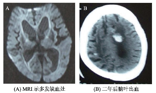
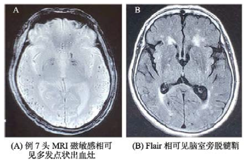
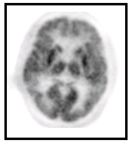
Objective: To review the clinical features at presentation in detail in outpatients with cerebral amyloid angiopathy (CAA) complicated by dementia. Methods: Seven patients were enrolled retrospectively from Department of Neurology of Peking Union Medical College Hospital were enrolled when they meet the Boston criteria. The clinical features and their CSF biomarkers and neuroimaging in detail were reviewed and analyzed. Results: There were 5 men and 2 women,and their mean age was(70.6±4.8)years old. The mean duration from memory impairment to presence of Parkinsonism was less than 1 year.Situational memory disorder,behavioral and psychiatric symptoms,and focal neurological signs were the major symptoms. MRI showed that Ventricular enlargement, white matter ischemia, white matter ischemia,and lobar hemorrhage in different patients. Neuroimage examination shows MAT grading of hippocampus I (0 case),II (2 case),III (3 case) and IV (2 case). PET showed the cingulate island sign in three patients. FDG PET showed significant lower FDG uptake in both frontal, parietal, and temporal lobe in two patients. The CSF markers (Aβ42, Aβ40, t-tau, and p-tau) have shown the same change in Alzheimer’s disease (AD) patients. The low Aβ42 content and the high t-tau and p-tau content exist in CAA patients. Conclusion: Taken together, these results suggest that dementia biomarkers might notbe useful to differentiate from AD. There is need to perform neuroimage examination on SWI to determine whether microbleeds exist in the patient with AD who have a prodigious cerebrovascular burden.

Cite this article

JIN Hong , GE Fangfang , ZHANG Zhenxin , LI Yanfeng . Clinical characteristicsof cerebral amyloid angiopathy complicated by dementia in a series of 7 cases[J]. Chinese Journal of Alzheimer's Disease and Related Disorders, 2020 , 3(1) : 12 -16 . DOI: 10.3969/j.issn.2096-5516.2020.01.005

脑淀粉样血管病(cerebral amyloid angiopathy,CAA)是一种β淀粉样蛋白(beta amyloid,Aβ)沉积在皮层和软脑膜小动脉及毛细血管中外膜为主要病理特征的一种脑小血管病[1-3]。一般软脑膜动脉先受累且程度更重。临床特点是反复多部位的血管破裂导致多灶性自发性脑叶出血[4-5]、认知功能障碍和淀粉样发作(amyloid spells)[6]。CAA有时与阿尔茨海默病(Alzheimer’s disease, AD)伴发,或以某种家族综合征的形式发生。由于该病与AD在病理和临床表现上相互重叠,特别是以痴呆为临床表现的脑淀粉样血管病易与AD痴呆的诊断混淆,临床上不易鉴别[7]。本文对7例临床诊断为脑淀粉样血管病痴呆患者的影像学特点和脑脊液痴呆标记物检查进行回顾分析总结,希望对于CAA痴呆与AD痴呆鉴别诊断有所帮助。

1 资料与方法

1.1 资料

本研究纳入7例就诊于北京协和医院门诊或住院患者,以记忆和社会行为功能下降为主诉就诊,并符合修订BOSTON淀粉样脑血管病诊断标准(见表1)的很可能(probable)CAA[8],即MRI显示皮层,脑叶,皮层下多发出血证据,年龄>55岁,除外其他出血原因。7例患者均经检查除外其他原因合并的痴呆,如HIV、梅毒感染,营养代谢障碍、中毒等。
表1 CAA诊断的修订BOSTON标准
1.确定的CAA(definite) 尸检结果显示
-脑叶、皮质或皮质下出血;
-严重的淀粉样血管病变;
-无其他诊断病变。
2.病理学支持的很可能的CAA 临床和病理(血肿抽吸或皮质活检)显示
(probable) -脑叶、皮质或皮质下出血;
-一定程度的淀粉样血管病变;
-无其他诊断病变。
3.很可能的CAA 临床以及MRI和(或)CT显示:
(probable) -局限于脑叶、皮质或皮质下的多发性出血灶(包括小脑出血),或局限于脑叶、皮质或皮质下的单个出血灶伴局限性(≤3个脑沟)或弥漫性(≥4个脑沟)皮质表面含铁血黄素沉积;
-年龄≤55岁;
-无其他原因的脑出血。
4.可能的CAA 临床以及MRI和(或)CT显示
(possible) -单纯性脑叶、皮质或皮质下出血,或局限性(≤3个脑沟)或弥漫性(≥4个脑沟)皮质表面含铁血黄素沉积。
-年龄≤55岁。
-无其他原因的脑出血。

1.2 方法

1.2.1 影像学检查

7例患者均经磁共振(MRI机型号为:Sigma 1.5T)常规序列扫描,再行SWI及Flair扫描。PET-CT检查示踪剂为18F-FDG,机型号为Biograph 64型PET/CT。

1.2.2 痴呆标志物检测方法

两组脑脊液标本(CSF)均经腰椎穿刺收集,2000 r/min离心10 min,除去细胞和不溶性的物质,立即冻存在-80 ℃冰箱,待测定时取出。采用ELISA法,按试剂盒要求进行实验准备和试验步骤。Aβ42、t-tau和p-tau蛋白检测试剂盒为Innogenetics公司产品。

1.3 统计学分析

使用SPSS13.0统计软件,两组间比较采用t检验P<0.05为差异有统计学意义。

2 结果

2.1 患者人口学特征

共有7例患者,平均年龄为(70.6±4.8)岁,男5例,女2例,文化程度大学3人,高中3人,初中1人。7例患者起病方式均为隐袭起病,7例患者均存在情景记忆障碍,3例患者存在精神行为症状,4例患者存在神经系统定位体征。患者基本临床特征见表2所示。
表2 患者临床资料
编号 年龄 性别 病程 文化 起病 情景记忆 精神行为 定位 认知评估
(岁) (年) 程度 方式 障碍 症状 体征 MMSE MoCA
1 74 5 高中 隐袭 11 7
2 72 4 大学 隐袭 21 16
3 69 2 大学 隐袭 26 12
4 75 3 高中 隐袭 21 16
5 83 2 大学 隐袭 22 14
6 50 2 高中 隐袭 19 17
7 71 3 初中 隐袭 17 14

2.2 患者影像学特征表现

所有7例患者均进行头MRI检查(见表3),7例患者存在脑室扩大;7例患者存在多发微出血灶;6例患者存在对称性脑白质缺血灶,7例患者存在多发性脑白质缺血灶;1例患者存在单发脑叶出血灶,2例患者存在多发脑叶出血病灶;海马MAT分级I级0例、II级2例、III级3例和IV级2例。其具体影像学特点总结见表3。例3患者MRI多发缺血灶(如图1A),两年后脑叶出血(如图1B);例7病人头MRI SWI相可见多发点状出血灶(如图2A),Flair相可见脑室旁脱髓鞘(如图2B)。
表3 患者影像学特点
影像特征 例数
脑室扩大 7
多发微出血灶 7
白质缺血灶
对称 6
多发 7
海马MAT分级
I级 0
II级 2
III级 3
IV级 2
脑叶出血病灶
单发 1
多发 2
图1 例3(A)MRI示多发缺血灶(B)两年后脑叶出血
图2 例7(A)头MRI磁敏感相可见多发点状出血灶(B)Flair相可见脑室旁脱髓鞘

2.3 FDG-PET检查

其中2例患者行PET-CT检查,可见到皮层代谢下降。其中例2患者PET-CT检查示双侧前额、双侧顶叶大部及双侧颞叶皮层代谢下降,其表现与AD不易区别(如图3)。
图3 例2患者PET-CT检查结果

2.4 CAA合并痴呆患者及20例非痴呆患者痴呆标志物检查

7例CAA合并痴呆患者与20例非痴呆疾病对照组患者进行脑脊液痴呆标志物检查(见表4)。对照组患者,男11例,女9例,平均66.4±7.4岁。两组受试者的年龄和性别比较差异无统计学意义(P>0.05)。对照组包括脑血管病5例,发作性疾病5例,神经系统肿瘤4例,脑转移瘤5例,脱髓鞘疾病3例。检查结果显示CAA合并痴呆患者的脑脊液痴呆标志物Aβ42下降,t-tau及p-tau升高,且差异有统计学意义。
表4 7例患者痴呆标志检查并与对照组比较
组别 例数 42 (ng/ml) t-tau(ng/ml) p-tau (ng/ml)
CAA 7 410.04 (199.23) 350.95 (167.22) 37.57 (24.10)
No-CAA 20 617.78 (239.40) 157.91 (100.99) 24.14 (10.07)
P <0.001 <0.001 <0.001

3 讨论

CAA合并痴呆的比例为40%以上,而AD合并CAA的比例则超过80%,CAA合并痴呆的原因既有血管性的机制,又可能与Aβ蛋白有关[9-11]。国内曾有1例病理报告未见有淀粉样血管病变继发脑实质病灶而出现痴呆的报告[7],国外的亦有文献讨论CAA合并痴呆。一般认为CAA认知功能缺陷与血管性认知功能障碍难以区分,即患者呈部分性的认知缺损,病情呈阶梯样发展等,痴呆的发展和转归依赖脑血管病的进程。而本文7例所报道的痴呆发生过程均为隐袭发展,影像学也看到类似阿尔茨海默病的海马萎缩的表现,但程度较轻,所有患者均有CAA的特征性影像学表现,如脑叶出血,白质脱髓鞘、磁敏感相多发小出血灶等。本组所报告患者的痴呆早期诊断不易与AD区分,均在进一步影像学检查后明确CAA的诊断。因此,从本组报道CAA合并痴呆过程更类似于阿尔茨海默病的疾病过程,所以CAA合并痴呆,不仅有血管因素参与,神经变性也是其中的原因之一。
有人认为,某些CAA合并痴呆可以看作是阿尔茨海默病的亚型,其淀粉样物来源相同,在分子影像检查不易区分[12],本研究中7例患者限于条件未作淀粉样蛋白PET(Aβ-PET)检查,但有两例患者的FDG- PET显示类似于阿尔茨海默病的表现。有报告CAA脑脊液的痴呆生物标志检查结果也类似于阿尔茨海默病,即痴呆标志物Aβ42下降,t-tau和p-tau升高,本研究的7例痴呆标志物检查结果支持以上结论,因此,单从痴呆标志物的检查无法鉴别CAA合并痴呆与AD[7,13]
因此,如果AD患者的影像学检查提示有较多的血管性病灶时,进一步的磁敏感相检查就非常有必要,有可能看到多发微出血(cerebral microbleeds,CMB)。SWI是一种新的磁共振成像技术,主要利用不同组织的磁敏感程度差异而产生图像对比的成像技术,对于静脉、出血和铁沉积高度敏感。因此,对于疑诊CAA的患者,有必要进行SWI显象检查。
[1]
Pantoni L. Cerebral small vessel disease: from pathogenesis and clinical characteristics to therapeutic challenges[J]. Lancet Neurol, 2010, 9(7): 689-701.

DOI

[2]
Vinters HV, Gilbert JJ. Cerebral amyloid angiopathy: incidence and complications in the aging brain. II. The distribution of amyloid vascular changes[J]. Stroke, 1983, 14(6):924-928.

PMID

[3]
Vinters HV. Cerebral amyloid angiopathy. A critical review[J]. Stroke, 1987, 18(2):311-324.

PMID

[4]
Cordonnier C, Al-Shahi SR, Wardlaw J, et al. Spontaneous brain microbleeds: systematic review, subgroup analyses and standards for study design and reporting[J]. Brain, 2007, 130(8):1988-2003.

DOI

[5]
Lee SH, Bae HJ, Kwon SJ, et al. Cerebral microbleeds are regionally associated with intracerebral hemorrhage[J]. Neurology, 2004, 62(1):72-76.

PMID

[6]
Coates R, Bell SM, Coley S, et al. Cerebral amyloid angiopathy: amyloid spells and cortical superficial siderosis[J]. Pract Neurol, 2015, 15(2):124-126.

DOI

[7]
王鲁宁, 许丹. 脑淀粉样血管病导致痴呆一例[J]. 中国卒中杂志, 2007, 2(6): 511-512.

[8]
Greenberg SM, Rebeck GW, Vonsattel JP, et al. Apolipoprotein E epsilon 4 and cerebral hemorrhage associated with amyloid angiopathy[J]. Ann Neurol, 1995, 38(2): 254-259.

PMID

[9]
Chao CP, Kotsenas AL, Broderick DF, et al. Cerebral amyloid angiopathy: CT and MR imaging findings[J]. Radiographics, 2006, 26(5): 1517-1531.

DOI

[10]
Love S. Contribution of cerebral amyloid angiopathy to Alzheimer’s disease[J]. Neurol Neurosurg Psychiatry, 2004, 75(1): 1-4.

[11]
van Rooden S, van der Grond J, van den Boom R, et al. Descriptive analysis of the Boston criteria applied to a Dutch-type cerebral amyloid angiopathy population[J]. Stroke, 2009, 40(9): 3022-3027.

DOI

[12]
Carandini T, Arighi A, Sacchi L, et al. Testing the 2018 NIA-AA research framework in a retrospective large cohort of patients with cognitive impairment: from biological biomarkers to clinical syndromes[J]. Alzheimers Res Ther, 2019, 11(1): 84.

DOI PMID

[13]
Ferreira D, Shams S, Cavallin L, et al. The contribution of small vessel disease to subtypes of Alzheimer’s disease: a study on cerebrospinal fluid and imaging biomarkers[J]. Neurobiol Aging, 2018, 70(10): 18-29.

DOI

Outlines

/