Home Journals Chinese Journal of Alzheimer's Disease and Related Disorders
Chinese Journal of Alzheimer's Disease and Related Disorders

Abbreviation (ISO4): Chinese Journal of Alzheimer's Disease and Related Disorders      Editor in chief: Jun WANG

About  /  Aim & scope  /  Editorial board  /  Indexed  /  Contact  / 
Research Articles

A study of social novelty of an Alzheimer's disease model

  • YE Xiao-lian , 1 ,
  • TAI Guang-ping , 2
Expand
  • 1. Model Animal Research Center, Medical College of Nanjing University, Nanjing, Jiangsu 210061, China
  • 2. Key Laboratory of Biofabrication of Anhui Higher Education Institutes, Centre for Advanced Biofabrication, Hefei 230601; Department of Biological and Environmental Engineering, Hefei University, Hefei 230601, China

Received date: 2021-08-22

  Revised date: 2021-09-15

  Online published: 2021-12-25

Abstract

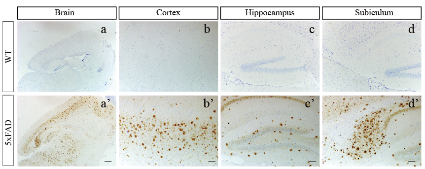
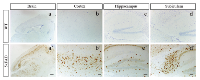
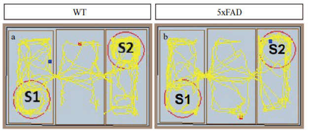
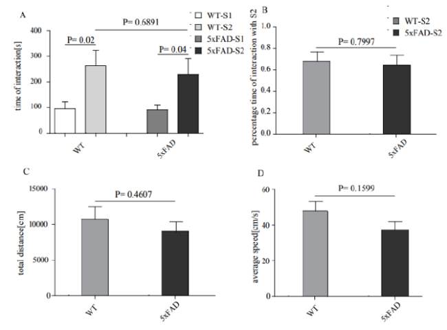
Objective: To explore the social novelty of 5×FAD transgenic mice, and to provide a theoretical basis for the rational application of 5×FAD mouse model to study AD-related behaviors. Methods: Aβ pathology of wild-type mice and 5×FAD mice was examined by immunohistochemical staining; Three chambers tests were carried out to evaluate wild-type and 5×FAD mice aged at 4-5 months. The interaction time between wild-type and 5×FAD mice with S2 mice, the total distance and time in the novelty exploration stage of three chamber tests were recorded. GraphPad Prism 5.0 software, unpaired-t test was performed for statistical analysis. Results: a large number of Aβ plaques were distributed in the cerebral cortex of 5×FAD mice; The average interaction time between wild-type and S1/2 was (95.5 ± 27.22/ 264.6 ± 58.38) s, P < 0.05. The average interaction time between 5×FAD mice and S 1/2 was (91.02 ± 19.49/ 230.2±60.18) s, P < 0.05. There was no difference in interaction time between wild-type and 5×FAD mice with S2, P > 0.05. The total distance of wild-type mice and 5×FAD mice during the tests was (10710 ± 1781/ 9017 ± 1402) mm respectively, P > 0.05. The average speed of wild-type and 5×FAD mice in the equipment was (47.7 ± 5.453/ 37.24 ± 4.590) mm/s respectively, P > 0.05. Conclusion: 5×FAD mice displayed severe Aβ plaques pathology at the age of 5 months, and their social novelty behavior is no different from that of wild-type mice.

Cite this article

YE Xiao-lian , TAI Guang-ping . A study of social novelty of an Alzheimer's disease model[J]. Chinese Journal of Alzheimer's Disease and Related Disorders, 2021 , 4(4) : 310 -313 . DOI: 10.3969/j.issn.2096-5516.2021.04.011

阿尔茨海默病(Alzheimer disease, AD)是一种中枢神经系统变性疾病,多发于65岁以上老年人,并且其发病率随着年龄增加而增加[1,2]。AD临床症状主要表现为记忆和认知损害。随着年龄增加,患者逐渐表现出痴呆相关的心理症状(psychological symptoms of dementia, PBSD),包括妄想、幻觉、躁动、攻击性、焦虑、社交淡漠和异常的运动行为等[3-6]。研究表明,大约有80%的AD患者兼具除认知和记忆损害症状之外的其他至少一种PBSD[7]。而社交障碍在AD患者中非常常见,但发病隐匿。因此,研究AD疾病模型中的社交行为对于理解和运用该模型研究AD具有重要意义。
AD的病理特征主要包括淀粉样蛋白(amyloid-Aβ)斑块沉积,神经炎症,tau蛋白过度磷酸化和神经元丢失等[8-11]。5×FAD小鼠模型是Oakley.等[12]成功构建的一种广泛用于研究Aβ病理的AD小鼠模型。相较于传统的APP/PS1小鼠模型,该模型小鼠Aβ斑块沉积更加快速和剧烈,是一种研究Aβ病理较为理想的AD疾病模型。已有研究表明不同的AD小鼠模型可能存在的行为学改变,为不同的AD模型的PBSD行为学实验研究提供了一定的理论支撑[13-15]
本研究旨在探究4~5月龄5×FAD小鼠的Aβ病理是否引起PBSD相关的新奇探索能力缺陷,同时,为合理应用5×FAD小鼠模型进行AD相关的研究提供理论依据。

1 材料与方法

1.1 小鼠来源及饲养

本研究中所用野生型小鼠和5×FAD小鼠均来自南京大学医学院模式动物研究所陈贵泉实验室[16]。实验小鼠饲养在南京大学模式动物研究所(specific pathogen free, SPF)级动物房中,饲养间的温度为(25±1)℃,光照时间为早7点至晚7点。实验小鼠均自由饮食和饮水。所有动物实验均已通过南京大学模式动物研究所动物伦理委员会 (IACUC) 批准。

1.2 小鼠脑组织处理

野生型小鼠(wild-type, WT)和5×FAD小鼠分别经二氧化碳安乐死、冷PBS充分灌流。将小鼠大脑置于新鲜的4% PFA中过夜固定。次日,利用梯度浓度乙醇(50%、70%、80%、90%、95%、100%)分别对小鼠大脑进行1 h脱水。随后浸蜡3 h后进行包埋。制作10 μm厚度的石蜡切片,用于免疫组化染色。

1.3 免疫组化染色

石蜡切片分别进行二甲苯和梯度乙醇(100 %、95 %、90 %、80 %、70 %、50 %)脱蜡复水,用柠檬酸钠溶液煮沸以进行抗原修复,随后经3 %过氧化氢溶液孵育15 min以充分暴露抗原决定簇。5 %牛血清封闭30 min,Aβ抗体(Biolegend, Cat# 803001) 4℃过夜孵育。次日,二抗孵育组织切片40 min,BCA试剂盒显色后,梯度乙醇(50 %、70 %、80 %、90 %、95 %、100 %)进行组织复水并用树脂封片。最后使用奥林巴斯光学显微镜采集图片。

1.4 三箱行为学实验

本实验中,野生型小鼠数量为8只,5×FAD小鼠数量为9只。同时使用雄鼠和雌鼠混合进行实验。小鼠三箱行为实验方法与Peng等已发表的文献中的方法相同[17]。实验设备分为左、中、右三箱,首先,分别在左箱和右箱放置空笼1和2。实验小鼠在装置中自由探索10 min,以充分熟悉设备环境。第一阶段,在左箱中的笼1中放入陌生小鼠1(stranger1, S1),实验小鼠继续在三箱设备中探索10 min。第二阶段,向右箱中的笼2放入陌生小鼠2(stranger2, S2)。记录实验小鼠在三箱设备中探索10 min内的运动路程、与S1和S2分别接触的时间。

1.5 统计学处理

利用GraphPad Prism5.0统计软件进行数据统计分析并作图。各组数据用(均数±标准差)表示,两组间统计利用非配对t检验统计分析。以P < 0.05为差异有统计学意义。

2 结果

2.1 小鼠大脑皮层Aβ病理分析

5月龄野生型和5×FAD小鼠大脑皮层的Aβ病理如图1。野生型小鼠大脑皮层未见Aβ斑块沉积,而5×FAD小鼠大脑皮层显示大量Aβ实质斑块分布。
图1 野生型小鼠(WT)与5×FAD小鼠大脑的Aβ 斑块沉积标尺:a'=500 μ m;'-d'=100 μ m;Cortex:大脑皮层;Hippocampus:海马;Subiculum: 海马下托

Fig.1 Aβ deposition of wide type (WT) and 5×FAD mice

2.2 三箱实验中小鼠新奇探索能力的轨迹分析

4~5月龄野生型小鼠和5×FAD小鼠新奇探索为轨迹如图2。该轨迹图显示,野生型小鼠在S2周围的运动轨迹明显多于在S1周围的运动轨迹(a)。同样,5×FAD小鼠在S2周围的运动轨迹明显多于其在S1周围的运动轨迹(b)。而5×FAD小鼠与野生型小鼠相比,其整体的运动轨迹有所减少。
图2 野生型小鼠与5xFAD小鼠的社交新奇性行为轨迹图 S1:陌生小鼠1;图S2:陌生小鼠2

Fig. 2 Trajectories of WT and 5×FAD mice in the social novelty

2.3 三箱实验中小鼠社交新异性偏好行为的统计分析

对野生型小鼠与S1和S2的接触时间进行统计分析,结果表明,对于野生型小鼠而言,其与S2的接触时间(264.6±58.38) s显著高于与S1的接触时间(95.5±27.22) s,差异有统计学意义(P < 0.05),具有统计学意义。对5×FAD小鼠与S1和S2的接触时间进行统计分析,结果表明,其与S2的接触时间(230.2±60.18) s显著高于与S1的接触时间(91.02±19.49) s,差异有统计学意义(P < 0.05),具有统计学意义。对野生型小鼠和5×FAD小鼠与S2的接触时间进行统计分析,二者差异无统计学意义(P > 0.05)。统计结果见图3
图3 野生型小鼠与5xFAD小鼠三箱社交实验中新奇探索能力分析 WT-S1:野生型小鼠与陌生小鼠1的接触;WT-S2:野生型小鼠与陌生小鼠2的接触;5xFAD-S1:5xFAD小鼠与陌生小鼠S1的接触;5xFAD-S2:5xFAD小鼠与陌生小鼠S2的接触

Fig.3 Analysis of WT and 5xFAD mice in the social novelty

分别对野生型小鼠和5×FAD小鼠与S2接触的时间百分比进行统计分析,结果表明,野生型小鼠与S2接触的时间百分比(0.6801±0.08749)和5×FAD小鼠与S2接触的时间百分比(0.6472±0.09114)之间差异无统计学意义(P > 0.05)。
分别对野生型小鼠和5×FAD小鼠在三箱设备中的总路程进行统计分析。结果表明,野生型小鼠的总路程(10710±1781) mm和5×FAD小鼠的总路程(9017±1402) mm之间差异无统计学意义(P > 0.05)。
分别对野生型小鼠和5×FAD小鼠在三箱设备中运动的平均速度进行统计分析,结果表明,野生型小鼠的平均速度(47.7±5.453) mm/s和5×FAD小鼠的平均速度(37.24±4.590) mm/s之间差异无统计学意义(P > 0.05)。

3 讨论

淀粉样斑块沉积是AD病人的典型病理特征。基于Aβ病理的转基因小鼠模型对研究AD的发病机制和治疗提供了重要基础。例如,Aβ PPSwe(APPNLh/NLh/PS-1P264L/P264L)小鼠于6月龄开始在大脑皮层区域加速产生Aβ沉积,而Aβ斑块在小鼠大脑白质及胼胝体区域分布较少[18]。3×Tg-AD模型小鼠大脑中同时具有Aβ斑块积累和神经纤维缠结的病理特征。Aβ斑块积累早于神经纤维缠结的形成,并广泛分布于海马、杏仁核和大脑皮层等脑区。另外,该模型小鼠的突触损伤早于淀粉样斑块积累和神经纤维缠结的发生[19]。而5×FAD小鼠则在1.5月龄开始形成细胞内Aβ1-42,2月龄开始在大脑的海马下托区域出现细胞外Aβ斑块积累和胶质细胞增生,并且,Aβ斑块积累随着小鼠年龄增加而逐渐增多,并进一步扩散至小鼠的海马、大脑皮层等脑区。该小鼠模型在9月龄开始出现突触损伤和神经元丢失病理[12]。本研究中,我们分析了5月龄5×FAD小鼠大脑中的Aβ分布情况,结果显示,在5月龄雌性5×FAD小鼠大脑中,Aβ斑块广泛分布于大脑皮层、内嗅皮层和海马等脑区。
研究表明,AD患者在疾病晚期,社交能力出现障碍[3]。因此,评价Aβ模型小鼠的新奇探索能力对于应用该模型进行AD相关的社交能力研究具有重要意义。本课题利用4~5月龄野生型和5×FAD小鼠在三箱设备中的新奇探索行为,观察、统计并分析了5×FAD小鼠模型的新奇探索行为。
本研究得到的小鼠在三箱设备中的轨迹及统计结果表明,首先,对于野生型小鼠和5×FAD小鼠,其与S2(新异小鼠)接触的时间均显著多于S1。这表明,5×FAD小鼠在5月龄尚未表现出社交障碍。即5×FAD具有正常的更倾向于与新异小鼠进行社交的偏好行为。并且,5×FAD小鼠和野生型小鼠与新异小鼠接触的时间及接触时间百分比差异无统计学意义(P > 0.05)。其次,根据小鼠运动的轨迹图可以看出,5×FAD小鼠整体的运动路程略少于野生型小鼠。但是统计学结果表明,二者各自在三箱设备中总的运动路程差异无统计学意义(P > 0.05)。推测可能的原因是5×FAD小鼠随着年龄的增加,会出现运动能力下降的趋势,但是在5月龄尚不明显。最后,野生型小鼠和5×FAD小鼠的平均速度之间差异无统计学意义(P > 0.05),表明,在此实验中,5×FAD小鼠的活动能力确实尚未发生改变。
因此,本研究结果表明,5月龄5×FAD小鼠大脑皮层、海马等脑区中大量分布淀粉样斑块,但是其新奇探索能力尚未发生损伤。这表明,在尚未发生神经元死亡的5×FAD 小鼠模型中,Aβ斑块沉积与小鼠的社交新奇探索行为之间可能不存在直接的相关性。即Aβ斑块沉积可能并不足以引起小鼠的社交新奇探索行为的异常。
5×FAD模型小鼠是基于Aβ学说建立的,以Aβ病理和神经炎症为主要病理特征的AD动物模型。而AD的发病机制复杂,除Aβ之外,tau蛋白过度磷酸化病理在AD的发病中也至关重要。本研究主要针对Aβ病理相关的社交行为,为基于5×FAD病理相关的早期社交行为提供理论证据。
[1]
Alzheimer's Association. 2015 Alzheimer's disease facts and figures[J]. Alzheimer's & dementia : the journal of the Alzheimer's Association, 2015, 11(3): 332-384.

[2]
Selkoe DJ. Preventing Alzheimer's disease[J]. Science (New York, NY), 2012, 337(6101): 1488-1492.

DOI

[3]
Chow TW, Liu CK, Fuh JL, et al. Neuropsychiatric symptoms of Alzheimer's disease differ in Chinese and American patients[J]. International journal of geriatric psychiatry, 2002, 17(1):22-28.

DOI PMID

[4]
Chung JA, Cummings JL. Neurobehavioral and neuropsychiatric symptoms in Alzheimer's disease: characteristics and treatment[J]. Neurologic clinics, 2000, 18(4): 829-846.

PMID

[5]
Cummings JL. The Neuropsychiatric Inventory: assessing psychopathology in dementia patients[J]. Neurology, 1997, 48(5 Suppl 6): S10-S16.

DOI PMID

[6]
Cummings JL, Mega M, Gray K, et al. The Neuropsychiatric Inventory: comprehensive assessment of psychopathology in dementia[J]. Neurology, 1994, 44(12): 2308-2314.

DOI PMID

[7]
Lyketsos CG, Lopez O, Jones B, et al. Prevalence of neuropsychiatric symptoms in dementia and mild cognitive impairment: results from the cardiovascular health study[J]. JAMA 2002, 288(12): 1475-1483.

DOI

[8]
Spillantini MG, Goedert M. Tau pathology and neurodegeneration[J]. The Lancet Neurology, 2013, 12(6): 609-622.

DOI

[9]
Tanzi RE, Bertram L. Twenty years of the Alzheimer's disease amyloid hypothesis: a genetic perspective[J]. Cell, 2005, 120(4): 545-555.

DOI

[10]
Heneka MT, Carson MJ, El Khoury J, et al. Neuroinflammation in Alzheimer's disease[J]. The Lancet Neurology, 2015, 14(4): 388-405.

DOI

[11]
Hardy J, Selkoe DJ. The amyloid hypothesis of Alzheimer's disease: progress and problems on the road to therapeutics[J]. Science (New York, NY), 2002, 297(5580): 353-356.

DOI

[12]
Oakley H, Cole SL, Logan S, et al. Intraneuronal beta-amyloid aggregates, neurodegeneration, and neuron loss in transgenic mice with five familial Alzheimer's disease mutations: potential factors in amyloid plaque formation[J]. Neurosci, 2006, 26(40): 10129-10140.

[13]
Filali M, Lalonde R, Rivest S, et al. Anomalies in social behaviors and exploratory activities in an APPswe/PS1 mouse model of Alzheimer's disease[J]. Physiology & behavior, 2011, 104(5): 880-885.

DOI

[14]
Flanigan TJ, Xue Y, Kishan Rao S, et al. Abnormal vibrissa-related behavior and loss of barrel field inhibitory neurons in 5xFAD transgenics[J]. Genes, brain, and behavior, 2014, 13(5): 488-500.

DOI

[15]
Deacon RM, Koros E, Bornemann KD, et al. Aged Tg2576 mice are impaired on social memory and open field habituation tests[J]. Behavioural brain research, 2009, 197(2):466-468.

DOI PMID

[16]
Wu J, Shao C, Ye X, et al. In vivo brain imaging of amyloid-β aggregates in Alzheimer's disease with a near-infrared fluorescent probe[J]. ACS Sensors, 2021, 6(3):863-870.

DOI

[17]
Peng SX, Wang YY, Zhang M, et al. SNP rs10420324 in the AMPA receptor auxiliary subunit TARP γ-8 regulates the susceptibility to antisocial personality disorder[J]. Scientific reports, 2021, 11(1): 11997.

DOI

[18]
Flood DG, Reaume AG, Dorfman KS, et al. FAD mutant PS-1 gene-targeted mice: increased A beta 42 and A beta deposition without APP overproduction[J]. Neurobiology of aging, 2002, 23(3): 335-348.

DOI

[19]
Oddo S, Caccamo A, Shepherd JD, et al. Triple-transgenic model of Alzheimer's disease with plaques and tangles: intracellular Abeta and synaptic dysfunction[J]. Neuron, 2003, 39(3):409-421.

DOI

Outlines

/