Home Journals Acta Academiae Medicinae Sinicae
Acta Academiae Medicinae Sinicae

Abbreviation (ISO4): Acta Academiae Medicinae Sinicae      Editor in chief: Xuetao CAO

About  /  Aim & scope  /  Editorial board  /  Indexed  /  Contact  / 
Review Articles

Research Progress in Neurogram and Neuro-Immune Interaction of Joints in the Case of Osteoarthritis

  • Jian GU 1 ,
  • Tao JIANG , 2
Expand
  • 1Graduate School,Nanjing University of Chinese Medicine,Nanjing 210023,China
  • 2Department Five of Orthopedics and Traumatology,Changzhou Hospital of Traditional Chinese Medicine,Changzhou,Jiangsu 213000,China
JIANG Tao Tel:0519-88124955,E-mail:

Received date: 2023-10-09

  Online published: 2024-12-03

Abstract

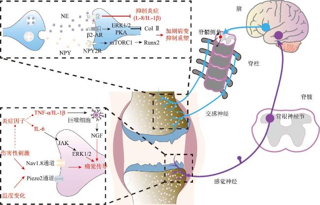
the joints have abundant sensory nerves and sympathetic nerve fibers,which convert physical and chemical stimuli in the joints into nerve impulses that are transmitted to the central nervous system and participate in the hypersensitivity reactions of inflammatory joint diseases such as osteoarthritis(OA).This paper summarizes the distribution and functional characteristics of intra-articular nerves and focuses on the mechanism of the vagus-sympathetic autonomic circuit in regulating the immune microenvironment in joints in the case of OA.in addition,intra-articular inflammatory cytokines represented by tumor necrosis factor-αand interleukin-6 directly or indirectly induce sensory nerve action potential and activate the pain transduction pathway from the local joint to the central nervous system.the sensory nerves in the joints in the case of OA are also involved in the recruitment of immune cells and inflammatory cytokines.This neuro-immune interaction model not only provides a variety of new targets for the treatment of OA but also suggests that the treatment of OA should adopt a holistic view with comprehensive consideration of the nerve and immune microenvironment in the bone and joint and their mutual influences。

Cite this article

Jian GU , Tao JIANG . Research Progress in Neurogram and Neuro-Immune Interaction of Joints in the Case of Osteoarthritis[J]. Acta Academiae Medicinae Sinicae, 2024 , 46(5) : 776 -782 . DOI: 10.3881/j.issn.1000-503X.15872

osteoarthritis (OA) is a common musculoskeletal disease, which often originates from the synovium and gradually involves the whole joint structure, such as joint capsule, ligament, cartilage and bone, eventually leading to limited joint movement and seriously affecting the quality of life of patients. Pain is the most common complaint and the primary clinical symptom of OA patients, but the degree of structural damage shown by X-ray and MRI is often not proportional to the level of pain response of patients[1-2]. Therefore, exploring the mechanism of pain in OA patients from a physiological point of view is the main concern of related research. At present, many studies have reported that nociceptive neurotransmitters released from sensory and sympathetic nerve endings in joint structures and inflammatory storms in the joint microenvironment are the main causes of allodynia in the course of OA[3-4]. Therefore, this article mainly introduces the main nerve types innervating joint function and their respective mechanisms regulating the pathological changes of OA joints, and summarizes the mechanisms of some inflammatory factors regulating nerve function and pain conduction pathways in the joint immune microenvironment, so as to provide more potential targets and directions for the treatment of OA.

1 neural map of induced OA allodynia

From the anatomical point of view,there is a wide range of nerve networks in each component of the joint structure,among which the fibrous layer of the joint capsule and the synovial membrane are the most abundant.Studies have shown that about 20%of articular nerves are myelinated nerve fibers with Aδ-nociceptors(Aδ-fibers),while the remaining 80%of articular nerves are unmyelinated C-fibers.According to the physiological characteristics of nerve fibers,sensory and sympathetic nerves are the main nerve types innervating joint function,and are also important participants in inducing neurogenic inflammatory response to OA[5]。 the following mainly reviews the physiological mechanisms and functional differences of these two nerves involved in the regulation of OA pain,and also reveals the therapeutic effect of peripheral vagus nerve stimulation on OA based on some recent studies。

1.1 Neural circuits of sensory-induced pain response

Sensory nerves innervating joint function are mainly composed of nerve fibers and receptors,which are mainly involved in the central transmission of pain and are the main source of local pain in OA patients。

1.1.1 Distribution characteristics of sensory nerves in the joint

There are abundant sensory nerves in the synovium,ligaments and bony structures of the joint[6]。 studies have shown that there are significant differences in the density of sensory nerves distributed in different parts.Although some Studies have suggested that the anterior synovium is the most sensitive area of the joint,immunohistochemical staining results show that the sensory nerves innervating the ligament are more dense than those in the synovium and are often associated with blood vessels[7][8]。 Among the bony structures,the sensory nerves on the periosteum are the most densely distributed,and these nerve axons are often closely connected with the germinal layer on the medial side of the periosteum and distributed linearly along the long axis of the femur[9]。 It was found that with the increase of age,the density of sensory nerve fibers in the joint of mice decreased significantly,but the density of nerve fibers around the periosteum was still very rich,which suggested that the treatment of targeted inhibition of sensory nerve function in the joint of OA patients should take into account the influence of age[10]。 It can be seen that the distribution of sensory nerves in different structures of the joint shows significant differences,and is affected by factors such as body condition and age.Follow-up studies should further describe the distribution map of sensory nerves in different parts of the body。

1.1.2 Intra-articular sensory nerves participate in pain conduction pathway

Existing studies have shown that sensory nerves are mainly involved in the transmission of pain signals in joints.According to the classical theory,there are many types of mechanoreceptors and chemoreceptors in sensory nerve endings[11]。 Among them,the former mainly captures physical signals such as pressure and traction at the joint,while the latter can not only capture noxious stimulation signals,but also monitor the local temperature change signals of the joint[12][13]。 in addition,some inflammatory factors in the joint can also activate the chemoreceptors of nerve endings and induce action potentials in the joint。
These receptors detect specific stimuli and transmit action potentials through the dorsal root ganglion to the dorsal horn of the spinal cord by triggering voltage-gated sodium and potassium channels[14]。 Voltage-gated sodium channel Nav1.8 is traditionally believed to play an important role in mechanical sensory transduction in the joint[15]。 However,recent studies have found that intra-articular Piezo2 ion channels also play an important role in pain transmission in peripheral sensory nerves.It not only participates in the information transmission between neurons and glial cells and between axons and Schwann cells in the peripheral sensory nervous system,but also connects the pain conduction pathway of the central nervous system through the dorsal root ganglion under external mechanical stimulation[16]。 pain signals are integrated by spinal dorsal horn cells and projected to the thalamus and brainstem through the spinothalamic tract and the spinoomentothalamic tract,eventually enabling patients to feel pain and accurately locate the pain site(Fig.1)[17]
图1 骨关节炎关节内的痛觉传导通路及神经免疫互作机制

NE: norepinephrine; NPY: neuropeptide Y; β2-AR: β2-adrenergic receptor; ERK: extracellular regulated protein kinase; PKA: protein kinase A; Col Ⅱ: type Ⅱ collagenase; IL: interleukin; mTORC1: mammalian target of rapamycin complex 1; JAK: Janus kinase; TNF-α: tumor necrosis factor-α; NGF: nerve growth factor

1.1.3 Amplification of sensory afferent signals by central sensitization

Although existing studies have explained the mechanism of local pain in OA patients,the degree of pain felt by patients is often not proportional to the degree of local injury detected by imaging techniques.This suggests that the central nervous system,in addition to integrating peripheral afferent pain signals,may also amplify and output their signals to local effectors in the joint,eventually exacerbating local pain in OA patients.This process is called central sensitization,which is mainly caused by the strong plasticity of nerve cells in the central nervous system[14]。 Studies have shown that under the action of long-term peripheral afferent signals,the spontaneous activity of glial cells increases,the activation threshold decreases,and the range of capturing sensory signals is significantly expanded,which ultimately shows that the sensitivity to pain signals increases,and at the same time,some non-sensory signals are integrated and output as pain signals[18-19]。 Therefore,the physiological mechanism of local joint pain in OA patients mainly involves the capture of stimulation signals by sensory nerve ending receptors,the integration of information in the central nervous system and the amplification of pain signals。

1.2 Autonomic nerve maintains joint homeostasis

in recent years,with the development of imaging and viral tracing technology,people have been able to directly observe the distribution characteristics of sympathetic nerve in the joint and the density changes of sympathetic nerve fibers in the pathological state of OA.Studies have shown that there are abundant sympathetic nerve fibers in subchondral bony structures,synovium,tendons and ligaments,which are similar to sensory nerves,and the density of sympathetic nerves in these different structures is also significantly different,while the density change of sympathetic nerves in joints has also been proved to be one of the characteristics of OA pathological response[20-21]

1.2.1 Functional properties of intra-articular sympathetic nerve and its bidirectional regulation on OA status

There is no clear agreement on the functional properties of intra-articular sympathetic nerves,which is mainly attributed to the extensive effects of neurotransmitters released from peripheral sympathetic nerve endings.Firstly,noradrenalin(NE),as the main catecholamine neurotransmitter released from sympathetic nerve endings in the joint,plays a two-way regulatory role in the occurrence and development of OA.on the one hand,NE in OA joints mainly acts onβ2-adrenergic receptors and interferes with the synthesis of type II collagenase in cartilage by activating extracellular regulated protein kinases(ERK)1/2-protein kinase A(PKA)signaling pathway,thus inhibiting the production of cartilage matrix,aggravating the local pathological changes of OA joints and interfering with the process of tissue repair[22]。 On the other hand,intra-articular NE also has anti-inflammatory effects.NE treatment can significantly reduce the expression of proinflammatory cytokines in mixed synoviocytes.NE stimulation of chondrocytes in OA patients can significantly reduce the expression of inflammatory cytokines IL-1βand IL-8[23][24]。 in addition,sympathetic nerve endings in the joint release neuropeptide Y and ATP in addition to NE.Among them,neuropeptide Y is overexpressed in the cartilage of OA patients,which activates the mammalian target of rapamycin complex 1 pathway by binding to its receptor and promotes the up-regulation of the downstream factor Runx2.This in turn promotes chondrocyte hypertrophy and cartilage degradation in the local joint(Fig.1);ATP has also recently been shown to be a key factor in the spread of local joint inflammation to distal sites,which promotes the upregulation of IL-6 expression in the joint after activation of nuclear factor-κB signaling in the local joint[25][26]
Defining the dominant effect of intra-articular sympathetic nerve bidirectional regulation is a hot research topic at present.in 2022,R Rösch et al.Found that when 6-hydroxydopamine was used to chemically ablate the peripheral sympathetic nerve of OA model mice,although it had no effect on the severity of cartilage degeneration and synovial inflammation In mice,it significantly aggravated the pathological changes of cartilage calcification and subchondral bone thickening,which are characteristic of OA[27]。 This indicates that although the local function of sympathetic nerve fibers in OA joints is not yet clear,the existence of sympathetic innervation in joints is still an important prerequisite for maintaining the homeostasis of the joint environment。

1.2.2 Peripheral Vagus Nerve Stimulation for OA

From an anatomical point of view,the osteoarticular structures lack vagal innervation.However,in recent years,many reports have shown that stimulation of peripheral vagal afferents can activate the anti-inflammatory function of intra-articular sympathetic nerves,and this cholinergic anti-inflammatory pathway activated by peripheral vagal nerves may become a cutting-edge approach for OA treatment。
Tracey proposed in 2002 that stimulating the peripheral vagus nerve can not only rapidly and effectively inhibit the systemic inflammatory response,but also improve the nerve function of the inflamed local to regulate the internal environment of the relevant tissue structure as a whole[28]。 Based on this theory,Akoolo et al.Used electroacupuncture at Zusanli(ST36)to stimulate the peripheral vagus nerve,activate the hypothalamic-pituitary-adrenal axis,and promote the adrenal gland to secrete dopamine to play a systemic anti-inflammatory role.So as to effectively reduce the expression of Th17 and Th1 cells in the joint,reduce the recruitment and infiltration of macrophages and neutrophils in the joint,and ultimately improve the symptoms of arthritis induced by Lyme disease[29]。 the above studies suggest that the cholinergic anti-inflammatory pathway induced by stimulation of the peripheral vagus nerve can not only effectively alleviate the infiltration of inflammatory cytokines in the joint,but also activate the anti-inflammatory properties of NE in the joint,and improve the inflammatory status and related symptoms of OA as a whole.This autonomic nerve loop,which is afferent from the vagus nerve and efferent from the sympathetic nerve,maintains the homeostasis of the joint and may be an important research target for exploring effective treatments for OA,but the specific mechanism of its neuroimmune interaction remains to be further studied。

2. The regulation of inflammatory factors in OA on nerve and pain conduction pathway

the complex immune microenvironment in OA joints is one of the important factors that limit its efficacy.Infiltrating immune cells in joints release a large number of inflammatory factors,which not only aggravate local tissue damage,but also participate in the regulation of pain conduction pathway in OA through two ways.On the one hand,the infiltration of inflammatory factors in the joint can reduce the activation threshold of sensory nerve endings,thereby enhancing the sensitivity of local sensory nerves to noxious stimuli[30]。 On the other hand,inflammatory factors can directly upregulate the proportion of sensory nerve activation in the joint[31]。 the main inflammatory factors of pain conduction pathway in OA joint play an important role in neuroimmune interaction,which provides a new perspective for understanding the neuroregulatory mechanism in the pathological process of OA。

2.1 Classical inflammatory factors represented by IL-6 and tumor necrosis factor-α

IL-6 and tumor necrosis factor-α(TNF-α),as classical inflammatory factors,are not only the main participants in the induction of inflammatory joint diseases such as OA and rheumatoid arthritis,but also the key factors regulating the pain conduction pathway in OA joints[32-33]。 A prospective cohort study has shown that the expression level of IL-6 in the joint is not only closely related to the volume of OA cartilage,but also proportional to the degree of pain in patients[34]。 Intra-articular IL-6 is involved in the induction of many key pathological features of OA,including driving proliferative osteoarticular remodeling,activating osteoclasts,and inhibiting bone and cartilage repair pathways[35]。 After IL-6 knockdown,the nerve density in the knee joint of OA model mice and the local pain perception of mice were significantly reduced,which was mainly due to the down-regulation of Janus kinase and ERK signaling pathway of IL-6 cascade[36]。 the above results confirm the regulatory effect of IL-6 on local nerve and pain conduction in OA joints,and reveal the key downstream signaling pathways and related molecules of IL-6.in addition,TNF-αcan also exert similar effects as IL-6,with a slightly different mechanism of action than IL-6.studies have shown that TNF-αinfiltrated into the joint can up-regulate the expression of nerve growth factor(NGF)in synovial fibroblasts and macrophages,and NGF has been confirmed to be involved in the induction of pain conduction pathway in mice after OA and in the late stage,and many Studies have shown that direct intra-articular injection of NGF can significantly aggravate the pain of OA model mice[37][38][39-40]。 Therefore,TNF-αregulates nerve density and pain conduction pathways in OA joints mainly by inducing the expression of NGF(Fig.1)。

2.2 Other inflammatory factors

in addition to IL-6 and TNF-α,some other inflammatory factors in OA joints are also involved in the regulation of nerve and pain conduction pathways.IL-1βis a common inflammatory factor in the synovium of OA patients.Studies have shown that injection of IL-1βinto synovial fibroblast explants can significantly promote the expression of NGF,which indicates that IL-1βmay regulate the distribution and function of intra-articular nerves in a similar way as TNF-α[37]。 In addition,IL-1R,the receptor that interacts with IL-1β,has been shown to be highly expressed in the terminals of transient receptor potential vanilloid 1+sensory neurons innervating joint function,and specific knockdown of IL-1R gene can significantly induce pain in mice,suggesting that IL-1βmay also be directly involved in the activation of sensory nerves in OA as a ligand[41]。 in addition to IL-1β,in recent years,researchers have also focused on the effects of Intra-articular IL-8 on the occurrence and development of OA.intra-articular IL-8 is not only a key indicator to distinguish OA from psoriatic arthritis,but also involved in inducing the expression of IL-6 and nitric oxide synthase in OA joints[42][33]。 Importantly,IL-8 can also promote the up-regulation of NGF in the joint by inducing the expression of annulus fibrosus cells,which may be the potential mechanism of IL-8 participating in the regulation of OA nerve and pain conduction pathway[43]。 However,the function of IL-8 in intra-articular infiltration of OA still needs further experimental verification。
to sum up,inflammatory factors such as IL-6 and TNF-αare important factors inducing the pain conduction pathway in OA,which is mainly related To their regulation of local nerve density and function in the joint.Improving the local inflammatory microenvironment of OA patients may be a necessary prerequisite for the long-term efficacy of nerve blockade therapy.in addition,sensory nerve fibers in the joint also participate in the recruitment of immune cells such as macrophages and monocytes and inflammatory factors such as IL-6,TNF-αand IL-1β[44-46]。 Many studies have found that a large number of calcitonin gene-related peptide and substance P can be detected in the synovial fluid of OA patients.These sensory neurotransmitters can not only play a powerful role in dilating blood vessels,but also have chemotactic and activating effects on a variety of immune cells,including macrophages[47]。 For example,SP can bind to the neurokinin-1 receptor on the surface of macrophages in the joint,promote the transition of M2 macrophages to M1 macrophages(pro-inflammatory),and release inflammatory factors such as IL-1βand TNF-a[48]。 Therefore,sensory and sympathetic nerves may play an antagonistic role in regulating the local inflammatory state of joints.At the same time,the interaction between inflammatory factors and local nerves in the joint,that is,inflammatory factors promote the pain conduction of sensory nerves and the recruitment of inflammatory factors by sensory nerves,forms a vicious circle of positive feedback in the joint,which is also one of the main factors limiting the clinical efficacy of OA。

3 Summary and Outlook

in recent years,the related functions of intra-articular nerves have been elucidated.Noxious and chemical stimuli in OA joints induce pain conduction pathways from local sensory nerve fibers to the central nervous system.in addition,sympathetic nerve fibers in the joint,Although associated with many key pathological features of OA,also exhibit unique anti-inflammatory properties locally in the joint.Based on this property,the role of vagus-sympathetic autonomic loop triggered by peripheral vagal stimulation in the treatment of OA is worth exploring.However,the clinical results translated from these basic studies have rarely achieved the desired efficacy,which also reveals the main challenges and limitations of targeted treatment of OA.on the one hand,it may be that the current method of directly stimulating the vagus nerve is still insufficient in anti-inflammatory efficacy.On the other hand,the mainstream concept of clinical treatment of OA is still anti-inflammatory and analgesic.although non-steroidal anti-inflammatory drugs,analgesics and sensory nerve block can relieve local joint pain in a short time,it is difficult to delay the progress of OA.Inflammation is a double-edged sword,which has both harmful effects and local protective effects under various pathological conditions.pain conduction pathway secondary to inflammatory storm is not only the key factor to induce local joint hypersensitivity,but also an important way to activate the body's own immune system.Therefore,targeted anti-inflammatory and analgesic methods can also inhibit the body's own immune response to pathological signals,which is also one of the important factors that cause many OA patients to prolong their illness.Follow-up studies can explore the treatment of OA from the following three directions:first,individualized treatment can be formulated according to the distribution and functional characteristics of intra-articular nerves in OA patients;Secondly,inflammatory factors in the joint are also the main factors inducing pain conduction,so the interaction between local nerve fibers and immune-related cytokines in the joint should be considered comprehensively.Finally,the concept of OA treatment should focus on"stimulation"on the basis of"targeting"and"inhibition",set up different treatment time windows according to the development of the disease,and dynamically use anti-inflammatory analgesics and immunopotentiators to restore the homeostasis of the local joint environment as a whole。
conflicts of interest All authors declare no Conflicts of Interest
Author's contribution statement Gu Jian:proposal of research ideas,literature retrieval and screening,collation of data and writing of articles;Jiang Tao:Paper revision,quality control and review,approval and integrity of research work
[1]
Hunter DJ, Bierma-Zeinstra S. Osteoarthritis[J]. Lancet, 2019, 393(10182):1745-1759.DOI:10.1016/S0140-6736(19)30417-9.

[2]
Sharma L. Osteoarthritis of the knee[J]. New Engl J Med, 2021, 384(1):51-59.DOI:10.1056/NEJMcp1903768.

[3]
Jiang W, Jin Y, Zhang S, et al. PGE2 activates EP4 in subchondral bone osteoclasts to regulate osteoarthritis[J]. Bone Res, 2022, 10(1):27.DOI:10.1038/s41413-022-00201-4.

[4]
Zhu S, Zhu J, Zhen G, et al. Subchondral bone osteoclasts induce sensory innervation and osteoarthritis pain[J]. J Clin Invest, 2019, 129(3):1076-1093.DOI:10.1172/JCI121561.

[5]
Grubb BD. Activation of sensory neurons in the arthritic joint[J]. Novartis Found Symp, 2004, 260:28-36,36-48,100-104,277-279.

[6]
Thai J, Kyloh M, Travis L, et al. Identifying spinal afferent (sensory) nerve endings that innervate the marrow cavity and periosteum using anterograde tracing[J]. J Comp Neurol, 2020, 528(11):1903-1916.DOI:10.1002/cne.24862.

[7]
Dye SF, Vaupel GL, Dye CC. Conscious neurosensory mapping of the internal structures of the human knee without intraarticular anesthesia[J]. Am J Sport Med, 1998, 26(6):773-777.DOI:10.1177/03635465980260060601.

[8]
Saxler G, Loer F, Skumavc M, et al. Localization of SP-and CGRP-immunopositive nerve fibers in the hip joint of patients with painful osteoarthritis and of patients with painless failed total hip arthroplasties[J]. Eur J Pain, 2007, 11(1):67-74.DOI:10.1016/j.ejpain.2005.12.011.

[9]
Liu S, Liu S, Li S, et al. Nerves within bone and their application in tissue engineering of bone regeneration[J]. Front Neurol, 2022, 13:1085560.DOI:10.3389/fneur.2022.1085560.

[10]
Chartier SR, Mitchell S, Majuta LA, et al. The changing sensory and sympathetic innervation of the young,adult and aging mouse femur[J]. Neuroscience, 2018, 387:178-190.DOI:10.1016/j.neuroscience.2018.01.047.

[11]
Mei H, Wu Y, Feng Q, et al. The interplay between the nerves and skeleton:a 30-year bibliometric analysis[J]. Ann Transl Med, 2023, 11(1):9.DOI:10.21037/atm-22-3323.

[12]
Laumonerie P, Dalmas Y, Tibbo ME, et al. Sensory innervation of the hip joint and referred pain:a systematic review of the literature[J]. Pain Med, 2021, 22(5):1149-1157.DOI:10.1093/pm/pnab061.

[13]
De Marziani L, Boffa A, Angelelli L, et al. Infrared thermography in symptomatic knee osteoarthritis:joint temperature differs based on patient and pain characteristics[J]. J Clin Med, 2023, 12(6):2319.DOI:10.3390/jcm12062319.

[14]
Basbaum AI, Bautista DM, Scherrer G, et al. Cellular and molecular mechanisms of pain[J]. Cell, 2009, 139(2):267-284.DOI:10.1016/j.cell.2009.09.028.

[15]
Malfait AM, Schnitzer TJ. Towards a mechanism-based approach to pain management in osteoarthritis[J]. Nat Rev Rheumatol, 2013, 9(11):654-664.DOI:10.1038/nrrheum.2013.138.

[16]
Shin SM, Moehring F, Itson-Zoske B, et al. Piezo2 mechanosensitive ion channel is located to sensory neurons and nonneuronal cells in rat peripheral sensory pathway:implications in pain[J]. Pain, 2021, 162(11):2750-2768.DOI:10.1097/j.pain.0000000000002356.

[17]
Finnerup NB, Kuner R, Jensen TS. Neuropathic pain:from mechanisms to treatment[J]. Physiol Rev, 2021, 101(1):259-301.DOI:10.1152/physrev.00045.2019.

[18]
Woolf CJ. Central sensitization:implications for the diagnosis and treatment of pain[J]. Pain, 2011, 152(3 Suppl):S2-S15.DOI:10.1016/j.pain.2010.09.030.

[19]
Clauw DJ, Hassett AL. The role of centralised pain in osteoarthritis[J]. Clin Exp Rheumatol, 2017, 35 Suppl 107(5):79-84.

[20]
Sohn R, Rosch G, Junker M, et al. Adrenergic signalling in osteoarthritis[J]. Cell Signal, 2021, 82:109948.DOI:10.1016/j.cellsig.2021.109948.

[21]
Grassel S, Muschter D. Peripheral nerve fibers and their neurotransmitters in osteoarthritis pathology[J]. Int J Mol Sci, 2017, 18(5):931.DOI:10.3390/ijms18050931.

[22]
Mitchell J, Lai LP, Peralta F, et al. β2-adrenergic receptors inhibit the expression of collagen type Ⅱ in growth plate chondrocytes by stimulating the AP-1 factor Jun-B[J]. Am J Physiol Endocrinol Metab, 2011, 300(4):E633-E639.DOI:10.1152/ajpendo.00515.2010.

[23]
Miller LE, Grifka J, Scholmerich J, et al. Norepinephrine from synovial tyrosine hydroxylase positive cells is a strong indicator of synovial inflammation in rheumatoid arthritis[J]. J Rheumatol, 2002, 29(3):427-435.

[24]
Lorenz J, Schäfer N, Bauer R, et al. Norepinephrine modulates osteoarthritic chondrocyte metabolism and inflammatory responses[J]. Osteoarthritis Cartilage, 2016, 24(2):325-334.DOI:10.1016/j.joca.2015.08.007.

[25]
Kang X, Qian Z, Liu J, et al. Neuropeptide Y acts directly on cartilage homeostasis and exacerbates progression of osteoarthritis through NPY2R[J]. J Bone Miner Res, 2020, 35(7):1375-1384.DOI:10.1002/jbmr.3991.

[26]
Hasebe R, Murakami K, Harada M, et al. ATP spreads inflammation to other limbs through crosstalk between sensory neurons and interneurons[J]. J Exp Med, 2022, 219(6):e20212019.DOI:10.1084/jem.20212019.

[27]
Rösch G, El Bagdadi K, Muschter D, et al. Sympathectomy aggravates subchondral bone changes during osteoarthritis progression in mice without affecting cartilage degeneration or synovial inflammation[J]. Osteoarthritis Cartilage, 2022, 30(3):461-474.DOI:10.1016/j.joca.2021.11.016.

[28]
Tracey KJ. The inflammatory reflex[J]. Nature, 2002, 420(6917):853-859.DOI:10.1038/nature01321.

[29]
Akoolo L, Djokic V, Rocha SC, et al. Sciatic-vagal nerve stimulation by electroacupuncture alleviates inflammatory arthritis in lyme disease-susceptible C3H mice[J]. Front Immunol, 2022, 13:930287.DOI:10.3389/fimmu.2022.930287.

[30]
Chakrabarti S, Pattison LA, Singhal K, et al. Acute inflammation sensitizes knee-innervating sensory neurons and decreases mouse digging behavior in a TRPV1-dependent manner[J]. Neuropharmacology, 2018, 143:49-62.DOI:10.1016/j.neuropharm.2018.09.014.

[31]
Ishihara S, Obeidat AM, Wokosin DL, et al. The role of intra-articular neuronal CCR2 receptors in knee joint pain associated with experimental osteoarthritis in mice[J]. Arthritis Res Ther, 2021, 23(1):103.DOI:10.1186/s13075-021-02486-y.

[32]
Schaible HG, von Banchet GS, Boettger MK, et al. The role of proinflammatory cytokines in the generation and maintenance of joint pain[J]. Ann Ny Acad Sci, 2010, 1193:60-69.DOI:10.1111/j.1749-6632.2009.05301.x.

[33]
Kapoor M, Martel-Pelletier J, Lajeunesse D, et al. Role of proinflammatory cytokines in the pathophysiology of osteoarthritis[J]. Nat Rev Rheumatol, 2011, 7(1):33-42.DOI:10.1038/nrrheum.2010.196.

[34]
Pan F, Tian J, Cicuttini F, et al. Prospective association between inflammatory markers and knee cartilage volume loss and pain trajectory[J]. Pain Ther, 2022, 11(1):107-119.DOI:10.1007/s40122-021-00341-1.

[35]
Kaaij MH, van Tok MN, Blijdorp IC, et al. Transmembrane TNF drives osteoproliferative joint inflammation reminiscent of human spondyloarthritis[J]. J Exp Med, 2020, 217(10):e20200288.DOI:10.1084/jem.20200288.

[36]
Liao Y, Ren Y, Luo X, et al. Interleukin-6 signaling mediates cartilage degradation and pain in posttraumatic osteoarthritis in a sex-specific manner[J]. Sci Signal, 2022, 15(744):eabn7082.DOI:10.1126/scisignal.abn7082.

[37]
Takano S, Uchida K, Miyagi M, et al. Nerve growth factor regulation by TNF-alpha and IL-1beta in synovial macrophages and fibroblasts in osteoarthritic mice[J]. J Immunol Res, 2016, 2016:5706359.DOI:10.1155/2016/5706359.

[38]
Vincent TL. Peripheral pain mechanisms in osteoarthritis[J]. Pain, 2020, 161 Suppl 1(1):S138-S146.DOI:10.1097/j.pain.0000000000001923.

[39]
LaBranche TP, Bendele AM, Omura BC, et al. Nerve growth factor inhibition with tanezumab influences weight-bearing and subsequent cartilage damage in the rat medial meniscal tear model[J]. Ann Rheum Dis, 2017, 76(1):295-302.DOI:10.1136/annrheumdis-2015-208913.

[40]
von Loga IS, El-Turabi A, Jostins L, et al. Active immunisation targeting nerve growth factor attenuates chronic pain behaviour in murine osteoarthritis[J]. Ann Rheum Dis, 2019, 78(5):672-675.DOI:10.1136/annrheumdis-2018-214489.

[41]
Mailhot B, Christin M, Tessandier N, et al. Neuronal interleukin-1 receptors mediate pain in chronic inflammatory diseases[J]. J Exp Med, 2020, 217(9):e20191430.DOI:10.1084/jem.20191430.

[42]
Chandran V, Abji F, Perruccio AV, et al. Serum-based soluble markers differentiate psoriatic arthritis from osteoarthritis[J]. Ann Rheum Dis, 2019, 78(6):796-801.DOI:10.1136/annrheumdis-2018-214737.

[43]
Moon HJ, Yurube T, Lozito TP, et al. Effects of secreted factors in culture medium of annulus fibrosus cells on microvascular endothelial cells:elucidating the possible pathomechanisms of matrix degradation and nerve in-growth in disc degeneration[J]. Osteoarthritis Cartilage, 2014, 22(2):344-354.DOI:10.1016/j.joca.2013.12.008.

[44]
Tewari D, Cook AD, Lee MC, et al. Granulocyte-macrophage colony stimulating factor as an indirect mediator of nociceptor activation and pain[J]. J Neurosci, 2020, 40(11):2189-2199.DOI:10.1523/JNEUROSCI.2268-19.2020.

[45]
Raoof R, Martin GC, Lafeber F, et al. Dorsal root ganglia macrophages maintain osteoarthritis pain[J]. J Neurosci, 2021, 41(39):8249-8261.DOI:10.1523/JNEUROSCI.1787-20.2021.

[46]
Baliu-Pique M, Jusek G, Holzmann B. Neuroimmunological communication via CGRP promotes the development of a regulatory phenotype in TLR4-stimulated macrophages[J]. Eur J Immunol, 2014, 44(12):3708-3716.DOI:10.1002/eji.201444553.

[47]
Suvas S. Role of substance P neuropeptide in inflammation,wound healing,and tissue homeostasis[J]. J Immunol, 2017, 199(5):1543-1552.DOI:10.4049/jimmunol.1601751.

[48]
Sun J, Ramnath RD, Zhi L, et al. Substance P enhances NF-kappaB transactivation and chemokine response in murine macrophages via ERK1/2 and p38 MAPK signaling pathways[J]. Am J Physiol-Cell Ph, 2008, 294(6):C1586-C1596.DOI:10.1152/ajpcell.00129.2008.

Outlines

/