Home Journals Acta Academiae Medicinae Sinicae
Acta Academiae Medicinae Sinicae

Abbreviation (ISO4): Acta Academiae Medicinae Sinicae      Editor in chief: Xuetao CAO

About  /  Aim & scope  /  Editorial board  /  Indexed  /  Contact  / 
Review Articles

Advances in the Pathogenesis of Hereditary Angioedema

  • Xiangyi CUI ,
  • Yuxiang ZHI
Expand
  • Department of Allergy,PUMC Hospital,CAMS and PUMC,Beijing 100730,China
ZHI Yuxiang Tel:010-69151601,E-mail:

Received date: 2023-11-06

  Online published: 2025-01-06

Abstract

Hereditary angioedema (HAE) is a rare,unpredictable,autosomal dominant disorder characterized by recurrent swelling in subcutaneous and submucosal tissue.In recent years,the pathophysiology and pathogenesis of HAE have been continuously studied and elucidated.In addition to the genes encoding complement 1 esterase inhibitors,new pathogenic variants have been identified in the genes encoding coagulation factor Ⅻ,plasminogen,angiopoietin-1,kininogen,heparan sulfate 3-O-sulfotransferase 6,and myoferlin in HAE.Moreover,different pathogenic variants have different mechanisms in causing HAE.In addition,the pathogenic genes of some patients remain unknown.This review summarizes the recent progress in the classification,epidemiology,pathophysiology,and pathogenesis of HAE,aiming to provide ideas for further fundamental research,clinical diagnosis,and drug development of HAE.

Cite this article

Xiangyi CUI , Yuxiang ZHI . Advances in the Pathogenesis of Hereditary Angioedema[J]. Acta Academiae Medicinae Sinicae, 2024 , 46(6) : 924 -931 . DOI: 10.3881/j.issn.1000-503X.15915

Hereditary angioedema (HAE) is an autosomal dominant genetic disorder, clinically rare and life-threatening, characterized by recurrent, unpredictable edema of the skin and submucosa[1]. The edema is asymmetric, non-pitting, and non-pruritic, with the most common symptoms being swelling of the face and limbs, as well as abdominal distension and pain. If laryngeal edema occurs, it can lead to patient asphyxiation and death due to untimely or inappropriate treatment[2]. Most HAE patients have mutations in the SERPING1 gene that encodes C1-inhibitor (C1-INH), leading to C1-INH deficiency and/or dysfunction, known as C1-INH deficiency type (hereditary angioedema with C1-inhibitor deficiency, HAE-C1-INH). A very small number of HAE patients have other genetic mutations, with normal C1-INH quantity and function, and no SERPING1 gene mutation, referred to as non-C1-INH deficiency type (HAE with normal C1-inhibitor, HAE-nC1-INH)[3]. Currently, the prevalence of HAE is 1:50,000 to 1:100,000, while the epidemiological situation of HAE in China remains unclear.
In 1882, Quincke[4] systematically described the characteristics of angioedema. In 1888, Osler, the father of modern medicine[5], further described the disease, confirming the hereditary nature of HAE and naming it. In 1963, Donaldson et al.[6] discovered that the deficiency of C1-INH could lead to the occurrence of HAE, which laid the theoretical foundation for C1-INH replacement therapy in HAE. In 1965, Rosen et al.[7] identified and defined HAE type 2, characterized by normal or elevated C1-INH concentration but reduced function. Subsequently, Frank et al.[8] proposed that hereditary and acquired angioedema are not mediated by histamine and should be distinguished from allergic angioedema associated with urticaria. Rosen et al.[9] reported on the clinical difficulty in distinguishing between HAE type 1 and HAE type 2. At the end of the 1990s, Nussberger et al.[10] demonstrated that bradykinin is the main mediator causing edema in HAE. In 2000, two studies first reported HAE with normal C1-INH levels and function, known as HAE-nC1-INH[11-12].
HAE is a life-threatening rare disease, and in recent years, research related to HAE has made rapid progress. This paper mainly reviews the latest research advances in the classification and epidemiology, pathophysiology, and pathogenesis of HAE, aiming to deepen the understanding of HAE and provide ideas for further basic research, clinical diagnosis, and drug development of HAE.

1 Classification and Epidemiology

The World Allergy Organization and the European Academy of Allergy and Clinical Immunology, in their 2021 updated guidelines for the management of HAE, proposed that the occurrence of HAE is related to mutations in the SERPING1 gene encoding C1-INH or other mechanisms, therefore, HAE can be classified into HAE-C1-INH and HAE-nC1-INH[13]. HAE-C1-INH, as the most common type of HAE, results from SERPING1 gene mutations leading to a deficiency in the concentration and/or function of C1-INH[14]. HAE-C1-INH can be further divided into HAE Type 1 and HAE Type 2. HAE Type 1, due to C1-INH deficiency, leads to a reduction in both the concentration and function of C1-INH, accounting for approximately 85% of cases reported internationally; HAE Type 2, caused by functional defects in C1-INH, results in normal or increased C1-INH concentrations but reduced function, making up about 15% of cases[15]. A study on Chinese HAE patients showed that the proportions of HAE Type 1 and HAE Type 2 patients were 98.73% and 1.27%, respectively, indicating that there are significantly more HAE Type 1 patients than HAE Type 2[13]. HAE-nC1-INH, a rare type characterized by normal levels and function of C1-INH, can be categorized based on the mutated genes involved: (1) HAE caused by mutations in the factor XII (FⅫ) gene (HAE-FⅫ); (2) HAE caused by mutations in the plasminogen (PLG) gene (HAE-PLG); (3) HAE caused by mutations in the kininogen-1 (KNG1) gene (HAE-KNG1); (4) HAE caused by mutations in the angiopoietin-1 (ANGPT1) gene (HAE-ANGPT1); (5) HAE caused by mutations in the heparan sulfate-3-O-sulfotransferase 6 (HS3ST6) gene (HAE-HS3ST6); (6) HAE caused by mutations in the Myoferlin (MYOF) protein gene (HAE-MYOF); (7) HAE with unknown mutations (HAE-UNK)[16], the pathogenesis of which requires further investigation[17].
In the published HAE-related literature, a prevalence of 1∶50 000~1∶100 000 for HAE is often cited, but in fact, the source of this data is not clear, and reports mainly come from Western countries, whether it applies to the global overall prevalence of HAE still needs further verification[17]. A study on the epidemiology of HAE-C1-INH in six European countries showed that the prevalence of HAE-C1-INH is about 1.5∶100 000[18]. Currently, there is a lack of epidemiological data on HAE in our country.

2 Pathophysiology and Mechanisms of Disease

2.1 HAE-C1-INH

HAE-C1-INH is caused by mutations in the SERPING1 gene located on chromosome 11 (11q12-q13.1)[17]. The SERPING1 gene encodes C1-INH, which belongs to the superfamily of serine protease inhibitors and has a highly conserved serine protease domain, playing a crucial role in regulating the coagulation cascade, plasma contact system, fibrinolysis system, and complement system[19](Figure 1). C1-INH is the primary inhibitor of various complement system proteases and contact system proteases, and also a secondary inhibitor of plasmin[20-21]. Currently, over 700 different variants of the SERPING1 gene are known. Among them, HAE type 1 often involves missense, nonsense, splicing mutations, as well as smaller deletions, insertions, and duplications, while HAE type 2 typically features missense mutations in the reactive center loop, such as at the reactive site Arg466[22-23].
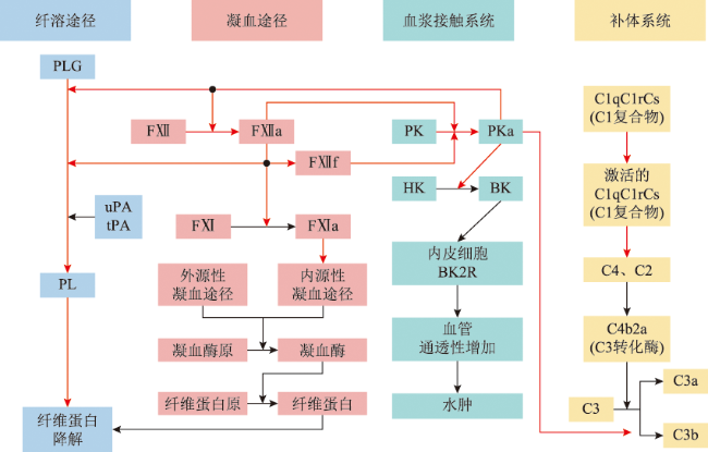
Figure 1 Schematic of C1-INH Interaction Sites in the Contact System, Complement System, Coagulation Pathway, and Fibrinolysis Pathway

C1-INH: C1 esterase inhibitor; PLG: plasminogen; PL: plasmin; uPA: urokinase-type plasminogen activator; tPA: tissue-type plasminogen activator; FⅫ: coagulation factor Ⅻ; FⅫa: activated coagulation factor Ⅻ; FⅫf: β-FⅫa (activated coagulation factor Ⅻ cleavage product); FⅪ: coagulation factor Ⅺ; FⅪa: activated coagulation factor Ⅺ; PK: plasma prekallikrein; PKa: plasma kallikrein; HK: high molecular weight kininogen; BK: bradykinin; BK2R: bradykinin 2 receptor; C1: complement C1; C2: complement C2; C3: complement C3; C4: complement C4; black arrows indicate the activation process of reactions; red arrows indicate inhibition of the process by C1-INH

The elevation of bradykinin levels in the blood is a key reason for the clinical manifestations such as edema in HAE-C1-INH patients[4]. Bradykinin, as a low molecular weight 9-peptide, is closely related to the plasma contact system and the complement system. The plasma contact system includes FⅫ, prekallikrein (PK), and high molecular weight kininogen (HK) among others[24]. In the plasma contact system, FⅫ can be activated into active FⅫ (activated FⅫ, FⅫa) by contacting with negatively charged surfaces. FⅫa further activates PK into active plasma kallikrein (plasma kallikrein, PKa). HK is cleaved into bradykinin under the action of PKa, and bradykinin causes vasodilation and increased vascular permeability through binding to the bradykinin B2 receptor (bradykinin receptor B2, B2R)[25]. Additionally, PKa can directly convert plasminogen (PLG) into plasmin or indirectly do so under the influence of urokinase-type plasminogen activator, while plasmin and PKa can, in turn, activate FⅫ into FⅫa, thus forming an auto-activation loop[26]. Under normal circumstances, some kininases rapidly metabolize bradykinin, such as kininase Ⅱ, aminopeptidase P, carboxypeptidase M, and N, etc.[27]. However, the SERPING1 gene mutation in HAE-C1-INH leads to decreased C1-INH levels or functional abnormalities, resulting in uncontrolled activation of the plasma contact system, leading to excessive production of bradykinin. After bradykinin binds to B2R, a member of the G protein-coupled receptor family, it triggers downstream signaling, causing phosphorylation of intravascular endothelial cadherin molecules, their internalization and degradation, thereby inducing actin cytoskeleton contraction, increasing the pore size between endothelial cells, and consequently causing vascular leakage, manifesting as angioedema and other clinical symptoms[28-29]. Moreover, studies speculate that the bradykinin B1 receptor (bradykinin receptor B1, B1R) may also be involved in the disease, contributing to angioedema. Although B1R is almost not expressed in normal tissues, inflammatory stimuli and the involvement of B2R can induce its increased expression[24]. Unlike B2R, which desensitizes quickly, B1R exhibits slow and partial desensitization upon agonist binding, which corresponds to the longer duration of edema episodes in HAE-C1-INH patients[30].
Research has shown that the critical functional threshold of C1-INH in controlling the plasma contact system is approximately 40%, and most HAE-C1-INH patients have heterozygous variants. Even with one normal allele, the C1-INH activity in these patients is only 5% to 30% of the normal level, not 50%[31-32]. Currently, it is believed that there are two reasons for this discrepancy: (1) C1-INH is continuously consumed by forming complexes with target proteases (such as C1s), and C1-INH is cleaved into an inactive form with a molecular weight of 94,000, leading to C1-INH activity below 50%; (2) under some missense mutations, abnormal C1-INH proteins form polymers in the endoplasmic reticulum of cells, which can embed normal C1-INH and partially inhibit its secretion, thereby reducing the secretion of normal C1-INH[33-34].
In the complement system, C1-INH deficiency/defect leads to uncontrolled overactivation of the classical complement activation pathway. After the activation of the C1 complex, a large amount of C4 and C2 are cleaved, resulting in increased generation of C4b and C2b, and a decrease in C4 levels[35]. Therefore, the level of C4 is of significant value for the diagnosis of HAE-C1-INH. There have been reports of abnormal activation of the coagulation pathway in HAE-C1-INH[36]. Research by Grover et al.[37] has shown that C1-INH deficiency in plasma promotes the generation of thrombin in human plasma and facilitates the growth of venous thrombi in mice. However, given that clinical patients with HAE rarely experience thrombotic events, the view that C1-INH deficiency/defect significantly increases the risk of venous thromboembolism in humans requires further research for validation[38].

2.2 HAE-nC1-INH

In addition to HAE-UNK as an unknown category, HAE-nC1-INH types now include HAE-FⅫ, HAE-PLG, HAE-KNG1, HAE-ANGPT1, HAE-HS3ST6, and HAE-MYOF. The mechanisms of HAE-nC1-INH have not been fully elucidated, but it is currently believed that bradykinin plays a crucial role in most types of HAE-nC1-INH[39-40].

2.2.1 HAE-FⅫ

HAE-FⅫ is the most common type of HAE-nC1-INH, which is an autosomal dominant inheritance with incomplete penetrance[41]. HAE-FⅫ is more common in females (male:female=1:10), especially in pregnant patients or those taking hormonal contraceptives, and studies have shown that it is closely related to increased estrogen levels[42]. There are four pathogenic mutations in the FⅫ gene, all located at sites adjacent to the Arg372-Val373 bond, these regions are associated with the activation process of FⅫ and are highly glycosylated[43]. The p.Thr328Lys mutation is the most common, while the other three mutations are relatively rare[44]. The gain-of-function mutation p.Thr328Lys can increase the enzymatic activity of FⅫ without altering the FⅫ antigen levels[45]. By identifying alleles in HAE-nC1-INH patients from different ethnicities, it was found that there may be ethnic differences in FⅫ variations[46-47]. It is currently believed that the molecular mechanism of HAE-FⅫ caused by p.Thr328Lys and p.Thr328Arg variants is as follows: thrombin, FⅪa, and plasmin cleave the FⅫ mutant at Lys/Arg328, generating a short, heavy chain-containing low molecular weight zymogen δFⅫ. δFⅫ can activate PK to PKa, and its efficiency is 15 times higher than that of full-length FⅫ, although both PKa and δFⅫ are inhibited by C1-INH, the accelerated mutual activation between PKa and δFⅫ exceeds the regulatory function of C1-INH to some extent, leading to the massive production of bradykinin[48]. In addition, the FⅫ p.Thr309Lys mutation not only lowers the activation threshold of the plasma contact system in vivo and in vitro but also creates a new plasmin cleavage site, thus presenting a potential pathway for contact system activation[49-50].

2.2.2 HAE-PLG

In 1971, Kaplan et al.[51] described the relationship between fibrinolysis and kinin generation. In 2018, Bork et al.[52] and Dewald[53] reported two cases of HAE-nC1-INH patients with a missense mutation site p.Lys311Glu (p.Lys330Glu if counted from the initiating methionine on the signal peptide). Currently, PLG-Glu311 has been found in over 150 individuals across more than 30 families on three continents, indicating a wide distribution[3,54]. This variant is inherited in an autosomal dominant manner and is more common in females (male:female = 1:3)[43].
PLG has five triple-loop structure regions, and the kringle 3 region is the only one among the five triple-loop structure regions of PLG that lacks the Asp-X-Asp/Glu motif. The p.Lys311Glu mutant is located in the triple-loop structure 3 region, which alters the structure of the wild-type protein, but its function remains unclear[55]. It is precisely Lys311 that disrupts the originally intact AspX-Asp/Glu motif (Asp-X-Lys), causing the p.Lys311Glu mutant to generate a new lysine/arginine binding site (Asp-X-Glu) in the triple-loop structure 3 region[56]. Researchers speculate that PLG/PLGlu311 can interact more effectively with HK and low molecular weight kininogen (LK) through these motifs, thereby increasing the production of bradykinin.
PLG-Glu311 carriers often exhibit edema in the head and neck, such as tongue edema, facial edema, and throat edema. Approximately 80% of symptomatic PLG-Glu311 patients experience tongue edema, which is typically their only clinical symptom, while erythema marginatum as a prodromal symptom is less common[39,57]. Angiotensin-converting enzyme inhibitors that inhibit bradykinin degradation may also cause tongue edema in patients[58]. Studies have shown that the lysine analog tranexamic acid successfully treated patients with angiotensin-converting enzyme inhibitor-induced angioedema, suggesting that local plasmin may mediate the production of bradykinin[59]. In vitro studies have demonstrated that the lysine analog ε-aminocaproic acid can inhibit plasmin-Glu311 from producing bradykinin[58].

2.2.3 HAE-ANGPT1

In 2018, Bafunno et al.[60] discovered a mutation site c.807G>T (p.Ala119Ser) in the gene encoding ANGPT1 in an Italian family. ANGPT1 is a ligand for tyrosine kinase receptor-2 (TIE2), which is expressed in vascular endothelial cells and subsets of hematopoietic cells[61]. The ANGPT1-TIE2 signaling pathway can inhibit various vascular permeability factors, including vascular endothelial growth factor (VEGF) and bradykinin, and contributes to the regulation of the barrier function of vascular endothelial cells[62]. The Ala119Ser mutation leads to a decrease in the amount of ANGPT1 in plasma, hinders the assembly of ANGPT1 multimers, and results in reduced binding to TIE2 due to haploinsufficiency[60,63]. Additionally, studies have shown that patients with the p.Ala119Ser mutation have a decreased ANGPT1/ANGPT2 ratio, and the ANGPT2 protein antagonizes the ANGPT1 protein, leading to increased vascular permeability[64]. Furthermore, the pathogenic mechanism of HAE-ANGPT1 has not been found to be associated with increased production of bradykinin but is believed to be related to alterations in intracellular signal transduction[56].

2.2.4 HAE-KNG1

The c.1136T>A (p.Met379Lys) mutation in KNG1 is inherited in an autosomal dominant manner. KNG1 produces HK and LK through alternative splicing. This mutation is located in both HK and LK. PKa specifically cleaves HK at the Lys380-Arg381 and Arg389-Ser390 sites, releasing a 9-amino acid bradykinin. Tissue kallikrein cleaves LK at the Met379-Lys380 and Arg389-Ser390 sites, releasing a 10-amino acid Lys-bradykinin[65]. Since the pathogenic mutation p.Met379Lys is located near the N-terminal cleavage site, it may affect the production of bradykinin and/or Lys-bradykinin[66].

2.2.5 HAE-MYOF

In 2020, a study reported the MYOF p.Arg217Ser variant in an Italian family[61]. MYOF encodes a type Ⅱ integral membrane protein with a relative molecular mass of 230,000, which is located on the plasma membrane of endothelial cells and can regulate VEGF signaling by inhibiting the ubiquitination and degradation of vascular endothelial growth factor receptor-2 (VEGFR2)[67]. The p.Arg217Ser mutation is located in the C2B domain, and it is speculated that if this mutation leads to the loss of MYOF function, it will result in decreased expression of VEGFR2. If the mutation results in gain of MYOF function, it will improve its localization on the plasma membrane and activate VEGF signaling, leading to increased vascular permeability[3]. Research has shown that myoferlin-Ser217 can express more VEGFR2 on the plasma membrane than the wild type, suggesting that MYOF p.Arg217Ser is a gain-of-function mutation that can transport VEGFR2 to the plasma membrane[47]. Additionally, in the study of the pathogenic mechanism of HAE-MYOF, no increase in bradykinin production was observed, suggesting that it may be related to changes in intracellular signal transduction[56].

2.2.6 HAE-HS3ST6

A p.Thr144Ser variant in HS3ST6 was reported in 4 HAE-nC1-INH patients from 1 family, showing an autosomal dominant inheritance, and all patients were female with an onset age of 1-20 years[68]. The 7 types of HS3STs expressed in the human body can regulate many biological processes, but there are differences in substrate specificity and tissue expression[69]. HS3STs transfer sulfate groups to the C3 position of glucosamine, forming 3-O-sulfated heparan sulfate, and syndecan-2, a transmembrane protein capable of binding to heparan sulfate, is expressed on the surface of endothelial cells. HK can form a complex by binding to heparan sulfate on syndecan-2 and enter the cell through endocytosis. The p.Thr144Ser mutation is located within the sulfotransferase domain, which may lead to incomplete biosynthesis of heparan sulfate, thereby potentially hindering the binding of HK to heparan sulfate on syndecan-2, making HK more likely to bind to the globular C1q receptor on endothelial cells, leading to mutual activation of FⅫ and PK, and promoting increased production of bradykinin[70].

3 Conclusion

By studying and exploring the pathophysiology and pathogenesis of HAE, our understanding of HAE has become more comprehensive and in-depth, which has also rapidly promoted the development of drugs for HAE and effective treatment strategies. It is currently believed that the occurrence of HAE is mainly due to the uncontrolled activation of the plasma contact system, leading to excessive production of bradykinin, which in turn triggers angioedema. With the rapid development of bioinformatics and computer technology, new pathogenic mutations have been discovered one after another, thereby promoting the research on the pathogenesis of HAE and the updating of its classification. However, the exact pathogenic mechanisms of many pathogenic mutations are still not very clear, and their relationship with the plasma contact system is also unclear, requiring further active research to provide a theoretical basis for the diagnosis and treatment of HAE.
Conflict of Interest All authors declare no conflict of interest
Author Contribution Statement Cui Xiangyi: responsible for the structural design, writing, and revision of the article, in addition to being accountable for their own research contributions, agrees to be responsible for the integrity of all aspects of the research work; Zhi Yuxiang: responsible for the conception, design, revision, review, and final approval of the article, in addition to being accountable for their own research contributions, agrees to be responsible for the integrity of all aspects of the research work
[1]
Aberer W. Hereditary angioedema:an orphan but an original disease[J]. J Allergy Clin Immunol, 2021, 148(4):994-995.DOI:10.1016/j.jaci.2021.07.026.

[2]
Launay D, Bouillet L, Boccon-Gibod I, et al. Hereditary angioedema and its new treatments:an update[J]. Rev Med Interne, 2023, 44(7):344-353.DOI:10.1016/j.revmed.2023.01.020.

[3]
Santacroce R, D’andrea G, Maffione AB, et al. The genetics of hereditary angioedema:a review[J]. J Clin Med, 2021, 10(9):2023.DOI:10.3390/jcm10092023.

[4]
Quincke HI. Über akutes umschriebenes Hautödem[J]. Monatsh Prakt Dermatol, 1882, 1:129-131.

[5]
Osler W. Hereditary angio-neurotic oedema[J]. Am J Med Sci, 1888, 95:362-367.

[6]
Donaldson VH, Evans RR. A biochemical abnormality in hereditary angioneurotic edema:absence of serum inhibitor of C’1-esterase[J]. Am J Med, 1963, 35(1):37-44.DOI:10.1016/0002-9343(63)90162-1.

[7]
Rosen FS, Pensky J, Donaldson V, et al. Hereditary angioneurotic edema:two genetic variants[J]. Science, 1965, 148(3672):957-958.DOI:10.1126/science.148.3672.957.

[8]
Frank MM, Gelfand JA, Atkinson JP. Hereditary angioedema:the clinical syndrome and its management[J]. Ann Intern Med, 1976, 84(5):580-593.DOI:10.7326/0003-4819-84-5-580.

[9]
Rosen FS, Alper CA, Pensky J, et al. Genetically determined heterogeneity of the C1 esterase inhibitor in patients with hereditary angioneurotic edema[J]. J Clin Invest, 1971, 50(10):2143-2149.DOI:10.1172/jci106708.

[10]
Nussberger J, Cugno M, Amstutz C, et al. Plasma bradykinin in angio-oedema[J]. Lancet, 1998, 351(9117):1693-1697.DOI:10.1016/s0140-6736(97)09137-x.

[11]
Binkley KE, Davis A 3rd. Clinical,biochemical,and genetic characterization of a novel estrogen-dependent inherited form of angioedema[J]. J Allergy Clin Immunol, 2000, 106(3):546-550.DOI:10.1067/mai.2000.108106.

[12]
Bork K, Barnstedt SE, Koch P, et al. Hereditary angioedema with normal C1-inhibitor activity in women[J]. Lancet, 2000, 356(9225):213-217.DOI:10.1016/s0140-6736(00)02483-1.

[13]
支玉香, 安利新, 赖荷, 等. 遗传性血管性水肿的诊断和治疗专家共识[J]. 中华临床免疫和变态反应杂志, 2019, 13(1):1-4.DOI:10.3969/j.issn.1673-8705.2019.01.001.

[14]
Proper SP, Lavery WJ, Bernstein JA. Definition and classification of hereditary angioedema[J]. Allergy Asthma Proc, 2020, 41(6):S03-S07.DOI:10.2500/aap.2020.41.200040.

[15]
曹阳, 刘爽, 支玉香. 遗传性血管性水肿发病机制研究进展[J]. 中国医学科学院学报, 2020, 42(5):686-690.DOI:10.3881/j.issn.1000-503X.11407.

[16]
Maurer M, Magerl M, Betschel S, et al. The international WAO/EAACI guideline for the management of hereditary angioedema-the 2021 revision and update[J]. Allergy, 2022, 77(7):1961-1990.DOI:10.1111/all.15214.

[17]
Wilkerson RG, Moellman JJ. Hereditary angioedema[J]. Emerg Med Clin North Am, 2022, 40(1):99-118.DOI:10.1016/j.emc.2021.09.002.

[18]
Aygören-Pürsün E, Magerl M, Maetzel A, et al. Epidemiology of bradykinin-mediated angioedema:a systematic investigation of epidemiological studies[J]. Orphanet J Rare Dis, 2018, 13(1):73.DOI:10.1186/s13023-018-0815-5.

[19]
Busse PJ, Christiansen SC. Hereditary angioedema[J]. N Engl J Med, 2020, 382(12):1136-1148.DOI:10.1056/NEJMra1808012.

[20]
Kaplan AP, Joseph K. Complement,kinins,and hereditary angioedema:mechanisms of plasma instability when C1 inhibitor is absent[J]. Clin Rev Allergy Immunol, 2016, 51(2):207-215.DOI:10.1007/s12016-016-8555-6.

[21]
De Maat S, Joseph K, Maas C, et al. Blood clotting and the pathogenesis of types Ⅰ and Ⅱ hereditary angioedema[J]. Clin Rev Allergy Immunol, 2021, 60(3):348-356.DOI:10.1007/s12016-021-08837-6.

[22]
Steiner UC, Keller M, Schmid P, et al. Mutational spectrum of the SERPING1 gene in Swiss patients with hereditary angioedema[J]. Clin Exp Immunol, 2017, 188(3):430-436.DOI:10.1111/cei.12941.

[23]
Ponard D, Gaboriaud C, Charignon D, et al. SERPING1 mutation update:mutation spectrum and C1 Inhibitor phenotypes[J]. Hum Mutat, 2020, 41(1):38-57.DOI:10.1002/humu.23917.

[24]
Kaplan AP, Joseph K. Pathogenesis of hereditary angioedema:the role of the bradykinin-forming cascade[J]. Immunol Allergy Clin North Am, 2017, 37(3):513-525.DOI:10.1016/j.iac.2017.04.001.

[25]
Busse PJ, Christiansen SC, Riedl MA, et al. US HAEA medical advisory board 2020 guidelines for the management of hereditary angioedema[J]. J Allergy Clin Immunol Pract, 2021, 9(1):132-150.e133.DOI:10.1016/j.jaip.2020.08.046.

[26]
Levi M, Cohn DM, Zeerleder S. Hereditary angioedema:linking complement regulation to the coagulation system[J]. Res Pract Thromb Haemost, 2019, 3(1):38-43.DOI:10.1002/rth2.12175.

[27]
Cicardi M, Zuraw BL. Angioedema due to bradykinin dysregulation[J]. J Allergy Clin Immunol Pract, 2018, 6(4):1132-1141.DOI:10.1016/j.jaip.2018.04.022.

[28]
Lopez Lera A. Pathophysiology and underlying mechanisms in hereditary angioedema[J]. Balkan Med J, 2021, 38(2):82-88.DOI:10.4274/balkanmedj.galenos.2020.2020.10.166.

[29]
Margaglione M, D’apolito M, Santocroce R, et al. Hereditary angioedema:looking for bradykinin production and triggers of vascular permeability[J]. Clin Exp Allergy, 2019, 49(11):1395-1402.DOI:10.1111/cea.13506.

[30]
Bossi F, Fischetti F, Regoli D, et al. Novel pathogenic mechanism and therapeutic approaches to angioedema associated with C1 inhibitor deficiency[J]. J Allergy Clin Immunol, 2009, 124(6):1303-1310.e1304.DOI:10.1016/j.jaci.2009.08.007.

[31]
Longhurst H, Cicardi M, Craig T, et al. Prevention of hereditary angioedema attacks with a subcutaneous C1 inhibitor[J]. N Engl J Med, 2017, 376(12):1131-1140.DOI:10.1056/NEJMoa1613627.

[32]
Zuraw BL, Cicardi M, Longhurst HJ, et al. Phase Ⅱ study results of a replacement therapy for hereditary angioedema with subcutaneous C1-inhibitor concentrate[J]. Allergy, 2015, 70(10):1319-1328.DOI:10.1111/all.12658.

[33]
Zuraw BL, Christiansen SC. HAE pathophysiology and underlying mechanisms[J]. Clin Rev Allergy Immunol, 2016, 51(2):216-229.DOI:10.1007/s12016-016-8561-8.

[34]
Haslund D, Ryo LB, Seidelin Majidi S, et al. Dominant-negative SERPING1 variants cause intracellular retention of C1 inhibitor in hereditary angioedema[J]. J Clin Invest, 2019, 129(1):388-405.DOI:10.1172/JCI98869.

[35]
Conigliaro P, Triggianese P, Ballanti E, et al. Complement,infection,and autoimmunity[J]. Curr Opin Rheumatol, 2019, 31(5):532-541.DOI:10.1097/bor.0000000000000633.

[36]
Cugno M, Zanichelli A, Bellatorre AG, et al. Plasma biomarkers of acute attacks in patients with angioedema due to C1-inhibitor deficiency[J]. Allergy, 2009, 64(2):254-257.DOI:10.1111/j.1398-9995.2008.01859.x.

[37]
Grover SP, Kawano T, Wan J, et al. C1 inhibitor deficiency enhances contact pathway-mediated activation of coagulation and venous thrombosis[J]. Blood, 2023, 141(19):2390-2401.DOI:10.1182/blood.2022018849.

[38]
Gailani D. Hereditary angioedema and thrombosis[J]. Blood, 2023, 141(19):2295-2297.DOI:10.1182/blood.2023019861.

[39]
Bork K, Wulff K, Witzke G, et al. Treatment for hereditary angioedema with normal C1-INH and specific mutations in the F12 gene (HAE-FXII)[J]. Allergy, 2017, 72(2):320-324.DOI:10.1111/all.13076.

[40]
Bork K, Wulff K, Witzke G, et al. Treatment of patients with hereditary angioedema with the c.988A>G (p.Lys330Glu) variant in the plasminogen gene[J]. Orphanet J Rare Dis, 2020, 15(1):52.DOI:10.1186/s13023-020-1334-8.

[41]
Serrano C, Guilarte M, Tella R, et al. Oestrogen-dependent hereditary angio-oedema with normal C1 inhibitor:description of six new cases and review of pathogenic mechanisms and treatment[J]. Allergy, 2008, 63(6):735-741.DOI:10.1111/j.1398-9995.2007.01579.x.

[42]
Miranda AR, Ue AP, Sabbag DV, et al. Hereditary angioedema type Ⅲ (estrogen-dependent) report of three cases and literature review[J]. An Bras Dermatol, 2013, 88(4):578-584.DOI:10.1590/abd1806-4841.20131818.

[43]
Bork K, Machnig T, Wulff K, et al. Clinical features of genetically characterized types of hereditary angioedema with normal C1 inhibitor:a systematic review of qualitative evidence[J]. Orphanet J Rare Dis, 2020, 15(1):289.DOI:10.1186/s13023-020-01570-x.

[44]
Bork K, Wulff K, Hardt J, et al. Characterization of a partial exon 9/intron 9 deletion in the coagulation factor Ⅻ gene (F12) detected in two Turkish families with hereditary angioedema and normal C1 inhibitor[J]. Haemophilia, 2014, 20(5):e372-e375.DOI:10.1111/hae.12519.

[45]
Cichon S, Martin L, Hennies HC, et al. Increased activity of coagulation factor Ⅻ (Hageman factor) causes hereditary angioedema type Ⅲ[J]. Am J Hum Genet, 2006, 79(6):1098-1104.DOI:10.1086/509899.

[46]
Hashimura C, Kiyohara C, Fukushi JI, et al. Clinical and genetic features of hereditary angioedema with and without C1-inhibitor (C1-INH) deficiency in Japan[J]. Allergy, 2021, 76(11):3529-3534.DOI:10.1111/all.15034.

[47]
Miyata T, Horiuchi T. Biochemistry,molecular genetics,and clinical aspects of hereditary angioedema with and without C1 inhibitor deficiency[J]. Allergol Int, 2023, 72(3):375-384.DOI:10.1016/j.alit.2023.04.004.

[48]
Ivanov I, Matafonov A, Sun MF, et al. A mechanism for hereditary angioedema with normal C1 inhibitor:an inhibitory regulatory role for the factor Ⅻ heavy chain[J]. Blood, 2019, 133(10):1152-1163.DOI:10.1182/blood-2018-06-860270.

[49]
Björkqvist J, De Maat S, Lewandrowski U, et al. Defective glycosylation of coagulation factor Ⅻ underlies hereditary angioedema type Ⅲ[J]. J Clin Invest, 2015, 125(8):3132-3146.DOI:10.1172/jci77139.

[50]
De Maat S, Björkqvist J, Suffritti C, et al. Plasmin is a natural trigger for bradykinin production in patients with hereditary angioedema with factor Ⅻ mutations[J]. J Allergy Clin Immunol, 2016, 138(5):1414-1423.e1419.DOI:10.1016/j.jaci.2016.02.021.

[51]
Kaplan AP, Austen KF. A prealbumin activator of prekallikrein.II.Derivation of activators of prekallikrein from active Hageman factor by digestion with plasmin[J]. J Exp Med, 1971, 133(4):696-712.DOI:10.1084/jem.133.4.696.

[52]
Bork K, Wulff K, Steinmüller-Magin L, et al. Hereditary angioedema with a mutation in the plasminogen gene[J]. Allergy, 2018, 73(2):442-450.DOI:10.1111/all.13270.

[53]
Dewald G. A missense mutation in the plasminogen gene,within the plasminogen kringle 3 domain,in hereditary angioedema with normal C1 inhibitor[J]. Biochem Biophys Res Commun, 2018, 498(1):193-198.DOI:10.1016/j.bbrc.2017.12.060.

[54]
Veronez CL, Csuka D, Sheikh FR, et al. The expanding spectrum of mutations in hereditary angioedema[J]. J Allergy Clin Immunol Pract, 2021, 9(6):2229-2234.DOI:10.1016/j.jaip.2021.03.008.

[55]
Law RH, Caradoc-Davies T, Cowieson N, et al. The X-ray crystal structure of full-length human plasminogen[J]. Cell Rep, 2012, 1(3):185-190.DOI:10.1016/j.celrep.2012.02.012.

[56]
Shamanaev A, Dickeson SK, Ivanov I, et al. Mechanisms involved in hereditary angioedema with normal C1-inhibitor activity[J]. Front Physiol, 2023, 14:1146834.DOI:10.3389/fphys.2023.1146834.

[57]
Kaplan AP. Hereditary angioedema:investigational therapies and future research[J]. Allergy Asthma Proc, 2020, 41(1):S51-S54.DOI:10.2500/aap.2020.41.200056.

[58]
Dickeson SK, Kumar S, Sun MF, et al. A mechanism for hereditary angioedema caused by a lysine 311-to-glutamic acid substitution in plasminogen[J]. Blood, 2022, 139(18):2816-2829.DOI:10.1182/blood.2021012945.

[59]
Wang K, Geiger H, Mcmahon A. Tranexamic acid for ACE inhibitor induced angioedema[J]. Am J Emerg Med, 2021, 43:292.e295-292.e297.DOI:10.1016/j.ajem.2020.10.029.

[60]
Bafunno V, Firinu D, D’apolito M, et al. Mutation of the angiopoietin-1 gene (ANGPT1) associates with a new type of hereditary angioedema[J]. J Allergy Clin Immunol, 2018, 141(3):1009-1017.DOI:10.1016/j.jaci.2017.05.020.

[61]
Ariano A, D’apolito M, Bova M, et al. A myoferlin gain-of-function variant associates with a new type of hereditary angioedema[J]. Allergy, 2020, 75(11):2989-2992.DOI:10.1111/all.14454.

[62]
Baffert F, Le T, Thurston G, et al. Angiopoietin-1 decreases plasma leakage by reducing number and size of endothelial gaps in venules[J]. Am J Physiol Heart Circ Physiol, 2006, 290(1):H107-H118.DOI:10.1152/ajpheart.00542.2005.

[63]
D’apolito M, Santacroce R, Colia AL, et al. Angiopoietin-1 haploinsufficiency affects the endothelial barrier and causes hereditary angioedema[J]. Clin Exp Allergy, 2019, 49(5):626-635.DOI:10.1111/cea.13349.

[64]
Zuraw BL. Hereditary angioedema with normal C1 inhibitor:four types and counting[J]. J Allergy Clin Immunol, 2018, 141(3):884-885.DOI:10.1016/j.jaci.2018.01.015.

[65]
Loules G, Parsopoulou F, Zamanakou M, et al. Deciphering the genetics of primary angioedema with normal levels of C1 inhibitor[J]. J Clin Med, 2020, 9(11):3402.DOI:10.3390/jcm9113402.

[66]
Bork K, Wulff K, Rossmann H, et al. Hereditary angioedema cosegregating with a novel kininogen 1 gene mutation changing the N-terminal cleavage site of bradykinin[J]. Allergy, 2019, 74(12):2479-2481.DOI:10.1111/all.13869.

[67]
Bernatchez PN, Acevedo L, Fernandez-Hernando C, et al. Myoferlin regulates vascular endothelial growth factor receptor-2 stability and function[J]. J Biol Chem, 2007, 282(42):30745-30753.DOI:10.1074/jbc.M704798200.

[68]
Bork K, Wulff K, Möhl BS, et al. Novel hereditary angioedema linked with a heparan sulfate 3-O-sulfotransferase 6 gene mutation[J]. J Allergy Clin Immunol, 2021, 148(4):1041-1048.DOI:10.1016/j.jaci.2021.01.011.

[69]
Chopra P, Joshi A, Wu J, et al. The 3-O-sulfation of heparan sulfate modulates protein binding and lyase degradation[J]. Proc Natl Acad Sci U S A, 2021, 118(3):e2012935118.DOI:10.1073/pnas.2012935118.

[70]
Mochizuki H, Futatsumori H, Suzuki E, et al. A quantitative method to detect non-antithrombin-binding 3-O-sulfated units in heparan sulfate[J]. J Biol Chem, 2021, 296:100115.DOI:10.1074/jbc.RA120.015864.

Outlines

/