Home Journals Progress in Chemistry
Progress in Chemistry

Abbreviation (ISO4): Prog Chem      Editor in chief: Jincai ZHAO

About  /  Aim & scope  /  Editorial board  /  Indexed  /  Contact  / 
Review

Microfluidic-Based Vasculatures on Chip: Methods and Recent Progress

  • Fangtian Wang 1 ,
  • Liang Zhao 1 ,
  • Guangsheng Guo 1, 2 ,
  • Xiayan Wang , 1, *
Expand
  • 1 Center of Excellence for Environmental Safety and Biological Effects, College of Chemistry and Life Science, Department of Chemistry, Beijing University of Technology, Beijing 100124, China
  • 2 College of Life and Environmental Science, Minzu University of China, Beijing 100081, China

Received date: 2024-01-24

  Revised date: 2024-02-26

  Online published: 2024-04-16

Supported by

National Natural Science Foundation of China(22174007)

National Natural Science Foundation of China(22127805)

Cultivating Fund of Faculty of Environment and Life(049000513203)

Beijing Outstanding Young Scientist Program(BJJWZYJH01201910005017)

Abstract

Microvasculature-on-a-chip,utilizing microfluidic technology,has emerged as a significant in vitro tool for simulating both the normal and disease states of blood vessel networks.in our review,we highlight the efficacy of microfluidic platforms in accurately reproducing the microenvironment of human blood vessels.we outline a range of methodologies employed to fabricate vascular networks in vitro,focusing on the use of endothelial cells within microfluidic structures.for each method,we provide an assessment of recent examples,critically evaluating their strengths and drawbacks.Furthermore,we delve into the outlook and the innovative advancements anticipated for next-generation vascular-on-a-chip models and the broader field of chip-based tissue engineering.

Contents

1 Introduction

2 The microfluidic approaches for recapitulating the vascular structure in vitro。

2.1 Monolayer-based culturing of the endothelial cells directly in the microfluidic device。

2.2 Hydrogel-based casting and fabricating of the lumen structure。

2.3 Mold or fugitive hydrogel sacrificially generates the endothelial lumen。

2.4 Self-assembling-based vascular network embedded in the hydrogel。

3 The conclusion and prospects

Cite this article

Fangtian Wang , Liang Zhao , Guangsheng Guo , Xiayan Wang . Microfluidic-Based Vasculatures on Chip: Methods and Recent Progress[J]. Progress in Chemistry, 2024 , 36(6) : 840 -850 . DOI: 10.7536/PC240121

1 Introduction

the vascular system is a vital physiological system in the human body.Its main function is to transport oxygen,nutrients(such as amino acids and electrolytes),carbon dioxide and hormones to the cells of the whole body through blood circulation,and to take away the waste produced by cell metabolism.This system not only plays a central role in maintaining homeostasis and regulating physiological processes,but is also closely related to the occurrence of a variety of diseases.Therefore,the study of vascular system dysfunction is essential for disease prevention,diagnosis,and treatment[1~4]
Traditional in vitro cell models,such as disk-based planar cell culture,provide a simplified environment to study cell behavior,but such models often fail to reproduce the complex interactions between cells and to simulate the response of whole tissues or organs to drug stimulation.in addition,in vitro cell culture,there is also a lack of blood vessels composed of cells,and it is difficult to restore the effect on cells in the case of existing blood vessel supply.This is particularly important for studying drug delivery and metabolism,as blood vessels are the main routes of drug distribution and clearance[5~7]。 to overcome these limitations,scientists often turn to animal models,especially to study disease development and drug efficacy.animal models,such as patient-derived xenograft models,can more closely mimic the biology of human disease and aid in the study of complex diseases such as tumors.However,animal models also have limitations,such as differences from human biology,selective limitations,ethical issues,and the difficulty of real-time monitoring and quantitative analysis of key cellular behaviors[8~10]。 in view of this,it is particularly important to develop in vitro models that are both economical and efficient and can more accurately predict human responses.such a model could provide a more ethical approach to research,reduce the reliance on animals,and potentially improve the accuracy of drug toxicity and efficacy predictions.Current research is moving towards the construction of more complex in vitro models,Such as three-dimensional vascular network models created using tissue engineering techniques,which can better simulate the microenvironment of the vascular system and allow real-time monitoring of cell behavior[11,12]
organ-on-a-chip technology and Micro-physiological system(MPS)are important advances in the field of bioengineering in recent years.Thanks to the integration of bioprinting technology,microfluidic chip technology and tissue engineering technology,Organ chip is a platform integrating bioengineering and biotechnology.Simulating the key functions of human organs at the microscale provides a model close to human physiological conditions for drug screening and disease research,aiming to simulate the physiological and pathological characteristics of human organs more accurately[13~16]。 the MPS platform further extends this concept,bridging the gap between traditional two-dimensional cell culture and animal models by simulating the three-dimensional environment of cells or tissues in the human body.Microfluidic systems have played a key role in the development of MPS,and their core lies in their ability to precisely control fluids at the microscale and provide cells with a dynamic hydrodynamic environment,which is essential for simulating the vascular system.in the human body,the diffusion of oxygen and nutrients is limited within tissues,usually limited to the range of 100 to 200μm.Beyond this distance,tissue cells rely on the microvascular network to obtain the necessary nutrients and eliminate metabolic waste to maintain growth and function.Therefore,the reconstruction of blood vessels in vitro is essential for the construction of organ chips and microphysiological systems.in order to reproduce the physiological functions of organs in vitro,the design of microvascular networks is a central challenge in the construction of vascularized organ chips[17~19]
Microfluidics is a key means to achieve this goal by precisely controlling fluid flow within tiny channels to create complex microfluidic geometries such as micropillars,semipermeable porous membranes,and even to construct endothelial cell monolayers to mimic vascular perfusion.these microfluidic structures not only provide physical support,but also mimic the functions of the vascular network,including acting as a semi-permeable barrier to control the bidirectional transport of small and large molecules such as gases,sugars,fatty acids,and proteins.in addition,the vascular system is also involved in the signaling between vascular and non-vascular cells in the tissue microenvironment,and this vascular secretory signaling is essential for maintaining tissue homeostasis,metabolism,and regeneration.in MPS,these physiological processes in the human body can be better understood and recapitulated by modeling these signaling pathways.the advantages of using microfluidic technology to reconstruct the vasculature in vitro are as follows:First,the microfluidic platform can precisely control the fluid in the channel in time and space,which makes it possible to produce controllable stimulation to endothelial cells and study the mechanism of the vasculature.Secondly,the channels of the microfluidic platform can be constructed to a size comparable to the specific microstructure in the vasculature,geometrically making these microchannels a repeat of the original blood vessel,which,combined with three-dimensional culture and tissue engineering,can selectively reconstruct the structure and function of the vascular network in vitro[20~23]
to sum up,microfluidics is of great significance in the construction of vascular network models in MPS.It not only enables the complexity of the vascular network to be reproduced in vitro,but also provides the possibility of simulating the internal signaling pathways and functions of the human body,which has important scientific and application value for the research of disease models and drug development.MPS models integrated with in vitro blood vessels allow researchers and clinicians to accurately mimic the physical structure and function of the human vasculature in vitro by tailoring the chemical microenvironment.the purpose of this paper is to summarize the methods and main strategies of microfluidic chip in the construction of in vitro vascular network model,to explore its application in the simulation of human vascular environment and its potential scientific and clinical value,and to make prospects for the future development of this technology。

2 Method for blood vessel reconstruction in vitro in microfluidic chip

Microfluidic chip,commonly known as"microvascular chip",has a wide range of applications in the reconstruction of blood vessels in vitro,which can accurately simulate the function of human vascular system,and can be used to build disease models,drug screening and toxicity assessment[24,25]。 in This paper,the construction methods in vitro(Fig.1)can be roughly divided into the following four types:(1)growing a monolayer of endothelial cells directly on a biocompatible wall or porous film,which is mostly used to construct endothelial barriers with certain physiological functions;(2)carrying out micro-shaping and processing on the hydrogel or collagen with biocompatibility by Using a soft photoetching and recasting mold mode to form a cavity structure formed by the hydrogel,and then pouring endothelial cells and the like to adhere to the wall to form a blood vessel lumen;(3)forming a hollow pipeline in the hydrogel in a mode of presetting and then removing by using sacrificial bioprinting or other template sacrificial modes,and then forming a blood vessel by using endothelial cell inoculation and adherence;(4)using the geometric structure and channel arrangement of microfluidic channels and chambers,endothelial cells mixed with collagen or fibrin are directly added to the designated microfluidic channels and chambers.Due to the geometric structure and channel arrangement,the encapsulated cells spontaneously form a vascular network under precisely defined microenvironmental conditions.this method is also known as self-assembly or self-morphogenesis[26]
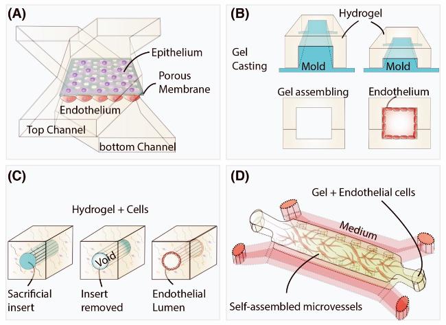
图1 体外构建血管以及血管网络用于在体外模拟类似于体内的血管和其他组织的方法归纳:(A) 利用多孔膜和与之配合的多层微流控管道构建二维单层培养的血管内皮细胞层,通常可以用来模拟血管屏障;(B) 利用微加工的模具(如聚二甲基硅氧烷(PDMS)等)将水凝胶类高分子通过塑形成为带有管腔的水凝胶微流控管道,并将血管内皮细胞通过灌注贴壁的方式培养在其中;(C) 利用打印或预先布置可牺牲凝胶或者模具的方式在凝胶中形成中空管道,并利用内皮细胞贴壁生长在凝胶中形成血管;(D) 通过将血管内皮细胞和其他支持细胞直接混合于水凝胶中,直接生长形成了毛细血管微网络。在芯片中,通过限制性结构分隔凝胶和培养基,实现对微环境的精确控制

Fig. 1 Schematic illustration of in vitro construction of blood vessels and vascular networks to simulate in vivo-like vessels. (A) The vascular endothelial layer is constructed using a porous membrane in conjunction with multilayer microfluidic channels, typically used to simulate the vascular barrier. (B) Using soft lithographically fabricated molds (such as PDMS), hydrogel polymers are cast to form hydrogel microfluidic channels with lumens, and vascular endothelial cells are cultured within them by perfusion. (C) Hollow channels are formed in the gel using printing or pre-arranged sacrificial gels or molds. After the removal of the mold or gel, the endothelial cells are cultured on the walls of these channels within the gel to form vessels. (D) Vascular endothelial cells and other supporting cells are directly mixed into the hydrogel and allowed to grow within the gel to form a capillary micro-network. Functional structures like micro-pillars are used to separate the gel and culture medium in different channels of the chip

This section will focus on several methods and technical strategies for in vitro vascular remodeling using microfluidic chips.the different methods and The application of microfluidic vascularization chip were summarized,and each method was analyzed and compared in detail。

2.1 Monolayer barrier formed by direct culture of vascular endothelial cells on a two-dimensional plane in a microfluidic chip

Culturing vascular Endothelial cells directly on microfluidic chips or with the help of porous membranes to form a monolayer barrier is one of the important methods to reconstruct blood vessels in vitro and simulate human vascular function.endothelial cells were attached to the inner surface of the microchannel and formed a continuous cell monolayer by specific surface treatment and cell culture techniques.in addition,by using porous membranes,researchers can create barriers with specific pore sizes and distributions that mimic the underlying structure of the vessel wall.These cells exhibit morphologies and functions similar to those in vivo,including the formation of tight junctions,which mimic the real vascular barrier,and can be used to study cell-cell interactions,vascular permeability,drug delivery,and vascular function under pathological conditions.This method allows cells to attach and grow,while also controlling the exchange of substances between cells。
In 2010,Ingber's team at Harvard University used a porous membrane made of PDMS combined with a microfluidic chip to simulate the anatomical structure of alveoli,and constructed a lung tissue chip with diastolic function[27]。 the device consists of three PDMS layers arranged And irreversibly bonded to form two sets of three parallel microchannels,with the upper and lower channels separated by a sheet of self-fabricated 10µm thick PDMS elastic porous membrane.After permanent bonding,the PDMS etchant flows through the two side channels to selectively etch the film in the channels,forming two large side chambers.Vascular endothelial cells and alveolar epithelial cells were seeded on both sides of the porous membrane,and the structure was highly similar to the actual alveolar structure in anatomy,forming the alveolar-capillary barrier.Furthermore,physiological respiratory motion was successfully mimicked by applying a periodic vacuum to the side chamber,causing mechanical stretching of the PDMS membrane(Fig.2A).Cyclic mechanical strain was found to enhance nanoparticle uptake by epithelial and endothelial cells and stimulate their trafficking to potential microvascular channels.the research team used the lung chip model to further study,that is,to simulate a variety of normal lung functions(infection-induced immune cell recruitment,respiratory-induced nanoparticle absorption and related inflammation,interleukin-2(IL-2)-induced pulmonary edema,inflammation-induced pulmonary thrombosis,etc.)and the observed response to drug treatment[28]。 In addition,Zhang et al.Constructed a human disease model on a lung chip based on this structure[29]。 The human alveolar chip model co-cultures human alveolar epithelium,microvascular endothelium,and circulating immune cells under fluid flow to reproduce the main features of the alveolar-capillary barrier in normal disease.From this,the model can reproduce the process of SARS-CoV-2-induced lung injury and immune response At the organ level.at the same time,they also found that COVID-19 infection of lung tissue may induce pulmonary microvascular endothelial injury by activating human immune cells to release a large number of inflammatory factors.Using this model,they also conducted preliminary testing and evaluation of the efficacy of antiviral compounds(Figure 2B).the Vunjak-Novakovic group used a vascular monolayer on a porous membrane in combination with multiple organs to simulate multiple organ physiology[30]。 in this system,mature human heart,liver,bone,and skin tissues and multiple iPSC-derived organs are connected by flow to form a cycle that mimics interdependent organ functions.each tissue was cultured In its own optimized environment,and a porous membrane-constructed endothelial barrier was integrated on Each organ unit.during 4 weeks of culture,these interconnected tissues maintained their molecular,structural,and functional phenotypes,reproducing the pharmacokinetic and pharmacodynamic profile of doxorubicin During normal culture(Figure 2C),confirming that vascular junctions and phenotypically stable mature human tissues may contribute to the clinical applicability of tissue chips。
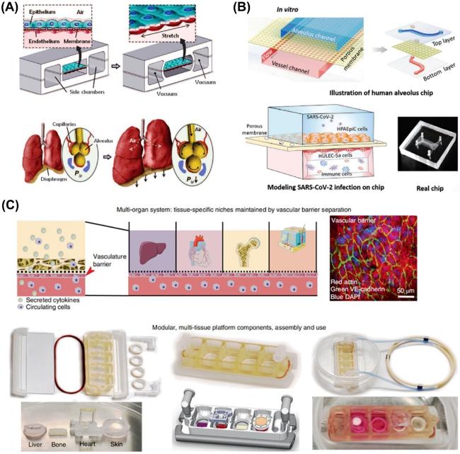
图2 基于多孔膜构建二维内皮屏障功能的微流控芯片模型:(A) 集成了弹性多孔膜以及侧翼抽气管道的微流控芯片构建和模拟肺泡-毛细血管屏障的生理呼吸过程[27];(B)受SARS-CoV-2感染的人肺泡芯片模型用于研究SARS-CoV-2病毒对人肺损伤过程和器官水平上的免疫反应[29];(C)利用多孔膜形成的血管屏障应用于多器官串联微生理系统,允许器官间的通讯与相互作用[30]

Fig. 2 Recapitulation of 2D endothelial barrier functions by using the porous membrane with the microfluidic device. (A) Biological-inspired design of a human breathing lung-on-a-chip. A microfluidic chip integrates compartmentalized PDMS microchannels and an ECM-coated elastic porous membrane to form an alveolar-capillary barrier[27]. (B) A human alveolar chip model infected with SARS-CoV-2 is used to study the process of lung damage caused by the SARS-CoV-2 virus and the immune response at the organ level[29]. (C) The vascular barrier formed by the porous membrane is applied to a multi-organ linked micro-physiological system, allowing communication and interaction between organs[30]

in addition,microfluidic systems can precisely control the flow of fluid and simulate the shear force of blood flow on endothelial cells,which is essential for the study of hydrodynamic effects in vascular biology.Huang's group used an integrated multilayer microfluidic system to analyze umbilical vein endothelial cells cultured in the chip from phenotype to whole transcriptome sequencing under single flow shear stress and double flow shear stress[31]。 Because the culture medium fluid can be controlled by the integrated micropump and microvalve In the chip,the direction and speed of the fluid can be accurately manipulated.Studies have shown that the expression of BMP4,BMPER,TEK,ADAM15,IL8,EDN1,ANGPT2 and CYR61 is sensitive to different fluid directions.in addition,the sequencing results showed that the RNA post-processing(alternative splicing)of NRG1 and IFI44 genes was different under different fluid conditions,which revealed that besides gene expression,RNA post-processing was also one of the strategies for endothelial cells to cope with mechanical environment。
Fabricating microfluidic chips by means of lithography and microfabrication and integrating porous membranes into them is one of the mainstream ways to form endothelial barriers in the field of organ chips because of its controllable integration process,good repeatability and convenient observation.However,the endothelial cell layer formed in this way is a two-dimensional monolayer,which does not have a three-dimensional lumen structure,and to some extent,it is difficult to reproduce the three-dimensional structure of blood vessels in vivo and the functions related to the three-dimensional structure.Moreover,due to the lack of extracellular matrix similar to the vascular structure in vivo,many physiological phenomena such as angiogenesis and sprouting are difficult to reconstruct and simulate on this model.Therefore,more and more attention has been paid to the techniques and methods that can controllably construct the three-dimensional lumen morphology of blood vessels in vitro。

2.2 Blood vessel formation by adherent culture of endothelial cells in hollow tubes with gel micromolding

Different from constructing a vascular endothelial cell monolayer on a two-dimensional plane or a porous membrane,a precise collagen or other matrix microstructure can be precisely and controllably manufactured on a silicon wafer or a PDMS mold by using a soft lithography technology and other micro-processing technologies,Removing the mold and encapsulating the gel to form the channel,vascular endothelial cells can be further seeded to form blood vessels and construct an in vitro vascular model.Through microfabrication techniques,researchers are able to create complex vascular network models,providing an important tool for in-depth understanding of the mechanisms of the vascular system and the development of new therapeutic approaches。
Based on this method,Zheng et al.Used a PDMS mold to prepare type I collagen gel with a groove structure[32]。 the two collagen layers were sealed together to form a closed fluid structure by applying pressure inside the Plexiglas apparatus.microvascular networks were formed by culturing microchannels seeded with human umbilical vein endothelial cells into collagen.in which native type I collagen has the appropriate stiffness to allow high reproducibility of vascular microarchitecture and can also be remodeled by degradation and deposition of extracellular matrix.the morphology,mass transfer process,and long-term stability of endothelial cells are described based on the vascular network formed in a native collagen matrix;The angiogenic activity of the endothelium and the differential interaction with perivascular cells in collagen were elucidated;The nature of the nonthrombotic vascular endothelium and its transition to a prethrombotic state in response to inflammation was also demonstrated(Figure 3A).The platform can successfully reproduce these Microvascular phenomena in vitro,indicating that in vitro vascular network models based on this method have broad potential in cardiovascular biology and pathophysiology.He research group can conveniently construct various complex channel structures in hydrogel,such as branched chain,helix,serpentine and so on,by using the inherent crosslinking characteristics of hydrogel materials[33]。 the authors attached human umbilical vein endothelial cells to the inner wall of the channel,forming a complete ring of cell layers along the entire length,while maintaining high survival and good propagation morphology(Figure 3 B).in addition,based on this cross-linking strategy,the research group established a complete blood circulation system model including large blood vessels and capillaries in the hydrogel.Compared with the single scale of other in vitro vascular models,a new concept based on multi-scale vascular model is proposed[34]。 in clinical translation,large solid tissues are difficult and cumbersome To form metabolites In vitro due to the lack of vascular network transport.to overcome this limitation,Zhang et al.Constructed a stable but biodegradable vascular chip scaffold using layer-by-layer stacking[35]。 this technique takes advantage of the UV polymerizability of poly(octylmethylene maleic anhydride citrate)(POMaC)to construct vascular chip scaffolds under mild conditions.A POMaC sheet with a blood vessel network pattern is superimpose on each other layer by lay under ultraviolet light irradiation,so that a complex suspended microstructure and an internal cavity can be formed.Finally,using This method,a myocardial patch with a vascular network was assembled and successfully implanted in mice。
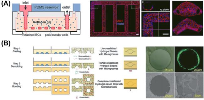
图3 利用凝胶微塑形的中空管道贴壁培养内皮细胞形成血管:(A)在胶原中微通道中接种内皮细胞形成具有完整管腔结构的血管网络并用于研究血管生成(angiogenesis)和血栓形成[32];(B)基于一种水凝胶的双交联机质在体外形成血管腔的制备工艺原理及其机理[33]

Fig. 3 Using gel micro-molding to create hollow channels and culture of endothelial cells to form blood vessels. (A) Endothelial cells are seeded in microchannels within collagen to form a vascular network with a complete lumen structure, which is used to study angiogenesis and thrombosis formation[32]. (B) The principle and mechanism of the preparation process for forming vascular lumens in vitro based on a hydrogel with a dual crosslinking mechanism[33]

vascular network formation based on microfabrication modeling enables precise control of the size,shape,and pattern of the vascular network,ensures a high degree of repeatability,and is compatible with a wide range of materials,making it very effective in simulating vascular structures in the human body,tissue engineering,disease models,and drug testing.However,this technique also suffers from cumbersome steps,high cost,non-circular tube geometry,and possible biocompatibility issues.in addition,the manufactured blood vessels are still composed of a single layer of cells,and it is still difficult to reconstruct and achieve special physiological phenomena such as vascular tissue regeneration in vivo[24]

2.3 Vessel formation by adherent culture of endothelial cells using a sacrificial mold or gel to form a lumen

in order to simplify the tedious steps of microfabrication molding and improve the experimental efficiency,the researchers designed a method to remove the template after template molding to form a microvascular model In vitro[36]。 this method usually requires two steps:first,a sacrificial mold is used to create a microchannel or network;Second,endothelial cells were seeded in the channel to form the endothelial layer.based on the current preparation methods,microvascular template formation and removal can be divided into needle-Based template formation and sacrificial template collagen formation.it is worth noting that this strategy is also the mainstream technical solution for the formation of blood vessels or other cavity structures in the field of bio-3D printing.Since this paper mainly discusses the use of microfluidic chip devices to form blood vessel models in vitro,It does not involve too much discussion on the scope of biological 3D printing。
Chen's group used a template method with removable microneedles to form a vascular endothelial cavity in collagen that could be perfused for a long time[37]。 this work demonstrates in vivo-like vascular sprouting and perfusable neovascularization in 3D collagen matrices.Microfluidic devices use acupuncture needles to form cylindrical channels of controlled size in the matrix.the two channels are respectively used as an endothelial channel and an angiogenic factor channel to display directional angiogenesis.Through the effects on angiogenesis under the action of different inducing factors,it is found that the angiogenesis process induced by different factors may have different mechanisms and targets on endothelial cells(Fig.4A).the method based on the presetting and removal of microneedles in collagen can not only form an endothelial lumen with a circular cross-section,but also allow physiological phenomena such as angiogenesis and sprouting of blood vessels in the three-dimensional structure of collagen.in order to better understand the interaction between tumor and vascular endothelium in pancreatic ductal adenocarcinoma(PDAC),Chen's team used microneedles to construct two parallel channels in collagen,one of which contained PDAC cells and was adjacent to the original blood vessel composed of endothelialized and perfused chambers.Using this model,it was observed that PDAC tumor cells could invade and remove the vascular endothelium during the process of endothelial cell ablation.the authors further used this model to confirm the critical role of the activin-ALK7 signaling pathway in mediating endothelial cell ablation in PDAC[38]。 Kim et al.Constructed a vessel-tumor model in a microfluidic chip by a combination of microfabrication and template-based removal of microneedles[39]。 by studying the interaction between the two,the effect of the position of lung cancer spheroids on the speed and growth direction of tumor angiogenesis was explored By controlling the direction of interstitial flow and the ratio and position of fibroblasts(Figure 4 B).Solid tumors create an immunosuppressive environment that places a tremendous burden on the immune system.Beebe's team used removable PDMS rod-like structures to create a 340-µm-diameter tube in collagen and seeded endothelial cells on the inner wall of the tube to form a vascular structure,which was perfused with culture medium to nourish the cells and mimic the vascular structure present in tumors[40]。 the authors used the chip to assess the impact of tumor environmental stress on NK cell function,exploring the plasticity of NK cells and their ability to reverse immune exhaustion。
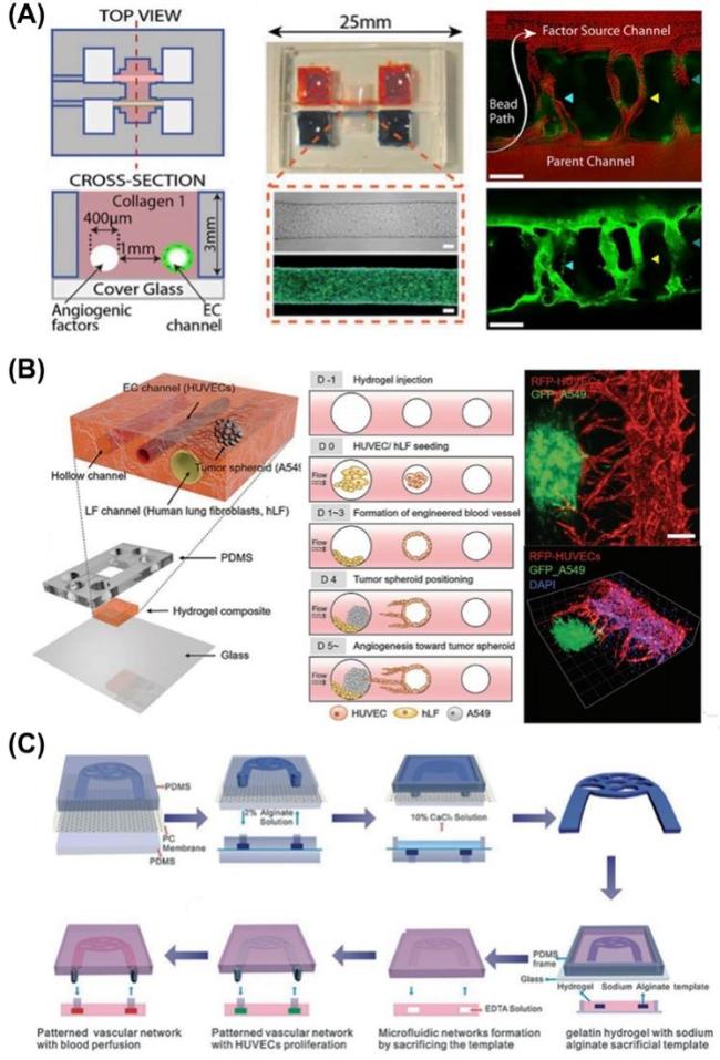
图4 利用可牺牲模具或凝胶形成管腔后贴壁培养内皮细胞形成血管:(A)基于微针去除法在体外重建血管模型用于研究新生血管芽形态发生以及相关的影响因素[37];(B)基于微针去除法在体外形成血管化肺癌肿瘤模型用于研究肿瘤微环境对血管生成的影响并评估新血管生成对促进抗癌药物给药效果[39];(C)海藻酸钠作为可牺牲模板在凝胶中快速形成相互连接的三维血管网络[41]

Fig. 4 Formation of blood vessels by culturing of endothelial cells after the creation of lumens using sacrificial molds or gels inside ECM hydrogel. (A) A vascular model is reconstructed in vitro based on the microneedle removal method for studying the morphogenesis of neovascular sprouts and the related influencing factors[37]. (B) A vascularized lung cancer tumor model is formed in vitro using the microneedle removal method to study the impact of the tumor microenvironment on angiogenesis and to evaluate the effectiveness of neovascularization in enhancing the delivery of anti-cancer drugs[39]. (C) Sodium alginate is used as a sacrificial template to rapidly form interconnected three-dimensional vascular networks within a gelatin hydrogel[41]

Compared with the single microvascular channel formed by the simple microneedle removal templating method,the sacrificial templating method can form a more complex vascular network structure in vitro.in this approach,a two-dimensional or three-dimensional vascular network mold is first formed using an easily soluble gel or solid material.the preformed template was encapsulated in a three-dimensional hydrogel.Finally,the degradable template is dissolved or melted and flows out of the solidified gel,leaving interconnected channels in the hydrogel structure.Huang's group used biocompatible sodium alginate as a sacrificial template to form an interconnected three-dimensional microfluidic vascular network in the hydrogel[41]。 The sodium alginate solution was crosslinked by Ca2+and then solidified to form a sacrificial template,which was encapsulated in the PDMS prepolymer.After the prepolymer is completely solidified,the sodium alginate mold encapsulated by PDMS is dissolved with EDTA solution to obtain a vascular network of interconnected channels(Fig.4C )。

2.4 Spontaneous formation of microvascular network by directly cultured cells in collagen on microfluidic chip

based on the mechanism of angiogenesis,it has been found that endothelial cells in collagen matrices such as laminin and fibronectin will organize themselves into tubular structures under specific culture conditions and signal induction,which are similar to real blood vessels in morphology and function.Hydrogels,such as collagen,Matrigel,gelatin and fibrin,contain 90%to 99%water and have high permeability to biomolecules.At present,the study of vascular network self-organization is mainly Based on the co-culture of vascular endothelial cells and fibroblasts in collagen or fibrin gel in vitro.endothelial cells spontaneously form microvessels and lumens in collagen under the action of cytokines such as vascular endothelial growth factor(VEGF)secreted by fibroblasts,and allow fine fluids to flow through vascular compartments in vitro[42~45]
Jeon et al.Designed a microfluidic device with five parallel microchannels to construct a perfusable microvascular network[46]。 the method involves mixing umbilical vein endothelial cells and fibroblasts separately In fibrinogen solution and introducing them into different channels to promote angiogenesis(Figure 5A).this design helps keep the endothelial and stromal cell suspensions separated within the central channel,encouraging endothelial cells to self-assemble to form a microvascular network.By adjusting parameters such as fibroblast ratio,they successfully established a perfusable physiological microvascular network in the microfluidic device.This method is of great significance in the construction of microvascular networks.Using This method,Jeon's team has constructed a sustainable perfusion microvascular network with physiological morphology on a three-dimensional scale and carried out many applications.in addition,Lee and Hughes established a physiological transport model from artery to vascularized tissue and then to vein[47]。 In this chip,the different stages of vascular development,including angiogenesis,endothelial cell lining,germinal angiogenesis and vascular anastomosis,are simulated by the spontaneous drive of endothelial cells and fibroblasts。
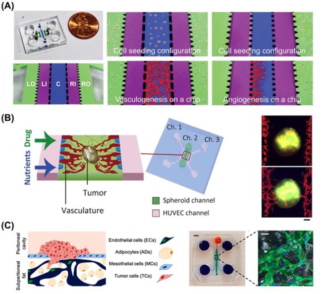
图5 基于内皮细胞与基质细胞自组织的方式在凝胶中自发形成血管网络:(A)基于内皮细胞与间质成纤维细胞在微流控可控的并列管道中共培养形成完整的三维微血管,模拟正常的血管生成过程[46];(B)体外血管化的人腹膜大网膜和卵巢肿瘤微环境微流控三维模型,用于研究卵巢癌转移中基质细胞对肿瘤细胞附着和生长的影响,以及基质细胞对早期和晚期转移瘤模型中血管和间皮通透性的影响[55];(C)基于微流控芯片在体外形成血管化肿瘤模型用于研究血管对肿瘤球状体生长和药物传递的影响[56]

Fig. 5 Spontaneous self-assembly formation of vascular networks within a gel based on the self-organization of endothelial cells and stromal cells. (A) The formation of complete three-dimensional microvessels through the co-culture of endothelial cells and fibroblasts within microfluidic parallel channels simulates the normal process of angiogenesis [46]. (B) The formation of a vascularized tumor model in vitro based on a microfluidic chip for studying the impact of vasculature on the growth of tumor spheroids and drug delivery [55]. (C) An in vitro vascularized human omentum and ovarian tumor microenvironment microfluidic 3D model for studying the effects of stromal cells on the attachment and growth of tumor cells during ovarian cancer metastasis, as well as the impact of stromal cells on vascular and mesothelial permeability in early and late-stage metastatic tumor models [56]

Another important application of in vitro vascular models is in vitro disease modeling,particularly in the field of tumor biology.the development of cancer can be divided into the following steps:initial growth of the primary tumor,infiltration of immune cells,angiogenesis,intravasation of the primary tumor cells,transvascular transport of cancer cells,distal extravasation of cancer cells,and growth of metastatic tumors[48]。 To realistically reproduce tumor progression in vitro,microvascular tumor models provide important tools for cancer research and serve as a low-cost anticancer drug screening platform.Kamm's research group studied the extravasation of tumor cells in blood vessels by using microvessels formed by self-organization in microfluidic chips,which provided a new research method for the study and analysis of the interaction between tumor cells and blood vessels in vitro[49]。 They demonstrated the process of tumor intravasation mediated by macrophages in a microfluidic system.the three-dimensional tumor microenvironment of this system,which mimics the interface of the tumor and endothelial layer,revealed that tumor necrosis factorα(TNF-α)secreted by macrophages directly impairs the barrier function of the endothelial layer and enhances the intravasation of breast tumors.in addition,the research team used a similar device To simulate the extravasation process of breast cancer cells in microvascularized osteoblastic tissue,and used confocal microscopic imaging to study the dynamic process of breast cancer cells entering,adhering,and metastasizing through the microvascular network.to demonstrate whether the secreted factor affects tumor extravasation,breast cancer cells were seeded on one side of the microfluidic channel and their migration through the vessel was monitored in real time.It was found that the extravasation rate of metastatic breast cancer cells into the bone microenvironment was four times higher than that of the skeletal muscle microenvironment in the control group,which is consistent with the phenomenon observed in vivo[50]。 in the tumor microenvironment,blood vessels play an indispensable role,especially In the delivery of anticancer drugs.Jeon's team proposed that individual parts of the device could be used to form a liquid guide without contact with the bottom surface,which could confine the colloidal solution to a designated area and eventually form a collagen shape with self-organized blood vessels inside[51]。 Using this method combined with 3D printing and micro-injection molding,Jeon's team studied the angiogenesis model induced by tumor spheroids and the migration and invasion detection of tumor spheroids,and used it for drug screening[52,53]。 Using a similar method,Wang's team at Shanghai Jiaotong University used a simpler laser cutting technology to process PMMA resin chip devices and used the device to form patterned fibrin collagen.vascular endothelial cells were cultured by self-organization to form a vascular network,which provides a new way to prepare microfluidic devices for vascular network research without microfabrication equipment[54]
in order to more accurately simulate the tumor microenvironment,researchers have tried to use different types of cells,but it is still a challenge to correctly form tumor tissue for blood flow.Yokokawa's team at Kyoto University has proposed a platform for vascularization of tumor spheroids using chips,which can simulate the tumor microenvironment In vitro[55]。 Fibroblasts within the tumor spheroid induce endothelial cells in fibrin collagen to form blood vessels and sprout structures,creating a perfusable vascular network.drug administration under perfusion conditions did not show a dose-dependent effect of anticancer drugs on tumor activity,in contrast to the results under static conditions,confirming the importance of flow in the vascular network for assessing tumor activity on a Drug screening platform(Figure 5B).in addition,Ibrahim et al.Established an in vitro model of vascularization of human peritoneal omentum and ovarian tumor microenvironment[56]。 Based on this model,the effect of stromal cells on tumor cell attachment and growth,and the effect of tumor cells on vascular and mesothelial cell permeability in early and late metastasis were studied(fig.5C).Jeon's team constructed tumor tissue for blood flow by implanting multicellular tumor spheroids into microfluidic devices[57]。 This work monitored blood perfusion,sphere growth,and vessel dynamics,and analyzed the contribution of internal and external vascular cells to sphere perfusion according to the cellular composition of the sphere。
Approaches to self-assemble vascular networks in collagen have achieved stable perfusable microvascular networks in vitro based on paracrine biological mechanisms intrinsic to endothelial and stromal cells[58]。 the main advantage of this vascular network construction method is that the relevant multicellular ecosystem can survive for a long time in the three-dimensional culture system,and tissue engineering experiments can be carried out on the microfluidic chip,thus more closely simulating the natural formation process of human microvasculature.It has important application value for drug screening and disease model research,especially in the field of cancer.However,this technology also faces certain challenges,such as the difficulty of precisely controlling the geometry and distribution of the vascular network,the limited structural complexity,the lack of long-term stability and physiological function,and the physical and chemical characteristics of hydrogel materials that may limit network formation and function。

3 Conclusion and prospect

Microfluidics-based modeling of the vascular system in vitro is a very active area of research because it can provide three-dimensional vascular structures that are closer to the in vivo environment than traditional two-dimensional cultures[19,59,60]。 This review recapitulates recent advances in microfluidics for in vitro modeling of the vasculature.the preparation of physiologically relevant vascular models is first reviewed.vascular models constructed by microfluidic technology usually use:directly using porous membrane to form a barrier;Collagen micromachining and shaping by soft lithography technology;It is fabricated by the formation and removal of sacrificial templates and the self-organization of vascular endothelial cells.each method and strategy has its own characteristics,and they complement and learn from Each other in the practical application process[61]。 These technologies and strategies can be used not only to study blood vessels and disease models,but also in the fields of drug screening,tissue engineering,and regenerative medicine.with the development of research,the complexity and physiological relevance of these in vitro vascular models are also increasing,providing new tools and opportunities for medical research and clinical application.However,these methods of model building still face challenges in terms of long-term stability,increased physiological relevance,and cost and technical complexity.in the future technology development,the integration of interdisciplinary cooperation,such as the combination of new microfluidic technologies,the use of artificial intelligence to optimize model design,and the comparison and verification With clinical data,will be the key to enhance the biomedical relevance and practical value of models.These efforts are expected to promote the application of microfluidic-based vascular network models in biomedical research,leading to significant scientific and clinical breakthroughs。
in conclusion,the construction of microfluidic chips for in vitro vascular network models provides a highly simulated platform for the study of human vascular diseases,and shows broad application prospects in the fields of tissue engineering,disease modeling,drug testing,and regenerative medicine.Through this technology,researchers can observe and analyze vascular function at the microscopic level,which provides new ideas and methods for disease prevention,diagnosis and treatment.Looking ahead,vascular network construction based on microfluidics is expected to play an increasingly important role in the biomedical field,bringing about significant changes in research and clinical applications。
[1]
Bogorad M I, DeStefano J, Karlsson J, Wong A D, Gerecht S, Searson P C. Lab a Chip, 2015, 15(22): 4242.

[2]
Pugsley M K, Tabrizchi R. J. Pharmacol. Toxicol. Meth., 2000, 44(2): 333.

[3]
Jain R K, Au P, Tam J, Duda D G, Fukumura D. Nat. Biotechnol., 2005, 23(7): 821.

[4]
Ni S Q, Liu Y Z, Tong S F, Li S H, Song X R. J. Anal. Test., 2023, 7(3): 260.

[5]
Paul S M, Mytelka D S, Dunwiddie C T, Persinger C C, Munos B H, Lindborg S R, Schacht A L. Nat. Rev. Drug Discov., 2010, 9(3): 203.

[6]
Breslin S, O’Driscoll L. Drug Discov. Today, 2013, 18(5/6): 240.

[7]
Duo Y Y, Zhao L, Wang Z G, Liu S L. J. Anal. Test., 2023, 7(3): 245.

[8]
Hidalgo M, Amant F, Biankin A V, Budinská E, Byrne A T, Caldas C, Clarke R B, de Jong S, Jonkers J, Mælandsmo G M, Roman-Roman S, Seoane J, Trusolino L, Villanueva A, Consortium F T E. Cancer Discov., 2014, 4(9): 998.

[9]
Low L A, Mummery C, Berridge B R, Austin C P, Tagle D A. Nat. Rev. Drug Discov., 2021, 20(5): 345.

[10]
Bhimani J, Ball K, Stebbing J. Br. J. Cancer, 2020, 122(5): 601.

[11]
Lancaster M A, Knoblich J A. Science, 2014, 345(6194): 1247125.

[12]
Garreta E, Kamm R D, Chuva de Sousa Lopes S M, Lancaster M A, Weiss R, Trepat X, Hyun I, Montserrat N. Nat. Mater., 2021, 20(2): 145.

[13]
Virumbrales-Muñoz M, Ayuso J M, Gong M M, Humayun M, Livingston M K, Lugo-Cintrón K M, McMinn P, Álvarez-García Y R, Beebe D J. Chem. Soc. Rev., 2020, 49(17): 6402.

[14]
Ewald M L, Chen Y H, Lee A P, Hughes C C W. Lab a Chip, 2021, 21(17): 3244.

[15]
Ma C, Peng Y S, Li H T, Chen W Q. Trends Pharmacol. Sci., 2021, 42(2): 119.

[16]
Zhang B Y, Korolj A, Lai B F L, Radisic M. Nat. Rev. Mater., 2018, 3(8): 257.

[17]
Bhatia S N, Ingber D E. Nat. Biotechnol., 2014, 32(8): 760.

[18]
Vunjak-Novakovic G, Ronaldson-Bouchard K, Radisic M. Cell, 2021, 184(18): 4597.

[19]
Tan S Y, Feng X H, Cheng L K W, Wu A R. Lab a Chip, 2023, 23(12): 2693.

[20]
Hu C, Chen Y F, Tan M J A, Ren K N, Wu H K. Anal., 2019, 144(15): 4461.

[21]
Fritschen A, Blaeser A. Biomaterials, 2021, 268: 120556.

[22]
Zhang S, Wan Z P, Kamm R D. Lab a Chip, 2021, 21(3): 473.

[23]
Quintard C, Tubbs E, Jonsson G, Jiao J, Wang J, Werschler N, Laporte C, Pitaval A, Bah T S, Pomeranz G, Bissardon C, Kaal J, Leopoldi A, Long D A, Blandin P, Achard J L, Battail C, Hagelkruys A, Navarro F, Fouillet Y, Penninger J M, Gidrol X. Nat. Commun., 2024, 15: 1452.

[24]
Shirure V S, Hughes C C W, George S C. Annu. Rev. Biomed. Eng., 2021, 23: 141.

[25]
Kim S, Kim W, Lim S, Jeon J. Bioengineering, 2017, 4(4): 8.

[26]
Lee S, Ko J, Park D, Lee S R, Chung M, Lee Y, Jeon N L. Lab a Chip, 2018, 18(18): 2686.

[27]
Huh D, Matthews B D, Mammoto A, Montoya-Zavala M, Hsin H Y, Ingber D E. Science, 2010, 328(5986): 1662.

[28]
Huh D, Leslie D C, Matthews B D, Fraser J P, Jurek S, Hamilton G A, Thorneloe K S, McAlexander M A, Ingber D E. Sci. Transl. Med., 2012, 4(159): 159ra14.

[29]
Zhang M, Wang P, Luo R H, Wang Y Q, Li Z Y, Guo Y Q, Yao Y L, Li M H, Tao T T, Chen W W, Han J B, Liu H T, Cui K L, Zhang X, Zheng Y T, Qin J H. Adv. Sci., 2021, 8(3): 2002928.

[30]
Ronaldson-Bouchard K, Teles D, Yeager K, Tavakol D N, Zhao Y M, Chramiec A, Tagore S, Summers M, Stylianos S, Tamargo M, Lee B M, Halligan S P, Abaci E H, Guo Z Y, Jacków J, Pappalardo A, Shih J, Soni R K, Sonar S, German C, Christiano A M, Califano A, Hirschi K K, Chen C S, Przekwas A, Vunjak-Novakovic G. Nat. Biomed. Eng, 2022, 6(4): 351.

[31]
Zheng C H, Zhang X N, Li C M, Pang Y H, Huang Y Y. Anal. Chem., 2017, 89(6): 3710.

[32]
Zheng Y, Chen J M, Craven M, Choi N W, Totorica S, Diaz-Santana A, Kermani P, Hempstead B, Fischbach-Teschl C, López J A, Stroock A D. Proc. Natl. Acad. Sci. U. S. A., 2012, 109(24): 9342.

[33]
Nie J, Gao Q, Wang Y D, Zeng J H, Zhao H M, Sun Y, Shen J, Ramezani H, Fu Z L, Liu Z J, Xiang M X, Fu J Z, Zhao P, Chen W, He Y. Small, 2018, 14(45): 1802368.

[34]
Nie J, Gao Q, Xie C Q, Lv S, Qiu J J, Liu Y D, Guo M Z, Guo R, Fu J Z, He Y. Mater. Horiz., 2020, 7(1): 82.

[35]
Zhang B Y, Montgomery M, Chamberlain M, Ogawa S, Korolj A, Pahnke A, Wells L, Massé S, Kim J, Reis L, Momen A, Nunes S, Wheeler A R, Nanthakumar K, Keller G, Sefton M, Radisic M. Nat. Mater., 2016, 15(6): 669.

[36]
Chrobak K M, Potter D R, Tien J. Microvasc. Res., 2006, 71(3): 185.

[37]
Nguyen D H T, Stapleton S C, Yang M T, Cha S S, Choi C K, Galie P A, Chen C S. Proc. Natl. Acad. Sci. U. S. A., 2013, 110(17): 6712.

[38]
Nguyen D H T, Lee E, Alimperti S, Norgard R J, Wong A, Lee J J K, Eyckmans J, Stanger B Z, Chen C S. Sci. Adv., 2019, 5(8): eaav6789.

[39]
Kim D, Hwang K S, Seo E U, Seo S, Lee B C, Choi N, Choi J, Kim H N. Adv. Healthc. Mater., 2022, 11(12): 2270073.

[40]
Ayuso J M, Rehman S, Virumbrales-Munoz M, McMinn P H, Geiger P, Fitzgerald C, Heaster T, Skala M C, Beebe D J. Sci. Adv., 2021, 7(8): eabc2331.

[41]
Wang X Y, Jin Z H, Gan B W, Lv S W, Xie M, Huang W H. Lab Chip, 2014, 14(15): 2709.

[42]
Newman A C, Nakatsu M N, Chou W, Gershon P D, Hughes C C W. Mol. Biol. Cell, 2011, 22(20): 3791.

[43]
Elshabrawy H A, Volin M V, Essani A B, Chen Z L, McInnes I B, Van Raemdonck K, Palasiewicz K, Arami S, Gonzalez M, Ashour H M, Kim S J, Zhou G F, Fox D A, Shahrara S. Angiogenesis, 2018, 21(2): 215.

[44]
Chan J M, Zervantonakis I K, Rimchala T, Polacheck W J, Whisler J, Kamm R D. PLoS One, 2012, 7(12): e50582.

[45]
van Duinen V, Zhu D, Ramakers C, van Zonneveld A J, Vulto P, Hankemeier T. Angiogenesis, 2019, 22(1): 157.

[46]
Kim S, Lee H, Chung M, Jeon N L. Lab a Chip, 2013, 13(8): 1489.

[47]
Wang X L, Phan D T T, Sobrino A, George S C, Hughes C C W, Lee A P. Lab a Chip, 2016, 16(2): 282.

[48]
Hanahan D, Weinberg R. Cell, 2011, 144(5): 646.

[49]
Zervantonakis I K, Hughes-Alford S K, Charest J L, Condeelis J S, Gertler F B, Kamm R D. Proc. Natl. Acad. Sci. U. S. A., 2012, 109(34): 13515.

[50]
Jeon J S, Bersini S, Gilardi M, Dubini G, Charest J L, Moretti M, Kamm R D. Proc. Natl. Acad. Sci. U. S. A., 2015, 112(1): 214.

[51]
Ko J, Lee Y, Lee S, Lee S R, Jeon N L. Adv. Healthc. Mater., 2019, 8(15): 1970063.

[52]
Lee S, Lim J, Yu J, Ahn J, Lee Y, Jeon N L. Lab a Chip, 2019, 19(12): 2071.

[53]
Ko J, Ahn J, Kim S, Lee Y, Lee J, Park D, Jeon N L. Lab a Chip, 2019, 19(17): 2822.

[54]
Li Q Y, Niu K, Wang D, Xuan L, Wang X L. Lab a Chip, 2022, 22(14): 2682.

[55]
Nashimoto Y, Okada R, Hanada S, ARIMA Y, Nishiyama K, Miura T, Yokokawa R. Biomaterials, 2020, 229: 119547.

[56]
Ibrahim L I, Hajal C, Offeddu G S, Gillrie M R, Kamm R D. Biomaterials, 2022, 288: 121728.

[57]
Ahn J, Kim D H, Koo D J, Lim J, Par T E, Lee J, Ko J, Kim S, Kim M, Kang K S, Min D H, Kim S Y, Kim Y, Jeon N L. Acta Biomaterialia, 2023, 165: 153.

[58]
Tan S Y, Jing Q Y, Leung Z, Xu Y, Cheng L K W, Tam S S T, Wu A R. Lab a Chip, 2022, 22(20): 3885.

[59]
Bang S, Tahk D, Choi Y H, Lee S, Lim J, Lee S R, Kim B S, Kim H N, Hwang N S, Jeon N L. Adv. Funct. Mater., 2022, 32(1): 2270001.

[60]
Wan Z P, Floryan M A, Coughlin M F, Zhang S, Zhong A X, Shelton S E, Wang X, Xu C G, Barbie D A, Kamm R D. Adv. Healthc. Mater., 2023, 12(14): 2201784.

[61]
De Lorenzi F, Hansen N, Theek B, Daware R, Motta A, Breuel S, Nasehi R, Baumeister J, Schöneberg J, Stojanović N, von Stillfried S, Vogt M, Müller-Newen G, Maurer J, Sofias A M, Lammers T, Fischer H, Kiessling F. Adv. Mater., 2023, e2303196.

Outlines

/