Home Journals Progress in Chemistry
Progress in Chemistry

Abbreviation (ISO4): Prog Chem      Editor in chief: Jincai ZHAO

About  /  Aim & scope  /  Editorial board  /  Indexed  /  Contact  / 
Review

Mechanism of hgcA/B Mediated Mercury Methylation and Application as Biomarkers

  • Bowei Chu 1, 2, 3 ,
  • Yingying Guo , 1, 2, * ,
  • Ligang Hu 1, 3 ,
  • Yanwei Liu 1, 2 ,
  • Yongguang Yin 1, 2, 3 ,
  • Yong Cai 4
Expand
  • 1 Laboratory of Environmental Nanotechnology and Health, Research Center for Eco-Environmental Sciences, Chinese Academy of Sciences,Beijing 100085, China
  • 2 State Key Laboratory of Environmental Chemistry and Ecotoxicology, Research Center for Eco-Environmental Sciences, Chinese Academy of Sciences,Beijing 100085, China
  • 3 University of Chinese Academy of Sciences,Beijing 100049, China
  • 4 Department of Chemistry and Biochemistry, Florida International University, Miami 33199, United States
*Corresponding author e-mail:

Received date: 2023-03-07

  Revised date: 2023-05-06

  Online published: 2023-05-30

Supported by

National Natural Science Foundation of China(21906168)

Key Projects for Frontier Sciences of the Chinese Academy of Sciences(QYZDB-SSW-DQC018)

Abstract

As a potent neurotoxin, methylmercury (MeHg) in the environment is primarily synthesized by anaerobic microorganisms such as methanogens, sulfate-reducing bacteria, and iron-reducing bacteria, which can bioaccumulate through aquatic trophic levels and affect human health. The identification of mercury methylation gene pair, i.e., hgcA and hgcB, not only broadens our understanding of potential mercury methylators but also opens up new avenues for investigating the molecular mechanism of biological mercury methylation. In this review, we outline the predicted structures of hgcA and hgcB genes and their expressed proteins HgcA and HgcB as well as their molecular role in mediating mercury methylation, discuss recent advances in environmental mercury methylation studies using hgcA and hgcB as biomarkers, summarize current limitations and challenges in hgcA and hgcB research, and prospect the research direction of mercury methylation gene field.

Contents

1 Introduction

2 Discovery of mercury methylation gene hgcA/hgcB and its functional validation

3 Predicted structures of HgcA and HgcB

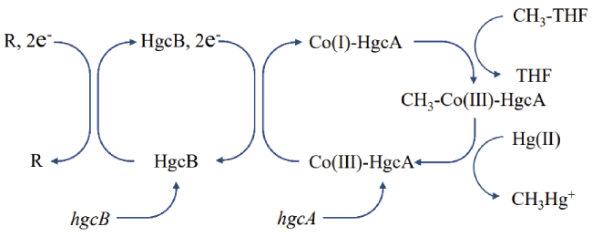
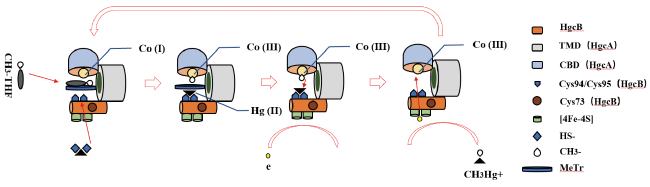
4 Biological mercury methylation processes involving HgcAB

5 Progress of hgcA/B-based environmental mercury methylation study

5.1 hgcA and hgcB can be used to identify new mercury methylation organisms and processes

5.2 Methylation of mercury in other media

5.3 the molecular biology techniques commonly used in The study of mercury methylation mediated by hgcA/B

5.4 The application of hgcA/B in ecological risk assessment

6 Limitations of current mercury methylation gene research

6.1 Lack of detailed molecular structures of HgcA and HgcB

6.2 Identification of HgcA and HgcB-interacting proteins

6.3 the relationship between The mercury methylation process mediated by hgcA/B and other metabolic pathways is not clear

7 Conclusion and perspectives

Cite this article

Bowei Chu , Yingying Guo , Ligang Hu , Yanwei Liu , Yongguang Yin , Yong Cai . Mechanism of hgcA/B Mediated Mercury Methylation and Application as Biomarkers[J]. Progress in Chemistry, 2023 , 35(10) : 1438 -1449 . DOI: 10.7536/PC230302

1 Introduction

Mercury (Hg) is recognized as a global persistent toxic pollutant, which exists in the environment in the form of inorganic mercury (Hg (Ⅱ), Hg (0)) and organic mercury (such as methylmercury (MeHg), ethylmercury (EtHg))[1]. Different speciation of mercury has different ecological and toxicological effects[2,3]. Methylmercury, as a strong neurotoxic substance, can be bioconcentrated and biomagnified along the food chain[4][5]. The outbreak of Minamata disease in Japan in 1956 was a large-scale public hazard caused by methylmercury poisoning[6]. In 2013, the Minamata Convention on Mercury was adopted under the auspices of the United Nations Environment Programme to limit the use and release of mercury. Although current global mercury exposure levels are generally much lower than the Minamata disease event, continuous mercury exposure at low doses has been shown to cause neurodevelopmental effects in organisms[7~10]. Therefore, the continuous production of micro-doses of methylmercury in natural water bodies and sediments can still pose a threat to human health through the food chain.
In 1968, Wood et al. Demonstrated that methylcobalamin in methanogen cell extracts could provide methyl groups to methylate mercury, suggesting that the process of mercury methylation might be related to microorganisms[11]. In 1969, Jensen et al. First confirmed that microorganisms in sediments were the main cause of mercury methylation by autoclaving control[12]. In 1985, Compeau et al. Found that the addition of molybdate (which interferes with the energy metabolism of sulfate-reducing bacteria) to low-salinity anoxic estuarine sediment samples could reduce the mercury methylation level by more than 95%, proving that sulfate-reducing bacteria are an important group of mercury methylating microorganisms[13]. In 2006, Fleming et al. Isolated an iron-reducing bacterium (Geobacter sp. CLFeRB) in iron-rich freshwater sediments, which confirmed that the bacterium could also methylate mercury[14].
Subsequent studies have identified that mercury methylation ability is closely related to the distribution of sulfate-reducing bacteria and iron-reducing bacteria in sediments through marker genes such as 16s rRNA and sulfite reductase gene (dsrAB)[15~17]. However, it should be pointed out that not all sulfate-reducing bacteria and iron-reducing bacteria can convert mercury into methylmercury, so the process of mercury methylation is not a unique ability evolved by a specific species of microorganisms to adapt to the environment[18~22]. Therefore, it is very important to analyze the formation pathway and molecular mechanism of methylmercury in microorganisms for exploring the process of biological mercury methylation.
In 2013, Parks et al. Identified the mercury methylation gene pair hgcA/B, which is closely related to the mercury methylation ability of microorganisms[23]. A lot of work has been done on hgcA/B in the follow-up studies, mainly focusing on the identification of mercury-methylating microorganisms and mercury-methylating genes in environmental media. In this paper, the hgcA/B mercury methylation gene pair, the predicted structure and molecular mechanism of its expression products hgcA and HgcB, and the application of mercury methylation genes in different environmental media were summarized, the existing problems of hgcA/B research were discussed, and the research direction of mercury methylation genes was put forward.

2 Discovery and Functional Verification of Mercury Methylation Gene hgcA/B

Two studies in 1994 showed that the methyl transfer process in biological mercury methylation requires the participation of a cobalamin protein with a size of 40 kDa[24]. Propyl iodide inhibited the production of methylmercury and acetyl-CoA to a similar extent, suggesting an association between the two[25]. The corrinoid iron-sulfur protein (CFeSP) in the reductive acetyl-CoA pathway is one of the few methyl transfer proteins that use metals as methyl transfer acceptors, which may be functionally and structurally similar to cobalamin proteins required for mercury methylation[26]. This CFeSP consists of three domains, an N-terminal domain bound to a [4Fe-4S] cluster, a TIM barrel domain in the middle, and a C-terminal domain bound to a cobalamin (corrnoid) cofactor. This protein can transfer the methyl group on methyltetrahydrofolate to the Ni-Ni- [4Fe-4S] cluster of acetyl-CoA synthetase.
On the basis of the above work, Parks et al. Performed homologous alignment of the CFeSP large subunit sequence (CfsA) in Carboxydothermus hydrogenoformans Z-2901 in the NCBI (National Center for Biotechnology Information) database, and found that the protein encoded by the DND132 _ 1056 sequence in sulfate-reducing bacteria was highly homologous to the C-terminal cobalamin binding domain of CfsA protein, but still lacked the TIM barrel structure and [4Fe-4S] binding domain of CfsA[23]. Continuous exploration of the gene sequence found that the DND132 _ 1057 sequence downstream of the DND132 _ 1056 sequence encodes an iron-sulfur protein, which can perform the same role as the N-terminal domain in CFeSP to transfer electrons. In addition, through comparative genomics, homologous sequences similar to DND132 _ 1056 and DND132 _ 1057 were found in a variety of methanogens, sulfate-reducing bacteria and iron-reducing bacteria with mercury methylation ability[19,23]. Knockout of DND132 _ 1056 and DND132 _ 1057 alone or together in Desulfovibrio desulfuricans ND132 reduced MeHg production to nearly zero, and subsequent anaplerotic experiments restored 97% of the methylation activity of the microorganism[23,27][23]. These two sequences and their homologues, which are highly related to mercury methylation, were named hgcA and hgcB, respectively.

3 HgcA, HgcB and their structure prediction

hgcA and hgcB are macromolecular proteins produced by transcription and translation of HgcA and HgcB gene pairs respectively. Heterologous overexpression of mercury methylation genes is affected by many factors, and purified HgcA and HgcB have not been obtained for protein structure analysis[28]. In the absence of experimental data, we can only rely on coevolutionary analysis, Rosetta modeling and other means to predict the protein structure of HgcA and HgcB[29~31].
HgcA is about 330 amino acids in length and consists of two domains, an N-terminal cobalamin-binding domain (corrinoid binding domain, CBD) and a C-terminal transmembrane domain (transm embrane domain, TMD). Similar to CFeSP, the N-terminal cobalamin-binding domain of HgcA protein is composed of five β sheets, four α helices and two short helices folded in Rossmann fold mode, which is responsible for binding cobalamin cofactor and is an important site for mercury methylation[31]. Cysteine (Cys93) located in the cap helix structure is essential for the mercury methylation process, and mutation of Cys93 to alanine (Ala93) or threonine (Thr93) will lead to the complete disappearance of microbial mercury methylation ability[32]. At the same time, when the tryptophan near Cys93 (Trp92) was mutated, the mercury methylation ability of microorganisms decreased by 70%, which was speculated to be related to the stabilization of the active site of HgcA or the localization of Hg (Ⅱ) on HgcA during mercury methylation[32][32]. The C terminus of HgcA is a hydrophobic transmembrane domain composed of transmembrane helices and shows no homology to any known protein (BLAST E < 10)[31]. This domain can anchor HgcA to the cell membrane structure, which is presumed to be related to the uptake of Hg (Ⅱ)[27]. In addition, when the C-terminal of HgcA was removed, the truncated HgcA protein with a size of 20 kDa and only containing the N-terminal cobalamin domain was not detected in bacteria, so the C-terminal may also be related to the stability of the N-terminal[32].
The N-terminal of HgcB is responsible for binding two [4Fe-4S] clusters, allowing electron transfer between the unknown electron donor and the cobalamin cofactor. In addition to the conserved cysteine in the [4Fe-4S] cluster, HgcB still contains 3 other conserved cysteines. Two of them are located at the C-terminal (Cys94, Cys95), and the other is Cys73[31]. The disulfide bond formed between Cys94 and Cys95 not only stabilizes the structure and promotes electron exchange, but also may be related to the binding of substrate Hg (Ⅱ)[33][31]. Experiments show that the coexistence of at least one amino acid in Cys94 and Cys95 and Cys73 is a necessary condition for microbial mercury methylation, and it is speculated that Cys73 is related to the departure of methylmercury from proteins[32]. In addition, when exposed to oxygen, [4Fe-4S] is converted into a stable [2Fe-2S] cluster, which makes HgcB inactive, which is one of the reasons why the complete molecular structure of HgcB is difficult to detect[34].
HgcA and HgcB may play the role of mercury methylation in organisms in the form of complexes. Fig. 1 is a predicted model of the structure of several HgcA, HgcB, and their complex HgcAB. The CBD core domain of HgcA was modeled mainly with reference to CFeSP, in which the cobalamin cofactor is bound to the CBD of HgcA by forming hydrogen bonds with 5 (Fig. 1 (A)) or 6 amino acids[31,35]. The C-terminal TMD has no known homologous structure, and five transmembrane helices are usually used to model the domain (Fig. 1 (a) (B)), but the prediction results in Alpha Fold structure prediction software also show that the TMD is composed of four transmembrane helices (Fig. 1 (C))[29,30]. The structure of HgcB is modeled according to the typical iron-sulfur protein, and the cofactor is bound to the protein by two CxxCxxCxCP sequences. The existing models all show the unique disordered end structure of the C terminus in HgcB. The mode of interaction between HgcA and HgcB refers to the complex structure predicted by Cooper et al. (Fig. 1 (a))[31]. In this structure, the distance between the [4Fe-4S] cluster in HgcB and the HgcA cofactor Co is 14.99 Å, which is smaller than the distance between Fe and Co in CFeSP (about 52 52 Å), suggesting a possible direct electron transfer between the two cofactors[36]. The CBD of HgcA is in polar contact with the [4Fe-4S] cluster binding domain of HgcB through residues, which is the main site of mercury methylation. The C-terminal α helix in HgcB interacts with helices 4 and 5 of the TMD in HgcA, forming the transmembrane structure of the HgcAB complex.
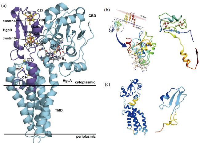
图1 HgcA、HgcB和HgcAB复合体的预测结构。(a)HgcAB复合体预测结构[31];(b)Marinimicrobia编码的HgcA和HgcB蛋白预测结构[37];(c)AlphaFold软件预测的HgcA和HgcB蛋白结构[29,30]

Fig. 1 Predicted structures of HgcA, HgcB, and HgcAB complex. (a) predicted structure of HgcAB complex [31]; (b) predicted structure of HgcA and HgcB protein encoded by Marinimicrobia [37]; (c) predicted structure of HgcA and HgcB protein by AlphaFold [29,30]. Adapted with permission from Ref. [31,37] under License CC BY 4.0

4 Biological mercury methylation process involving HgcAB

The biological mercury methylation process involving HgcAB is shown in Figure 2. The methyl group is mainly derived from methyltetrahydrofolate (CH3-THF), in which Co in the HgcA-bound cobalamin cofactor is an important site for the binding and transfer of the methyl group. Because the d z 2 orbital of Co (Ⅰ) has a pair of electrons and is perpendicular to the corrin ring, it has strong nucleophilicity, so the methyl radical can combine with Co (Ⅰ) to form Co (Ⅲ) complex[38]. The main difference between the mercury methylation process and other pathways involving methyl transfer in organisms is that the methyl radical combines with Co (Ⅲ) and then leaves in the form of methyl anion (C H 3 -). It has been shown that C H 3 - can be transferred from methylcobalamin to Hg (Ⅱ) without enzyme catalysis[39]. When the strictly conserved Cys93 in HgcA was replaced by His93, a cobalamin ligand commonly used by other CFeSPs, the mercury methylation ability was greatly reduced. The model calculation results also show that when Cys93 is used as the lower axial ligand of Co (Ⅲ) in the corrin ring, the Co-C will be cleaved heterogeneously under the electrophilic attack of Hg (Ⅱ), and the carbon atom will leave in the form of C H 3 - and combine with Hg (Ⅱ) to form a CH3Hg+[32,40,41][39]. The HgcB-bound [4Fe-4S] cluster is the simplest electron transfer group found in biological systems, which can accept electrons from an unknown substrate (R) and reduce Co (Ⅲ) in the corrin ring to a more active Co (Ⅰ), thereby promoting the completion of the cycle[42].
图2 HgcA、HgcB蛋白在汞甲基化过程中负责甲基和电子转移 [23,43]

Fig. 2 The function of HgcA and HgcB in the transfer of methyl group and electron in mercury methylation process[23,43]

5 Research progress of environmental mercury methylation based on hgcA/B

5.1 hgcA/B can be used to identify new mercury methylation organisms and processes

Early identification of mercury-methylating microorganisms relied on the addition of specific inhibitors. For example, molybdate is used to inhibit sulfate reducing bacteria and bromoethane sulfonate is used to inhibit methanogens. However, the above methods have a great dependence on the environmental samples and the dominant mercury methylating microorganisms in the samples. As mentioned above, the mercury methylation ability of sulfate-reducing bacteria (Cheese quake State Park (New Jersey, U. S.)) and iron-reducing bacteria (Clear Lake (California, U. S.)) was confirmed for specific environmental samples[13][14]. Due to the lack of environmental samples in which methanogens are dominant, the mercury methylation ability of methanogens in the environment has not been confirmed[44]. With the discovery of hgcA and hgcB and the confirmation of the dominant environmental sample (St. Lawrence River (Quebec, Canada)), Yu et al. Verified the mercury methylation ability of methanogenic strain (Methanospirillum hungatei JF -1) under experimental conditions, and clarified the important factors of methylmercury production in anoxic methanogenic environment[45][46]. Fish are the main source of methylmercury intake in humans, and researchers have long noted endogenous mercury methylation in fish, but do not know the specific sites of mercury methylation. In the early days, it was considered that the liver of fish was the main organ of mercury methylation. Recent studies have observed the mercury methylation process mediated by hgcA-containing microorganisms in the intestines of freshwater and marine fish with the help of mercury methylation gene probes, suggesting that intestinal microorganisms play an important role in mercury methylation in fish[47][48][49]. As a typical representative of the extreme environment of the earth, the source of methylmercury in the polar marine oxygenated aquifer has been lacking a strong explanation[50,51]. Gionfriddo et al. Identified Nitrospina microorganisms containing hgcA homologous genes in Antarctic sea ice, identified them as potential mercury methylating microorganisms in sea ice, and considered that methylmercury produced by Nitrospina in sea ice was the main source of methylmercury in polar oceans[35].
Environmental metagenome analysis using hgcA as a probe has greatly enriched the population of mercury-methylating microorganisms. So far, more than 1000 microorganisms with hgcA/B gene pairs have been found in 30 phyla[52]. Spirochaetes, Actinobacteria, PVC superphylum belonging to the bacterial domain, and Asgard superphylum belonging to the archaeal domain, which have not been reported before, have also identified potential mercury methylating microorganisms[53][54]. However, there is no obvious cluster of these mercury methylating microorganisms in race. In addition, because most microorganisms are difficult to be isolated and cultured under laboratory conditions, their mercury methylation ability can not be confirmed one by one. Therefore, only a small number of microorganisms with hgcA/B genes were identified to have the ability to methylate mercury, mainly in Euryarchaeota, Firmicutes, and Proteobacteria[21,46,55].
As a large number of microorganisms with hgcA/B gene pairs have been identified, researchers have also analyzed the distribution of mercury methylation genes hgcA/B in microbial genomes in an attempt to clarify the evolutionary law of hgc A/B. The distribution of hgcA/B in the genome has three situations: 1) coexistence and intergenic spacer: this hgcA/B clustering is not obvious in the phylogenetic tree, mainly concentrated in Proteobacteria and Firmicutes. 2) The presence of hgcA or hgcB alone: The number of microorganisms identified as containing only hgcA is about 30 times that of microorganisms containing only hgcB. In addition to the more stringent detection conditions, the presence of hgc B alone is rarely discussed in the study[52]. Some studies speculate that the existence of hgcA alone may imply an evolutionary process of hgcA/B disappearance. First, hgcB may disappear earlier than hgcA in this process. Second, the hgcA key sequence will be replaced before it disappears completely. For example, Desulfovibrio vietnamensis G3 100T, which is in the lower branch of the phylogenetic tree, not only lacks HgcB, but also replaces the key amino acid Cys93 of HgcA with lysine[56]. In addition, the existence of hgcA or hgcB alone may also be related to horizontal gene transfer, which is a phenomenon of exchange of genetic material between individuals of different organisms. 3) Fusion hgcA/B: This is a new fusion model hgcAB gene proposed by Podar et al. This class of genes does not have a common intergenic spacer[57]. The subsequent hgcA/B gene bank amplified the hgcAB microorganisms to 52 species, but only two microorganisms with hgcAB genes were tested for mercury methylation under laboratory conditions: Pyrococcus furiosus and Methanococcoides methylutens[52][57][55]. Studies have shown that these two microorganisms do not have the ability to methylate mercury. Although the size of the hgcA/B interval is not related to the efficiency of mercury methylation, it is not clear whether the disappearance of the intergenic region is the direct cause of the loss of microbial methylation ability due to the lack of other controls[56]. At the same time, the phylogenetic tree based on 16s rRNA showed that the four microorganisms with hgcAB gene were in the middle of the phylogenetic tree, which may imply that the fused hgcAB gene is the manifestation of the middle stage of the evolution process of hgcA/B gene[55,57]. However, due to the small number of confirmed strains, it is impossible to accurately speculate the phylogeny of microorganisms with hgcAB genes.

5.2 Methylation of Mercury in Different Environmental Media

Microorganisms with hgcA/B genes are mainly concentrated in anaerobic aquatic sediments, wetlands and saturated soil environments, which match the methylation ability of environmental mercury[58]. Environmental conditions are the key factors controlling the methylation of Hg (Ⅱ), for example, the complexation of Hg (Ⅱ) by chlorides, sulfides, thiols and natural organic matter can affect the uptake of mercury by microorganisms[58~60]. However, the effects of environmental conditions on biological mercury methylation are very complex, and the use of hgcA gene to explore the composition and structure of mercury methylating microbial communities under different environmental conditions can provide a new understanding of the effects of environmental factors on mercury methylation.

5.2.1 Mercury Methylation in Rice Field

Rice ingestion is an important way of human intake of methylmercury, especially in areas where it is the staple food[61,62]. MeHg accumulated in rice mainly comes from anoxic soil, and the bioconcentration factor is between 0. 2 and 1 100, especially in some mining areas with serious mercury pollution[63][64][65,66]. As a typical freshwater anoxic environment, methylmercury in paddy soil mainly comes from anaerobic microbial transformation[67]. Liu et al. Used hgcA/B as a probe to determine that iron-reducing bacteria and methanogens were the main mercury-methylating microorganisms in paddy soils in Wanshan and Fenghuangshan areas, while sulfate-reducing bacteria accounted for a small proportion (< 3%).MeHg content in paddy soil was positively correlated with the relative abundance of Geobacter with hgcA/B gene pairs, suggesting that the importance of iron-reducing bacteria in MeHg production in paddy soil may be underestimated[68]. Another study found that the dominant mercury methylating microorganisms in the hgcA-containing microbial communities in paddy soils from different mercury mining areas were different, and pH and organic matter were the most important factors[45]. Sulfur reducing bacteria (SRB), as the most widely studied mercury methylating microorganisms, have complex effects on mercury methylation. On the one hand, as the main electron acceptor of SRB,Sulfate (S O 4 2 -) can enhance the efficiency of mercury methylation in soil environment by affecting the abundance of microorganisms and thus increasing the abundance of hgcA. On the other hand, sulfide as a product can also affect the efficiency of mercury methylation by changing the bioavailability of mercury[69]. The change of water storage is the main characteristic of paddy field environment. Rothenberg et al. Found that paddy environments with different water storage methods had different oxidation periods, which would affect the community of mercury methylating microorganisms in paddy fields[70]. The interaction of microbial populations in paddy soil can affect the formation of methylmercury, for example, changing the ratio of nitrogen and sulfur in paddy soil can increase the proportion of N and S cycle microbial groups, thereby reducing the abundance of mercury methylation microorganisms dominated by C metabolism, and ultimately affecting the formation of methylmercury[71][72]. Plant rhizosphere exudates also affected the diversity of hgcA-containing microorganisms in soil, and the diversity of hgcA-containing organisms in rhizosphere and near-rhizosphere soils was higher than that in non-rhizospheric soils[73]. It is speculated that rice root exudates, especially acetate, can increase the activity of microorganisms, thereby increasing the copy number of hgcA[74]. It is worth noting that the current hgcA-based research on paddy field environment is mainly focused on the vicinity of mercury-contaminated mining areas, which makes it difficult to ignore the impact of other metal pollutants, such as zinc and cadmium, on biological mercury methylation[75]. In addition, hgcA is only used as an indicator to quantify microorganisms, which leads to the fact that the factors affecting the efficiency of mercury methylation are mainly attributed to the bioavailability of mercury and the activity of mercury-methylating microorganisms, lacking the explanation of the impact on the intracellular mercury methylation process.

5.2.2 Methylation of mercury in freshwater sediments

Freshwater sediments are the main formation sites of methylmercury in freshwater bodies. So far, most of the isolation of mercury methylating microorganisms has been carried out in freshwater sediments. Sulfate-reducing bacteria were once considered to be the main mercury-methylating microorganisms in sediments. However, studies have shown that mercury methylation rates and methylmercury production are the highest in sediments with low sulfate content, and hgcA/B diversity also varies with sulfide concentration in pore water[76]. This may be related to the interaction between microbial populations. Studies have shown that the interaction between sulfate-reducing bacteria and methanogens in low sulfate environment can increase the methylation efficiency of Hg (Ⅱ) by 2 to 9 times[77]. In high-sulfate estuarine sediments, the sequencing results of hgcA/B genes showed that methanogens had a higher contribution to mercury methylation when Hg (0) was the sole source of mercury, even higher than sulfate-reducing bacteria[78]. Similarly, iron-reducing bacteria have been shown to be the major carriers of mercury methylation in iron-rich river sediments[79]. The complex organic matter in freshwater sediments, especially the dissolved organic matter, plays an important role in the mercury cycle in sediments and even in freshwater systems[80]. Sulfur-containing ligands in dissolved organic matter can form complexes with Hg (Ⅱ), thus affecting the rate of mercury methylation[81,82]. Algae organic matter (algal organic matter, AOM) is the most recently studied organic matter, which can affect the rate of methylmercury production by increasing the abundance of methanogen hgcA[83]. In addition, hgcA can also provide clues for the prevention and control of mercury pollution in sediments. Although the dominance of sulfate-reducing bacteria in sediments is not significant, the proportion of sulfate-reducing bacteria in sediments can be quickly determined based on the ratio of dsrAB to hgcA, and then the methylmercury pollution can be conveniently treated[79,84]. The abundance of hgcA gene can also be used to reveal the risk of methylmercury formation in sediments when mercury pollution occurs in eutrophic waters, and to evaluate the efficiency of mercury pollution control[85].

5.2.3 Methylation of Mercury in the Ocean

Marine sediments are ideal living places for anaerobic mercury-methylating microorganisms, which are rich in hgcA/B genes[57]. However, the methylmercury enriched in pelagic fish does not come from marine sediments, but mainly from the in situ generation of methylmercury in the marine water column[86~88]. However, the hgcA genes of the major environmental mercury methylating microorganisms, such as iron-reducing bacteria, sulfate-reducing bacteria, and methanogens, were not found in the oxygenated pelagic water column[89]. There are several explanations for marine in situ mercury methylation: one is that the abiotic mercury methylation pathway mediates the formation of methylmercury[90~92]; One is mercury methylation involving unknown microorganisms; Finally, biological mercury methylation in an anaerobic microenvironment in an oxygenated water column. Recent studies have reported mercury methylating microorganisms in sinking particles in an oxygenated water column, suggesting the possibility of this view[93]. In addition to the microaerophilic nitrospira with hgcA/B gene pair mentioned above, which are possible mercury methylating microorganisms in the upper ocean, Lin et al. Found potential mercury methylating microorganisms Calditrichaeota sp., Deltaproteobacteria SAR324 and Marinimicrobia sp. In the marine hypoxic environment, which may be the main producers of higher concentrations of methylmercury in this region[94][37][95,96]. However, the mercury methylation ability of the above microorganisms has not been proved under experimental culture conditions, so further discussion is needed. In addition, the study based on hgcA/B also provides a strong proof for the enrichment of endogenous methylmercury in marine organisms. The hgcA gene belonging to Proteus and Firmicutes was detected in the gut of copepod zooplankton[97]. The hgcA/B gene pair of uncultured Deltaproteobacteria was also detected in the gut of a marine Olavius algarvensis[57]. The above studies have enriched the potential mercury methylation sites in the ocean, further indicating that hgcA/B is a powerful exploration tool for the study of mercury methylation in a wide range of environmental media such as the ocean.

5.2.4 Methylation of mercury in other media

Mountain glaciers are one of the important mercury sinks and sources of MeHg, and glacier meltwater is also a possible source of MeHg in freshwater systems. It has been proved that the mercury methylation process mediated by hgcA-containing microorganisms also exists in relatively extreme environments, and can cause changes in the concentration of methylmercury in the surrounding environment. A study on the genome of glaciers on the Qinghai-Tibet Plateau showed that the mercury methylation gene hgcA was present in all glacier samples except supraglacial ice samples, and the sediment-containing samples generally had a high abundance of hgcA, revealing a strong potential for mercury methylation at the relevant sites, suggesting the potential contribution of glacier meltwater to methylmercury in freshwater systems[98]. The anaerobic environment and carbon accumulation in alpine peatland can provide electron acceptors such as polysaccharides and amino acids for biological mercury methylation, so it has more potential for mercury methylation than other wetlands[99]. By measuring the ratio of hgcA abundance to qPCR (Real-time Quantitative PCR) results of marker genes dsrA, gltA and mcrA (hgcA/qPCR), it was found that although sulfate-reducing bacteria were the most abundant population based on 16s rRNA, iron-reducing bacteria accounted for the highest proportion of mercury-methylating microorganisms (hgc A/qPCR = 6. 52)[100]. In addition, the study found that redox potential and C/N ratio were strongly correlated with the distribution of mercury methylation communities.
Other studies have detected hgcA in typical anaerobic environments in production and living. The species of hgcA-methylating microorganisms in the landfill mainly changed with depth, and there was a significant positive correlation between total mercury and hgcA gene distribution[101,102]. In the process of sludge composting, the composition of hgcA-containing microbial community changed with the composting time, and Desulfonobacteria and Euryarchaeota were the main mercury-methylating microorganisms in the process of sludge composting[103]. The above studies have greatly enriched the basic knowledge of the mercury cycle at environmental sites.

5.3 Molecular biological techniques commonly used in the study of hgcA/B-mediated mercury methylation

5.3.1 PCR (Polymerase Chain Reaction) and qPCR

PCR is a conventional method for detecting low-abundance genes in biomolecules, and it is also the earliest technique used to detect microorganisms with hgcA. Depending on the design of primers, the biological information that can be obtained in environmental samples is also different. On the one hand, a wide range of primers can amplify most of the hgcA genes in environmental samples, and the results can reflect the mercury methylation potential of the relevant environmental sites and the diversity of hgcA. The primers designed and used by Schaefer et al. And Christensen et al. Are the commonly used hgcA broad range amplification primers at this stage (Table 1)[104][105]. However, there are still some differences in the sequences of hgcA genes in bacteria and archaea, and the efficiency of simultaneous amplification with the same primer is low. In 2020, on the basis of the original primer, Gionfriddo et al. Improved the amplification efficiency of the primer by improving the reverse primer (ORNL-HgcAB-uni-32R)[106]; On the other hand, the use of highly specific hgcA primers for a single species to amplify a sample allows the identification of a population of microorganisms that undergo mercury methylation. Using specially designed PCR primers, the researchers confirmed the presence of microorganisms containing the hgcA gene in Antarctic sea ice[35]. The results of qPCR for hgcA in the plankton gut showed that the hgcA gene was contained in the genome of all copepod plankton gut microorganisms in this environmental locus[97]. The above results not only expand the formation sites of methylmercury in the environment, but also further improve the biogeochemical cycling pathway of mercury in the environment.
表1 hgcA常用引物及适用范围

Table 1 Common primers and scope of application of hgcA

Primer Primer sequences 5'-3' Scope of application ref
hgcA_261F CGGCATCAAYGTCTGGTGYGC Broad-range hgcA/B primer 104
hgcA_912R GGTGTAGGGGGTGCAGCCSGTRWARKT 105
ORNL-HgcAB-uni-F AAYGTCTGGTGYGCNGCVGG 106
ORNL-HgcAB-uni-R CABGCNCCRCAYTCCATRCA
ORNL-HgcAB-uni-F AAYGTCTGGTGYGCNGCVGG
ORNL-HgcAB-uni-32R CAGGCNCCGCAYTCSATRCA
ORNL-Delta-HgcA-F GCCAACTACAAGMTGASCTWC Primers for Deltaproteobacteria hgcA 105
ORNL-Delta-HgcA-R CCSGCNGCRCACCAGACRTT Primers for methanogenic Archaea hgcA
ORNL-Archaea-HgcA-F AAYTAYWCNCTSAGYTTYGAYGC Primers for Firmicutes hgcA
ORNL-Archaea-HgcA-R TCDGTCCCRAABGTSCCYTT
ORNL-SRB-HgcA-F TGGDCCGGTDARAGCWAARGATA
ORNL-SRB-HgcA-R AAAAGAGHAYBCCAAAAATCA
Nitro_SP14_1F GGGGACTAATGTCTGGTGTG Primers for Nitrospina hgcA 35
Nitro_SP14_2F GGRACYAATGTCTGGTGTG
Nitro_SP14_1R AACAGGGTCTGTTATTGACGT
Amplification of hgcA using primers is highly accurate, but the results depend on the reported hgcA gene pool and cannot be used to explore unknown mercury-methylating microbial populations. Although the latest gene bank contains 1053 unique hgcA/B genes and the amplification efficiency of the primers used is improving, this method still can not cover all hgcA/B sequences in the environment[52].

5.3.2 Metagenomic technology

Related omics techniques are often used to explore the phylogeny of a larger range of mercury-methylating microorganisms or species. Metagenomic technology is the most widely used detection technology at present. Among them, sequencing sequence classification and metagenome assembly have been widely used to explore the abundance and diversity of hgcA in rice fields, sediments and seawater. The conserved sequence of hgcA (NVWCA (A/G/S) GK, N (V/I) WCA (A/G) (A/G) K) is now the most commonly used and stringent probe, which can recognize the conserved cap helix of HgcA and characterize its abundance[37,101]. Sequencing sequence classification is mainly based on the matching of metagenomic libraries constructed from raw data with existing microbial gene banks to explore the diversity of mercury-methylating microorganisms, relying more on the established hgcA gene bank. In contrast, metagenome assembly can obtain the complete gene sequence of microorganisms and explore the diversity of microorganisms through microbial marker genes, such as dsrA, gltA and mcrA. In 2022, Capo et al. Introduced a single-sample assembly channel (marky-coco) for finding and quantifying hgcA/B in environmental samples, which can achieve the same hgcA/B recovery efficiency as co-assembly in most cases[52]. Christensen et al. Used the Pfam database to translate the raw metagenome data into amino acid sequences, and classified hgcA according to the results of multiple sequence alignment and hidden Markov model, which can be used to classify undetected mercury-methylating microorganisms[107]. Metagenomics has undoubtedly expanded the scale of hgcA research. Podar et al. Summarized the global distribution of microorganisms with hgcA gene by using relevant metagenomic data, suggesting that methylmercury can enter the food chain through a variety of ways, and pointed out that methanogens may be the first microbial population with mercury methylation function through phylogenetic analysis[57]. In a recent study on mangrove sediments, researchers used metagenomics to discover new mercury-methylating microorganisms that may play an important role in the evolution of hgcA genes, and improved the phylogenetic law of mercury-methylating genes hgcA[54].
However, metagenomics can only analyze hgcA at the DNA level, and can not reflect the mercury methylation activity of microorganisms by analyzing the expression of hgcA or related enzyme activity. Moreover, the existing studies can not prove that there is a significant correlation between the abundance of hgcA gene and the production of methylmercury, so they can only characterize the mercury methylation potential of environmental sites, and can not reflect the interaction between environmental factors and biological mercury methylation process[107].

5.3.3 Metatranscriptome and Metaproteome Technology

Compared with metagenome, metatranscriptomics and metaproteomics can directly reflect the mercury methylation activity of microorganisms by detecting RNA or protein, which is helpful to clarify the metabolic mechanism of microbial mercury methylation. Early studies used metatranscriptomics and metaproteomics to detect the abundance of hgcA at environmental sites, but no significant factors affecting microbial mercury methylation were found, and the results showed that there was no correlation between the concentration of total mercury and methylmercury at environmental sites and the expression of hgcA[37,107,108]. Until recently, Capo et al. First demonstrated the strong correlation between the abundance of hgcA transcripts and dissolved Hg (II) sulfide and biological mercury methylation using metatranscriptome, reiterating the important role of the above omics technology in exploring the influencing factors of mercury methylation[109].

5.4 Application of hgcA/B in ecological risk assessment

Although the relationship between the abundance of hgcA/B and the amount of mercury methylation has not been confirmed, the detection of hgcA gene can still be used to indicate potential methylmercury production sites, such as PCR or omics detection of hgc A gene at risk sites such as sludge and municipal landfills, which reveals the possible risk of methylmercury exposure[110,111][101,102]. The formation of methylmercury in paddy field environment has always been the focus of attention, so researchers have carried out a lot of research on the methylation process of biological mercury in paddy soil.It was found that the formation of methylmercury in paddy field could be affected by the water storage mode, the input proportion of different elements and the interaction of microbial populations, suggesting that the optimization of paddy field cultivation conditions could reduce the formation of methylmercury to a certain extent[70][72][71]. It can be seen that hgcA/B genes have great potential in indicating the formation site of methylmercury, and the combination of more advanced environmental monitoring methods in the follow-up study can provide reliable data for ecological risk assessment, which is more conducive to the development of effective environmental management strategies.

6 Limitations of current studies on mercury methylation genes

6.1 Lack of detailed molecular structure of HgcA and HgcB

Under both experimental culture and natural conditions, hgcA/B was in an inactive state of low expression[112]. In addition, heterologous overexpression of HgcAB has some problems. Because the content of methylcobalamin and iron-sulfur clusters in commonly used competent E. coli is strictly regulated, no protein crystals can be used to solve the structure through experiments[113]. In this case, it is a very effective research direction to predict the structure of HgcA and HgcB by algorithm. However, this approach has limitations: although the CBD of HgcA is homologous to the C-terminal domain of the large subunit of cobalamin iron-sulfur protein (CFeSP), the TMD does not have any known homologous sequences; And in different homologous sequences, the tail structure of the C-terminal of HgcB is also different. All these bring difficulties to the accurate construction of the prediction model.
In addition, the function of the TMD in HgcA has not been confirmed because of the lack of a homologous structure. Cooper et al. Searched for membrane protein structures similar to the TMD in the protein library, and the results suggested that the TMD in HgcA might be an ion-conducting structure[31]. Whether the transmembrane structure of HgcAB complex can control the concentration balance of Hg (Ⅱ) or methylmercury in microbial cells still needs further discussion. However, it has been found that sulfate-reducing bacteria (G. sulfurreducens, D. desulfuricans) have a significant efflux of methylmercury, and the microorganisms always maintain the concentration of methylmercury in the cytoplasm at a low level (G. Sulfurreducens), suggesting that there may be a concentration control mechanism for methylmercury or Hg (Ⅱ)[114,115]. However, due to the lack of homologous structure and experimental data, it is not clear which molecular mechanism of the transmembrane domain is responsible for the transmembrane behavior of which molecule. In addition, due to the lack of detailed structure of HgcAB complex, it is difficult to confirm the key molecular steps of the methylation process, such as whether Cys94/Cys95 in HgcB is the initial binding site of Hg (II) and whether it is responsible for binding Hg from Hg(SR)2.

6.2 Identification of HgcA-HgcB-interacting protein

Methylation of biological mercury involves the transfer of methyl group to HgcA, the binding of HgcAB complex to Hg (Ⅱ), the transfer of methyl radical to Co (Ⅰ), and the transfer of C H 3 - to Hg (Ⅱ). In contrast to the methyl transfer process in the cobalamin iron-sulfur protein in Moorella thermoacetica, the HgcA protein itself does not contain a methyltetrahydrofolate-binding domain[116]. Therefore, we speculate that the biological mercury methylation process also requires an additional HgcA-interacting methyltransferase (MeTr) (Fig. 3), which is responsible for binding methyltetrahydrofolate and transferring the methyl radical derived from methyltetrahydrofolate to Co (I) in the cobalamin cofactor to form a methyl-Co (III) complex. The identification of this enzyme may be the key to understanding the mercury methylation process.
图3 hgcA/B介导的生物汞甲基化中的甲基转移和Hg(Ⅱ)结合过程

Fig. 3 Methyl transfer and Hg (Ⅱ) binding process in biogenic mercury methylation mediated by hgcA/B

6.3 The relationship between hgcA/B-mediated mercury methylation and other metabolic pathways is not clear.

There are many methyltransferases dependent on cobalamin cofactor in microorganisms, such as methionine synthetase, cobalamin iron-sulfur protein and so on. Most of the methyl acceptors of these methyltransferases are involved in well defined biological processes or pathways. However, as a new methyltransferase, the methylated product of HgcA lacks association with other biological processes or pathways, and does not have a clear biological function. Isotope tracing and proteomics methods show that the process of biological mercury methylation may be closely related to carbon metabolism. 14C isotope tracer showed that the methyl group of methylmercury synthesized by microorganisms originated from methyltetrahydrofolate[25]. At the same time, proteomics showed that the abundance of key proteins involved in carbon metabolism such as glycolysis and pyruvate fermentation, especially the one-carbon metabolism pathway represented by the reductive acetyl-Coa pathway, was significantly increased in the ΔhgcA/B knockout mutant[43,117]. However, this association is not necessary, because some sulfate-reducing strains (D. propionicus 1pr3, D. propionicus MUD, isolate BG-8, D. multivorans DSMZ 2059) that do not use the reductive acetyl-CoA pathway and use propionate as a carbon source can also produce methylmercury[118,119]. In addition, transcriptomics-based studies have found that the biological methylation of Hg (II) is related to the overall activity of microorganisms and their specific metabolic capabilities (such as sulfate reduction, fermentation, and hydrogen oxidation), suggesting that the mercury methylation process may have different pathways in different microorganisms[109]. Moreover, genes expressing three subunits of nitrogenase complex (nifH and nifD-nifK) were detected in most mercury-methylating microorganisms, and combined with potential mercury-methylating microorganisms in sea ice, it was speculated that biological mercury methylation might be related to microbial nitrogen fixation pathway[53][35]. There are also some biological pathways involved in methyl transfer, such as the dimethylmercaptopropionate (DMSP) degradation pathway, which are predicted to be associated with hgcA/B-mediated biological mercury methylation, but there is a lack of sufficient evidence[19]. In addition, proteomic studies on mercury methylation model microorganisms (G. sulreducens PCA, D. desulfuricans ND132) showed that hgcA/B gene knockout resulted in changes in RND (resistance-nodulation-cell division) family transporters related to metal efflux pumps, suggesting that hgcA/B-mediated biological mercury methylation process may be related to microbial metal homeostasis[43,117]. A recent study further confirmed that the transcription of hgcA/B is related to arsenic resistance through transcriptomics and qPCR, and proposed that it is regulated by AsrR transcription factor[120].
Omics technology is undoubtedly an important tool to explore the interaction of metabolic pathways in microorganisms, but the current data based on a single omics tool can not accurately locate the mercury methylation process in the metabolic network. Subsequent studies should consider targeted labeling of specific metabolites or key enzymes (such as HgcA) in the mercury methylation process, supplemented by multi-omics (proteomics, metallomics, metabolomics, genomics) analysis to find metabolites or enzymes interacting with them in other metabolic pathways.

7 Conclusion and prospect

Biological mercury methylation plays a vital role in the regional and global mercury cycle. Using the hgcA/B gene as a probe, not only new mercury-methylating microorganisms can be identified, but also mercury-methylating microorganisms at environmental sites can be identified, which will bring new understanding to the mercury methylation at this environmental site. In addition, the hgcA gene is also very promising in the quantitative evaluation of the mercury methylation ability of environmental samples, and the net production potential of environmental methylmercury can be directly obtained by co-detection with the mercury-reducing mer series genes.
However, there are still many limitations in the study of hgcA/B genes. The protein structure and molecular mechanism are not clear enough, and the biological significance is not clear, which limits the further application of hgcA/B genes in the environment. Future research should focus on the above contents, explore the environmental factors affecting biological mercury methylation in combination with relevant omics technologies, and strengthen the exploration of HgcA and HgcB association pathways.
[1]
Pacyna J M. Sci. Total Environ., 2020, 738: 139955.

[2]
Clarkson T W, Magos L. Crit. Rev. Toxicol., 2006, 36(8): 609.

[3]
Boening D W. Chemosphere, 2000, 40(12): 1335.

[4]
Wood J M. Science, 1974, 183: 1049.

[5]
Reinfelder J R, Fisher N S, Luoma S N, Nichols J W, Wang W X. Sci. Total Environ., 1998, 219(2/3): 117.

[6]
Harada M. Crit. Rev. Toxicol., 1995, 25: 1.

[7]
Cheng J P, Fujimura M, Bo D D. J. Environ. Sci., 2015, 38: 36.

[8]
Driscoll C T, Mason R P, Chan H M, Jacob D J, Pirrone N. Environ. Sci. Technol., 2013, 47(10): 4967.

[9]
Zahir F, Rizwi S J, Haq S K, Khan R H. Environ. Toxicol. Pharmacol., 2005, 20(2): 351.

[10]
Ceccatelli S, DarÉ E, Moors M. Chem. Biol. Interact., 2010, 188(2): 301.

[11]
Wood J M, Kennedy F S, Rosen C G. Nature, 1968, 220(5163): 173.

[12]
Jensen S, Jernelöv A. Nature, 1969, 223(5207): 753.

[13]
Compeau G C, Bartha R. Appl. Environ. Microbiol., 1985, 50(2): 498.

[14]
Fleming E J, Mack E E, Green P G, Nelson D C. Appl. Environ. Microbiol., 2006, 72(1): 457.

[15]
Shao D D, Kang Y, Wu S C, Wong M H. Sci. Total Environ., 2012, 424: 331.

[16]
Devereux R, Winfrey M R, Winfrey J, Stahl D A. FEMS Microbiol. Ecol., 1996, 20(1): 23.

[17]
Macalady J L, Mack E E, Nelson D C, Scow K M. Appl. Environ. Microbiol., 2000, 66(4): 1479.

[18]
Gilmour C C, Elias D A, Kucken A M, Brown S D, Palumbo A V, Schadt C W, Wall J D. Appl. Environ. Microbiol., 2011, 77(12): 3938.

[19]
Ma M, Du H X, Wang D Y. Crit. Rev. Environ. Sci. Technol., 2019, 49: 1893.

[20]
Ma M, Du H X, Wang D Y, Sun T. J. Soils Sediments, 2018, 18(3): 1100.

[21]
Gilmour C C, Podar M, Bullock A L, Graham A M, Brown S D, Somenahally A C, Johs A, Hurt R A, Bailey K L, Elias D A. Environ. Sci. Technol., 2013, 47(20): 11810.

[22]
Kerin E J, Gilmour C C, Roden E, Suzuki M T, Coates J D, Mason R P. Appl. Environ. Microbiol., 2006, 72(12): 7919.

[23]
Parks J M, Johs A, Podar M, Bridou R, Hurt R A, Smith S D, Tomanicek S J, Qian Y, Brown S D, Brandt C C, Palumbo A V, Smith J C, Wall J D, Elias D A, Liang L Y. Science, 2013, 339(6125): 1332.

[24]
Choi S C, Chase T, Bartha R. Appl. Environ. Microbiol., 1994, 60(4): 1342.

[25]
Choi S C, Chase T, Bartha R. Appl. Environ. Microbiol., 1994, 60(11): 4072.

[26]
Doukov T I, Iverson T M, Seravalli J, Ragsdale S W, Drennan C L. Science, 2002, 298: 567.

[27]
Lin H, Hurt R A, Johs A, Parks J M, Morrell-Falvey J L, Liang L Y, Elias D A, Gu B H. Environ. Sci. Technol. Lett., 2014, 1(5): 271.

[28]
Date S S, Parks J M, Rush K W, Wall J D, Ragsdale S W, Johs A. Appl. Environ. Microbiol., 2019, 85(13): e00438-19.

[29]
Jumper J, Evans R, Pritzel A, Green T, Figurnov M, Ronneberger O, Tunyasuvunakool K, Bates R, Zidek A, Potapenko A, Bridgland A, Meyer C, Kohl S A A, Ballard A J, Cowie A, Romera-Paredes B, Nikolov S, Jain R, Adler J, Back T, Petersen S, Reiman D, Clancy E, Zielinski M, Steinegger M, Pacholska M, Berghammer T, Bodenstein S, Silver D, Vinyals O, Senior A W, Kavukcuoglu K, Kohli P, Hassabis D. Nature, 2021, 596: 583.

[30]
Varadi M, Anyango S, Deshpande M, Nair S, Natassia C, Yordanova G, Yuan D, Stroe O, Wood G, Laydon A, Zidek A, Green T, Tunyasuvunakool K, Petersen S, Jumper J, Clancy E, Green R, Vora A, Lutfi M, Figurnov M, Cowie A, Hobbs N, Kohli P, Kleywegt G, Birney E, Hassabis D, Velankar S. Nucleic Acids Res., 2022, 50: D439.

[31]
Cooper C J, Zheng K Y, Rush K W, Johs A, Sanders B C, Pavlopoulos G A, Kyrpides N C, Podar M, Ovchinnikov S, Ragsdale S W, Parks J M. Commun. Biol., 2020, 3: 320.

[32]
Smith S D, Bridou R, Johs A, Parks J M, Elias D A, Hurt R A, Brown S D, Podar M, Wall J D. Appl. Environ. Microbiol., 2015, 81: 3205.

[33]
Carugo O, Cemazar M, Zahariev S, Hudaky I, Gaspari Z, Perczel A, Pongor S. Protein Eng., 2003, 16: 637.

[34]
Beinert H, Holm R H, Munck E. Science, 1997, 277: 653.

[35]
Gionfriddo C M, Tate M T, Wick R R, Schultz M B, Zemla A, Thelen M P, Schofield R, Krabbenhoft D P, Holt K E, Moreau J W. Nat. Microbiol., 2016, 1: 12.

[36]
Svetlitchnaia T, Svetlitchnyi V, Meyer O, Dobbek H. Proc. Natl. Acad. Sci. U. S. A., 2006, 103: 14331.

[37]
Lin H Y, Ascher D B, Myung Y, Lamborg C H, Hallam S J, Gionfriddo C M, Holt K E, Moreau J W. ISME J., 2021, 15: 1810.

[38]
Ludwig M L, Matthews R G. Annu. Rev. Biochem., 1997, 66: 269.

[39]
Desimone R E, Penley M W, Charbonneau L, Smith S G, Wood J M, Hill H A O, Pratt J M, Ridsdale S, Williams R J P. Biochim. Biophys. Acta, 1973, 304: 851.

[40]
Zhou J, Riccardi D, Beste A, Smith J C, Parks J M. Inorg. Chem., 2014, 53(2): 772.

[41]
Demissie T B, Garabato B D, Ruud K, Kozlowski P M. Angew. Chem. Int. Edit., 2016, 55: 11503.

[42]
Nzuza N, Padayachee T, Chen W P, Gront D, Nelson D R, Syed K. Curr. Issues Mol. Biol., 2021, 43: 1374.

[43]
Qian C, Johs A, Chen H M, Mann B F, Lu X, Abraham P E, Hettich R L, Gu B H. J. Proteome Res., 2016, 15(10): 3540.

[44]
Pak K R, Bartha R. Appl. Environ. Microbiol., 1998, 64(3): 1013.

[45]
Hamelin S, Amyot M, Barkay T, Wang Y P, Planas D. Environ. Sci. Technol., 2011, 45(18): 7693.

[46]
Yu R Q, Reinfelder J R, Hines M E, Barkay T. Appl. Environ. Microbiol., 2013, 79(20): 6325.

[47]
Fu Q, Fan X C, Sun J H, Tan H W, Wang Y, Ouyang J, Na N. Small, 2020, 16(33): 2000072.

[48]
Tan S, Xu X W, Cheng H, Wang J J, Wang X. Environ. Res., 2022, 204: 10.

[49]
Yang T T, Liu Y, Tan S, Wang W X, Wang X. Environ. Pollut., 2021, 277: 10.

[50]
Schartup A T, Balcom P H, Soerensen A L, Gosnell K J, Calder R S D, Mason R P, Sunderland E M. Proc. Natl. Acad. Sci. U. S. A., 2015, 112(38): 11789.

[51]
Lehnherr I, St Louis V L, Hintelmann H, Kirk J L. Nat. Geosci., 2011, 4(5): 298.

[52]
Capo E, Peterson B D, Kim M, Jones D S, Acinas S G, Amyot M, Bertilsson S, Björn E, Buck M, Cosio C, Elias D A, Gilmour C, Goñi-Urriza M, Gu B H, Lin H Y, Liu Y R, McMahon K, Moreau J W, Pinhassi J, Podar M, Puente-Sánchez F, Sánchez P, Storck V, Tada Y Y, Vigneron A, Walsh D A, Vandewalle-Capo M, Bravo A G, Gionfriddo C M. Mol. Ecol. Resour., 2023, 23(1): 190.

[53]
McDaniel E A, Peterson B D, Stevens S L R, Tran P Q, Anantharaman K, McMahon K D. mSystems, 2020, 5(4): 00299-20.

[54]
Zhang C J, Liu Y R, Cha G H, Liu Y, Zhou X Q, Lu Z Y, Pan J, Cai M W, Li M. ISME J., 2023, 17(3): 478.

[55]
Gilmour C C, Bullock A L, McBurney A, Podar M, Elias D A. mBio, 2018, 9(2): 02403-17.

[56]
Goñi-Urriza M, Klopp C, Ranchou-Peyruse M, Ranchou-Peyruse A, Monperrus M, Khalfaoui-Hassani B, Guyoneaud R. Res. Microbiol., 2020, 171(1): 3.

[57]
Podar M, Gilmour C C, Brandt C C, Soren A, Brown S D, Crable B R, Palumbo A V, Somenahally A C, Elias D A. Sci. Adv., 2015, 1(9): e1500675.

[58]
Cai Y. Biogeochemistry of Environmentally Important Trace Elements, 2003, 835: 1.

[59]
Barkay T, Gillman M, Turner R R. Appl. Environ. Microbiol., 1997, 63(11): 4267.

[60]
Golding G R, Kelly C A, Sparling R, Loewen P C, Rudd J W M, Barkay T. Limnol. Oceanogr., 2002, 47(4): 967.

[61]
Zhao L, Meng B, Feng X B. Ecotox. Environ. Safe, 2020, 195: 9.

[62]
Rothenberg S E, Feng X B, Li P. Environ. Pollut., 2011, 159(4): 1017.

[63]
Meng B, Feng X B, Qiu G L, Cai Y, Wang D Y, Li P, Shang L H, Sommar J. J. Agric. Food Chem., 2010, 58(8): 4951.

[64]
Tang W L, Liu Y R, Guan W Y, Zhong H, Qu X M, Zhang T. Sci. Total Environ., 2020, 714: 136827.

[65]
Qiu G L, Feng X B, Li P, Wang S F, Li G H, Shang L H, Fu X W. J. Agric. Food Chem., 2008, 56(7): 2465.

[66]
Meng M, Li B, Shao J J, Wang T, He B, Shi J B, Ye Z H, Jiang G B. Environ. Pollut., 2014, 184: 179.

[67]
Liu Y R, Yu R Q, Zheng Y M, He J Z. Appl. Environ. Microbiol., 2014, 80(9): 2874.

[68]
Liu Y R, Johs A, Bi L, Lu X, Hu H W, Sun D, He J Z, Gu B H. Environ. Sci. Technol., 2018, 52(22): 13110.

[69]
Lei P, Tang C, Wang Y J, Wu M J, Kwong R W M, Jiang T, Zhong H. Sci. Total Environ., 2021, 778: 146325.

[70]
Rothenberg S E, Anders M, Ajami N J, Petrosino J F, Balogh E. Sci. Total Environ., 2016, 572: 608.

[71]
Zhou X Q, Qu X M, Yang Z M, Zhao J T, Hao Y Y, Feng J, Huang Q Y, Liu Y R. J. Hazard. Mater., 2022, 439: 129578.

[72]
Li Y Y, Dai S S, Zhao J T, Hu Z C, Liu Q, Feng J, Huang Q Y, Gao Y X, Liu Y R. J. Hazard. Mater., 2023, 448: 130983.

[73]
Wang J, Xiang Y P, Tian X S, Zhang C, Gong G Q, Xue J P, Jiang T, Wang D Y, Wang Y M. J. Environ. Sci., 2022, 119: 139.

[74]
Zhao J Y, Ye Z H, Zhong H. Environ. Pollut., 2018, 242: 1921.

[75]
Li Z Y, Ma Z W, van der Kuijp T J, Yuan Z W, Huang L. Sci. Total Environ., 2014, 468/469: 843.

[76]
Jones D S, Johnson N W, Mitchell C P J, Walker G M, Bailey J V, Pastor J, Swain E B. Environ. Sci. Technol., 2020, 54(22): 14265.

[77]
Yu R Q, Reinfelder J R, Hines M E, Barkay T. ISME J., 2018, 12(7): 1826.

[78]
Wang Y W, Roth S, Schaefer J K, Reinfelder J R, Yee N. FEMS Microbiol. Lett., 2020, 367(23): fnaa196.

[79]
Bravo A G, Peura S, Buck M, Ahmed O, Mateos-Rivera A, Herrero Ortega S, Schaefer J K, Bouchet S, Tolu J, Björn E, Bertilsson S. Appl. Environ. Microbiol., 2018, 84(23): e01774-18.

[80]
Branfireun B A, Cosio C, Poulain A J, Riise G, Bravo A G. Sci. Total Environ., 2020, 745: 140906.

[81]
Liem-Nguyen V, Skyllberg U, Björn E. Environ. Sci. Technol., 2017, 51(7): 3678.

[82]
Chiasson-Gould S A, Blais J M, Poulain A J. Environ. Sci. Technol., 2014, 48(6): 3153.

[83]
Lei P, Zhang J, Zhu J J, Tan Q G, Kwong R W M, Pan K, Jiang T, Naderi M, Zhong H. Environ. Sci. Technol., 2021, 55(15): 10811.

[84]
Bravo A G, Zopfi J, Buck M, Xu J Y, Bertilsson S, Schaefer J K, PotÉ J, Cosio C. ISME J., 2018, 12(3): 802.

[85]
Ji X N, Liu C B, Pan G. Ecotox. Environ. Safe, 2020, 188: 9.

[86]
Sunderland E M, Krabbenhoft D P, Moreau J W, Strode S A, Landing W M. Glob. Biogeochem. Cycles, 2009, 23(2): GB2010.

[87]
Monperrus M, Tessier E, Amouroux D, Leynaert A, Huonnic P, Donard O F X. Mar. Chem., 2007, 107(1): 49.

[88]
Kirk J L, St Louis V L, Hintelmann H, Lehnherr I, Else B, Poissant L. Environ. Sci. Technol., 2008, 42(22): 8367.

[89]
Bowman K L, Collins R E, Agather A M, Lamborg C H, Hammerschmidt C R, Kaul D, Dupont C L, Christensen G A, Elias D A. Limnol. Oceanogr., 2020, 65: S310.

[90]
Wang K, Liu G L, Cai Y. Crit. Rev. Environ. Sci. Technol., 2022, 52(22): 3997.

[91]
Celo V, Lean D R S, Scott S L. Sci. Total Environ., 2006, 368(1): 126.

[92]
Hu H Y, Lin H, Zheng W, Tomanicek S J, Johs A, Feng X B, Elias D A, Liang L Y, Gu B H. Nat. Geosci., 2013, 6(9): 751.

[93]
Capo E, Cosio C, GascÓn Díez E, Loizeau J L, Mendes E, Adatte T, Franzenburg S, Bravo A G. Water Res., 2023, 229: 119368.

[94]
Tada Y Y, Marumoto K, Takeuchi A. Front. Microbiol., 2020, 11: 1369.

[95]
Lamborg C H, Hammerschmidt C R, Bowman K L, Swarr G J, Munson K M, Ohnemus D C, Lam P J, Heimbürger L E, Rijkenberg M J A, Saito M A. Nature, 2014, 512(7512): 65.

[96]
Cossa D, Knoery J, Bǎnaru D, Harmelin-Vivien M, Sonke J E, Hedgecock I M, Bravo A G, Rosati G, Canu D, Horvat M, Sprovieri F, Pirrone N, Heimbürger-Boavida L E. Environ. Sci. Technol., 2022, 56(7): 3840.

[97]
Gorokhova E, Soerensen A L, Motwani N H. PLoS One, 2020, 15(3): e0230310.

[98]
Zhang B L, Chen T, Guo J M, Wu M H, Yang R Q, Chen X M, Wu X K, Zhang W, Kang S C, Liu G X, Dyson P. Sci. Total Environ., 2020, 708: 135226.

[99]
Haynes K M, Kane E S, Potvin L, Lilleskov E A, Kolka R K, Mitchell C P J. Glob. Biogeochem. Cycles, 2017, 31(2): 233.

[100]
Liu C T, Liu J L, Zhou C Y, Huang X Y, Wang H M. Geoderma, 2021, 403: 115375.

[101]
An Y W, Zhang R, Yang S, Wang Y Q, Lei Y, Peng S H, Song L Y. Waste Manag., 2022, 145: 102.

[102]
Yang J, Takaoka M, Sano A, Matsuyama A, Yanase R. Int. J. Environ. Res. Public Health, 2018, 15(6): 1252.

[103]
Liu J B, Bao Z, Wang C L, Wei J Y, Wei Y S, Chen M X. Water Res., 2022, 226: 119204.

[104]
Schaefer J K, Kronberg R M, Morel F M M, Skyllberg U. Environ. Microbiol. Rep., 2014, 6(5): 441.

[105]
Christensen G A, Wymore A M, King A J, Podar M, Hurt R A, Santillan E U, Soren A, Brandt C C, Brown S D, Palumbo A V, Wall J D, Gilmour C C, Elias D A. Appl. Environ. Microbiol., 2016, 82(19): 6068.

[106]
Gionfriddo C M, Wymore A M, Jones D S, Wilpiszeski R L, Lynes M M, Christensen G A, Soren A, Gilmour C C, Podar M, Elias D A. Front. Microbiol., 2020, 11: 541554.

[107]
Christensen G A, Gionfriddo C M, King A J, Moberly J G, Miller C L, Somenahally A C, Callister S J, Brewer H, Podar M, Brown S D, Palumbo A V, Brandt C C, Wymore A M, Brooks S C, Hwang C, Fields M W, Wall J D, Gilmour C C, Elias D A. Environ. Sci. Technol., 2019, 53(15): 8649.

[108]
Vigneron A, Cruaud P, AubÉ J, Guyoneaud R, Goñi-Urriza M. Npj Biofilms Microbiomes, 2021, 7: 83.

[109]
Capo E, Feng C Y, Bravo A G, Bertilsson S, Soerensen A L, Pinhassi J, Buck M, Karlsson C, Hawkes J, Bjorn E. Environ. Sci. Technol., 2022, 56: 13119.

[110]
Liu J B, He X L, Zhong H, Lei P, Zhang J Y, Xu Y F, Wei Y S. Bioresour. Technol., 2022, 347: 126394.

[111]
Liu J B, He X L, Xu Y F, Zuo Z, Lei P, Zhang J Y, Yin Y G, Wei Y S. J. Hazard. Mater., 2021, 406: 124310.

[112]
Goñi-Urriza M, Corsellis Y, Lanceleur L, Tessier E, Gury J, Monperrus M, Guyoneaud R. Environ. Sci. Pollut. Res., 2015, 22(18): 13764.

[113]
Nou X, Kadner R J. Proc. Natl. Acad. Sci. U. S. A., 2000, 97(13): 7190.

[114]
Schaefer J K, Morel F M M. Nat. Geosci., 2009, 2(2): 123.

[115]
Schaefer J K, Rocks S S, Zheng W, Liang L Y, Gu B H, Morel F M M. Proc. Natl. Acad. Sci. U. S. A., 2011, 108(21): 8714.

[116]
Kung Y, Ando N, Doukov T I, Blasiak L C, Bender G, Seravalli J, Ragsdale S W, Drennan C L. Nature, 2012, 484(7393): 265.

[117]
Qian C, Chen H M, Johs A, Lu X, An J, Pierce E M, Parks J M, Elias D A, Hettich R L, Gu B H. Proteomics, 2018, 18(17): 1700479.

[118]
Ekstrom E B, Morel F M M. Environ. Sci. Technol., 2008, 42(1): 93.

[119]
Ekstrom E B, Morel F M M, Benoit J M. Appl. Environ. Microbiol., 2003, 69(9): 5414.

[120]
Gionfriddo C M, Soren A B, Wymore A M, Hartnett D S, Podar M, Parks J M, Elias D A, Gilmour C C. Appl. Environ. Microbiol., 2023, 89(4): e01768-22.DOI:10.1128/aem.01768-22.

Outlines

/