
New Dressings Based on Chitosan and Its Application
Received date: 2023-03-09
Revised date: 2023-05-20
Online published: 2023-07-18
Supported by
Basic Scientific Research Funds for Universities Affiliated to Hebei Province(KY2022056)
Innovation and Entrepreneurship Training Program for Undergraduate Students of Hebei Agricultural University(2023114)
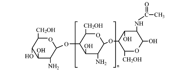
Chitosan has great potential in the fields of materials science and biomedicine because of its advantages such as coagulation, antibacterial, biocompatibility and biodegradation. This paper introduces the coagulation and bacteriostatic mechanism of chitosan, and lists the research progress of new dressings based on chitosan. According to the different morphology, the new dressings can be divided into the following types: fabric dressings based on chitosan, hydrogel dressings based on chitosan, spongy dressings based on chitosan, hydrocolloid dressings based on chitosan, asymmetric wettable dressings based on chitosan and frozen gel dressings based on chitosan. The experimental results of the new dressings based on chitosan in terms of antibacterial properties, in vitro coagulation properties, waterproof properties, breathable properties and mechanical properties were summarized. The application of new dressings based on chitosan in the treatment of diabetic foot ulcer, burn wound, inferior vena cava injury and endoscopic sinus surgery was summarized in detail. Finally, based on some problems existing in the new dressings based on chitosan (for example, the preparation process is greatly affected by the external environment conditions, some working mechanism of chitosan is still in the preliminary stage), the future development of the new dressings and their application are prospected.
1 Introduction
2 Working mechanism of new dressings based on chitosan
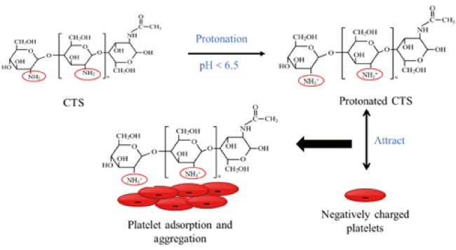
2.1 Hemostasis effect of chitosan
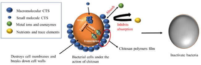
2.2 Bacteriostatic effect of chitosan
3 Research progress of new dressings based on chitosan
3.1 Fabric dressing based on chitosan
3.2 Hydrogel dressing based on chitosan
3.3 Spongy dressing based on chitosan
3.4 Hydrocolloidal dressing based on chitosan
3.5 Asymmetric wettability dressing based on chitosan
3.6 Frozen gel dressing based on chitosan
4 Application of new dressing based on chitosan
4.1 Diabetic foot ulcer
4.2 Burn wound
4.3 Inferior vena cava injury
4.4 Endoscopic sinus surgery
5 Conclusion and outlook

Key words: chitosan; new dressings; application
Quan Zhang , Siyu Duan , Zhongyuan Huo , Xinwang Meng , Jun Wang , Guohe Xu . New Dressings Based on Chitosan and Its Application[J]. Progress in Chemistry, 2023 , 35(10) : 1450 -1460 . DOI: 10.7536/PC230303
表1 基于壳聚糖的不对称膜的制备和优缺点Table 1 Preparation, advantages and disadvantages of asymmetric membranes based on chitosan |
| Preparation method | Preparation | Advantages | Disadvantages |
|---|---|---|---|
| Wet-phase inversion method | The casting polymer is soaked in a non-solvent coagulant bath to promote the polymer to precipitate, thus forming a film. | Easy to operate | The top layer of the asymmetric membrane thickness reduction(<1 μm) |
| Dry/wet-phase inversion method | Membrane production is initiated by a pre-evaporation process before the cast polymer is immersed in the coagulation bath. | Producing a denser top layer | The evaporation process uses a volatile solvent; Time-consuming |
| scCO2-phase inversion method | Under the supercritical condition of CO2, the precipitation of polymer solution is promoted to produce an asymmetric membrane. | Simple; fast; eco-friendly | Professional high-pressure equipment is required. |
| Electrospinning method | A polymer solution is loaded into a syringe and subjected to a high-voltage electric field, which promotes the polymer to be sprayed toward the collector, resulting in nanofibers. | The operation is simple; the asymmetric membrane based on chitosan can be optimized by adjusting the parameters; a variety of polymers can be used. | A professional DC power supply and injection pump are required, and the control requirements for processing variables and environmental conditions are demanding. |
| Bioprinting method | Asymmetric membranes can be produced by printing different layers containing the respective skin cells (keratinocytes om top, fibroblasts on bottom). | Asymmetric membranes based on chitosan can be customized to the specific needs of the patient. | A professional 3D printer is required; a sterile environment is required; The number of polymers and solvents available is limited. |
表2 基于壳聚糖的新型敷料的对比Table 2 Comparison of novel dressing based on chitosan |
| Type of dressing | Preparation technology | Advantages | ref |
|---|---|---|---|
| Fabric dressing | The fabric is thoroughly soaked in a chitosan solution. | It is easy to prepare, soft, tailoring and biocompatibility. | 39⇓⇓~42 |
| Hydrogel dressing | Cross-linking of positively charged chitosan with negatively charged ions, and polymers can be used to synthesize chitosan-based hydrogel dressings. | The treatment effect is good, with super stretching, fast self-healing and good antibacterial activity. | 45⇓~47 |
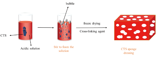
| Spongy dressing | The chitosan solution is stirred in an acidic environment to foam, then cross-linked and transformed into a chitosan-based spongy dressing by freeze-drying technology. | With excellent permeability and fluid absorption capacity, the material is soft and can be stored for a longer time, and is easier to carry. | 53,54 |
| Hydrocolloidal dressing | The polymer elastomer and chitosan are heated together. Plasticizer, viscosifying resin, antioxidant and crosslinking agent are added to make it fully crosslinked. | It has good water vapor permeability, effective antibacterial activity and a good enzymatic degradation effect. | 57,58 |
| Asymmetric wettability dressing | Wet-phase inversion method; dry/wet-phase inversion method; scCO2-phrase inversion method; Electrospinning method; Bioprinting method | It has lower clotting index, high hydrophobic and hydrophilic activity and antibacterial activity, and can effectively protect against water, blood and bacterial contamination. | 63 |
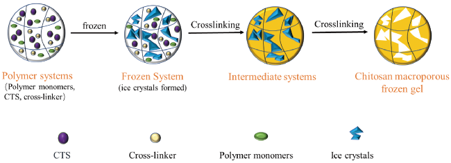
| Frozen gel dressing | Using water as the solvent, in a sub-zero environment, most of the water turns into ice crystals and a small part remains in the liquid phase. Chitosan and other polymers in the liquid phase are concentrated and cross-linked to form polymer networks. When the ice crystals melt, frozen gels with highly intercommunicating microporous structures can be obtained. | It has high porosity, rapid water absorption, high blood-sucking ability, excellent mechanical strength and fatigue resistance, whole blood coagulation ability and red blood cell and platelet adhesion ability | 69,72,73 |
| [1] |
|
| [2] |
|
| [3] |
|
| [4] |
|
| [5] |
|
| [6] |
|
| [7] |
|
| [8] |
|
| [9] |
|
| [10] |
|
| [11] |
|
| [12] |
|
| [13] |
|
| [14] |
|
| [15] |
|
| [16] |
|
| [17] |
|
| [18] |
|
| [19] |
|
| [20] |
|
| [21] |
|
| [22] |
|
| [23] |
|
| [24] |
|
| [25] |
|
| [26] |
|
| [27] |
|
| [28] |
|
| [29] |
|
| [30] |
|
| [31] |
|
| [32] |
|
| [33] |
|
| [34] |
|
| [35] |
|
| [36] |
|
| [37] |
|
| [38] |
|
| [39] |
|
| [40] |
|
| [41] |
|
| [42] |
|
| [43] |
|
| [44] |
|
| [45] |
|
| [46] |
|
| [47] |
|
| [48] |
|
| [49] |
|
| [50] |
|
| [51] |
|
| [52] |
|
| [53] |
|
| [54] |
|
| [55] |
|
| [56] |
|
| [57] |
|
| [58] |
|
| [59] |
|
| [60] |
|
| [61] |
|
| [62] |
|
| [63] |
|
| [64] |
|
| [65] |
|
| [66] |
|
| [67] |
|
| [68] |
|
| [69] |
|
| [70] |
|
| [71] |
|
| [72] |
|
| [73] |
|
| [74] |
|
| [75] |
|
| [76] |
|
| [77] |
|
| [78] |
|
| [79] |
|
| [80] |
|
| [81] |
|
| [82] |
|
| [83] |
|
| [84] |
|
| [85] |
|
| [86] |
|
| [87] |
|
| [88] |
|
| [89] |
|
| [90] |
|
| [91] |
|
| [92] |
|
| [93] |
|
| [94] |
|
| [95] |
|
| [96] |
|
| [97] |
|
| [98] |
|
/
| 〈 |
|
〉 |