Home Journals Progress in Chemistry
Progress in Chemistry

Abbreviation (ISO4): Prog Chem      Editor in chief: Jincai ZHAO

About  /  Aim & scope  /  Editorial board  /  Indexed  /  Contact  / 
Review

Applications of Metallomics and Metalloproteomics Techniques in Biomedical Research

  • Yuchuan Wang , *
Expand
  • Heibei Key Laboratory for Chronic Diseases, School of Basic Medical Sciences, North China University of Science and Technology,Tangshan 063210, China
*Corresponding author e-mail:

Received date: 2023-03-07

  Revised date: 2023-04-09

  Online published: 2023-06-12

Supported by

Fundamental Research Funds for the Central Universities(JQN2022026)

Abstract

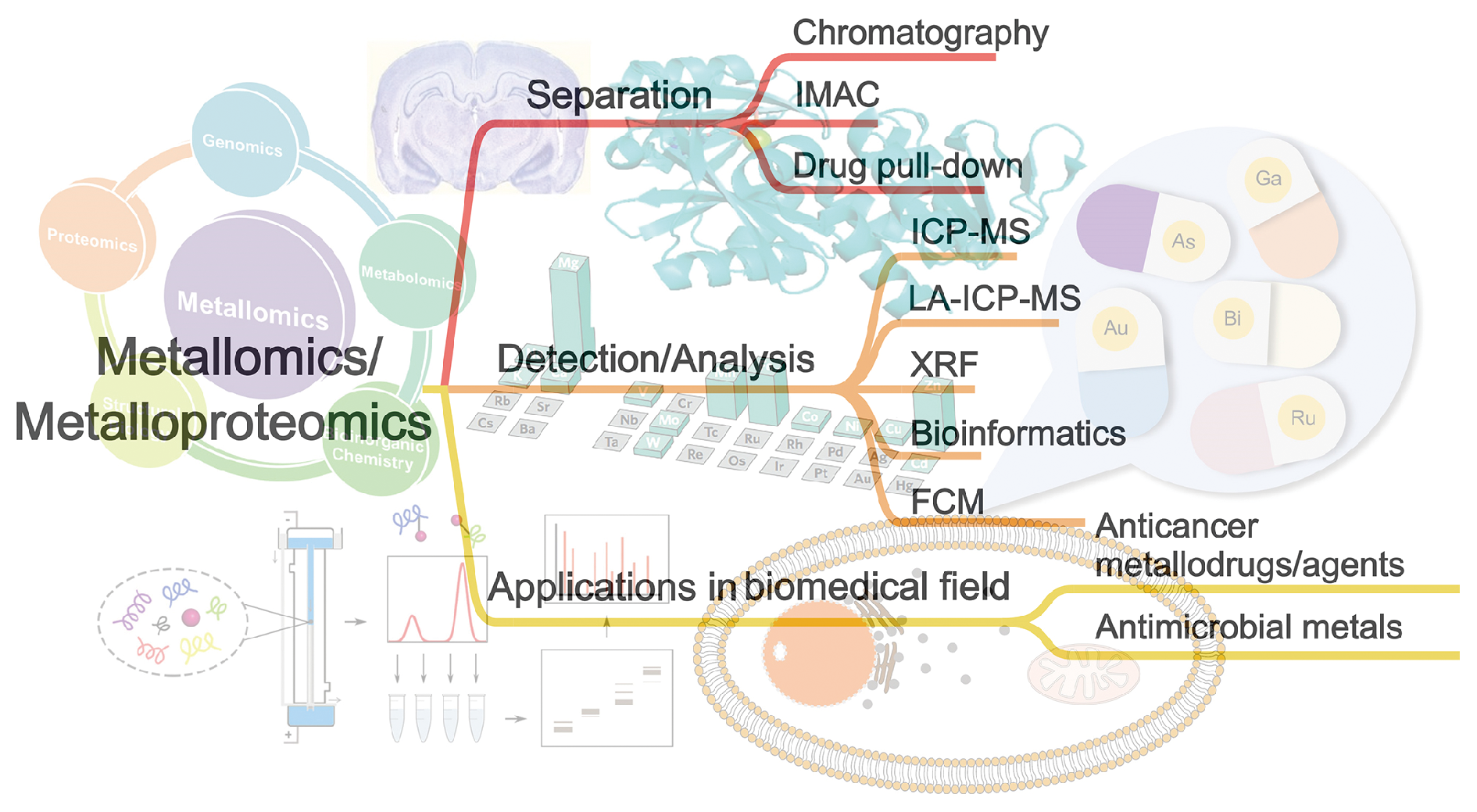
Metals are recognized as essential cofactors in life processes and are fundamental elements in many key cellular processes. Metallomics, as an emerging research field, aims to understand and reveal the functions of bio-active metals and the molecular mechanisms of metal-based life processes, and the related studies have received growing attention due to its rapid development as a frontier science. In this review, we first introduce the concept of metallomics and the related research technologies, and focuses on an important research branch in this field, metalloproteomics, which aims to recognize the relationships between biometals and cellular proteins in a systematic manner. The development of this field has provided a number of practical research tools. We summarize and highlight the recent applications, major progress and important research findings of metallomics and metalloproteomics in biomedical research, which cover the studies of metals/metallodrugs uptake at the single-cell level, the distributions of metals/metallodrugs in cells, tissues and organs, the identification and characterization of intracellular metal-binding proteins, as well as the bioinformatics analysis of metalloproteins. Based on the current research status, the challenges and prospects of the applications of metallomics techniques in biomedical research are further discussed. Moreover, popularization of the metalloproteomics research would be an innovate and efficient way to obtain a complete understanding of the role of bioactive metals in cells. We believe that the development of new methodologies in metallomics and metalloproteomics, as well as the discovery of novel metal-related biological mechanisms will facilitate, support and expand the research perspectives in biomedicine and clinical research.

Contents

1 Introduction

2 Metallomics and metalloproteomics

2.1 Definition

2.2 Research methods and techniques

3 Applications and progress in biomedical research

3.1 Metals/metallodrugs uptake studies at single cell levels

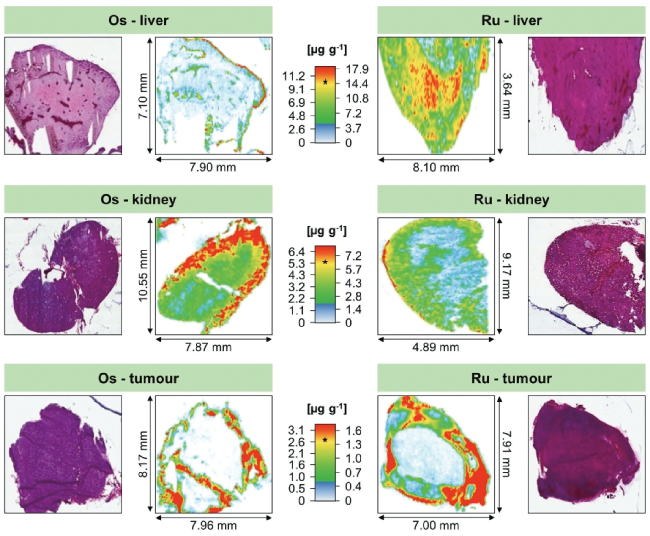
3.2 Distribution studies of metals/metallodrugs in cells and tissues

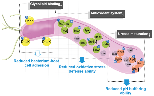
3.3 Identification of metallodrug-targeting proteins in cells

4 Conclusion and outlook

Cite this article

Yuchuan Wang . Applications of Metallomics and Metalloproteomics Techniques in Biomedical Research[J]. Progress in Chemistry, 2023 , 35(10) : 1492 -1504 . DOI: 10.7536/PC230301

1 Introduction

Metals play an important role in maintaining the normal physiological functions of cells and living individuals. For example, metals are irreplaceable cofactors in some biological enzymes in a variety of important biochemical reactions, including energy metabolism, signal transduction, and cell proliferation[1~3]. Metalloomics and metalloproteomics are emerging disciplines to study metal elements and their interacting proteins in biological systems, which provide abundant research tools to reveal the molecular mechanism of bioactive metals and their complexes[4,5]. As a comprehensive research strategy, metalloomics and metalloproteomics effectively overcome the limitations of traditional methods to characterize metal drugs and their potential molecular targets separately.This is essential for a comprehensive understanding of the biological roles of metal complexes, providing important information for the design and development of novel metal-based drugs[6~8].
Although many components of the homeostatic mechanism regulating transition metals have been extensively studied, the metal cation itself has not received much attention, and important questions about metal speciation, etc., remain to be investigated. How is the metal ion delivered intracellularly before it is incorporated as a cofactor into the target metalloprotein? How are metal ions "recycled" during protein degradation? What is the fate of foreign metal ions such as toxic heavy metals or therapeutic metal complexes in organisms? To answer these questions, metal ions need to be identified and quantified in the context of natural cellular environments such as organelles, vesicles, or cytosol[9,10]. As an important analytical tool, mass spectrometry (MS) has been rapidly developed and widely used in medicinal chemistry, environmental science and life science since its inception. In recent years, inductively coupled plasma mass spectrometry (ICP-MS) has played an important role in metalloomics and metalloproteomics[11]. In addition, the development and application of laser ablation inductively coupled plasma mass spectrometry and X-ray fluorescence in metal bioimaging are introduced.
With the rapid development of analytical methods, immobilized metal affinity chromatography based on metal/metal drug-protein affinity and drug pull-down method combined with other protein detection analytical techniques have also been successfully applied to selectively separate cellular proteins related to metal homeostasis, metal-related diseases, and metal cytotoxicity[12,13]. In addition to the successful application of metalloproteomics in the field of pharmaceutical inorganic chemistry to explore the molecular mechanism of metal-based compounds, metalloproteomics has also been applied to clinical studies such as microbial metal homeostasis and metal-related disease processes. In this article, we focus on the main progress of metalloproteomics at the technical and application levels, including the important research results on the molecular targets and modes of action of the major metallodrugs found in metalloproteomics research in recent years, including gold-based, platinum-based, ruthenium-based, arsenic-based and bismuth-based drugs.

2 Metalloomics and metalloproteomics

2.1 Definition

Metallomics is a discipline that studies the interactions and functional relationships between metals and genes, proteins, metabolites and other biomolecules in organisms and ecosystems, focusing on all metals and metalloids in biological systems.The concept of metallome was first proposed by Williams in 2001 to describe the distribution pattern of elements in organisms, and metallomics was proposed by Haraguchi in 2002 to integrate various disciplines of biological trace element research[14~17]. As an important research branch of metalloomics, metalloproteomics is mainly used to characterize the metalloproteome in organisms, and it is an important way to explore the biological function of proteins with metal-binding ability and to fully understand the role of bioactive metals in cells[18,19]. With the development of metalloomics research technology, considerable progress has been made in the rapid identification and characterization of metal-binding proteins, the analysis of the functional mechanism of metalloproteins, and the definition of the distribution and use of metal elements in biological systems[20~22]. In addition, the proposal of metallomics provides valuable experimental tools and implementation strategies for revealing the molecular mechanism of metallodrugs from the perspective of systems biology, including the study of their physiological processes (such as cellular distribution and biotransformation) and molecular targets[6,23].

2.2 Research Methods and Techniques

2.2.1 Metalloprotein detection based on inductively coupled plasma-mass spectrometry (ICP-MS

As one of the classical methods of metalloproteomics, ICP-MS-based analytical techniques mainly separate biological samples by chromatography, then use ICP-MS for metal analysis and LC-MS/MS for protein analysis, after which the analytical data of metals and proteins are integrated separately to discover and describe the functional metalloproteome (Fig. 1A)[24,25]. A series of analytical methods based on ICP-MS, such as gel electrophoresis-inductively coupled plasma mass spectrometry (GE-ICP-MS), high performance liquid chromatography-inductively coupled plasma mass spectrometry (HPLC-ICP-MS), capillary electrophoresis-inductively-coupled plasma mass spectroscopy (CE-ICP-MSc) and protein component prefractionation-ICP-MSc, have been widely used (Figure 1B). GE-ICP-MS combines gel electrophoresis and a metal-specific detection system by using a T-junction to divide the eluate of the column gel system into two parts, one for metal identification by ICP-MS and the other for protein identification by biological mass spectrometry of the collected fractions[20]. The recently developed 3D printing GE-ICP-MS can print gel tubes, gel electrophoresis tanks and elution chambers at low cost in the laboratory, and can adjust the printing accuracy to achieve efficient separation of metalloproteins[26]. The 2-D (WAX-GE) ICP-MS system enables the sample to be fractionated by ion-exchange chromatography WAX column in the first dimension, and the collected fraction is subsequently separated by gel electrophoresis in the second dimension and finally detected by ICP-MS. Two-dimensional separation provides an effective technical path to solve the complex separation of metalloprotein mixtures[27]. The combination of the high specificity of high performance liquid chromatography separation in HPLC-ICP-MS and the high sensitivity of ICP-MS detection has obvious advantages for the detection of metalloproteins[28,29]. The advantages of CE-ICP-MS are that the capillary used in the CE device has a large surface-to-volume ratio, which allows the Joule heat generated by the application of an electric field to dissipate quickly without adversely affecting electrophoretic separation, and that CE-ICP-MS has a short analysis time and high separation efficiency, and can process very few samples (even single cells)[30]. In addition, the PVP covalent coating of the capillary can prevent the interaction between the protein and the capillary surface and improve the separation efficiency[31].
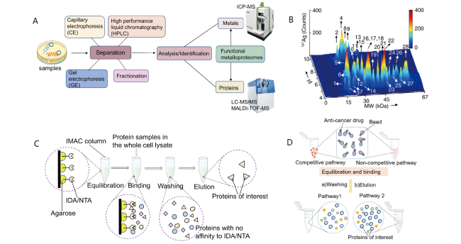
图1 金属蛋白质组学方法及应用。(A) 由ICP-MS和LC-MS/MS技术组成的传统整合分析方法;(B) 大肠杆菌胞浆中与Ag+相关的蛋白质图谱;(C)固定化金属亲和色谱法(IMAC);(D) 药物下拉实验的工作流程

Fig.1 Illustration of metalloproteomics approaches and the applications. (A) Conventional integrated analytical methods consisted of ICP-MS and LC-MS/MS; (B) Map of Ag+-associated proteins in the E. coli cytosol; (C) The workflow of immobilized metal affinity chromatography (IMAC); (D) The workflow of drug pull-down assay

Although there may be some inevitable disadvantages in the combination of ICP-MS and other techniques, such as the low resolution limitation of 1D gel column, the loss of protein in gel electrophoresis and the measurement bias caused by isotopic interference, there is no denying their convenience and potential for improvement. Related technologies have good application examples in the analysis of intracellular metal-protein binding, pre-enrichment of proteins and the analysis of metal selectivity of intracellular metalloproteins[26][32][33]. At present, ICP-MS-based metalloprotein detection techniques have been successfully used to identify Ag-binding proteins in Escherichia coli (E. coli), Gd-binding proteins in NIH-3T3 cells, Ru (Ⅲ) drug-binding proteins in human serum and plasma samples, Bi-binding protein profiles in Helicobacter pylori (H. pylori), and nanosilver-binding protein in bacteria and cancer cells[32][29][34][20][28,35].

2.2.2 Metal bioimaging technique

X-ray fluorescence microscopy (XFM) is a microscopic imaging technique for elemental analysis. The research areas opened up using XFM fall into two main categories — the study of "natural" metal and trace element distributions in health and disease, and the study of the distribution of metals and trace elements introduced into organisms or cells. synchrotron X-ray fluorescence microscopy (SXRF) is a Microanalytical technique for quantitative mapping of elements in organisms, and is a practical analytical technique for whole-cell quantitative elemental imaging with submicron resolution[36,37].
As an elemental imaging technique for solid biological samples, laser ablation-inductively coupled plasma-mass spectrometry (LA-ICP-MS) is capable of quantitative spatially resolved analysis of elemental distribution with millimeter-scale lateral resolution. The main characteristics of LA-ICP-MS include low detection limit, multi-element capability, standard sample preparation process, and high resolution and transmission rate[38]. Technological advances in LA devices can result in significant improvements in instrument sensitivity, analysis speed, and sample throughput to achieve the parameters required for high-throughput analysis of clinical samples[39]. In currently available LA systems, cell-level spatial resolution can be achieved at pixel acquisition rates > 250 pixels/s[38]. In the field of metal bioimaging, LA-ICP-MS is mainly used in the study of cancer, single cell, metal absorption and accumulation, and neuroimaging.

2.2.3 IMAC (immobilized metal affinity chromatography) and drug pull-down assay

Immobilized metal affinity chromatography IMAC is an affinity separation method based on the interaction of a protein with metal binding ability with a specific metal ion immobilized on a solid matrix material. By changing the experimental conditions, such as pH and eluent composition, IMAC can efficiently separate metal-binding proteins for further specific metalloproteome characterization[40]. The operation process of IMAC includes four key steps: (1) selecting the corresponding solid-phase ligand (such as IDA and NTA) according to the nature of the metal-binding protein to be isolated and identified; (2) loading the extracted protein sample into a column and incubating with the immobilized metal; (3) washing with buffer to remove non-target metal-binding proteins; (4) The target metal-binding protein was eluted by changing the pH of the buffer or using a high concentration of imidazole (fig. 1 C)[41,42].
Although in the enrichment process of IMAC, proteins with low abundance and hydrophobicity are not identified, some protein information is lost or some false positive protein information is obtained[41]. However, as one of the most widely used chromatographic methods, the performance of IMAC can be greatly improved by optimizing the production procedures of resin and ligand materials[42]. In addition, IMAC combined with protein analysis and identification technology has been successfully used to characterize the binding proteins and specific binding peptides, amino acid selectivity and protein-binding motifs of Co and Ni in Streptococcus pneumoniae (S. pneumoniae) and Bi in Helicobacter pylori[43,44]. Metal-binding protein and metal-binding motif databases will be able to provide a rational basis for the molecular mechanism of drug action, providing valuable information for novel drug development[44].
Similar to the IMAC technique, the drug pulldown method is based on affinity chromatography, combined with shotgun proteomics and bioinformatics applications to identify proteins targeted by metal anticancer agents. Metallodrug pull-down experiments use both non-competitive and competitive approaches to identify the main drug-binding target proteins. Non-competitive pull-down is the exposure of biotin/streptomycin-modified beads to cell lysate; Competitive pull-down is to expose biotin/streptomycin modified beads and metal anticancer drugs to cell lysate, which saturates the selective binding sites of drugs, and the proteins bound to drugs under this condition may be the main target proteins of drugs. Then, according to the changes in the abundance of drug-binding proteins under non-competitive and competitive experimental conditions, the enrichment map was drawn, and it was preliminarily determined that the main target protein of the drug might change the most (Fig. 1D). After that, the target protein of the drug can be further accurately analyzed by integrating the enrichment analysis results of the drug pull-down experiment and the experimental data of the differentially expressed protein under the action of the drug[13,45].

2.2.4 Single Cell Metal Research Technique

The results of conventional analysis methods for multiple cells usually fail to retain the information of individual differences of cells, and single-cell analysis has attracted more and more attention. However, due to the small size of a single cell and the low accumulation of metals, the quantitative analysis of the content of metals or metalloproteins in a single cell is relatively complex. In recent years, it has become a research hotspot to explore more efficient and sensitive methods for single cell metal analysis. This paper introduces two commonly used methods for single cell metal analysis.
Inductively coupled plasma mass spectrometry (ICP-MS) is widely used to obtain quantitative information of elements because of its high sensitivity and low interference. Taking the quadrupole mass spectrometer (ICP-QMS) as an example, the sample to be analyzed usually enters the plasma center in the form of aqueous solution or aerosol, enters the mass spectrometer after ionization, distributes in the quadrupole according to the mass-to-charge ratio of each ion, and then enters the detector to calculate the number of ions.
This technique is often used to analyze the content of metal elements in multiple cells as a whole, but it can be used to study single cells by reducing the concentration of cell suspension, lengthening the residence time of mass spectrometer or improving the sampling system. The feasibility of this method was confirmed in the tracking experiment of the mercury content in a single Tetrahymena[46]. The sample injection system uses a microsyringe pump with a syringe, and the sample is introduced into the nebulizer through a 100 μm quartz capillary to ensure the feasibility of the experiment. Similarly, in the dynamic detection of AuNP in mouse blood, the purpose of analyzing metal nanoparticles in single cells at a lower concentration was also achieved by improving the sampling system[47].
However, this method has some limitations because the use of a specific metal tag to label cells cannot measure different target molecules at the same time, and ICP-MS cannot distinguish between metal signals and other related elements from cells. In contrast, flow cytometry (FCM) is becoming a more effective method for single-cell research. The flow cytometer is composed of a liquid flow system, an optical system and an electronic system. The liquid flow system achieves the purpose of analyzing single cells by scanning the single cell suspension with a laser, while the electronic system can further convert the signal received in the optical system into an electronic signal, and finally reflect the relative size and particle complexity of the cells.
Flow cytometry combined with ICP-MS and time-of-flight mass spectrometry (ICP-TOF-MS) can analyze the characteristics of single cells through elemental spectroscopy[48]. In addition, the emergence of mass cytometry not only inherits the characteristics of high-speed analysis of traditional flow cytometry, but also has the high-resolution ability of mass spectrometry detection, without the interference of autofluorescence signals, and can analyze multiple parameters at the same time. These qualities make mass spectrometry flow cytometry an attractive technique currently available for highly multiplexed single-cell analysis[49].

2.2.5 Bioinformatics

With the development of computer science and information technology, bioinformatics has been gradually applied to metalloproteomics, which provides a fast and simple way to mine the metalloproteome in cells, and makes up for the obstacles of the complexity of experimental work and the high demand for resources. At present, many bioinformatics algorithms have been used for the analysis and prediction of metalloproteins, which can be roughly divided into two directions: one is to use databases,The metal binding information of the unknown protein is obtained by comparing the unknown protein with the domain of the protein which has been manually annotated and experimentally proved to have metal binding sites in the database. At present, there are many databases for searching the Metal binding sites of proteins, such as PDB, Uniprot, MetalPDB, Metal-MACiE, dbTEU, Mespeus and so on[50]. The use of these databases provides important information support for the study of metalloproteomics.
Another direction is to develop new bioinformatics tools to predict undiscovered metalloprotein-binding sites. Zincfinder is used to predict zinc binding sites. It can not only obtain known metal binding sites by comparing with existing protein information, but also analyze some unknown zinc binding sites, which can be further verified by homology modeling[51]. In addition, MetalDetector, developed by Frasconi Laboratory, is the first server that can predict the metal binding geometry of new folds only from sequence information, which makes good use of the characteristics of cysteine and histidine residues binding to metals to infer the geometry of peptide fragments near the binding site[52]. In addition, HemeBIND and SCMHBP can be used to predict heme-binding sites, while MetSite uses sequence profiles and approximate structural information from prediction models to identify metal-binding sites[53]. More and more new tools have been applied to the prediction of metalloproteins, which makes the research of metalloproteomics more in-depth.

3 Progress of Applied Research in Biomedicine

3.1 Cell uptake of metal/metal drugs at single cell level

Studies have confirmed that even homologous cells in the same culture environment have differences in their size, growth characteristics and gene expression levels[54]. Individual cells have differences in structure, composition and metabolism, and the effects of these differences are reflected in the functions of tissues and organs. Most of the 25 essential elements for life are metals or metalloids, and metals and metalloproteins are involved in many important physiological processes in the body. In addition, the analysis of single cell access to metal or metal drugs will help to promote the exploration of related biological processes involving metal or metal proteins, promote the development of related biological drugs, help the rapid diagnosis of metal-related diseases and the study of cell heterogeneity.
For a long time, bismuth drugs have been used to treat gastric diseases caused by Helicobacter pylori infection, which has important clinical application value. The inhibition of Helicobacter pylori by bismuth drugs is related to the destruction of key protein functions in bacteria. Tsang et al. Used time-resolved ICP-MS to monitor the absorption of bismuth by H. Pylori under different treatment conditions[55]. The results of quantitative and statistical analysis at the single cell level showed that the differential uptake of metals by bacteria may be related to their cell cycle, which provided valuable biological insights into the uptake of bismuth drugs by Helicobacter pylori. In addition, the interference of iron ions on bismuth absorption indicates that bismuth drugs can be taken up by bacteria by using the iron transport pathway in pathogens.
The uptake of metals or metallodrugs by cells varies from cell to cell and from cell cycle to cell cycle in the same cell. Zhou et al. Synchronized the cell cycle with time-resolved ICP-MS analysis by using double thymine to block the cell division process, and combined with flow cytometry to detect the DNA content of cells treated with two arsenic-based drugs (ATO and ZIO-101) in different cycles.It was demonstrated that the uptake of both arsenic-based drugs in single NB4 and HL60 cells was cell cycle dependent, suggesting that cell cycle may need to be considered in the treatment of leukemia and the development of new leukemia drugs to accurately assess the uptake and cytotoxicity of anticancer drugs[56].
The abuse of nanoparticles (NPs) can cause some pollution problems. Studies have found that NPs produced by the reduction of metal ions represent one of the sources of pollution, and some single-cell organisms can internalize these metal nanoparticles to spread to the human body along the food chain and cause health problems. Wu et al. Simulated metal NPs with very low concentration in natural environment, and set metal particles of different sizes as controls to culture a model unicellular organism (Tetrahymena thermophila), and used high-throughput mass spectrometry to detect the uptake of metal nanoparticles AuNPs by Tetrahymena thermophila[48]. The results show that some Tetrahymena thermophila can enrich AuNPs even at very low concentrations of AuNPs, indicating that even if the water source is polluted by only a small amount of NPs, there is a real environmental problem.
As a heavy metal, the harm of lead is gradually being taken seriously, but because it can not be replaced in some production and life, there are not a few cases of lead poisoning. According to statistics, about one third of children's blood lead content is higher than normal. At present, the distribution pattern of chemical pollutants in individual cells of higher organisms is still unclear, resulting in very low therapeutic efficiency. Liu et al. collected blood samples from 79 patients with lead poisoning before, 24 H after, and 72 H after medication, as well as blood samples from 36 healthy subjects.The single cell analysis of different types of cells at different stages in blood samples by mass spectrometry and flow cytometry showed that there was heterogeneity in the distribution of lead in blood cells, and the probability distribution of lead in different States could be well described by a unified gamma distribution[57]. Reticulocytes contain more lead than normal red blood cells, and with the aging of red blood cells, the content of lead is also decreasing, which also gives new hints to explore the transport pathway of lead in the human body. These findings improve the understanding of lead accumulation and toxicity in humans, and provide a basis for research on the prevention and treatment of heavy metal poisoning.
Drug uptake and retention in cancer cells are important research parameters in cancer drug development, and quantifying the content of metal-based anticancer drugs in individual cancer cells remains a challenge. By using single cell inductively coupled plasma mass spectrometry (SC-ICP-MS) to study the absorption and retention of mononuclear (Ir1) and binuclear (Ir2) iridium (III) photoredox catalysts in cells, Fan et al. Can rapidly and accurately quantify the content of drugs in individual cancer cells[58]. By studying the retention of Ir1 and Ir2 in cancer cells, it is shown that Ir2 has a high retention ability in HepG2 cells, which is expected to help overcome the problem of drug resistance.

3.2 Distribution of Metal/Metal Drugs in Cells and Tissues

Trace metal imbalances, particularly those of copper, iron, and zinc, play an important role in several classes of neurodegenerative diseases, including amyotrophic lateral sclerosis (ALS), Alzheimer's disease (AD), and Parkinson's disease (PD). SXRF technology can non-destructively display the distribution of metal ions in brain tissue or nerve cells, which provides a favorable tool for the study of neurodegenerative diseases[59,60]. For example, SXRF and X-ray absorption near-edge structure (XANES) analysis of substantia nigra specimens from patients with Parkinson's disease showed that the ratio of Fe (Ⅲ): Fe (Ⅱ) in glial cells around neuromelanin granules was significantly increased[61]. LA-ICP-MS also plays an important role in tissue imaging of metal elements in the study of Parkinson's disease. Matusch et al. Used LA-ICP-MS to analyze 11 adjacent sections of the substantia nigra of the cerebral hemisphere of wild-type C57BL6 male mice with unilateral 6-OHDA lesions in the medial forebrain bundle, and derived the average metal concentration in the region of interest, including the cortex, substantia nigra, and characteristic structures with high concentrations of their respective metals. In the damaged substantia nigra, the mean concentrations of iron, copper, and manganese were significantly increased by 21, 17, and 20%, respectively, whereas zinc remained unchanged (− 1%)[62].
In the field of metal drug research, LA-ICP-MS has been successfully used to study the distribution of metals and metal drugs in tumor tissues, tumor spheroids and single cells, providing scientific data for the tissue penetration depth, adverse side effects and accumulation of metal drugs at the cellular level[63][64]. Because of the nephrotoxicity of cisplatin and the complex histological structure of the kidney, LA-ICP-MS is widely used to study the accumulation of cisplatin in the kidney. A LA-ICP-MS-based study suggested that platinum accumulation at the level of renal cortical tubular epithelial cells may be responsible for multifocal renal injury after cisplatin treatment[65].
Because of the complexity and heterogeneity of tumor tissues, LA-ICP-MS has also been used to study the accumulation of metal drugs in different parts of the tumor microenvironment, and the heterogeneous distribution of elements corresponding to the histological characteristics of tumors has been observed[66]. Enrichment of metal drugs was mainly found in the loose soft tissue and the periphery or margin of tumor samples, while lower metal content was observed in the solid fraction of tumor samples corresponding to deeper cell layers[63]. In another study, the uptake and spatial distribution of structurally identical organometallic anticancer drug candidates based on ruthenium and osmium in organs and tumors of treated mice were evaluated by LA-ICP-MS, which showed that the highest metal content was in the liver, followed by kidney, lung, and tumor tissue. The two metallodrugs showed different tissue spatial distributions, with osmium compounds mainly accumulating at the organ margins and ruthenium compounds penetrating deeper into organs and tumor tissues (Fig. 2)[67]. The potential of related methods in the study of metallodrugs remains to be exploited.
图2 单剂量钌和锇化合物在CT-26肿瘤小鼠组织中的分布。图像为连续H&E染色组织切片图与LA-ICP-MS分析得到的图像

Fig.2 Metal distribution in tissues of mice bearing a CT-26 tumour treated with a single dose of ruthenium and osmium compounds. The images are obtained from consecutive H&E stained tissue sections and LA-ICP-MS analysis

Spatial metallomics can provide two-dimensional or three-dimensional in situ and migration distribution of elements, and has great potential in exploring the distribution of metals in cells and tissues. Xie et al. Used SRXRF and MALDI-MSI to visualize the in situ 2D/3D distribution and morphology of selenium in the seeds of Cardamine violifolia, a selenium hyperaccumulator[68]. Studies have reported selenium content in bulk seeds, mainly selenocysteine SeCys and selenomethionine SeMet. Further in situ studies of individual seeds showed that methylated selenium was mainly distributed in the ectoderm. This study deepens the understanding of the mechanism of selenium tolerance and hyperaccumulation in sensitive reproductive tissues such as Cardamine violacea seeds.

3.3 Identification of metal drug target protein

3.3.1 Au, Pt, As based anti-tumor metal drugs

Traditionally, the mode of action of (metal) drugs has often been evaluated on the basis of accumulated empirical data, involving numerous in vitro studies of individual putative drug-targeting proteins or other biomolecules. Given the inherent complexity of biological systems, such goal-oriented studies cannot provide a systematic evaluation of cellular coping (metal) drugs. Metalloomics provides an invaluable tool to reveal the molecular mechanism of metallodrugs, including physiological processes such as cellular distribution and biotransformation of drugs, as well as molecular targets from the perspective of systems biology[5,69,70]. The following is an introduction to the recent important progress in the research of metal drug targets.
The gold-based drug auranofin is used in the clinical treatment of arthritis. Because of their antiproliferative properties, gold complexes have been extensively studied in recent years for their therapeutic use as anticancer drugs. With the help of photoaffinity probes and proteomic analysis, Zou et al. Identified multiple molecular target proteins of the pincer compound [AuIII(C^N^C)(NHC)]OTf, including mitochondrial heat shock protein 60 (HSP60), vimentin (VIM), nucleoside diphosphate kinase A (NDKA), nucleophosmin (NMP), nuclease sensitive element binding protein (YB-1) and peroxiredoxin 1 (PRDX1)[71]. Subsequently, whether the specific binding of gold compounds to target proteins interferes with the function of each protein in cancer cells was experimentally investigated. Bioinformatic analysis of the proteomic data showed that the eukaryotic initiation factor 2 (eIF2) signaling pathway was predominantly regulated in HeLa cells treated with [AuIII(C^N^C)(NHC)]OTf. Normally, VIM, NMP, and YB-1 can inhibit eIF2α kinase, resulting in dephosphorylation of eIF2α and ensuring normal protein synthesis[72]. [AuIII(C^N^C)(NHC)]OTf treatment causes VIM degradation, NMP oligomer disruption, and YB-1 down-regulation, resulting in inhibition of eIF2α, which reduces protein synthesis and inhibits cancer cell proliferation[73]. The dysregulation of the activity of several target proteins in the resulting cancer cells was identified to be able to individually or synergistically trigger the downstream anticancer mechanisms of gold compounds, including inhibition of epidermal growth factor receptor (EGFR), induction of tumor suppressor p53 production, and activation of apoptotic caspases in apoptosis[71]. The above target proteins identified in this study are all potential anti-tumor target proteins, indicating that the pincer-type gold (Ⅲ) NHC complex is expected to be developed as a multi-target anticancer drug with potential low drug resistance.
Platinum-based drugs are an important part of cancer chemotherapy and represent a contribution to the field of inorganic chemical drugs. In a recent study, Wan et al identified vimentin as the intracellular target protein of the clamp Pt (II) NHC compound cellular thermal shift assay by cellular thermal shift assay (CETSA) and a series of in vivo and in vitro experiments based on the previous proteomic target identification[74]. VIM is a key cytoskeletal component of mesenchymal cancer cells, often with tumor-initiating and metabolic properties. VIM expression enhances cell motility and invasiveness, prompting the spread of cancer cells from the primary tumor to distant organs through the bloodstream or lymphatic system, leading to a metastatic cascade[75]. [PtII(C^N^C)(NHC2Bu)]PF6 exhibits high anticancer activity, and its high affinity binding to VIM can destroy its cytoskeleton and effectively inhibit tumorigenesis and metastasis[74].
Arsenic trioxide (ATO) is an effective chemotherapeutic agent used alone or in combination in the treatment of acute promyelocytic leukemia (APL), while it has also shown efficacy in other hematologic malignancies and other solid tumors. The toxicity of arsenic may be attributed to the interaction of arsenic with proteins, resulting in conformational and functional changes of the target proteins. A deeper understanding of the arsenic-binding proteome has important implications for the evaluation and development of arsenic-based therapies. Hu et al. Combined metalloproteomics with quantitative proteomics by developing a novel organoarsenic probe, AS-AC, and identified 37 arsenic-binding proteins and 250 arsenic-regulated proteins in human APL cell line NB4[76]. Bioinformatics analysis of the identified arsenic-based drug-related proteins and their PPI networks showed that ATO could disrupt a variety of physiological processes, especially chaperone-related protein folding and cellular response to stress, and found that heat shock protein 60 (Hsp60) was an important target of ATO. The binding of ATO to Hsp60 leads to the loss of the refolding ability of Hsp60, and the binding of ATO to Hsp60 disrupts the formation of Hsp60-p53 and Hsp60-survivin complexes, resulting in the degradation of p53 and survivin.
However, the toxicity of ATO and its clinical administration dose limit its use in the treatment of non-APL cancers. Compared with inorganic arsenic, some new arsenic compounds have better antitumor activity and lower toxicity. The organoarsenic drug Darinaparsin (ZIO-101) showed better anticancer performance against a variety of solid tumors and hematological malignancies in vitro and in vivo. Xu et al. Identified histone H3.3 as an arsenic-binding protein in the nuclei of ZIO-101-treated NB4 and HL60 cells using GE-ICP-MS[77]; Arsenic binding to the target protein was not found in the proteome analysis of ATO-treated cells. Histone H3.3 and nucleosomes are closely associated with active transcription, and the binding of ZIO-101 to histone H3.3 and the subsequent destabilization of nucleosomes in cells may significantly affect transcriptional regulation[78]. In the genome of NB4 cells, 53 genes highly related to histone H3.3 were screened, and the dynamic changes of gene expression levels after drug treatment were detected. The results showed that ZIO-101 treatment induced the activation of transcription factors and regulatory factors in NB4 cells in a time-dependent manner. Protein interaction network (PPI) analysis showed that histone H3.3 was closely related to histone deacetylase 1 (HDAC1), and inhibition of HDACs could increase the acetylation level of histone, resulting in the up-regulation of cyclin-dependent kinase inhibitor 1A (CDKN1A), which in turn stimulated the overexpression of TNF family-related genes, triggered TRAIL-induced apoptosis, and inhibited tumor growth[77]. The above two studies explored the mechanism of action of arsenic-based drugs from the perspective of metalloproteomics, and provided research basis and theoretical guidance for the rational design and application of arsenic-based anticancer drugs.
Inorganic arsenic nanomedicine also has great potential in cancer inhibition and anti-cancer. Arsenene, a 2D single-element layered nanosheet composed of arsenic, has recently emerged as a promising new 2D material for biomedical applications due to its excellent optical and electronic properties. Wang et al. Synthesized novel two-dimensional arsenene nanosheets and proved that the material could effectively inhibit NB4 promyelocytic leukemia cells and induce apoptosis without toxicity to normal cells[79]. Mass spectrometric studies showed that arsenene was mainly bound to nucleotide-binding proteins in the nucleus of NB4 cells. Label-free proteome analysis showed that arsenene affects nuclear DNA replication, nucleotide excision repair, and purine metabolism pathways by down-regulating DNA polymerases POLE, POLD1, POLD2, and POLD3, and significantly reduces the expression of TXNL1 nuclear protein, which may disrupt intracellular redox homeostasis. In another study, Wang et al. Analyzed the mechanism of action of arsenene in vivo and in vitro by a multi-omics approach[80]. From the single cell transcriptome level in the tumor microenvironment, it is known that arsenene has a highly efficient immunomodulatory ability by recruiting a high proportion of anti-tumor immune cells. Further analysis clarified that in the tumor microenvironment, arsenene not only effectively inhibited DNA replication and TCA cycle in tumor cells, but also activated the antigen presentation process and T cell receptor pathway, thereby regulating the anti-cancer process. At the cellular level, proteomic analysis was employed to show that arsenene specifically inhibits thioredoxin TXNL1 to disrupt redox balance. Overloaded reactive oxygen species further trigger the endoplasmic reticulum stress response, leading to the activation of antigen presentation processes to induce subsequent effector tumor-specific CD8+T cellular immune responses. The above two studies used single-cell transcriptomic tools and proteomic methods to reveal the mode of action and target proteins of new arsenic nanomedicines, and led to the discovery of the immunomodulatory function of arsenic-based drugs. These works demonstrate that combining single-cell transcriptomic tools on the basis of proteomics can clarify the relationship between tumor microenvironment patterns and therapeutic efficacy.

3.3.2 Identification of drug target proteins based on Ru and Ir

Ruthenium compounds are considered to be a promising alternative to platinum compounds because of their lower toxicity and greater selectivity for tumors. The anti-tumor mode of action of ruthenium compounds is quite different from that of other commonly used chemotherapeutic drugs. Recently, several studies have identified a variety of intracellular target proteins of ruthenium compounds related to cancer by drug pull-down method[13,45,81].
A recent study by Meier et al. Identified Plectin as the target protein of the ruthenium compound Plecstatin[45]. Plectin, a scaffolding protein, is an important cytosolic protein that regulates the keratin and tubulin network and has a significant effect on the organization of nonmitotic microtubules[82]. Non-mitotic microtubules are drug targets, and their interference by plectin targeting agents can affect the motility of cancer cells, which may be a promising anti-cancer strategy[45]. Plectin targeting reorganizes the nonmitotic microtubule network into a tightly curved submembranous network that surrounds the nucleus, resulting in G0/G1 arrest. In several cancer types, plectin loss down-regulates Src/RhoA/CDC42 signaling, inhibiting the migration and invasion of pancreatic, bladder, and colon cancer cells, that is, drug targeting of plectin leads to decreased motility and invasiveness of cancer cells[83][84][85]. In another research work, Neuditschko et al. Identified ribosomal proteins RPL10, RPL24, and transcription factor GTF2 as target proteins of the clinical candidate antitumor metal ruthenium reagent BOLD-100/KP1339[81]. Intracellular target protein profiling experiments were performed by using the naturally formed adduct between BOLD-100 and human serum albumin as an immobilization strategy. Direct interaction of BOLD-100 with ribosomal proteins is accompanied by ER stress induction and down-regulation of the ER molecular chaperone GRP78 in cancer cells. The identification of drug target proteins of ruthenium compounds will help to clarify the mechanism of action of metal drugs and lay a theoretical foundation for their application in biomedicine.
immunogenic cell death (immunogenic cell death, ICD) is a unique mode of cell death response that provokes durable antitumor immunity by inducing damage-associated molecular pattern (DAMP) signaling, such as calreticulin (CRT) surface exposure, ATP release, and secretion of HMGB1[86]. Chemotherapeutic agents with anti-tumor immune function have good clinical value: they are cytotoxic to rapidly growing cancer cells, thus producing a "first hit", while the host immune system produces a "second hit" to stimulate tumor-specific immune responses. Given that sustained induction of ER stress is a key feature of ICD, Xiong et al. Investigated a series of cyclometalated iridium (Ⅲ) compounds and successfully revealed the ICD activity of an ER-targeted iridium (Ⅲ) compound containing a bis-N-heterocyclic carbene (bisNHC) ligand[87]. The compound was able to induce all the hallmarks of ICD and showed vaccine effects similar to oxaliplatin in a mouse model. Subsequent target analysis and validation experiments identified the involvement of binding immunoglobulin (BiP), a key protein regulating ER homeostasis, which is closely related to the ICD resistance of tumors. Further studies showed that the cyclometalated iridium (III) compound effectively destroyed the intracellular stability of BiP, and demonstrated the important role of BiP protein in inducing anti-tumor immunity[87]. In this work, the direct target information of ICD inducers was obtained by chemical biology methods, which laid the foundation for the design of new ICD inducers.
Metalloproteomics technology also provides an important basis for the analysis of target proteins of iridium anti-tumor compounds. Label-free quantitative proteomics analysis of the anticancer mechanism of iridium (Ⅲ) hydride complex 2-N3 showed that 2-N3 activated the extracellular matrix-receptor interaction pathway, increased the expression of laminin LAMC1 and LAMB2 to activate the ECM-receptor interaction pathway, and affected cellular DNA transcription, post-translational glycosyl modification, and redox homeostasis. 2-N3 damages several key proteins and enzymes in the mitochondria and nucleus, leading to disturbances in cellular processes[88]. In another study, the proteomic mechanism of the mitochondria-targeted iridium (Ⅲ) compound IrFN showed that the iron death process mediated by heme oxygenase 1 (HMOX1) was activated by IrFN, which was confirmed by mRNA transcription quantification, in vitro HMOX1 overexpression, and RNAi-mediated knockdown[89]. In addition, a recent differential proteomic analysis of iridium compounds containing Py-RSL ligands showed that the compounds significantly inhibited a series of ErbB signaling pathway-related proteins, inhibited the expression of eukaryotic translation initiation factor 2A (EIF2A) protein, and promoted cell iron death to exert their anti-tumor activity[90].

3.3.3 Identification of target protein of metal antibacterial agent

Metal ions have been used as antibacterial agents for a long time because of their inherent broad-spectrum antibacterial properties and less resistance. As a potential alternative to solve the crisis of antimicrobial resistance, the antibacterial properties and mechanisms of metal-based compounds have attracted wide attention[91,92]. The critical role played by metalloproteins in biological systems and the relative lack of knowledge of the microbial metalloproteome have stimulated interest in analytical techniques capable of characterizing metal-protein interactions in bacteria,It is expected to reveal the main target proteins of metal antibacterial agents in pathogenic bacteria and their mechanisms of action through related research methods.
Metal chelate affinity chromatography is one of the most effective techniques for the separation of various metalloproteins in organisms, which is based on the interaction between amino acid residues on the surface of proteins and metal ions on solid carriers. It has the characteristics of simple ligand, large adsorption capacity, mild separation conditions and strong versatility[93]. IMAC coupled with gel electrophoresis-based proteomics was successfully used to enrich and identify seven bismuth-binding proteins in H. Pylori, including HspA, HspB, NapA, TsaA, EF-Tu[94]. Furthermore, more than 300 bismuth-binding peptides corresponding to 166 proteins in Helicobacter pylori were successfully identified by Bi-IMAC and high-throughput liquid chromatography-mass spectrometry. Bioinformatics analysis of the identified peptides showed that Bi was highly selective for Cys and His containing peptides in Helicobacter pylori, and the proteomic data obtained were helpful for further understanding of the antibacterial mechanism of bismuth agents[44]. RpoB and RpoC, the two subunits of RNA polymerase in Pseudomonas aeruginosa, were successfully identified as the main gallium nitrate binding proteins by Fe competitive Ga-IMAC. Biochemical experiments showed that gallium antibacterial agent could inhibit RNA polymerase-mediated bacterial transcription by binding to the target protein[95].
Accurate identification of metal-binding proteins in cells is a key step in the comprehensive elucidation of protein function, but it remains a challenge for analytical chemistry and metalloproteomics. In recent years, the high resolution gel electrophoresis coupled with ICP-MS (GE-ICP-MS) has been developed, which can realize the matching of metal ions and their binding proteins in complex biological samples, and can prepare denaturing or non-denaturing gel electrophoresis columns according to the types and characteristics of the metal proteins to be analyzed[20,26][33]. Hu et al. successfully characterized the binding protein spectrum of bismuth drugs in Helicobacter pylori using GE-ICP-MS system, observed the interaction between bismuth drugs and Helicobacter pylori proteome through metal-based analysis, and clarified the antibacterial mechanism of bismuth drugs acting on multiple target proteins of Helicobacter pylori from the perspective of metalloomics (Fig. 3)[20][96]. This research strategy combines metalloomics with proteomics, which can analyze metals and their related intracellular proteins at the whole proteome level, and is of great significance for high-throughput omics research.
图3 铋剂药物抑制幽门螺旋杆菌的多靶点作用模式示意图

Fig.3 A model for the multi-targeted mode of action of Bi drug in eradicating H. pylori

By adding a second dimension to improve the protein separation system of this technology, that is, using liquid chromatography to pre-separate the protein sample, the resolution of this technology for the detection of metalloproteins can be improved, and the information of molecular weight, pI and protein-bound metal content of metalloproteins can be obtained simultaneously. Wang et al. Used an improved LC-GE-ICP-MS system to successfully identify more than 30 kinds of silver ion binding proteins in Escherichia coli and Staphylococcus aureus, and then clarified the antibacterial mechanism of silver ions[21,32]. Silver ions exert their bactericidal activity by targeting key proteins or enzymes in pathogenic bacteria and disrupting important biochemical metabolic pathways.
In view of the principle that the elucidation of the antibacterial mechanism of metal antibacterial agents at the protein level can be translated into clinical antibacterial methods, the combination of antibiotics with silver ions or other metal compounds or metal nanomaterials (such as colloidal bismuth citrate or auranofin) can be used as a promising strategy to inhibit the selective effect of bacterial antibiotics.So as to prevent the occurrence of primary antibiotic resistance and prolong the service life of conventional antibiotics, so as to alleviate the current antibiotic resistance crisis[97][98].

4 Conclusion and outlook

The convergence of omics technologies has begun to provide us with a systems view of microorganisms, microbial communities, complex organisms, and ecosystems, that is, all known living systems require the unique chemical and physical properties of metals to maintain life activities and homeostasis[19]. Although the above metalloomics and metalloproteomics strategies have been used to characterize active metals and metal-binding proteins in a variety of organisms worldwide, the resolution of their analysis and detection still needs to be improved. In addition, the researchers propose that other types of information can be used to discover new metalloprotein classes in the proteome, such as protein co-evolution signals, to solve the problem of finding proteins with new metal-binding sequences and structural patterns[50]. At the same time, even in the same culture environment, the uptake of metals or metal drugs by different cells will be different, and the degree of metal loss, enrichment or exchange after extraction of biological samples should be paid attention to when analyzing these differences.This helps to understand the biochemical problems of individual cells during dynamic changes and the extent to which current analytical methods bias toward specific proteins. In addition, researchers should make more efforts in the identification and development of drug mechanisms and reliable drug targets to fully understand the biological responses of metal drugs. The combination of metallomics with chemical biology and other omics methods, which enables the systematic evaluation of the molecular targets of more metal compounds, will greatly promote the in-depth understanding of the functional links between metal drugs and complex biological proteomes, and enable the functional, toxic, and resistance mechanisms of metal drugs to be elucidated at the molecular level. Combined with clinical data, this knowledge will facilitate the design and development of more effective metallodrugs with high therapeutic index and low toxicity. Future applications of metallomics in biological research will continue to expand the understanding of metals in microbial physiology, ecology, and human disease. In conclusion, we strongly believe that the fields of metalloomics and metalloproteomics will continue to flourish in the future and provide more tools to advance human understanding of metal biology and related diseases.
[1]
Valdez C E, Smith Q A, Nechay M R, Alexandrova A N. Acc. Chem. Res., 2014, 47(10): 3110.

[2]
Foster A W, Young T R, Chivers P T, Robinson N J. Curr. Opin. Chem. Biol., 2022, 66: 102095.

[3]
Chandrangsu P, Rensing C, Helmann J D. Nat. Rev. Microbiol., 2017, 15(6): 338.

[4]
Mounicou S, Szpunar J, Lobinski R. Chem. Soc. Rev., 2009, 38(4): 1119.

[5]
Xiong X L, Liu L Y, Mao Z W, Zou T T. Coord. Chem. Rev., 2022, 453: 214311.

[6]
Wang Y C, Li H Y, Sun H Z. Inorg. Chem., 2019, 58(20): 13673.

[7]
Wang H, Zhou Y, Xu X, Li H, Sun H. Curr. Opin. Chem. Biol., 2020, 55: 171.

[8]
Scalese G, Kostenkova K, Crans D C, Gambino D. Curr. Opin. Chem. Biol., 2022, 67: 102127.

[9]
Waldron K J, Rutherford J C, Ford D, Robinson N J. Nature, 2009, 460(7257): 823.

[10]
Waldron K J, Robinson N J. Nat. Rev. Microbiol., 2009, 7(1): 25.

[11]
Xu X H, Wang H B, Li H Y, Sun H Z. Chem. Lett., 2020, 49(6): 697.

[12]
She Y M, Narindrasorasak S, Yang S Y, Spitale N, Roberts E A, Sarkar B. Mol. Cell. Proteom., 2003, 2(12): 1306.

[13]
Babak M V, Meier S M, Huber K V M, Reynisson J, Legin A A, Jakupec M A, Roller A, Stukalov A, Gridling M, Bennett K L, Colinge J, Berger W, Dyson P J, Superti-Furga G, Keppler B K, Hartinger C G. Chem. Sci., 2015, 6(4): 2449.

[14]
Williams R J P. Coord. Chem. Rev., 2001, 216/217: 583.

[15]
Haraguchi H. J. Anal. At. Spectrom., 2004, 19(1): 5.

[16]
Maret W. Metallomics: the Science of Biometals and Biometalloids, 2018.

[17]
Maret W. Metallomics, 2022, 14 (8): mfac051.

[18]
da Silva M A, Sussulini A, Arruda M A. Expert Rev. Proteom., 2010, 7(3): 387.

[19]
Yannone S M, Hartung S, Menon A L, Adams M W, Tainer J A. Curr. Opin. Biotechnol., 2012, 23(1): 89.

[20]
Hu L G, Cheng T F, He B, Li L, Wang Y C, Lai Y T, Jiang G B, Sun H Z. Angew. Chem. Int. Ed., 2013, 52(18): 4916.

[21]
Wang H B, Wang M J, Xu X H, Gao P, Xu Z L, Zhang Q, Li H Y, Yan A X, Kao R Y T, Sun H Z. Nat. Commun., 2021, 12: 3331.

[22]
de Jonge M D, Holzner C, Baines S B, Twining B S, Ignatyev K, Diaz J, Howard D L, Legnini D, Miceli A, McNulty I, Jacobsen C J, Vogt S. Proc. Natl. Acad. Sci. U. S. A., 2010, 107(36): 15676.

[23]
Shi W X, Chance M R. Curr. Opin. Chem. Biol., 2011, 15(1): 144.

[24]
Cvetkovic A, Lal Menon A, Thorgersen M P, Scott J W, Poole F L, Jenney F E, Lancaster W A, Praissman J L, Shanmukh S, Vaccaro B J, Trauger S A, Kalisiak E, Apon J V, Siuzdak G, Yannone S M, Tainer J A, Adams M W W. Nature, 2010, 466(7307): 779.

[25]
JimÉnez-Lamana J, Szpunar J. Metallomics, 2017, 9(8): 1014.

[26]
Wang D Y, He B, Yan X T, Nong Q Y, Wang C, Jiang J, Hu L G, Jiang G B. Talanta, 2019, 197: 145.

[27]
Yan X T, He B, Wang D Y, Hu L G, Liu L H, Liao C Y, Jiang G B. Talanta, 2018, 184: 404.

[28]
Xu M, Yang Q Y, Xu L N, Rao Z Y, Cao D, Gao M, Liu S J. Part. Fibre Toxicol., 2019, 16(1): 46.

[29]
Nong Q Y, Chen X, Hu L G, Huang Y S, Luan T G, Liu H T, Chen B W. Talanta, 2020, 219: 121281.

[30]
Prange A, Pröfrock D. Anal. Bioanal. Chem., 2005, 383(3): 372.

[31]
Holtkamp H U, Movassaghi S, Morrow S J, Kubanik M, Hartinger C G. Metallomics, 2018, 10(3): 455.

[32]
Wang H B, Yan A X, Liu Z G, Yang X M, Xu Z L, Wang Y C, Wang R M, Koohi-Moghadam M, Hu L G, Xia W, Tang H R, Wang Y L, Li H Y, Sun H Z. PLoS Biol., 2019, 17(6): e3000292.

[33]
Wang Y C, Hu L G, Yang X M, Chang Y Y, Hu X Q, Li H Y, Sun H Z. Metallomics, 2015, 7(10): 1399.

[34]
Timerbaev A R, Pawlak K, Aleksenko S S, Foteeva L S, Matczuk M, Jarosz M. Talanta, 2012, 102: 164.

[35]
Yan X T, He B, Liu L H, Qu G B, Shi J B, Hu L G, Jiang G B. Metallomics, 2018, 10(4): 557.

[36]
Yang L C, McRae R, Henary M M, Patel R, Lai B, Vogt S, Fahrni C J. Proc. Natl. Acad. Sci. U. S. A., 2005, 102(32): 11179.

[37]
Fahrni C J. Curr. Opin. Chem. Biol., 2007, 11(2): 121.

[38]
Doble P A, de Vega R G, Bishop D P, Hare D J, Clases D. Chem. Rev., 2021, 121(19): 11769.

[39]
Van Malderen S J M, Managh A J, Sharp B L, Vanhaecke F. J. Anal. At. Spectrom., 2016, 31(2): 423.

[40]
Sun X S, Chiu J F, He Q Y. Methods in Molecular Biology?. Totowa, NJ: Humana Press, 2008. 205.

[41]
Sun X S, Chiu J F, He Q Y. Expert Rev. Proteom., 2005, 2(5): 649.

[42]
Block H, Maertens B, Spriestersbach A, Brinker N, Kubicek J, Fabis R, Labahn J, Schäfer F. Methods in Enzymology. Amsterdam: Elsevier, 2009. 439.

[43]
Sun X S, Yu G C, Xu Q, Li N, Xiao C L, Yin X F, Cao K, Han J L, He Q Y. Metallomics, 2013, 5(7): 928.

[44]
Wang Y C, Tsang C N, Xu F, Kong P W, Hu L G, Wang J W, Chu I K, Li H Y, Sun H Z. Chem. Commun., 2015, 51(92): 16479.

[45]
Meier S M, Kreutz D, Winter L, Klose M H M, Cseh K, Weiss T, Bileck A, Alte B, Mader J C, Jana S, Chatterjee A, Bhattacharyya A, Hejl M, Jakupec M A, Heffeter P, Berger W, Hartinger C G, Keppler B K, Wiche G, Gerner C. Angew. Chem. Int. Ed., 2017, 56(28): 8267.

[46]
Shi J B, Ji X M, Wu Q, Liu H W, Qu G B, Yin Y G, Hu L G, Jiang G B. Anal. Chem., 2020, 92(1): 622.

[47]
Sun Y Z, Liu N, Wang Y Y, Yin Y G, Qu G B, Shi J B, Song M Y, Hu L G, He B, Liu G L, Cai Y, Liang Y, Jiang G B. Anal. Chem., 2020, 92(22): 14872.

[48]
Wu Q, Shi J B, Ji X M, Xia T, Zeng L, Li G T, Wang Y Y, Gao J, Yao L L, Ma J J, Liu X L, Liu N, Hu L G, He B, Liang Y, Qu G B, Jiang G B. ACS Nano, 2020, 14(10): 12828.

[49]
Bendall S C, Simonds E F, Qiu P, Amir E A D, Krutzik P O, Finck R, Bruggner R V, Melamed R, Trejo A, Ornatsky O I, Balderas R S, Plevritis S K, Sachs K, Pe’er D, Tanner S D, Nolan G P. Science, 2011, 332(6030): 687.

[50]
Zeng X, Cheng Y, Wang C. Biochemistry, 2021, 60(46): 3507.

[51]
Passerini A, Andreini C, Menchetti S, Rosato A, Frasconi P. BMC Bioinform., 2007, 8(1): 39.

[52]
Passerini A, Lippi M, Frasconi P. Nucleic Acids Res., 2011, 39(2): W288.

[53]
Zhang Y, Zheng J G. Molecules, 2020, 25(15): 3366.

[54]
Hasty J, Pradines J, Dolnik M, Collins J J. Proc. Natl. Acad. Sci. U. S. A., 2000, 97(5): 2075.

[55]
Tsang C N, Ho K S, Sun H Z, Chan W T. J. Am. Chem. Soc., 2011, 133(19): 7355.

[56]
Zhou Y, Wang H B, Tse E, Li H Y, Sun H Z. Anal. Chem., 2018, 90(17): 10465.

[57]
Liu N, Huang Y S, Zhang H Z, Wang T, Tao C, Zhang A Q, Chen B W, Yin Y G, Song M Y, Qu G B, Liang Y, Shi J B, He B, Hu L G, Jiang G B. Environ. Sci. Technol., 2021, 55(6): 3819.

[58]
Fan Z X, Rong Y, Sadhukhan T, Liang S X, Li W Q, Yuan Z X, Zhu Z L, Guo S W, Ji S M, Wang J Q, Kushwaha R, Banerjee S, Raghavachari K, Huang H Y. Angew. Chem. Int. Ed., 2022, 61(23): e202202098.

[59]
Tomik B, Chwiej J, Szczerbowska-Boruchowska M, Lankosz M, WÓjcik S, Adamek D, Falkenberg G, Bohic S, Simionovici A, Stegowski Z, Szczudlik A. Neurochem. Res., 2006, 31(3): 321.

[60]
Yoshida S, Ide-Ektessabi A, Fujisawa S. Struct. Chem., 2003, 14(1): 85.

[61]
Ide-Ektessabi A, Fujisawa S, Yoshida S. J. Appl. Phys., 2002, 91(3): 1613.

[62]
Matusch A, Fenn L S, Depboylu C, Klietz M, Strohmer S, McLean J A, Becker J S. Anal. Chem., 2012, 84(7): 3170.

[63]
Theiner S, Schweikert A, Haberler C, Peyrl A, Koellensperger G. Metallomics, 2020, 12(8): 1246.

[64]
Theiner S, Schweikert A, Van Malderen S J M, Schoeberl A, Neumayer S, Jilma P, Peyrl A, Koellensperger G. Anal. Chem., 2019, 91(13): 8207.

[65]
van Acker T, van Malderen S J M, van Heerden M, McDuffie J E, Cuyckens F, Vanhaecke F. Anal. Chim. Acta, 2016, 945: 23.

[66]
Theiner S, Kornauth C, Varbanov H P, Galanski M S, Van Schoonhoven S, Heffeter P, Berger W, Egger A E, Keppler B K. Metallomics, 2015, 7(8): 1256.

[67]
Klose M H M, Theiner S, Kornauth C, Meier-Menches S M, Heffeter P, Berger W, Koellensperger G, Keppler B K. Metallomics, 2018, 10(3): 388.

[68]
Xie H X, Tian X, He L N, Li J C, Cui L W, Cong X, Tang B C, Zhang Y, Guo Z Y, Zhou A Y, Chen D L, Wang L M, Zhao J T, Yu Y L, Li B, Li Y F. J. Agric. Food Chem., 2023, 71(5): 2658.

[69]
Wang Y C, Wang H B, Li H Y, Sun H Z. Dalton Trans., 2015, 44(2): 437.

[70]
Holtkamp H U, Hartinger C G. Trac Trends Anal. Chem., 2018, 104: 110.

[71]
Fung S K, Zou T T, Cao B, Lee P Y, Fung Y M E, Hu D, Lok C N, Che C M. Angew. Chem. Int. Ed., 2017, 56(14): 3892.

[72]
Wehner K A, Schütz S, Sarnow P. Mol. Cell. Biol., 2010, 30(8): 2006.

[73]
Chen T, Ozel D, Qiao Y, Harbinski F, Chen L M, Denoyelle S, He X Y, Zvereva N, Supko J G, Chorev M, Halperin J A, Aktas B H. Nat. Chem. Biol., 2011, 7(9): 610.

[74]
Wan P K, Tong K C, Lok C N, Zhang C L, Chang X Y, Sze K H, Wong A S T, Che C M. Proc. Natl. Acad. Sci. U. S. A., 2021, 118(17), e2025806118.

[75]
Satelli A, Li S L. Cell. Mol. Life Sci., 2011, 68(18): 3033.

[76]
Hu X Q, Li H Y, Ip T K Y, Cheung Y F, Koohi-Moghadam M, Wang H B, Yang X M, Tritton D N, Wang Y C, Wang Y, Wang R M, Ng K M, Naranmandura H, Tse E W C, Sun H Z. Chem. Sci., 2021, 12(32): 10893.

[77]
Xu X H, Wang H B, Li H Y, Hu X Q, Zhang Y, Guan X Y, Toy P H, Sun H Z. Chem. Commun., 2019, 55(87): 13120.

[78]
Henikoff S, Smith M M. Cold Spring Harb. Perspect. Biol., 2015, 7(1): a019364.

[79]
Wang X X, Hu Y, Mo J B, Zhang J Y, Wang Z Z, Wei W, Li H L, Xu Y, Ma J, Zhao J, Jin Z, Guo Z J. Angew. Chem. Int. Ed., 2020, 59(13): 5151.

[80]
Wang X, Zhang J, Hu Y, Zhao X, Wang Z, Zhang W, Liang J, Yu W, Tian T, Zhou H, Li J, Liu S, Zhao J, Jin Z, Wei W, Guo Z. ACS Appl. Mater. Interfaces., 2022, 14(40): 45137.

[81]
Neuditschko B, Legin A A, Baier D N, Schintlmeister A, Reipert S, Wagner M, Keppler B K, Berger W, Meier-Menches S M, Gerner C. Angewandte Chemie Int. Ed., 2021, 60(10): 4954.

[82]
Wiche G, Osmanagic-Myers S, CastañÓn M J. Curr. Opin. Cell Biol., 2015, 32: 21.

[83]
Shin S J, Smith J A, Rezniczek G A, Pan S, Chen R, Brentnall T A, Wiche G, Kelly K A. Proc. Natl. Acad. Sci. U. S. A., 2013, 110(48): 19414.

[84]
Sutoh Yoneyama M, Hatakeyama S, Habuchi T, Inoue T, Nakamura T, Funyu T, Wiche G, Ohyama C, Tsuboi S. Eur. J. Cell Biol., 2014, 93(4): 157.

[85]
McInroy L, Määttä A. Exp. Cell Res., 2011, 317(17): 2468.

[86]
Li J B, Cai W X, Yu J, Zhou S, Li X L, He Z G, Ouyang D F, Liu H Z, Wang Y J. Biomaterials, 2022, 287: 121651.

[87]
Xiong X L, Huang K B, Wang Y, Cao B, Luo Y L, Chen H W, Yang Y, Long Y, Liu M Y, Chan A S C, Liang H, Zou T T. J. Am. Chem. Soc., 2022, 144(23): 10407.

[88]
Wang X X, Zhang J Y, Zhao X Y, Wei W, Zhao J. Metallomics, 2019, 11(8): 1344.

[89]
Wang X X, Chen F, Zhang J Y, Sun J X, Zhao X Y, Zhu Y L, Wei W, Zhao J, Guo Z J. Sci. China Chem., 2020, 63(1): 65.

[90]
Zhao X Y, Zhang J Y, Zhang W, Guo Z J, Wei W, Wang X X, Zhao J. Chem. Sci., 2023, 14(5): 1114.

[91]
Lemire J A, Harrison J J, Turner R J. Nat. Rev. Microbiol., 2013, 11(6): 371.

[92]
Turner R J. Microb. Biotechnol., 2017, 10(5): 1062.

[93]
Gaberc-Porekar V, Menart V. J. Biochem. Biophys. Methods, 2001, 49(1/3): 335.

[94]
Ge R G, Sun X S, Gu Q, Watt R M, Tanner J A, Wong B C Y, Xia H H, Huang J D, He Q Y, Sun H Z. JBIC J. Biol. Inorg. Chem., 2007, 12(6): 831.

[95]
Wang Y C, Han B J, Xie Y X, Wang H B, Wang R M, Xia W, Li H Y, Sun H Z. Chem. Sci., 2019, 10(24): 6099.

[96]
Wang Y C, Hu L G, Xu F, Quan Q, Lai Y T, Xia W, Yang Y, Chang Y Y, Yang X M, Chai Z F, Wang J W, Chu I K, Li H Y, Sun H Z. Chem. Sci., 2017, 8(6): 4626.

[97]
Wang R M, Lai T P, Gao P, Zhang H M, Ho P L, Woo P C Y, Ma G X, Kao R Y T, Li H Y, Sun H Z. Nat. Commun., 2018, 9: 439.

[98]
Sun H Z, Zhang Q, Wang R M, Wang H B, Wong Y T, Wang M J, Hao Q, Yan A X, Kao R Y T, Ho P L, Li H Y. Nat. Commun., 2020, 11: 5263.

Outlines

/