Home Journals Progress in Chemistry
Progress in Chemistry

Abbreviation (ISO4): Prog Chem      Editor in chief: Jincai ZHAO

About  /  Aim & scope  /  Editorial board  /  Indexed  /  Contact  / 
Review

Construction and Application of 3D Microfluidic Liver-On-A-Chip

  • Xueping Lu ,
  • Liang Zhao ,
  • Xiayan Wang ,
  • Guangsheng Guo , *
Expand
  • Center of Excellence for Environmental Safety and Biological Effects, Faculty of Environment and Life, Department of Chemistry, Beijing University of Technology,Beijing 100124, China

Received date: 2023-02-03

  Revised date: 2023-04-01

  Online published: 2023-04-20

Supported by

The National Natural Science Foundation of China(22174007)

The National Natural Science Foundation of China(22127805)

The Beijing Outstanding Young Scientist Program(BJJWZYJH01201910005017)

Abstract

As the largest internal organ in the human body, the liver plays an essential role in the metabolism. The liver or relevant diseases are one of the leading causes of death in the world, with the number of cases surging each year. Therefore, an in-depth understanding of the physiological and biochemical processes and pathological mechanisms of the liver is of great significance for the research, prevention, diagnosis, and treatment of liver-related or metabolism-related diseases. The in vitro liver culture model is an important experimental platform for the study of liver-related biological mechanisms. However, the traditional two-dimensional in vitro cell culture model makes it difficult to reproduce the complex physiological structure and microenvironment of the liver, and lack of disease characteristics. More importantly, the cell structure, gene expression, substance metabolism, and so on in the process of planar culture are significantly different from those in vivo. Microfluidic technology can simulate the physiological structure of liver by designing appropriate micro-structure, providing a microenvironment more like that in vivo by combining with three-dimensional liver tissue culture. Therefore, this paper summarizes the methods and latest progress in constructing 3D liver chips in vitro based on microfluidic technology, including porous membrane culture, hydrogel culture, cell spheroid-based culture, and 3D bioprinting. The applications of 3D cultured liver microchips in remodeling liver physiological structure, exploring mechanism and pathological mechanism, drug screening, and toxicity testing are further summarized. Finally, the potential value and challenges of 3D liver-on-a-chip are discussed.

Contents

1 Introduction

2 Construction methods for 3D microfluidic liver-on-a-chip

2.1 Porous membrane

2.2 Cell spheroids

2.3 Gel-based 3D culture

2.4 3D bioprinting

3 Application of 3D microfluidic liver-on-a-chip

3.1 Disease models

3.2 Drug screening

4 Conclusion and prospects

Cite this article

Xueping Lu , Liang Zhao , Xiayan Wang , Guangsheng Guo . Construction and Application of 3D Microfluidic Liver-On-A-Chip[J]. Progress in Chemistry, 2023 , 35(9) : 1357 -1368 . DOI: 10.7536/PC230116

1 Introduction

Liver is one of the most important organs of the human body, which is responsible for excretion, metabolism, detoxification, synthesis and many other functions in the body. Liver disease is life-threatening, and millions of people die every year due to liver-related diseases and their complications[1~3]. In addition to the disease of the liver itself, the compound may cause toxicity to the liver or other organs after metabolism in the liver, which is also the key factor that makes a large number of potential compounds difficult to pass clinical safety assessment in the current drug development process[4]. The use of animal models is an extremely important experimental method and means in modern biomedical research, which is helpful to understand the occurrence and development of human diseases and to evaluate the efficacy and safety of clinical new drugs. Therefore, people have been using animals that are highly similar to human genes in a biological sense and may have similar reactions to viruses and bacteria as human beings as anatomical and physiological models. However, animal-based experimental studies are not perfect: because drugs are absorbed, distributed, and metabolized differently in different species, and genomic differences ultimately still interfere with molecular-level drug research, it is difficult to predict clinical evaluation results from animal results. In 2022, the U.S. Senate unanimously passed the FDA Modernization Act 2.0, which removed the federal authorization for animal testing of new drugs and ended the "animal testing mandate" to promote a significant reduction in the use of experimental animals. The FDA Modernization Act changed animal testing to non-clinical tests and trials: cell-based assays, organoids, microphysiological systems, computer models, etc.
A microfluidic chip, also known as a lab-on-a-chip, is a functional device fabricated using microfabrication technology and having a channel or chamber structure with a submillimeter structure in at least one dimension[5,6]. Microfluidics involves scales broadly consistent with those of mammalian cells, making it possible to accommodate molecules, cells, biomimetic tissues, and even organ units. And its unique fluid dynamic manipulation system enables it to provide a physiological environment closer to the body[7]. Therefore, microfluidic systems are considered to be an ideal technology platform for extremely important in vitro cell research[8]. We call the bionic microphysiological system with specific functions, complex structures and microenvironment of human organs based on microfluidic technology as organ chip[9~12]. With the rapid development of microfluidic technology and in vitro cell culture technology, organ chip has gradually become a new method for the development of in vitro biomimetic models, the study of disease mechanisms, drug screening, biomedicine and other related fields[13][14][15].
Liver organ chip based on microfluidic technology can be used to study the dynamic changes of liver cells and related tissues and their functions under physiological and pathological conditions in an environment similar to living organs[16]. In vitro cell culture with microfluidic chip as the carrier is an important platform technology in the study of liver organ chip[17]. At present, in vitro cell culture technology is mainly divided into two-dimensional culture (2D) and three-dimensional culture (3D). As shown in Table 1, the cells cultured in the traditional two-dimensional in vitro model grow in a monolayer on the substrate, which has a short culture cycle and low cost, but it cannot simulate the structure of the tissue or tumor itself, nor can it reflect the physiological function, and the cell morphology will be changed during the culture process, and the phenotype and polarity will be lost, which cannot well reflect the physiological environment in vivo[18,19]. In addition, cells will also prefer to be maintained in the same phase of the cell cycle due to the lack of concentration gradient and fluid state, which can lead to significant changes in the response of cells to drugs. Three-dimensional cell culture models are mainly cell culture through porous membranes, cell clusters, and collagen or hydrogel[20][21][22,23]. Three-dimensional culture can enhance cell-cell interaction, polarity, phenotype and function, and can make up for the shortcomings of two-dimensional culture mode[24~26]. Therefore, compared with the liver organ chip based on two-dimensional in vitro culture, the liver organ chip based on three-dimensional in vitro culture can better reshape the liver physiological model, and can also more accurately judge the disease mechanism and predict the response of drugs in the human body[27,28]. Here, we made a preliminary summary of the basic in vitro liver cell culture models, and summarized and compared the advantages and disadvantages of different methods (Table 1).
表1 不同体外肝脏实验模型之间优缺点的比较

Table 1 Comparison of different liver models: the advantages and limitations

in vitro
liver models
Advantages/benefits Limitations The throughput/Similarity ref
2D hepatocytes culture (monolayer) Easy to handle; low cost; high accessibility Hard to recapitulate native liver tissue and microenvironment; different cell morphology and gene expression with native liver; loss of cell diversity High/Low
69
3D hepatocytes culture 3D cell organization enables cell-cell interactions and similar architecture of native tissue; enhanced cellular functions Incapable of controlling nutrient and oxygen gradient; inappropriate for mimicking liver sinusoid architecture High/Medium 24,25,26
31,37,43
3D liver-on-a-chip Capable of recapitulating similar hepatic structure (liver sinusoid); able to imitate liver microenvironment and chemical gradient; highly spatial and temporal controllable manner Difficult to fabricate the chip and seed the cell; high costs; require sophisticated equipment; limited testing assays Medium/High

80
3D bioprinting Feasible reconstruction of organ/tissue in vitro;providing an experimental platform that simulates the real environment and improve the efficiency of biomedical research Technical limitations; high cost; requiring the stability and sustainability of materials Medium/High 52,53,54,55
At present, there is no review on the systematic summary and elaboration of three-dimensional liver tissue culture and related biological research using microfluidic chip system. Based on this, this paper comprehensively summarizes the methods of constructing liver tissues and organs in chip microphysiological systems, as well as the latest progress in the application of these systems to simulate and study liver-related physiological and pathological processes in vitro. Furthermore, the related methods of constructing three-dimensional liver tissue and maintaining its physiological activity in the chip were classified and compared. In addition, we also summarized the studies on the efficacy evaluation of drugs or prodrugs using the microfluidic chip system containing three-dimensional liver tissue.

2 Construction method of microfluidic three-dimensional liver chip

The liver is one of the most complex organs in the body: at the microscopic level, different cells form basic units such as hepatic lobules and sinusoids through a complex and orderly structure. Microfluidic chip technology enables spatially controllable manipulation and arrangement of cells at the micron level. Therefore, organ chip, especially liver organ chip, is one of the frontier hotspots in the field of microfluidics. It is necessary to construct an in vitro model that can simulate the liver organ in vivo for the study of human liver diseases and the development of related therapeutic drugs. The early liver organ chip is mainly based on the culture of liver cells or other related cells in the chamber or tube of the chip, and the culture form is mainly simple two-dimensional plane culture. With the deepening understanding of the relationship between liver microenvironment and function in vivo, it is believed that the traditional planar culture has been difficult to achieve in vitro reconstruction and simulation of liver structure and function at the tissue and organ level. Therefore, how to realize the complex three-dimensional architecture of cell populations on the chip and how to accurately organize and arrange cells in three-dimensional space has become a new research hotspot. At present, based on different chip manufacturing technologies and microfabrication modes, the mainstream construction methods of three-dimensional liver organ chips are mainly divided into the following types: I) different liver-related cells are cultured on both sides of the membrane based on the way that the porous membrane is sandwiched in the chip; ii) culturing a three-dimensional liver based on a multicellular cluster-based technique and integrating it in a chip; (iii) culturing the liver and related cells in the hydrogel with good biocompatibility to realize a three-dimensional liver organ chip; iv) Using a bio-3D printer with various heat-cured or light-cured gels and bio-inks, liver cells or other cells are mixed and printed out to form a special three-dimensional structure and integrated into the chip device. In this paper, we will summarize the construction methods of three-dimensional liver microfluidic organ chip in vitro, and introduce the latest research progress using the above methods.

2.1 Porous membrane

Based on its pore size and interface effect, the porous membrane material can provide a support surface for cell culture, realize the free permeation of solution, and especially can culture different cells separately without affecting the exchange of chemical substances between cells. Therefore, cell culture based on porous membrane has been widely used in the fields of tissue engineering and biomedicine[29,30]. Hegde et al. Developed a microfluidic device based on a porous membrane for "sandwich" culture of hepatocytes[31]. The device is designed to separate two layers of PDMS chip channels by a porous membrane, thus forming a two-chamber device separated by a porous membrane. Among them, the top chamber is used to continuously perfuse the culture medium, and the bottom channel is used to culture hepatocytes. Compared with the static model, the hepatocytes cultured in dynamic fluid have stronger function and can maintain the function of the liver for a long time. Physiologically, the liver is mainly composed of hepatocytes, Kupffer cells, hepatic stellate cells and hepatic sinusoidal endothelial cells. Because of the interaction between various cells in vivo and the specific physiological and biochemical environment, it is difficult to simulate the specific structure and function of liver tissue in vitro[32~34]. Therefore, Long et al. Developed a microfluidic liver organ chip based on porous membrane to simulate liver sinusoidal structure in vivo (Fig. 1A)[35]. Liver related cells (including vascular endothelial cells, hepatic stellate cells and Kupffer cells) were cultured in the upper channel of the chip, and liver parenchymal cells were cultured in the lower channel, and the dynamic culture of cells was carried out by external fluid of the chip. The chip integrates two key factors of hydrodynamic and visual tracking and four types of primary hepatocytes, highly integrates the key structures of the liver, realizes the simulation of the microphysiological environment of the liver, and can be used to study the immune response in a short time. In a word, the construction of microfluidic three-dimensional liver chip based on porous membrane method is one of the common methods, which can well simulate the liver sinusoidal structure in vivo, and the dynamic culture environment can provide similar physical and chemical conditions as in vivo, which has a wide range of applications in exploring the relationship between liver physiological structure and function.
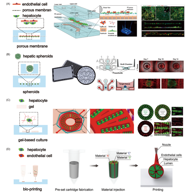
图1 几种在体外利用微流控技术构建三维肝脏生理模型的方法。(A) 基于多孔膜的方法[35];(B) 基于细胞团簇培养[42];(C) 基于凝胶胶原中的细胞培养[49];(D)基于 3D生物打印的构建方法[54]

Fig.1 Microfluidic-based approaches to construct 3D liver-on-chip in vitro. (A) using porous permeable membrane[35]; (B) applying cell spheroids[42]; (C) the gel-based 3D culture[49]; (D) the 3D-printing based construction[54]

2.2 Cell cluster

The method of multicellular clusters is also an important three-dimensional cell culture technique. The basic principle is that under the condition of inhibiting cell attachment, cells tend to form multicellular clusters, which have a multi-layer cell structure and are closely connected with each other. Compared with two-dimensional monolayer cells, it presents a three-dimensional structure close to the body, and can maintain the phenotype and function of cells, and has similar characteristics with solid tissues. Cell clusters can be formed quickly and efficiently based on the micro-pit structure on the microfluidic chip[36]. Lee et al. Designed honeycomb-arrayed dimple arrays for the formation and culture of three-dimensional hepatocyte clusters[37,38]. Compared with two-dimensional culture, three-dimensional clustered hepatocytes showed higher cell activity and function. Luo and Liang et al. Developed a method for dynamic culture of hepatocyte clusters on a chip. The results showed that the metabolic function of hepatocyte clusters in dynamic culture was stronger than that in static culture[39]. Shen et al. Designed a microfluidic liver chip based on hydrogel materials[40]. In the hydrogel chip, liver cells formed hepatocyte clusters in the micropores of the channel outside the chip, and the endothelial cells were cultured with collagen in the inner channel. The results showed that the liver cells showed higher activity within 8 days, and compared with the liver cell cluster culture alone, the co-culture of liver cells and endothelial cells could better reproduce the physiological function of the liver in vivo. Khademhosseini et al. Proposed to construct a pathological model of nonalcoholic fatty liver disease by co-culturing hepatocytes and endothelial cells into multicellular clusters in microchip pits. The results showed that when the hepatocyte cluster contained 20% endothelial cells, the pathogenesis of nonalcoholic fatty liver disease could be better simulated[41]. In addition, the construction of vascular system in vitro is an important method to simulate human tissues and realize the functions of tissues and organs. Therefore, Bonanini et al. Developed a microfluidic organ chip platform integrated with a three-dimensional vascular network and primary liver cells (Fig. 1B)[42]. The platform mainly consists of 384-well plates and 64 microfluidic chips, each of which can induce angiogenesis. On top of the chip is an open system for manual placement of primary hepatocyte clusters. Hepatocyte clusters continue to culture in collagen and eventually interact with vascular endothelial cells to form a stable, perfusable vascular network. In conclusion, three-dimensional culture based on cell clusters can mimic the characteristics of tissues and organs in vivo, and can maintain cell activity and tissue-related functions. The micropit array integrated in the microfluidic chip can quickly and efficiently form hepatocyte clusters, which will play a positive role in the exploration and construction of liver in vitro models and high-content drug screening.

2.3 Gel culture

Polymer gels with good biocompatibility are widely used in three-dimensional cell culture in vitro, and their three-dimensional network structure is very similar to the structure and properties of extracellular matrix molecules in vivo. Cells cultured by gel method are more likely to adhere, promote cell growth and enhance the interaction between cells and matrix, which is a commonly used three-dimensional cell culture method. Compared with traditional gel-cultured cells, cells cultured on microfluidic chips can maintain higher activity. Revzin et al. Developed a microfluidic device for long-term liver culture and maintenance of liver function based on collagen culture[43]. Collagen and hepatocytes are directly injected into the culture chamber through a syringe, and the cells in the collagen can be continuously cultured for more than one month in a device with fluid flow and maintain good physiological function. However, the liver tissue cultured in collagen in 96-well plates lost its phenotype and function within 3 to 5 days. Therefore, this method can be used for future liver disease modeling or personalized liver-directed therapy. In addition, Qin et al. Combined with droplet microfluidic devices, human induced pluripotent stem cells were encapsulated in droplets to form hydrogel capsules, which were continuously cultured and eventually differentiated into liver organoids[44]. The method is easy to operate, scalable and stable, and the cultured liver organs show homogeneity and good cell viability, providing a technical platform for stem cell organoids with higher controllability and fidelity. Three-dimensional liver microarray based on gel method is often used to construct liver lobule structure and explore the interaction between liver parenchymal cells and vascular endothelial cells, hepatic astrocytes or fibroblasts[45][46][47][48]. For example, Banaeiyan et al. Designed a high-throughput liver lobule chip[22]. The chip includes a tissue culture region, a feed region, and a central exit port with a diffusion path between the tissue culture region and the feed region to protect the cells from fluid shear forces. The liver tissue cultured by this method could be continuously cultured for 21 days, and the cells could maintain morphology and function during the culture period. Wang et al. Used an integrated microfluidic chip to fabricate 3D liver lobular microtissue in vitro (Fig. 1C), in which hepatocytes and vascular endothelial cells were cultured in collagen[49]. The experiment proves that the liver tissue cultured by the device retains great drug metabolism capacity, can accurately analyze the interaction between clinical drugs and the potential adverse drug reaction causing liver injury, and provides a new method for the analysis of drug metabolism and hepatotoxicity. In conclusion, three-dimensional cell culture based on gel method has higher controllability and fidelity, and is widely used in the construction of liver organ chip.

2.4 Bioprinting

Bio-3D printing technology is an emerging technology for liver tissue engineering using layered biomanufacturing methods, which is conducive to precise printing of three-dimensional structures and high-throughput manufacturing. Compared with the above three cell culture methods, bio-3D printing technology can directly print personalized tissue cells or organs, which provides convenience for solving the problems of donor shortage, immune rejection and drug treatment[50]. Lin et al. Used a homemade inkjet printer to print microscale cell patterns and used them to achieve cell culture, stimulation, and analysis on a chip[51]. Cells were encapsulated in sodium alginate solution, and the cells were precisely distributed on the slide by software control, and then calcium chloride solution was poured into the microfluidic chip channel to form gel immediately and fix it on the bottom of the chip. This method helps to improve cell patterning efficacy in microfluidic chips and reduce laborious experimental work. In order to achieve a variety of cell layered printing, Cho et al. Used 3D bioprinting to wrap liver cells and endothelial cells in different hydrogels, and finally layered printing. The results showed that compared with the traditional liver cell culture, the liver function of the cells cultured after 3D bioprinting was significantly enhanced[52]. Because 3D printed hepatocytes lack the bile system necessary for bile acid excretion, they can not overcome the drawbacks of in vitro liver testing in drug development, and they have developed liver chips with multiple cell types[53]. The chip has a vascular and biliary system that shows better liver function and is expected to be used for in vitro liver organ testing. In order to control the spatial distribution of cells more accurately, Jin et al. Developed an advanced 3D bioprinting method to realize the presetting of multiple cell cultures and structures at the same time, and successfully constructed the liver lobule structure (Fig. 1D)[54]. The chip has structures similar to hepatic lobules, including liver parenchymal cells, endothelial cells and lumens, showing higher liver-related metabolic activity, such as enhanced secretion of urea and albumin, which proves that this method can maintain structural integrity and improve cell function. In order to print the structure of liver lobule more conveniently, Mandal et al. Developed a bioink based on liver extracellular matrix[55]. Hepatic parenchymal cells, endothelial cells and hepatic stellate cells were encapsulated in bioink in advance, and the structure of hepatic lobules was printed by 3D bioprinter. The results show that the 3D printed liver structure has high metabolic capacity and can predict drug hepatotoxicity more accurately. This method can help accelerate drug development and provide a powerful platform for hepatotoxicity screening. In conclusion, 3D bioprinting technology can produce more anatomically accurate liver structures, including liver-specific spatial structures and vascular networks, which provides a favorable tool for the construction of in vitro liver models for the study of liver diseases and drug screening.

3 Application of microfluidic three-dimensional liver chip

3.1 Liver related disease model

Liver is the place of human metabolism, and its common pathological changes include alcoholic liver disease (ALD), non-alcoholic fatty liver disease (NAFLD), viral hepatitis and metabolic disease type II diabetes mellitus. The establishment of microfluidic liver chip, which can simulate the physiological function and cell state of the liver, provides a good in vitro model and research platform for in-depth understanding of disease mechanisms and finding appropriate treatment options.
ALD is a liver disease caused by long-term heavy drinking, which is a widespread liver disease and seriously endangers human health. The research and development of targeted treatment for ALD has been one of the research hotspots[56]. As shown in Fig. 2A, Lee et al. Constructed a three-dimensional in vitro model of alcoholic liver based on microfluidic technology[57]. They placed rat primary hepatocytes and hepatic stellate cells into a microfluidic chip for dynamic culture under flow, using a self-made osmotic pump to provide a dynamic culture environment. Hepatocytes were treated with different concentrations of alcohol for 3 days, and the results showed that the cells were significantly damaged when the concentration of alcohol was 60 μL/mL and 80 μL/mL, which verified the reversibility and irreversibility of liver function recovery under different conditions. Studies have shown that liver injury can promote the proliferation of hepatic stellate cells, and co-culture of hepatocytes and hepatic stellate cells is more conducive to the recovery of hepatocyte function after alcohol stimulation. This method is helpful to reproduce the physiological environment of ALD, and has great potential in the study of physiological and pathological mechanisms and the testing of drug toxicity.
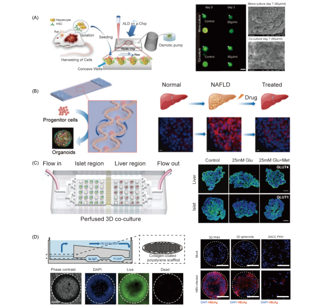
图2 基于微流控技术的体外肝脏模型用于肝脏功能与病理机制的研究。(A) 基于微流控技术的酒精肝三维体外模型[57];(B) 基于微流控技术的非酒精性脂肪肝药物高通量筛选平台[61];(C) 基于微流控多类器官系统用于研究肝脏代谢类疾病II型糖尿病[62];(D) 基于微流控芯片研究乙型肝炎病毒感染过程[64]

Fig.2 The microfluidic-based 3D liver-on-chip for studying the hepatic diseases. (A) the microfluidic technology-based 3D liver-on-chip for simulating alcoholic liver disease (ALD)[57]; (B) the microfluidic technology-based high-throughput screening platform for emulating nonalcoholic fatty liver disease (NAFLD)[61]; (C) the microfluidic multi-organoid system for recapitulating type II diabetes mellitus (T2DM)[62]; (D) the liver-on-chip platform for studying the hepatitis B virus (HBV) infection process[64]

In recent years, with the change of dietary structure, NAFLD has become the most common liver disease worldwide. The disease is not associated with alcohol intake, but with excessive intake of high-sugar and high-fat foods, which often lead to liver fibrosis, liver failure and even liver cancer. Due to the lack of drug screening platforms for NAFLD, the understanding of its causes has been very limited[58~60]. As shown in Fig. 2B, Yu et al. Developed a 100-well NAFLD drug screening platform based on microfluidics. The chip can generate liver organoid clusters with uniform size and reshape the main structure of NAFLD[61]. Compared with the organoid cultured on the chip after pre-differentiation, the organoid differentiated in situ in the chip has a better chemical microenvironment, which can promote liver differentiation, improve liver function, and enhance cell polarity and bile duct structure. The results showed that the cells were induced with free fatty acids, and the cells showed a high level of lipid accumulation, affecting blood glucose regulation and reducing AKT phosphorylation in organoids. As a sensitive high-throughput drug testing platform, this method can help accelerate drug development for NAFLD.
Type 2 diabetes mellitus is a chronic metabolic disease, which accounts for 90% of diabetic patients. Qin et al. Proposed a microfluidic multi-organoid system[62]. As shown in Figure 2C, human induced pluripotent stem cells (hiPSCs) self-organize to form clusters that induce differentiation to form liver and islet organoids. Organoid clusters were perfused in this system and co-cultured continuously for one month. The results showed that the cells of the liver and islet organoids were active and the mediators and secreted metabolites between the organs could be exchanged, which was suitable for exploring the insulin and glucose regulation of the human liver-islet axis in disease. This method is helpful to explore the interaction of multiple organs, and provides a platform for the study of disease mechanism and the screening of disease-related drugs.
Hepatitis B (HBV) is an infectious disease of the liver caused by the hepatitis B virus[63]. Traditionally, cell lines and two-dimensional cultured primary cells are often unable to simulate the complexity of the liver, resulting in limited research on the process of HBV infection. Dorner et al. Used 3D microfluidic chips to study the process of hepatitis B virus infection[64]. As shown in Figure 2D, collagen was coated on the styrene scaffold for the culture of primary human hepatocytes, and the system could last for 40 days, thus allowing the reconstruction of various steps of the HBV life cycle. This work proves that the co-culture of liver parenchymal cells and non-parenchymal cells can facilitate the identification of the source of cells producing immune effector factors, and provides a valuable research platform for preclinical HBV.

3.2 Liver drug screening model

The development of safe and effective clinical drugs is a time-consuming, difficult and expensive process. Studies have shown that it takes more than 10 years and costs about $1.7 billion from the development of new drugs to their approval for marketing[65]. In this process, most of the compounds are eliminated due to organ toxicity or lack of efficacy. Hepatotoxicity of drugs is a problem that must be faced and solved in preclinical drug research and development, and it is also an important reason why drugs are withdrawn after being introduced into the market[66]. Animal models similar to humans are usually used for safety verification such as hepatotoxicity before clinical practice, but the use of animal models in the early stage of drug development is not only expensive and time-consuming, but also has species differences, and the effects of drugs on the cellular level can not be observed at any time, only through the observation of animal phenotype, so the mechanism of disease can not be explored[67]. Therefore, the development of efficient, convenient and reliable microfluidic liver organ chips for rapid evaluation of drug hepatotoxicity has become an urgent need in current research.
Lin et al. Made a microfluidic device that can simulate the metabolic function of the liver and detect the metabolism of prodrugs[68]. Capecitabine (CAP), as a prodrug, is non-toxic to cells and can only be converted into 5-fluorouracil (5-FU), an effective chemotherapeutic drug that is toxic to cells, after being metabolized into an active intermediate by the liver. They achieved two-dimensional co-culture of liver cells (HepG2) and breast cancer cells (MCF-7) on a microfluidic chip, and integrated a solid phase extraction column on the chip to extract drug components, which can prove the process of drug metabolism by the liver and the toxic effect of drugs on tumors through online detection by mass spectrometer. During metabolism in vivo, the drug may produce side effects in other organs, even though it appears to have no toxic effect on the liver[65]. Therefore, the development of multi-organ chip system is conducive to early detection of drug off-target side effects and other risks in the process of drug testing. Hickman et al. Of Hesperos, an organ chip company, built a multi-organ chip system to predict preclinical target efficacy, liver metabolic conversion, and drug off-target toxicity (Figure 3A)[69]. Chamber 1 is used to culture liver cells to simulate the first step of drug metabolism when drugs enter the human body, and leukemia cells can be directly cultured in other chambers to evaluate the inhibitory effect of drugs on bone marrow proliferation and the relationship between liver and bone marrow. In addition, the chip can also culture cardiomyocytes and microelectrode arrays for measuring myocardium in chambers 2 and 4, respectively, and culture different tumor cells in chambers 3 and 5 to assess the effects of drugs metabolized by the liver on the heart and tumors. This method provides a new platform and protocol for preclinical drug efficacy and safety assessment.
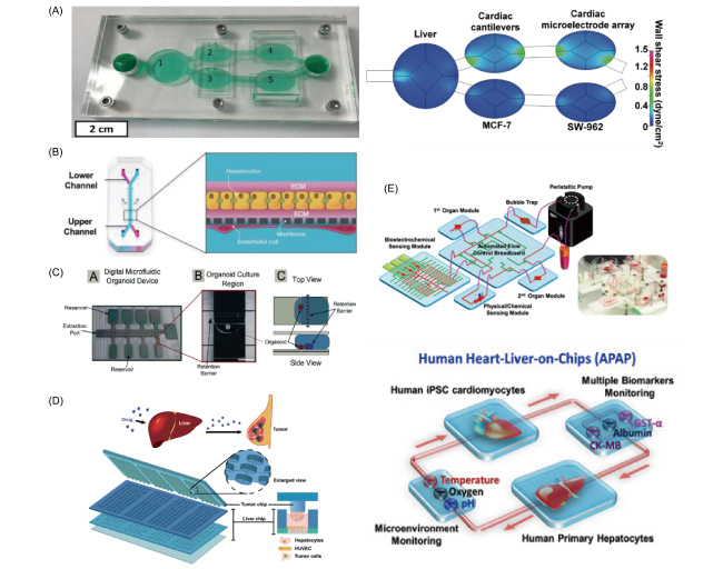
图3 基于微流控技术的体外肝脏器官芯片模型用于药物代谢与筛选以及安全性评估研究。(A)基于二维培养原代肝细胞、诱导分化的心肌细胞以及不同肿瘤细胞共同构成的多器官芯片用于药物研究[69];(B) 基于集成多孔膜方法的三维肝脏器官芯片用于测试药物对于不同物种(人、鼠、狗)细胞的毒性[70];(C) 集成有微电极的高通量数字化微流控芯片装置用于肝脏器官芯片的研究[72];(D) 利用阵列微孔设计的集成高通量肝脏-肿瘤器官芯片用于多种前体药物活性和效用的研究[77];(E) 集成化器官芯片用于原代肝细胞、诱导分化的心肌细胞的药物生物效应的研究。芯片上集成有包括电化学传感器在内的多种生物传感器[80]

Fig.3 3D liver-on-chip system for drug metabolism and efficacy screening. (A) a multi-organ chip system for studying drug efficacy and safety. Different cells including primary hepatocytes, iPSC derived cardiomyocytes, and tumor cells can be cultured on a single chip[69]; (B) the porous membrane was used to construct 3D liver-on-chip for testing drug toxicities to different species (human, rat, dog)[70]; (C) a high throughput digital microfluidic device for study of liver-on-chip[72]; (D) an integrated high throughput biomimetic system for studying liver and tumor interaction. The device was designed with array microwells for testing prodrug activity and efficacy[77]; (E) multisensor-integrated organs-on-chips for studying the drug induced biological effects in primary hepatocytes and induced differentiation of cardiomyocytes. The chip integrated various microbioreactors including electrochemical biosensors[80]

Two-dimensional cell culture is the main culture method for drug screening in vitro, but the cells cultured in two dimensions do not have a specific tissue structure, so three-dimensional cell models are usually used for secondary screening. Therefore, scientists have gradually shifted their focus to the development of three-dimensional liver organ microfluidic chips. As shown in Figure 3B, Hamilton et al. Of Emulate, an organ chip company, developed a microfluidic chip based on a porous membrane to simulate the physiological structure of the liver[70]. The chip is divided into an upper layer and a lower layer by using a porous membrane, wherein the upper layer is used for culturing liver cells by a sandwich method, and the lower layer is used for culturing liver-related cells (including endothelial cells, Kuffer cells and hepatic stellate cells), so that the complex physiological structure and the physical and chemical conditions in the liver are simulated. This method provides a more effective way to detect hepatotoxicity, hepatocyte injury, steatosis, cholestasis and fibrosis, and species-specific aspects after treatment with drugs. In early drug screening, it is very important and necessary for the overall process of drug research and development to be able to screen out drugs with greater side effects in time. Therefore, it is of great necessity to develop a high-throughput and high-content drug screening platform[71]. Wheeler et al. Developed a microfluidic organoid platform based on a digital microfluidic system for drug screening, as shown in Fig. 3C[72]. The device can generate addressable free-floating liver organoids to explore the effects of different experimental conditions on liver organoids. The experimental results proved that the organoids exhibited fibroblast-dependent contractile behavior, showing high cell activity and metabolic function. The platform could be a cost-effective tool in the early drug discovery phase. A prodrug is a substance that is less active than the original drug but becomes active after hepatic metabolism, which enhances drug targeting and reduces drug toxicity and side effects[73~76]. As shown in Fig. 3D, Jiang et al. Developed an integrated biomimetic array chip for prodrug screening[77].
The chip consists of two functional chips: a liver chip and a tumor chip, and both the liver and the tumor are cultured in collagen in three dimensions. Using this device, they evaluated the anticancer bioactivity induced by prodrug metabolism and studied the interaction between different drugs. The chip can be used to simultaneously evaluate the anticancer bioactivity and possible hepatotoxicity of prodrugs. Overall, this approach provides a liver organ-on-chip platform with integrated functionality and operational simplicity and high throughput, which can be used as an auxiliary screening tool in the drug development process. In addition to hepatotoxicity, cardiovascular toxicity is also a side effect of drugs that can not be ignored[78,79]. As shown in Fig. 3e, Khademhosseini et al. Developed a multi-organ system integrated with microfluidic biosensors to detect the toxic effects of drugs on liver and heart organs[80]. The device contains a multi-organ chip and multiple biosensors. Liver and cardiomyocyte culture is integrated on the multi-organ chip, which is mainly used to detect the toxic effects of drugs on liver and heart. The biosensor is mainly used for in situ continuous online detection of small doses of biomarkers (such as albumin, CK-MB, etc.) secreted by cells to determine the hepatotoxicity and cardiotoxicity of drugs. In addition, the device can also detect the physiological parameters of the system, such as oxygen, pH and temperature. This method effectively combines multi-organ chips with sensors for online detection, providing a technical platform for studying multi-organ interactions and simultaneously detecting biomarkers.

4 Conclusion and prospect

Liver is the most important detoxification organ in human body, which is responsible for the metabolic transformation of almost all compounds and drugs. In recent years, animal models have been widely criticized for their limitations in predicting the toxicity, safety, and efficacy of new drugs in humans. China has also issued some guidelines for non-clinical research and evaluation, suggesting that cell-based and tissue-based models (such as 2D or 3D tissue models, organoid and microfluidic models) can be used to provide useful supplementary information for the evaluation of efficacy and safety when relevant animal models are lacking. Therefore, emerging technologies, such as in vitro microphysiological systems based on microfluidics and organ chips, are attracting more and more attention. The construction of organ chips that can simulate the structure and function of the liver in vitro can better explore the physiological and pathological mechanisms of the liver and the response of drugs in the process of liver metabolism. In this paper, we preliminarily summarized four ways to construct three-dimensional liver organ chips: among them, the porous membrane method and 3D bioprinting method are convenient to reshape the physiological structure of the liver and are widely used to explore the physiological and pathological mechanisms of the liver; The liver organ cultured by the scaffold-free cell cluster method and the scaffold-free gel method has stronger function, which provides a good method for exploring the response of drugs in vivo and the study of drug hepatotoxicity. At present, there are still some problems in organ chip technology: for example, the organs on the chip simply simulate the structure and function of human organs, and their complexity is ultimately difficult to compare with real internal organs. Secondly, the physiological and biochemical processes in the body are extremely complex, often involving multiple organ interactions. Therefore, it is a challenge to simulate multiple organs and link them to each other on the chip at the same time. Finally, the standardization of organ chips and integrated sensor detection are still in the exploratory stage. In the future, we need to increase investment to promote the development of organ chip research and related industries, so that China can occupy a favorable position in the new round of international competition in the field of organ chips.
[1]
Stanger B Z. Annu. Rev. Physiol., 2015, 77: 179.

[2]
Trefts E, Gannon M, Wasserman D H. Curr. Biol., 2017, 27(21): R1147.

[3]
Polidoro M A, Ferrari E, Marzorati S, Lleo A, Rasponi M. Liver Int., 2021, 41(8): 1744.

[4]
Kaplowitz N. Nat. Rev. Drug Discov., 2005, 4(6): 489.

[5]
Duncombe T A, Tentori A M, Herr A E. Nat. Rev. Mol. Cell Biol., 2015, 16(9): 554.

[6]
Whitesides G M. Nature, 2006, 442(7101): 368.

[7]
Huh D, Matthews B D, Mammoto A, Montoya-Zavala M, Hsin H Y, Ingber D E. Science, 2010, 328(5986): 1662.

[8]
Zhao L, Wang X Y. Trac Trends Anal. Chem., 2023, 158: 116864.

[9]
Bhatia S N, Ingber D E. Nat Biotechnol., 2014, 32: 760.

[10]
Zhang B., Radisic M. Lab Chip, 2017, 17: 2395.

[11]
Vunjak-Novakovic G, Ronaldson-Bouchard K, Radisic M. Cell, 2021, 184(18): 4597.

[12]
Zhang B Y, Korolj A, Lai B F L, Radisic M. Nat. Rev. Mater., 2018, 3(8): 257.

[13]
Kaur S, Kidambi S, Ortega-Ribera M, Thuy L T T, Nieto N, Cogger V C, Xie W F, Tacke F, Gracia-Sancho J. Cell. Mol. Gastroenterol. Hepatol., 2023, 15(3): 559.

[14]
Polini A, Prodanov L, Bhise N S, Manoharan V, Dokmeci M R, Khademhosseini A. Expert Opin. Drug Discov., 2014, 9(4): 335.

[15]
Ingber D E. Nat. Rev. Genet., 2022, 23(8): 467.

[16]
Li X, George S M, Vernetti L, Gough A H, Lansing Taylor D. Lab Chip, 2018, 18(17): 2614.

[17]
Moradi E, Jalili-Firoozinezhad S, Solati-Hashjin M. Acta Biomater., 2020, 116: 67.

[18]
Lin R Z, Chang H Y. Biotechnol. J., 2008, 3(9/10): 1172.

[19]
ZiÓłkowska K, Kwapiszewski R, BrzÓzka Z. New J. Chem., 2011, 35(5): 979.

[20]
Agrawal A A, Nehilla B J, Reisig K V, Gaborski T R, Fang D Z, Striemer C C, Fauchet P M, McGrath J L. Biomaterials, 2010, 31(20): 5408.

[21]
Zhao L, Xiu J D, Liu Y, Zhang T Y, Pan W J, Zheng X N, Zhang X J. Sci. Rep., 2019, 9: 19717.

[22]
Banaeiyan A A, Theobald J, Paukštyte J, Wölfl S, Adiels C B, Goksör M. Biofabrication, 2017, 9(1): 015014.

[23]
Wu J, Chen Q S, Liu W, He Z Y, Lin J M. TRAC Trends Anal. Chem., 2017, 87: 19.

[24]
Underhill G H, Khetani S R. Cell. Mol. Gastroenterol. Hepatol., 2018, 5(3): 426.

[25]
Deng J, Wei W B, Chen Z Z, Lin B C, Zhao W J, Luo Y, Zhang X L. Micromachines, 2019, 10(10): 676.

[26]
Fischbach C, Chen R, Matsumoto T, Schmelzle T, Brugge J S, Polverini P J, Mooney D J. Nat. Methods, 2007, 4(10): 855.

[27]
Fernandes R, Luo X L, Tsao C Y, Payne G F, Ghodssi R, Rubloff G W, Bentley W E. Lab Chip, 2010, 10(9): 1128.

[28]
Zheng Y B, Ma L D, Wu J L, Wang Y M, Meng X S, Hu P, Liang Q L, Xie Y Y, Luo G A. Talanta, 2022, 241: 123262.

[29]
Kim M Y, Li D J, Pham L K, Wong B G, Hui E E. J. Membr. Sci., 2014, 452: 460.

[30]
Rahimnejad M, Rasouli F, Jahangiri S, Ahmadi S, Rabiee N, Ramezani Farani M, Akhavan O, Asadnia M, Fatahi Y, Hong S, Lee J, Lee J M, Hahn S K. ACS Biomater. Sci. Eng., 2022, 8(12): 5038.

[31]
Hegde M, Jindal R, Bhushan A, Bale S S, McCarty W J, Golberg I, Usta O B, Yarmush M L. Lab Chip, 2014, 14(12): 2033.

[32]
Kim J, Lee C, Kim I, Ro J, Kim J, Min Y, Park J, Sunkara V, Park Y S, Michael I, Kim Y A, Lee H J, Cho Y K. ACS Nano, 2020, 14(11): 14971.

[33]
Kang Y B, Sodunke T R, Lamontagne J, Cirillo J, Rajiv C, Bouchard M J, Noh M. Biotechnol. Bioeng., 2015, 112(12): 2571.

[34]
Rennert K, Steinborn S, Gröger M, Ungerböck B, Jank A M, Ehgartner J, Nietzsche S, Dinger J L, Kiehntopf M, Funke H, Peters F T, Lupp A, Gärtner C, Mayr T, Bauer M, Huber O, Mosig A S. Biomaterials, 2015, 71: 119.

[35]
Du Y, Li N, Yang H, Luo C H, Gong Y X, Tong C F, Gao Y X, S Q, Long M. Lab Chip, 2017, 17(5): 782.

[36]
Bhise N S, Manoharan V, Massa S, Tamayol A, Ghaderi M, Miscuglio M, Lang Q, Shrike Zhang Y, Shin S R, Calzone G, Annabi N, Shupe T D, Bishop C E, Atala A, Dokmeci M R, Khademhosseini A. Biofabrication, 2016, 8(1): 014101.

[37]
Lee G H, Lee J S, Lee G H, Joung W Y, Kim S H, Lee S H, Park J Y, Kim D H. Biofabrication, 2017, 10(1): 015001.

[38]
Lee G, Lee J, Oh H, Lee S. PLoS One, 2016, 11(8): e0161026.

[39]
Ma L D, Wang Y T, Wang J R, Wu J L, Meng X S, Hu P, Mu X, Liang Q L, Luo G A. Lab Chip, 2018, 18(17): 2547.

[40]
Meng Q, Wang Y, Li Y, Shen C. Biotechnol. Bioeng., 2021, 118: 612.

[41]
Lasli S, Kim H J, Lee K J, Suurmond C A E, Goudie M, Bandaru P, Sun W J, Zhang S M, Zhang N Y, Ahadian S, Dokmeci M R, Lee J M, Khademhosseini A. Adv. Biosys., 2019, 3(8): 1900104.

[42]
Bonanini F, Kurek D, Previdi S, Nicolas A, Hendriks D, Ruiter S, Meyer M, ClapÉs Cabrer M, Dinkelberg R, García S B, Kramer B, Olivier T, Hu H L, LÓpez-Iglesias C, Schavemaker F, Walinga E, Dutta D, Queiroz K, Domansky K, Ronden B, Joore J, Lanz H L, Peters P J, Trietsch S J, Clevers H, Vulto P. Angiogenesis, 2022, 25(4): 455.

[43]
de Hoyos-Vega J M, Hong H J, Loutherback K, Stybayeva G, Revzin A. Adv. Mater. Technol., 2023, 8(2): 2201121.

[44]
Wang Y Q, Liu H T, Zhang M, Wang H, Chen W W, Qin J H. Biomater. Sci., 2020, 8(19): 5476.

[45]
Ya S N, Ding W P, Li S B, Du K, Zhang Y Y, Li C P, Liu J, Li F F, Li P, Luo T Z, He L Q, Xu A, Gao D Y, Qiu B S. ACS Appl. Mater. Interfaces, 2021, 13(28): 32640.

[46]
Jin Y, Kim J, Lee J S, Min S, Kim S, Ahn D H, Kim Y G, Cho S W. Adv. Funct. Mater., 2018, 28(37): 1801954.

[47]
Kuang J J, Sun W, Zhang M, Kang L, Yang S L, Zhang H Y, Wang Y R, Hu P. Chin. Chem. Lett., 2023, 34(3): 107573.

[48]
Cui J, Wang H P, Zheng Z Q, Shi Q, Sun T, Huang Q, Fukuda T. Biofabrication, 2018, 11(1): 015016.

[49]
Ma C, Zhao L, Zhou E M, Xu J, Shen S F, Wang J Y. Anal. Chem., 2016, 88(3): 1719.

[50]
Chang R, Emami K, Wu H L, Sun W. Biofabrication, 2010, 2(4): 045004.

[51]
Zhang J, Chen F M, He Z Y, Ma Y, Uchiyama K, Lin J M. Analyst, 2016, 141(10): 2940.

[52]
Lee H, Cho D W. Lab Chip, 2016, 16(14): 2618.

[53]
Lee H, Chae S H, Kim J Y, Han W, Kim J, Choi Y, Cho D W. Biofabrication, 2019, 11(2): 025001.

[54]
Kang D G, Hong G, An S, Jang I, Yun W S, Shim J H, Jin S W. Small, 2020, 16(13): 1905505.

[55]
Janani G, Priya S, Dey S, Mandal B B. ACS Appl. Mater. Interfaces, 2022, 14(8): 10167.

[56]
Deng J, Chen Z Z, Zhang X L, Luo Y, Wu Z Z, Lu Y, Liu T J, Zhao W J, Lin B C. Biomed. Microdevices, 2019, 21(3): 57.

[57]
Lee J, Choi B, No D Y, Lee G, Lee S R, Oh H, Lee S H. Integr. Biol., 2016, 8(3): 302.

[58]
Wang Y Q, Wang H, Deng P W, Tao T T, Liu H T, Wu S, Chen W W, Qin J H. ACS Biomater. Sci. Eng., 2020, 6(10): 5734.

[59]
Du K, Li S B, Li C P, Li P, Miao C G, Luo T Z, Qiu B S, Ding W P. Acta Biomater., 2021, 134: 228.

[60]
Gori M, Simonelli M C, Giannitelli S M, Businaro L, Trombetta M, Rainer A. PLoS One, 2016, 11(7): e0159729.

[61]
Teng Y, Zhao Z X, Tasnim F, Huang X Z, Yu H. Biomaterials, 2021, 275: 120904.

[62]
Tao T T, Deng P W, Wang Y Q, Zhang X, Guo Y Q, Chen W W, Qin J H. Adv. Sci., 2022, 9(5): 2270029.

[63]
Lee S W L, Adriani G, Ceccarello E, Pavesi A, Tan A T, Bertoletti A, Dale Kamm R, Wong S C. Front. Immunol., 2018, 9: 416.

[64]
Ortega-Prieto A M, Skelton J K, Wai S N, Large E, Lussignol M, Vizcay-Barrena G, Hughes D, Fleck R A, Thursz M, Catanese M T, Dorner M. Nat. Commun., 2018, 9: 682.

[65]
Cook D, Brown D, Alexander R, March R, Morgan P, Satterthwaite G, Pangalos M N. Nat. Rev. Drug Discov., 2014, 13(6): 419.

[66]
Mirahmad M, Sabourian R, Mahdavi M, Larijani B, Safavi M. Drug Metab. Rev., 2022, 54(2): 161.

[67]
Ma C, Peng Y S, Li H T, Chen W Q. Trends Pharmacol. Sci., 2021, 42(2): 119.

[68]
Zhang J, Wu J, Li H, Chen Q, Lin J M. Biosens. Bioelectron., 2015, 68: 322.

[69]
McAleer C W, Long C J, Elbrecht D, Sasserath T, Bridges L R, Rumsey J W, Martin C, Schnepper M, Wang Y, Schuler F, Roth A B, Funk C, Shuler M L, Hickman J J. Sci. Transl. Med., 2019, 11(497): eaav1386.

[70]
Jang K J, Otieno M A, Ronxhi J, Lim H K, Ewart L, Kodella K R, Petropolis D B, Kulkarni G, Rubins J E, Conegliano D, Nawroth J, Simic D, Lam W, Singer M, Barale E, Singh B, Sonee M, Streeter A J, Manthey C, Jones B, Srivastava A, Andersson L C, Williams D, Park H, Barrile R, Sliz J, Herland A, Haney S, Karalis K, Ingber D E, Hamilton G A. Sci. Transl. Med., 2019, 11(517): eaax5516.

[71]
Chen Y L, Gao D, Liu H X, Lin S, Jiang Y Y. Anal. Chim. Acta, 2015, 898: 85.

[72]
Au S H, Dean Chamberlain M, Mahesh S, Sefton M V, Wheeler A R. Lab Chip, 2014, 14(17): 3290.

[73]
Ai X N, Zhao L, Lu Y Y, Hou Y, Lv T, Jiang Y, Tu P F, Guo X Y. Anal. Chem., 2020, 92(17): 11696.

[74]
Frey O, Misun P M, Fluri D A, Hengstler J G, Hierlemann A. Nat. Commun., 2014, 5: 4250.

[75]
Oleaga C, Riu A, Rothemund S, Lavado A, McAleer C W, Long C J, Persaud K, Narasimhan N S, Tran M, Roles J, Carmona-Moran C A, Sasserath T, Elbrecht D H, Kumanchik L, Bridges L R, Martin C, Schnepper M T, Ekman G, Jackson M, Wang Y I, Note R, Langer J, Teissier S, Hickman J J. Biomaterials, 2018, 182: 176.

[76]
Rajan S A P, Aleman J, Wan M M, Pourhabibi Zarandi N, Nzou G, Murphy S, Bishop C E, Sadri-Ardekani H, Shupe T, Atala A, Hall A R, Skardal A. Acta Biomater., 2020, 106: 124.

[77]
Hou Y, Ai X N, Zhao L, Gao Z, Wang Y J, Lu Y, Tu P F, Jiang Y. Lab Chip, 2020, 20(14): 2482.

[78]
Ronaldson-Bouchard K, Teles D, Yeager K, Tavakol D N, Zhao Y M, Chramiec A, Tagore S, Summers M, Stylianos S, Tamargo M, Lee B M, Halligan S P, Abaci E H, Guo Z Y, JackÓw J, Pappalardo A, Shih J, Soni R K, Sonar S, German C, Christiano A M, Califano A, Hirschi K K, Chen C S, Przekwas A, Vunjak-Novakovic G. Nat. Biomed. Eng, 2022, 6(4): 351.

[79]
Herland A, Maoz B M, Das D, Somayaji M R, Prantil-Baun R, Novak R, Cronce M, Huffstater T, Jeanty S S F, Ingram M, Chalkiadaki A, Benson Chou D, Marquez S, Delahanty A, Jalili-Firoozinezhad S, Milton Y, Sontheimer-Phelps A, Swenor B, Levy O, Parker K K, Przekwas A, Ingber D E. Nat. Biomed. Eng., 2020, 4(4): 421.

[80]
Zhang Y S, Aleman J, Shin S R, Kilic T, Kim D, Ali Mousavi Shaegh S, Massa S, Riahi R, Chae S, Hu N, Avci H, Zhang W J, Silvestri A, Sanati Nezhad A, Manbohi A, De Ferrari F, Polini A, Calzone G, Shaikh N, Alerasool P, Budina E, Kang J, Bhise N, Ribas J, Pourmand A, Skardal A, Shupe T, Bishop C E, Dokmeci M R, Atala A, Khademhosseini A. Proc. Natl. Acad. Sci. U. S. A., 2017, 114(12): E2293.

Outlines

/