
Design and Application of Chiral Plasmonic Core-Shell Nanostructures
Received date: 2022-12-28
Revised date: 2023-05-26
Online published: 2023-07-18
Supported by
National Natural Science Foundation of China(22072181)
Innovation Fund Project for Graduate Student of China University of Petroleum(23CX04031A)
Fundamental Research Funds for the Central Universities.
Chirality describes the geometrical feature of an object that cannot overlap with its mirror image and has been a crucial concept in chemistry and biology since the 19th century. With the development of nanotechnology, chiral plasmonic nanomaterials are becoming the research focuses for scientists to develop chiral functional materials due to the special chiral optical properties and good biocompatibility. However, the relatively weak chiral signals limit their applications. Chiral plasmonic core-shell nanostructures combine the chiral plasmonic properties and core-shell structures, which is an effective strategy to amplify chiral signals. In addition, the core-shell nanostructure integrates the properties of both internal and external materials to complement each other, which can further improve the physicochemical properties and enhance the performance in various fields. This paper summarizes the design strategies of chiral plasmonic core-shell nanostructures based on the spatial distribution of chiral molecules, and reviews their applications in the fields of ultrasensitive sensing and chiral catalysis. We analyze the existing problems and their possible solutions, and make an outlook on their future development.
1 Introduction
2 Design strategies for chiral plasmonic core-shell nanostructures
2.1 Chiral molecules distributed on the shell
2.2 Chiral molecules distributed on the core
2.3 Chiral molecules distributed in the core-shell gap
3 Application of chiral plasmonic core-shell nanostructures
3.1 Ultra-sensitive sensing
3.2 Chiral catalysis
4 Conclusion and outlook

Wenliang Liu , Yuqi Wang , Xiaohan Li , Xuanyu Zhang , Jiqian Wang . Design and Application of Chiral Plasmonic Core-Shell Nanostructures[J]. Progress in Chemistry, 2023 , 35(8) : 1168 -1176 . DOI: 10.7536/PC221222
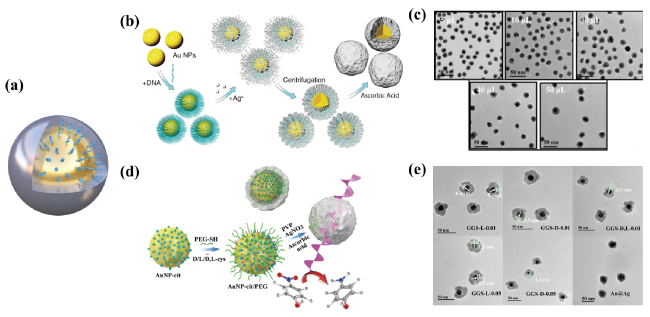
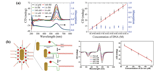
图1 (a)Au@DNA修饰的Ag的示意图;(b)Au@DNA修饰的Ag的制备示意图;(c)Au@DNA修饰的Ag的TEM图;(d)DNA以及Au@DNA修饰的Ag的紫外-可见吸收光谱;(e)DNA、Au@Ag、Au@DNA修饰的Ag的圆二色光谱[38]Fig.1 (a)Schematic diagram of Au@DNA modified Ag. (b)Schematic diagram of the preparation of Au@DNA modified Ag. (c)TEM image of Au@DNA modified Ag. (d)UV-vis absorption spectra of DNA and Au@DNA modified Ag. (e)CD spectra of DNA, Au@Ag, and Au@DNA modified Ag[38] |
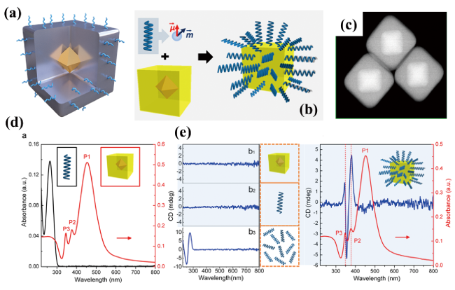
图3 (a)Au@半胱氨酸修饰的Ag的示意图;(b)Au@半胱氨酸修饰的Ag的TEM图;(c)Au@半胱氨酸修饰的Ag的圆二色光谱;(d~f)Au@半胱氨酸修饰的Ag的手性响应信号放大策略[43]Fig.3 (a)Schematic diagram of Au@cysteine modified Ag. (b)TEM images of Au@cysteine modified Ag(c)CD spectra of Au@cysteine modified Ag. (d~f)Chiral response signal amplification strategy for Au@cysteine modified Ag[43] |
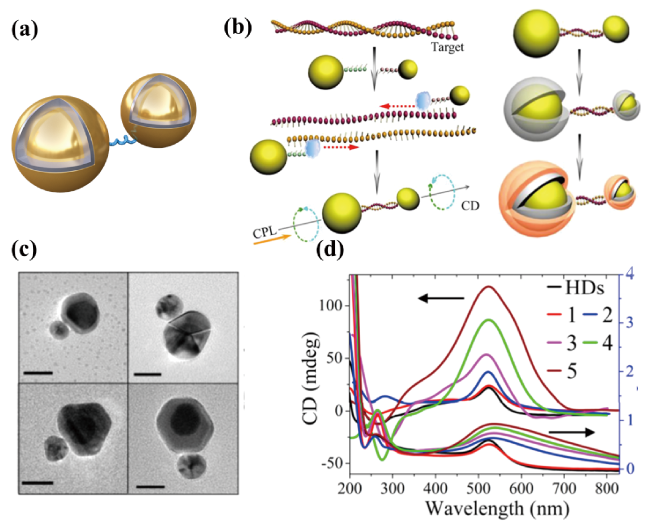
图4 (a)手性分子修饰的Au@Ag的示意图;(b)DNA修饰的Au@Ag的制备示意图;(c)DNA修饰的Au@Ag的TEM图[44] ;(d)半胱氨酸修饰的Au@Ag的制备示意图;(e)半胱氨酸修饰的Au@Ag的TEM图[45]Fig.4 (a)Schematic diagram of chiral molecules modified Au@Ag. (b)Schematic diagram of the preparation of DNA modified Au@Ag.(c)TEM images of DNA modified Au@Ag[44]. (d)Schematic diagram of the preparation of cysteine modified Au@Ag.(e)TEM images of cysteine modified Au@Ag[45] |
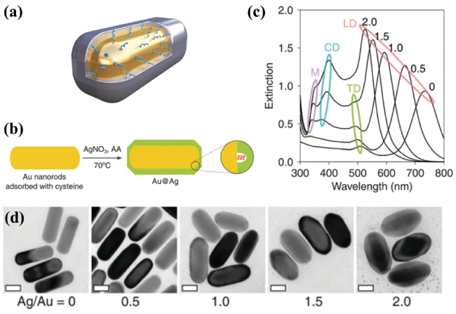
图5 (a)半胱氨酸修饰的Au@Ag的示意图;(b)半胱氨酸修饰的Au@Ag的制备示意图;(c)Au@半胱氨酸修饰的Ag的圆二色光谱;(d~f)Au@半胱氨酸修饰的Ag的手性响应信号放大策略[46]Fig.5 (a)Schematic diagram of cysteine modified Au@Ag. (b)TEM image of cysteine modified Au@Ag.(c)CD spectra of cysteine modified Au@Ag. (d~f)Chiral response signal amplification strategy for cysteine modified Au@Ag[46] |
表1 手性等离子体核壳纳米结构中手性分子的空间分布及其不对称因子Table 1 Spatial distribution of chiral molecules in chiral plasmonic core-shell nanostructures and their g factors |
| Spatial distribution of chiral molecules | Materials | g-factors | ref |
|---|---|---|---|
| Chiral molecules distributed on the shell | Au@DNA modified Ag | 4.4×10⁻³ | 38 |
| DNA bridged Au@AgAu | 1.21×10⁻² | 39 | |
| Au@cysteine modified Ag | 1.45×10⁻³ | 43 | |
| Chiral molecules distributed on the core | DNA modified Au@Ag | 1.93×10⁻² | 44 |
| cysteine modified Au@Ag | 1×10⁻² | 45 | |
| cysteine modified Au@Ag | 1.3×10⁻³ | 46 | |
| Chiral molecules distributed in the core-shell gap | penicillamine modified Au@AgAu | 2.1×10⁻² | 47 |
| [1] |
|
| [2] |
|
| [3] |
|
| [4] |
|
| [5] |
|
| [6] |
|
| [7] |
|
| [8] |
|
| [9] |
|
| [10] |
|
| [11] |
|
| [12] |
|
| [13] |
|
| [14] |
|
| [15] |
|
| [16] |
|
| [17] |
|
| [18] |
|
| [19] |
|
| [20] |
|
| [21] |
|
| [22] |
|
| [23] |
|
| [24] |
|
| [25] |
|
| [26] |
|
| [27] |
|
| [28] |
|
| [29] |
|
| [30] |
|
| [31] |
|
| [32] |
|
| [33] |
|
| [34] |
|
| [35] |
|
| [36] |
|
| [37] |
|
| [38] |
|
| [39] |
|
| [40] |
|
| [41] |
|
| [42] |
|
| [43] |
|
| [44] |
|
| [45] |
|
| [46] |
|
| [47] |
|
| [48] |
|
| [49] |
|
| [50] |
|
| [51] |
|
| [52] |
|
| [53] |
|
| [54] |
|
| [55] |
|
| [56] |
|
| [57] |
|
| [58] |
|
| [59] |
|
| [60] |
|
/
| 〈 |
|
〉 |
