Home Journals Progress in Chemistry
Progress in Chemistry

Abbreviation (ISO4): Prog Chem      Editor in chief: Jincai ZHAO

About  /  Aim & scope  /  Editorial board  /  Indexed  /  Contact  / 
Review

Controlled Synthesis of Silver Nanomaterials and Their Environmental Applications

  • Ziyu Pan ,
  • Haodong Ji , *
Expand
  • School of Environment and Energy, Peking University Shenzhen Graduate School,Shenzhen 518055, China
*Corresponding author e-mail:

Received date: 2022-12-28

  Revised date: 2023-02-27

  Online published: 2023-05-15

Supported by

National Natural Science Foundation of China(52100069)

Shenzhen Science and Technology Program(JCYJ20220531093205013)

Abstract

Silver nanomaterials have been widely used in catalysis, medicine, environment and other fields due to their high catalytic activity, fine biocompatibility, unique physical and chemical properties. This review first introduced the species, properties and synthetic strategy of silver nanomaterials, summarized controllable synthesis method in detail, and discussed the new achievements of machine learning in the synthesis of silver nanomaterials. Then, we reviewed the applications of silver nanomaterials in the environment such as pollutant removal, sterilization and virus inactivation, sensor and so on. Based on this, the species, controlled synthesis and environmental applications of silver nanomaterials were reviewed and prospected in this paper.

Contents

1 Introduction

2 Types and synthesis methods of silver nanomaterials

2.1 Types and synthesis methods of silver nanomaterials composed of only silver element

2.2 Types and synthesis methods of silver nanomaterials of containing two or more elements

2.3 The types and synthesis methods of silver nanomaterials with different carriers

2.4 Types and synthesis methods of silver oxide,silver halide and other nanomaterials

3 Environmental applications of silver nanomaterials

3.1 Application of silver nanomaterials in pollutants-adsorption and catalytic degradation

3.2 Application of silver nanomaterials in water purification,antibacterial and antiviral

3.3 Application of silver nanomaterials in the treatment of toxic metal wastewater-sensor

4 Summary and prospects for the future

Cite this article

Ziyu Pan , Haodong Ji . Controlled Synthesis of Silver Nanomaterials and Their Environmental Applications[J]. Progress in Chemistry, 2023 , 35(8) : 1229 -1257 . DOI: 10.7536/PC221218

1 Introduction

Silver functional materials are widely used in electronics, chemical industry, biomedicine, medicine, daily necessities and other industries because of their unique physical and chemical properties, such as good conductivity, catalysis and biocompatibility. Similarly, they also have high application value in SERS substrates, optical tags and other frontier research fields[1~5]. Among them, silver nanomaterials have attracted much attention because of their unique nano-size effects (such as small size effect: 1 ~ 100 nm), surface and interface effects, quantum size effect and macroscopic quantum tunnel effect. Based on this, more and more researchers are committed to using new strategies to controllably synthesize silver nanomaterials with various shapes to meet the needs of various fields, such as spherical, cubic, rod-like, trigonal bipyramidal, triangular, trigonal prismatic, linear and tubular, etc. However, silver nanomaterials with different sizes and shapes show different physical and chemical properties[6~8][9~11][12~14][15~17][18~20][21][12,22][23][24,25]. In addition, the better bactericidal effect and the localized surface plasmon resonance property endow silver nanomaterials with unique properties. Based on this, silver nanomaterials have been widely used in many fields, such as the removal of pollutants in the environment, broad-spectrum antibiotics, chemical/biological sensors, biomarkers and biomedical materials[26][27][28][29][30]. Therefore, the characteristics of silver nanomaterials (composition, size, morphology, surface properties and controlled synthesis) make them key materials for environmental applications[31,32].
At present, silver nanomaterials have been paid attention to as a research hotspot and frontier, and new technologies, new concepts and new strategies developed in recent years have also been applied to silver nanomaterials, such as machine learning methods to guide the synthesis of silver nanoparticles, and new strategies for the synthesis of more efficient single-atom silver catalytic materials[31][32]. The continuous breakthrough of nanotechnology has endowed silver nanomaterials with more novel properties and a wide range of applications. As early as 2011, Nowack et al. Described in a published article that for more than a hundred years, silver nanoparticles in the form of colloids have been more effective in the application process than traditional silver blocks, which is enough to illustrate the extraordinary performance of nanoscale silver materials[33]. Bigioni et al. Reported the synthesis, structure, reactivity, and initial origin of silver nanomaterials[34]. David et al. Summarized the strategies to stabilize silver nanomaterials and described their application in enhancing plasmonic properties, aiming at the shortcomings of nanomaterials such as easy agglomeration[35].
Up to now, many articles and reviews have summarized the synthesis, mechanism and practical application of silver nanomaterials[28,36~39]. For example, Cinelli et al. Proposed a classical green synthesis model for the synthesis of silver nanomaterials[40]. Xue et al. Summarized the synthesis methods of silver sulfide quantum dots and their applications in the field of solar energy[41]. In addition, many articles have also reported the application of silver nanomaterials in electrochemical reactions, photocatalysis and other fields[42,43][44,45]. Recently, Xu et al pointed out that the excellent localized surface plasmon resonance effect of silver nanomaterials makes them efficient photocatalysts, and explored the influence of their physicochemical properties on the catalytic mechanism[46]. In this paper, we first classify silver nanomaterials according to their morphology, composition and structure. Then, according to the preparation strategy (chemical method or physical method), the main methods and means of controllable synthesis of silver nanomaterials are introduced in detail. After that, the environmental applications of silver nanomaterials (such as adsorption, antibacterial, catalysis, etc.) Were focused on. A summary is shown in Fig. 1. Finally, the future development and environmental applications of silver nanomaterials are prospected. It is hoped that this review can inspire more researchers to develop more efficient and environmentally friendly functional materials.
图1 银纳米材料的种类,合成策略,近年来银纳米材料在环境中的应用(包括污染物去除、杀菌和病毒灭活、传感器)

Fig.1 The types and synthesis strategies, and recent applications of silver nanomaterials in the environment, including pollutant removal, sterilization and virus inactivation, sensors

2 Types and Synthesis Methods of Silver Nanomaterials

Since the advent of silver nanomaterials, there have been endless types of silver nanomaterials synthesized, which can be divided into the following four categories: first, silver nanomaterial containing only silver element, such as silver nanoclusters, silver nanospheres, silver nanocubes, silver nanoflowers, silver nanorods, silver nano-triangular sheets, silver nanowires, silver nano-triangular bipyramids, and silver nano-triangular prisms; Second, silver nanomaterials containing two or more elements, such as bimetallic, nanocage, Janus and core-shell silver nanomaterials; Third, silver nanomaterials with different carriers; Fourth, silver oxide nanoparticles and silver halide nanomaterials. Fig. 2 shows the types of silver nanomaterials.
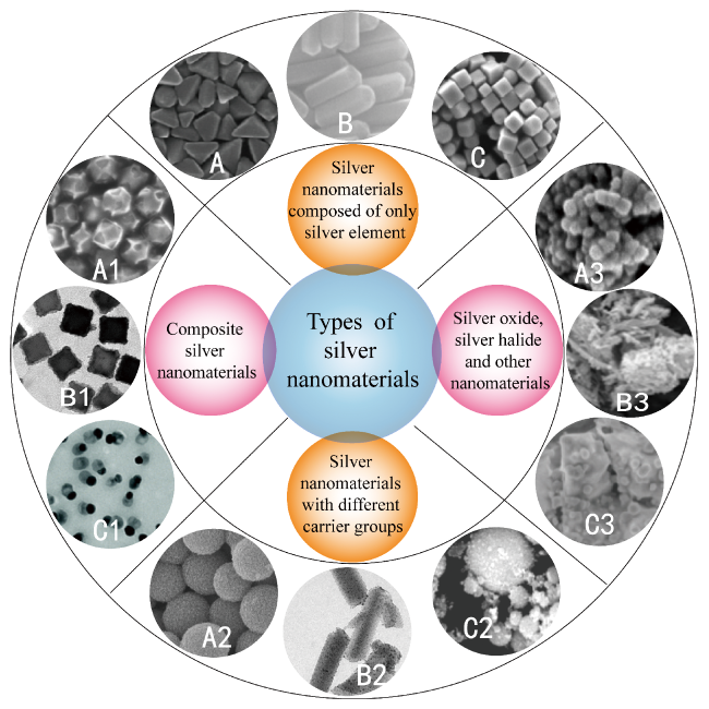
图2 银纳米材料分类图:(A,B,C)银纳米三角双锥[47],银纳米棒[14],银纳米立方体[48];(A1,B1,C1)金包银凹形立方八面体[49],银铑核框纳米立方体[50], Janus Ag/AgClBr纳米结构[51];(A2,B2,C2)多孔TiO2-Ag核壳复合材料[52],铽金属有机骨架(Tb-MOF)作为银纳米颗粒载体的复合材料[53],介孔二氧化硅负载银纳米颗粒[54];(A3,B3,C3)硫化银纳米立方体团簇[55], Ag/AgBr/AgVO3复合材料[56],Ag/AgCl材料[57]

Fig.2 Classification diagram of silver nanomaterials. (A, B and C) Silver right triangular bipyramids[47], Copyright 2010, American Chemical Society; silver nanorods[14], Copyright 2009, American Chemical Society; silver nanocubes[48], Copyright 2017, American Chemical Society. (A1, B1 and C1) The Ag@Au concave cuboctahedra[49], Copyright 2016, American Chemical Society; the Ag-Rh core-frame nanocubes[50], Copyright 2018, American Chemical Society; Janus Ag/AgClBr nanostructures[51], Copyright 2021, The Royal Society of Chemistry. (A2, B2 and C2) Porous TiO2-Ag core-shell composite material[52], Copyright 2013, The Royal Society of Chemistry; Tb-MOF support of Ag nanoparticles[53], Copyright 2017, Wiley; Silver nanoparticles supported on mesoporous silica (Ag/HMS)[54], Copyright 2017, Wiley. (A3, B3 and C3) Clusters formed by Ag2S nanocubes[55], copyright 2017, The Royal Society of Chemistry; The 2% Ag/AgBr/AgVO3 composite[56], Copyright 2021, Elsevier; the Ag/AgCl nanostructures[57], Copyright 2017, The Royal Society of Chemistry.

There are numerous synthesis methods of silver nanomaterials, which can be divided into two categories: chemical synthesis methods and physical synthesis methods. Chemical synthesis methods include chemical reduction, heat treatment, seed growth, green synthesis, etc. Physical synthesis methods can be subdivided into atomic layer deposition, microwave-assisted synthesis, ultrasonic synthesis, etc. No matter chemical synthesis method or physical synthesis method, the basic synthesis rule is top-down or bottom-up. Top-down synthesis strategy, as its name implies, uses bulk or larger silver to achieve nanoscale size by chemical or physical methods (such as grinding, etching, etc.). The silver nanomaterials prepared by the synthesis strategy have the advantages that the required materials can be prepared in a large amount in a short time; However, the disadvantage is also obvious: it will destroy the surface structure of silver nanomaterials, which determines the final properties of the materials, and then affect the application of the prepared silver nanomaterials[58]. Relatively speaking, the bottom-up synthesis strategy is also based on chemical or physical methods (such as chemical reduction, vapor deposition, electrochemistry, polyol reduction, etc.), so that the size of the material can finally reach the nanometer level.The silver nanomaterials prepared based on this synthesis strategy can effectively control the kinetic and thermodynamic conditions in the synthesis process, so as to obtain silver nanomaterials with more excellent properties[59]. This review summarizes common and easy-to-operate methods for the controlled synthesis of silver nanomaterials, as shown in Figure 3.
图3 银纳米材料的合成方法

Fig.3 Synthetic methods of silver nanomaterials

Chemical reduction method: Chemical reduction method is the most common and widely used synthesis method for silver nanomaterials. The three main components of silver nanoparticles synthesized by this method are precursor silver salt, reducing agent and stabilizing agent. The most commonly used precursor silver salt is silver nitrate (AgNO3), and silver ions can be reduced to the desired silver nanomaterials when a suitable reducing agent is added to silver nitrate. After long-term exploration by the scientific research community, many kinds of reducing agents can reduce silver ions to silver nanomaterials, such as sodium citrate, alcohols, sodium borohydride, glucose, ascorbic acid and so on[60~62][63~65][61,62][63,66][8,67]. Because of the large specific surface area and easy agglomeration of nanoparticles, the addition of appropriate stabilizers is the key to the successful synthesis of nanomaterials. The commonly used stabilizers are surfactants, ligands or polymers, such as acetonitrile, ethylene glycol (PEG), polyvinylpyrrolidone (PVP), polymethacrylic acid (PMAA), lignin, glycerol, oleylamine and collagen[68][8,69][61,64,66][70][71][8,72][73][74].
Seeded growth method: Seeded growth method is another method for the synthesis of silver nanomaterials. This method has good control over the size, morphology, structure and composition of silver nanomaterials in the synthesis process. Therefore, it is the most commonly used method for the synthesis of silver nanoscale materials[75,76]. The synthesis method involves two steps: firstly, seed nanoparticles are synthesized, and then the seed nanoparticles are added into a growth solution containing a metal precursor silver salt, a reducing agent, a stabilizing agent or a shape directing agent, or the precursor solution is injected into a container containing a mixture of seeds, the reducing agent, a capping agent and a colloidal stabilizing agent. The metal precursor is reduced (or decomposed) to form zero-valent atoms, which nucleate heterogeneously on the surface of the seed, and the seed continues to grow through one of the many possible growth pathways to form well-defined nanocrystals, as shown in Figure 4[76]. Therefore, the nucleation and growth processes for the synthesis of silver nanomaterials using this method are effectively isolated into two distinct synthesis steps, allowing for better control of the size, shape, and morphology of the nanostructures produced in the latter step[76].
图4 使用晶种调节生长法合成金属胶体的一般策略[76]

Fig.4 General strategy for the synthesis of metal colloid by the seed-mediated growth method[76], Copyright 2017, Wiley

Heat treatment method: a method for preparing nanomaterials by thermal decomposition of a solid precursor. As the name suggests, thermal decomposition is a thermally driven process in which the precursor chemically decomposes at a specific temperature, resulting in the formation of nanoparticles. The thermal decomposition of solids is essentially controlled by the geometry, the nature of the metal ion, and the various physicochemical processes occurring at the reaction interface[77]. This method is simple and economical, and can obtain a large number of stable monodisperse nanoparticles by heating once, which has become a classical method for the synthesis of nanomaterials[78].
Green synthesis method: Although the advantages of the above methods for the synthesis of silver nanomaterials are outstanding, there are also disadvantages. These unavoidable disadvantages limit the widespread use of nanomaterials, especially in biomedical and pharmaceutical applications, due to the use of chemical reagents such as strong reducing agents and stabilizers, or the need for a large amount of energy (heat), and the generation of by-products harmful to the environment. Therefore, researchers are committed to exploring new synthesis methods in order to use nanomaterials in specific medical fields, based on which green synthesis methods emerge as the times require. The synthesis method has the advantages of cleanness, good biocompatibility, low cost, high benefit, environmental friendliness and the like, and is in line with the sustainable development trend of society and environment[79]. Related researchers have also devoted more and more energy to the in-depth study of this synthesis method[74,80,81].
Atomic layer deposition (ALD) is a surface engineering-dependent method in which a substance is deposited on a substrate layer by layer in the form of a monatomic film. This process requires the construction of a thin film on the substrate through a continuous gas-phase chemical reaction in which a gas-phase precursor is alternately pulsed into a reactor and reacts with the substrate to form a deposited film. During each of these individual gas surface reactions, called half-reactions, the precursor is pulsed into the reactor and maintained at a specific temperature and pressure for a set period of time to ensure complete reaction of the precursor with the substrate.And then the chamber is purged with an inert carrier gas (nitrogen or argon) to eliminate unreacted precursors and by-products formed during the reaction, and the process is cycled to achieve the desired film thickness. The key point of this synthesis method is that by using layer-by-layer deposition, the thickness of the film can be changed by changing the number of atomic layer deposition cycles. There are two kinds of atomic layer deposition methods: plasma enhanced atomic layer deposition and thermal atomic layer deposition[82,83].
Microwave-assisted synthesis: Microwave-assisted synthesis of silver nanomaterials is a promising synthesis method, which has attracted much attention from both academia and industry[84]. The synthesis of nanomaterials is based on the direct interaction of electromagnetic radiation with molecules in the reaction medium (via dielectric heating mechanisms: dipole polarization and ionic conduction processes)[83].
Ultrasonic synthesis method: The solution is irradiated by 20 kHz ~ 10 MHz ultrasonic wave. Under the action of the ultrasonic wave, the liquid is hollow and forms small bubbles. Then, the small bubbles grow and burst, releasing huge energy, which makes the liquid produce high temperature, high pressure and strong micro-jet locally, thus driving the chemical reaction. The method has the advantages of short reaction time and low requirements on temperature and reaction system, so the method can be used for synthesizing nanomaterials with different forms[85,86]. Recently, Dong et al. Used ultrasonic enhancement to improve the preparation of silver nanoparticles by chemical reduction, so as to controllably synthesize monodisperse spherical silver nanoparticles, and elaborated the mechanism of the traditional chemical method and the method of synthesizing silver nanoparticles after ultrasonic enhancement. After in-depth study and comparison, it is concluded that in the traditional chemical synthesis method,The high reactivity and growth process of Ag+ is uncontrolled, while ultrasonic irradiation promotes the crystallization and digestion maturation process, therefore, both the chemical reaction rate and the mass transfer rate are enhanced, thereby accelerating the primary nucleation and inhibiting the growth of uncontrolled particles to form monodisperse spherical silver nanoparticles[31]. Finally, the synthesis of silver nanoparticles was quickly evaluated by the frontier machine learning method: the machine learning method, Decision tree regression, combined with Shapley value analysis, revealed that the concentration of reactants was a more important condition affecting the synthesis of silver nanoparticles, as shown in Figure 5.
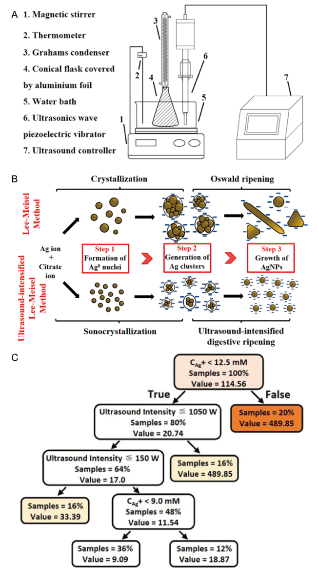
图5 超声强化法合成单分散球形银纳米颗粒[31]。(A)超声强化化学还原法制备银纳米颗粒的实验示意图;(B)有无超声增强作用化学合成银纳米颗粒的机理图;(C)机器学习分析:拟合决策树回归

Fig.5 Synthesis of monodisperse spherical AgNPs by ultrasound-intensified Lee-Meisel method[31], copyright 2021, Elsevier. (A) Schematic diagram of ultrasound-intensified Lee-Meisel method. (B) Mechanism of conventional Lee-Meisel method and ultrasound-intensified Lee-Meisel method. (C) Machine learning analysis: fitted decision tree regressor

2.1 Types and synthesis methods of silver nanomaterials containing only silver element

There are many kinds of silver nanomaterials containing only silver element, such as silver nanospheres, silver nanocubes, silver nanoflowers, silver nanorods, silver nanosheets, silver nanowires, silver nanobipyramids, and silver nanoprisms. In addition, there are many kinds of synthesis methods. Table 1 summarizes the synthesis methods, size, and shape characteristics of such silver nanomaterials, which are classified in detail as follows.
表1 银纳米颗粒的合成方法及特性

Table 1 The synthesis methods and properties of silver nanoparticles (Ag NPs)

Method Process Ag NPs size and shape ref
Chemical
Methods
Photochemical 7 nm, sphere 134
Chemical reduction 10, 12, 14 nm, spheres 87
Seed-mediated growth 42 nm, rod, 1~4 μm, nanowire 12
Photoinduced 100 nm, nanoprism 123
Seed-mediated growth 60 nm, nanodisk 128
Soft, solution-phase approach Lateral dimension:30~40 nm, length: ~50 μm, nanowire 117
Chemical reduction 50, 80, 95, 115 nm, nanocubes 65
Chemical reduction Lateral dimension:30~40 nm, length: ~50 μm, nanowire 116,118
Chemical reduction Lateral dimension:35 nm, length: 166 nm~12 μm, nanowire 60
“Green” Synthesis 5.3 nm, sphere 92
Silver mirror reaction Mean edge length:55 nm, nanocube 109
Chemical reduction Nanowire:30~40 nm, nanowire thin film, 129
Thermal method 39 nm, nanoprism 61
Chemical reduction 25~45 nm, nanocubes 105
Polyol method Nanocube: 80 nm; truncated nanocube: 120 nm;
cubocta hedras: 150~200 nm; octahedras: 250~300 nm
135
Chemical reduction 90, 170, 250, 350 nm, triangular nanoplates 111
Seed-mediated growth 75~150 nm, right bipyramids 15
Seed-mediated growth 64 nm, 81 nm, triangular nanoplates 19
Sulfide-mediated polyol method 45, 90 nm, nanocubes 64
Chemical reduction 146 nm, nanorod 13
Solvothermal reduction Nanorod:40 nm;triangulars:50,150nm;nanocubes:50~80 nm; quasi-spherical polyhedrons:60~80 nm; hexagonal nanoplates:50, 30 nm; 132
Seed-based method 20, 33, 46, 65 nm, nanoprisms 112
Green approach 20~60 nm, spheres 91
Thermal regrowth 50 nm~2 μm, pentagonal silver nanorods 14
Photoinduced synthesis 107, 132, 165, 192 nm, right-triangular bipyramids 121
Seed-catalyzed reduction 11~200 nm, triangular silver nanoplates 113
Green approach 8~71 nm, spheres 81
Photomediated synthesis Various triangular bipyramids and prisms 47
Seed-mediated 30~200 nm, nanocubes 75
Chemical reduction 30~70 nm, nanocubes 108
Seed-mediated growth Octahedral:80 nm; various concave nanocrystals 67
Chemical reduction Hierarchical assemblies of silver nanostructures 125
Seed-mediated approach 52, 67, 460, 870, 1010 nm, nanorods 115
Chemical reduction Various nanoplates 20
Green method 10.60, 11.23, 15.30 nm, spheres 63
Chemical reduction 20~100 nm, quasi-spherical 8
Seed-mediated growth Various nanocubes and octahedrons 130
Seed-mediated growth 20~72 nm, octahedra 131
Chemical reduction 4~8 nm, spheres 88
Seed-mediated growth 30~100 nm, nanocubes 102
Chemical reduction Silver nanoparticle with various shapes 66
Seeded growth method 150 nm~1.5μm, triangular silver nanoplates 68
Greener synthesis 5~150 nm, spheres and triangular 97
Seed-mediated growth 20~120 nm, quasi-spherical 99
Biogenic synthesis 2~15 nm, quasi-spherical 89
Seed-mediated growth 23~60 nm, nanocubes 103
Chemical reduction 15~90 nm, spherical; 150 nm, triangular 62
Green synthesis 40~70 nm, quasi-spherical 93
Green synthesis 15 nm, sphere 96
Seed-mediated growth coupled with oxidative etching 37~68 nm, sphere 101
Green synthesis 17~27 nm, pherical/quasispherical 94
Chemical reduction 59.84 nm, 75.70 nm, 110.32 nm, nanocubes 69
Lithography 90, 120, 145 nm, elliptical, triangular 58
Physical
Methods
Ultrasonic-Assisted Synthesis 120 nm, nanoplate 110
Sonochemical approach Less than 2 nm, nanocluster 70
Sonochemical synthesis Mean diameters:100 nm, lengths: 4~7 μm, nanorods 114
Sonochemical synthesis 1.3 μm, microflowers 124
Conventional thermal treatment 10.4 nm, sphere 95
Microwave treatment 12 nm, sphere 95
Microwave irradiation Nanowires diameters: 50~100 nm, 100~200 nm 119
Microwave-assisted polyol Lateral dimension:60~480 nm, length: 10~30 μm, nanowires 120

2.1.1 Ynthesis method of spherical silver nano material

There are numerous synthesis methods of spherical silver nanomaterials, such as seed growth, chemical reduction, green synthesis, heat treatment, ultrasonic synthesis and so on. Monodisperse spherical silver nanoparticles can be synthesized by a two-step process using citrate and ascorbic acid as reducing agents and compounds with hydroxyl groups (such as glycerol, ethylene glycol, agarose, or sucrose) as dispersing agents[8]. In addition, sodium borohydride is also the most frequently used reducing agent. As early as 1998, the mechanism of using NaBH4 as a reducing agent to synthesize silver nanoparticles has been studied in depth. Evidence shows that the reaction pathway does not follow the classical nucleation and growth theory, but is dominated by colloidal interactions[87]. Spherical silver nanoparticles with narrow size distribution can also be synthesized by using NaBH4 as a reducing agent[88]. Baker et al. Used oleylamine as a surfactant and reducing agent to synthesize organic-soluble silver nanoparticles for the first time by a simple and convenient microwave-assisted chemical reduction method in a specific environment, halide-free, deep eutectic solvent[73]. Biosynthetic preparation of metal nanomaterials has attracted much attention due to its many advantages, such as cleanliness, good biocompatibility, non-toxicity, low cost, etc. For example, Prasad et al. Used Chlorella pyrenoidosa as an algae platform for the design and synthesis of silver nanoparticles, and used a specific capping agent to synthesize silver nanoparticles with controllable size (2 ~ 15 nm), stable existence, and highly consistent morphology[89]. More than that, green synthesis of nanomaterials is considered to be a promising synthesis strategy due to the abundance of precursor materials. For example, phytosynthesis can also be used to prepare metal nanomaterials, generally using components in plant extracts as reducing and stabilizing agents: alkali lignin is used to synthesize silver nanoparticles of 17 ~ 27 nm.Cell free extract of Pseudomonas aeruginosa M6 from mangrove and Streptophyllum leaf extract were used to synthesize 15 nm silver nanoparticles, and antioxidants from blackberry, blueberry, pomegranate and turmeric extracts were used to synthesize silver nanoparticles[90~93][94][95][96][97]. In addition, spherical silver nanoparticles can also be synthesized by thermal decomposition. For example, Koga et al. Solved the problem of controlling the shape and size of nanoparticles in the synthesis process by adjusting the annealing temperature, heating time, heating rate and reactant concentration to decompose silver acetate[98]. Coincidentally, seed-growth based synthesis methods also have amazing control over the size, morphology, structure and composition of silver nanomaterials, such as the use of environmentally friendly glucose as a reducing agent.Due to its weak reducibility, the secondary nucleation is prevented during the seed-mediated growth process, resulting in the synthesis of monodisperse silver nanoparticles with particle size in the range of 20 ~ 120 nm and narrow particle size distribution[99]. Silver nanoclusters are used as seeds for the subsequent growth of silver nanoparticles, and spherical silver nanoparticles can also be synthesized by using a seed growth method combined with an oxidative etching method through silver nanocubes[100][101].

2.1.2 Synthesis of cubic silver nanomaterials

Compared with spherical silver nanomaterials, the synthesis methods of cubic silver nanomaterials are less, and the most common method is seed growth. During the synthesis process, the growth rate on the { 100 } facet is reduced due to the selective adsorption of the protectant/stabilizer on this facet, resulting in the formation of silver nanocubes. For example, silver nanocubes with uniform size can be synthesized by seed growth method, and the edge length range (30 ~ 100 nm) of silver nanocubes can be easily controlled by changing the reaction time, the number of seed particles and the concentration of AgNO3[102]. Silver nanocubes with adjustable size (23 ~ 60 nm) can be obtained by adding silver seed solution to the mixed solution of cetyltrimethylammonium chloride (CTAC), silver trifluoroacetate and ascorbic acid, and adjusting the volume of seed crystal[103]. In addition, silver nanocubes can be synthesized in large quantities by using PVP as a dispersant and ethylene glycol to reduce silver nitrate[65]. The experimental parameters (such as the concentration of silver nitrate, the concentration of polyvinylpyrrolidone, the molar ratio of precursor silver to covering agent, etc.) And the growth mechanism of silver nanocubes prepared by polymer-mediated polyol method were explored.The researchers found that determining the crystallinity of the seed (single crystal decahedron, multiple twin particles) and the range of PVP coverage helped to control the morphology of the final silver nanomaterials[104]. With the in-depth exploration of the synthesis methods of silver nanocubes, the most commonly used and mature synthesis method is to improve the above polyol-mediated rapid synthesis method, using trace sodium sulfide or sodium hydrosulfide as a reducing agent, and many subsequent synthesis methods of silver nanocubes are based on this reference and improvement[64,105][69,106,107]. Later, Xia et al. Synthesized silver nanocubes with controllable edge length of 30 ~ 200 nm based on seed growth method[75]. Silver nanocubes with controllable edge length of 30 ~ 70 nm can also be easily synthesized by using silver trifluoroacetate as precursor[108]. In addition to the above common synthesis methods, a novel method for the synthesis of silver nanocubes has been reported: cetyltrimethylammonium bromide (HTAB) was introduced into the synthesis method, and monodisperse silver nanocubes with an average side length of 55 ± 5 nm were controllably synthesized through the silver mirror reaction[109].

2.1.3 Synthesis of Triangular Silver Nanomaterials

The synthesis methods of triangular silver nanoparticles include ultrasound-assisted synthesis, chemical reduction, etc. For example, in N, N-dimethylformamide solution, in the presence of PVP, uniform triangular silver nanoparticles can be grown by a simple sonochemical method.Among them, PVP can be adsorbed on different planes of silver nanoparticles, thus inducing the selective growth of plate-like crystals, and the synthesized silver triangular nanoplatelets are single crystals with (111) lattice plane as the basal plane[110]. Xia et al. First used the hydroxyl group of PVP to reduce silver nitrate in water, and quickly and directly prepared silver nano-triangular sheets through kinetic control, specifically, after heating the aqueous solution of AgNO3 and PVP to 60 ℃,The terminal hydroxyl groups of PVP reduce the AgNO3 at a sufficiently slow rate to allow kinetically controlled growth of silver nanocrystals, and it has been demonstrated that this kinetically controlled synthesis allows the preparation of silver nanosheets with controlled edge lengths by varying the reaction time[111]. Kelly et al. Proposed a seed growth method for the rapid, easy to repeat, and high yield synthesis of silver triangular nanosheets at room temperature, and proposed a new insight into the anisotropic growth mechanism of silver triangular nanosheets.Specifically, there are many defects in the (111) direction perpendicular to the plane of the silver nanosheet, and these defects can be combined between the two fcc layers to form an H CP layer with a periodicity of 2.5050 Å, providing an explanation for the common lattice fringes in nanoparticles[112,113]. In addition, the formation of the triangular shape of the nanometric triangular sheet is driven by the asymmetric distribution of the crystal plane edges, which in turn is determined by the asymmetric thickness of the fcc layer on both sides of the hcp layer, and thus, this two-dimensional hcp layer can explain the two-dimensional lateral growth, they also pointed out.The silver halide model may be a good starting point for understanding anisotropic growth, as it identifies the crucial defects, but it clearly does not adequately explain the growth mode of metallic nanosheets, and thus, they suggest, defects induce the arrangement of silver atoms into continuous hcp domains, which represents a growth mechanism for anisotropic metallic nanoparticles. In addition, Yin et al. Conducted a systematic study on the synthesis of silver triangular nanosheets, and proposed that in the process of preparing silver triangular nanosheets by chemical reduction,It is generally believed that the surface energy and the growth rate on the { 111 } plane are reduced due to the selective adsorption of the protective agent (usually sodium citrate), so citrate is more critical in the synthesis of silver triangular nanosheets. However, Yin et al. Found that the reagent playing a key role is not citrate as generally believed, but hydrogen peroxide. Contrary to the previous conclusion that citrate is the key component, they have shown that the ligand with selective adsorption to Ag (111) crystal plane can be extended to many dicarboxylic and tricarboxylic acid compounds. Moreover, in addition to the general knowledge that NaBH4 can be used as a reducing agent, they also found that NaBH4 as a capping agent can stabilize silver nanoparticles, prolong the initiation time required for nanoplate nucleation, and help to control the thickness and aspect ratio of silver nanoplates. Based on the re-recognition of this mechanism, his research group has developed a new method for the synthesis of silver nano-triangular sheets with repeatability, high efficiency and high yield[20,68].

2.1.4 Method for synthesizing rod-like and linear silver nano material

Initially, Murphy prepared silver nanorods with different aspect ratios from near-spherical 4 nm silver nanoparticles by seed growth method[12]. Kitaev et al. Used citrate as a reducing agent, PVP as a dispersing agent, and decahedral silver nanoparticles as a precursor to prepare monodispersed silver nanorods with controllable size, five-fold twin structure, and variable width by heat treatment[14]. Xia et al. Used another strategy to synthesize silver nanorods with rectangular sides and an average aspect ratio of 2.7 by changing the concentration of bromide ions in the silver nanomaterials synthesized by the polyol method[13]. Wang et al. Synthesized silver nanorods with a length of 4 ~ 7 μm and an average diameter of about 100 nm by sonochemical method by irradiating silver nitrate, methylaniline (HMTA) and PVP aqueous solution with ultrasonic wave for 60 min. The effects of irradiation time, PVP concentration and reaction temperature on the morphology of silver nanorods were discussed, and the formation mechanism of silver nano-rods was finally speculated[114]. Later, Mirkin et al. Also synthesized silver nanorods by seed growth method. In the presence of silver ions and trisodium citrate, silver nanorods were synthesized by plasmon excitation of silver seed crystals irradiated by light of 600 ~ 700 nm[115].
Xia et al. Used a seed growth method to synthesize silver nanowires with a diameter of 30 ~ 40 nm and a length of about 50 μm on a large scale. First, ethylene glycol was heated to about 160 ℃ and used to reduce silver nitrate to form silver nanoparticles.Then, in the presence of PVP, the generated silver nanoparticles are used as seeds for heterogeneous nucleation and growth to form uniform nanowires with an aspect ratio of up to 1000.By adjusting the reaction conditions (PVP to silver nitrate ratio, reaction temperature, and number of seed particles, etc.), the morphology and aspect ratio of silver nanostructures can range from nanoparticles, nanorods, to long nanowires,Although the feasibility of this method can be proved by preparing silver nanowires with diameters of 30 ~ 60 nm and lengths of 1 ~ 50 μm, the growth mechanism has not yet been clarified. The following year, the mechanism of this method was further studied and clarified in another report[116,117][118]. Varma et al. Used cheap, abundant and environmentally friendly glycerol as reducing agent and solvent to prepare silver nanowires with controllable diameter rapidly (1 min) under microwave irradiation without stirring[119]. In addition, silver nanowires with pentagonal cross-section were also synthesized by a simple and rapid microwave-assisted, polyol method in the presence of polyvinylpyrrolidone (PVP) and sodium sulfide (Na2S)[120].

2.1.5 Synthesis of trigonal bipyramidal and cylindrical silver nanomaterials

Triangular bipyramids of silver nanoparticles can be synthesized by seed growth method. Silver nanoparticles with a single (111) twin are selectively nucleated and then grow into right bipyramids with an edge length of 75 ~ 150 nm. The key to produce single and twin seeds is whether sodium bromide is added during the synthesis of triangular bipyramids of silver nanoparticles by polyol method[15]. Monodisperse silver nanotrigonal bipyramids can be synthesized by photoinduction with high yield, and the edge length can be controlled by adjusting the wavelength of the excitation light. The mechanism of Plasmon-assisted synthesis is that silver nanomaterials absorb light energy through surface Plasmon resonance (SPR).O as to cause the enhancement of the electromagnetic field of certain parts on the surface of the particle, and the enhanced electromagnetic field can influence the reaction activity of the reaction substance, thereby simultaneously accelerating the etching, re-reduction and re-growth processes of silver, and growing the silver into the particle with a specific shape under the action of the protective agent and in the framework of thermodynamics[121]; In addition, Mirkin et al. Also used the photoinduced method to synthesize trigonal bipyramidal and columnar silver nanoparticles, and then van Duyne et al. Used this method to synthesize monodisperse trigonal columnar silver nanoparticles, and their optical properties were studied in detail and in depth[47][122]. In addition, silver nanospheres can also be converted into silver nanopillars under light-induced conditions[123].

2.1.6 Ynthesis of silver nanomaterials of other shapes

In addition to the above common shapes, other silver nanomaterials with complex shapes have also been successfully synthesized. Tao et al. Reported a simple, surfactant-free method for the synthesis of three-dimensional honeysuckle flowers at room temperature, which has the characteristics of high yield and good size distribution[124]. In addition, the 3D honeysuckle has a special structure of nano-scale wrinkles, and the shape, size, and surface structure (controllable roughness of surface morphology) of the honeysuckle can be adjusted by controlling the experimental parameters. With the assistance of acid molecules (citric acid, mandelic acid, etc.), directional self-assembled honeysuckle flowers with complex structures can also be synthesized without any surfactant or capping agent. Subsequently, according to this method, honeysuckle flowers can still be formed after the addition of succinic acid, and have a good hierarchical structure[125][126]. In addition, Liu et al. Designed a simple method to synthesize monodisperse and hierarchical peony-like silver micro-nanomaterials assembled by nanostructures with specific surface morphology without any surfactant, and their size and surface roughness can be well controlled[127].
A novel silver nanostructured material, silver nanosheets, can be prepared by seed growth method: silver nanosheets can be prepared by using silver particles as seeds in the presence of concentrated cetyltrimethylammonium bromide (CTAB)[128]. A novel silver nanowire film composed of monocrystalline silver nanowire bundles with a diameter of 30 ~ 40 nm can be successfully synthesized in an aqueous solution of polymethacrylic acid at room temperature by chemical reduction[129]. Xia's group prepared silver octahedra by seed growth method using silver trifluoroacetate, ascorbic acid and sodium citrate as precursors and cubic and quasi-spherical silver nanoparticles as seeds[130,131]. In addition, various silver nanomaterials with concave-convex surfaces, hexagonal plates, didecahedrons and other silver nanomaterials with complex shapes have also been successfully synthesized[67][24,132,133].

2.2 Types and synthesis methods of silver nanomaterials containing two or more elements

Silver nanomaterials containing two or more elements are also a class of functional materials that have been extensively studied in recent years, which can be roughly divided into three categories: bimetallic alloys, core-shell structures, and Janus nanoparticles. They exhibit superior thermal, electrical, optical, and catalytic properties compared to the previously described monometallic materials. Fig. 6 is a synthesis schematic[136]. A is a schematic diagram of the preparation of Ag @ Hg nanoalloys from four typical silver nanoparticles. All Ag nanoparticles undergo the following three growth stages: (1) rapid adsorption of Hg atoms onto Ag nanoparticles; (2) Hg atoms diffuse into Ag nanoparticles in the early stage, making the particles shorter or rounder; (3) Hg atoms further diffuse to form spherical Ag @ Hg nanoalloy with uniform distribution of Ag and Hg[137]. Fig. B is a schematic diagram of one-pot synthesis of copper/silver bimetallic nanoparticles: silver nitrate and copper chloride were used as precursors of silver and copper, respectively, glucose was used as a reducing agent, and hexadecylamine (HDA) was used as a selective capping agent for copper (100).Glucose ring opening produces aldehyde, which can reduce Ag+ to Ag0. However, at room temperature, the equilibrium reaction of the produced glucal is disordered, which makes it difficult for Ag+ to be reduced to Ag0. Thus, the reaction of Cu2+ with glucose (capable of reducing CuII to Cu0) formed Cu NPs. Cu/Ag bimetallic nanoparticles were formed due to the Cu+2Ag+→Cu2++2Ag substitution reaction[138]. Fig. C is a schematic diagram of the formation of Au @ Ag half-shell Janus nanoparticles: Au @ Ag half-shell Janus nanoparticles were prepared by selective etching of silver with H2O2+NH3 using Au @ Ag core-shell nanoparticles as raw materials[139]. Fig. D is a schematic diagram of two methods for depositing the second metal M on Ag nanocubes: Ag @ M core-frame and core-shell nanocubes above, Ag @ Ag-M core-frame nanocubs below, and M-based nanoframes/nanoboxes and Ag-M nanoframes with ridges of different thicknesses after removing the Ag core, respectively[140]. Figure E shows the three steps of the synthesis of colloidal gold-silver-gold and core-shell-shell nanoparticles: firstly, 12 nm gold nanoseeds are prepared and used as gold cores; Then, in the presence of sodium hydroxide, silver nitrate is reduced by ascorbic acid, so that the first silver shell grows from the gold seed; Finally, gold shells with different sizes were prepared in the presence of sodium citrate and hydroquinone[141]. Table 2 is a summary of the synthesis methods and characteristics of such silver nanomaterials, which are described in detail below.
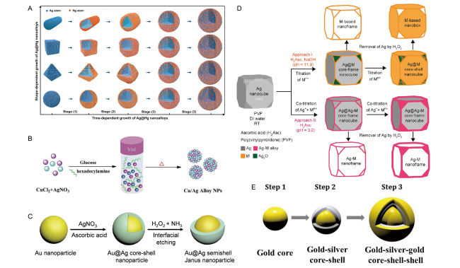
图6 双金属合金、核壳结构和Janus纳米颗粒的合成示意图。(A)银汞合金的生长示意图[137];(B)一锅合成铜/银双金属纳米颗粒示意图[138];(C)形成Au@Ag半壳Janus纳米粒子示意图[139];(D)在银纳米立方体种子上沉积第二种金属M的两种方法示意图[140];(E)胶体金-银-金、核-壳-壳纳米粒子的合成示意图[141]

Fig.6 Schematic diagram of synthesis of bimetallic alloy, core-shell structure and Janus nanoparticles. (A) The growth of Ag@Hg nanoalloys from four typical Ag nanoparticles[137],Copyright 2013, American Chemical Society. (B) Schematic illustration of the one-pot synthetic procedure of Cu/Ag bimetallic NPs[138], Copyright 2015, The Royal Society of Chemistry. (C) Schematic illustration of forming Au core@Ag semishell Janus nanoparticles[139], Copyright 2016, The Royal Society of Chemistry. (D) Schematic illustration of two proposed pathways for the deposition of a second metal M on a Ag nanocube seed[140], Copyright 2017, American Chemical Society. (E) Three steps synthesis of colloidal Gold-Silver-Gold Core-Shell-Shell nanoparticles[141], Copyright 2015, American Chemical Society

表2 含有两种或两种以上元素的银纳米材料的合成方法及特性总结

Table 2 The synthesis methods and properties of silver nanomaterials containing two or more elements

Method Process Constituent elements ref
Chemical Methods Galvanic replacement reactions Pd-Ag, Pt-Ag nanoboxes 147
Microwave-polyol method Au-Ag core-shell nanoparticles 153
Sonochemical co-reduction Au-Ag core-shell nanoparticles 154
Aqueous reduction Fe-Ag core-shell nanoparticles 160
Thermal decomposition Janus Ag-Ag2S nanoparticles 165
Galvanic exchange reactions Ag-Au Janus nanoparticles 166
Galvanic replacement reaction Pt-Ag nanobox, heterodimer, multimer, popcorn-shaped nanoparticles 150
Phytosynthesis Au-Ag nanoparticles 145
Chemical reduction Ag-Hg nanoalloys 137
Chemical reduction Au-Ag core-shell nanoparticles 155
Green synthesis Au-Ag bimetallic nanoparticles 146
Coreduction reaction Au-Ag multispiked nanoparticles 143
Galvanic replacement-free deposition Au-Ag core-shell nanocubes 156
Coreduction reaction Au-Ag-Au core-shell-shell nanoparticles 141
One-pot reduction Cu-Ag nanoalloys 138
Overgrowth of seed-mediated growth Au-Ag nanorods 157
Chemical etching Au-Ag semishell Janus nanoparticles 139
Co-reduction Ag-Pd nanoframes 148
Chemical reduction Ag-Au concave cuboctahedra 49
Chemical reduction Ag-Ni snowman and Ag@Ni core-shell nanoparticles 162
Impregnation-reduction method Ag-Pd alloy nanoparticles 149
Chemical reduction Au-Ag nanoboxes 144
Seed-mediated-growth method Au-Ag core-shell nanoparticles 158
Chemical reduction Ag-Rh core-frame nanocubes 50
Chemical reduction Janus Ag/AgClBr nanostructures:Janus silver/ternary silver halide nanostructures 51
Physical Methods Laser-induced heating Au-Ag alloy nanoparticles 142
Room-temperature radiolysis Ag-Ni, Pd-Ni alloy nanoparticles 151
Combination of “grafting from” and “grafting to” approaches Hairy Janus particles with immobilized Ag or Au nanoparticles 169
One-pot reaction Janus Ag-MSN@CTAB: Janus silver mesoporous silica nanobullets 167
Ultrasonic treatment Janus silver/silica nanoplatforms 168
Deposition Hairy Janus silver nanoparticles 170
Electrostatic adsorption Janus plasmonic silver nanoplatelets 171

2.2.1 Ynthesis method of bimetallic alloy nano material

Au-Ag alloy is the most common bimetallic alloy. Kleinermanns et al. Synthesized Au-Ag bimetallic nanoalloy by laser-assisted method[142]. Sharma et al. Synthesized Au-Ag bimetallic nanoalloy with high monodispersity and multiple sharp corners by co-reduction method using ascorbic acid as reducing agent and PVP as stabilizer[143]. Zhang et al. First used ascorbic acid (AA), a strong reducing agent, to directly deposit gold atoms on the surface of silver nanocubes to prepare Ag @ Au nanocubes, and then used Fe(NO3)3 to corrode the interior of silver nanocube.The hollow gold nanocapsules were obtained, and finally, Au-Ag alloys with different Ag/Au ratios were prepared by electrochemical replacement reaction by adding HAuCl4 aqueous solution into the boiling silver nanocube suspension[144]. With the progress of technology, the direction of simple and environmentally friendly synthesis is changing. Abdel-Mottaleb et al. Established a green and simple method for the synthesis of Au-Ag bimetallic nanoparticles by using the extract of sago (Potamogeton pectinatus L.)[145]. Li et al. Also developed a simple, economically feasible and green method to synthesize Au-Ag bimetallic nanoparticles with good stability, which used non-toxic and renewable degraded Pueraria starch as reducing agent and capping agent[146].
Silver-palladium and silver-platinum bimetallic alloys have also been widely studied and used in the field of catalysis because of their superior catalytic performance. Xia et al. Can convert silver nanocubes dispersed in water into Pd-Ag or Pt-Ag nanoboxes by adding Na2PdCl4 or Na2PtCl4, and can adjust the surface plasmon resonance peak of the nanostructure in the whole visible spectrum range (440 ~ 730 nm) by controlling the addition amount of noble metal salt and the molar ratio of Na2PdCl4 or Na2PtCl4 to silver[147]; Qin et al. Reported an easily synthesized Ag-Pd bimetallic nanoframe with ridges as thin as 1.7 nm[148]; Silver-palladium (Ag-Pd) alloy nanoparticles have a strong ability to absorb visible light, and their photocatalytic activity is higher than that of pure palladium (Pd) and silver (Ag) nanoparticles. Based on this, Zhu et al. Prepared Ag-Pd alloy nanoparticles supported on ZrO2 and Al2O3 carriers[149]; Later, Lu et al. Reported that templated silver nanocubes underwent a displacement reaction with platinum ions in the presence of HCl, resulting in the synthesis of gap-controlled platinum-silver (Pt-Ag) bimetallic nanostructures[150]. In addition, silver amalgam, silver-copper alloy, silver-rhodium alloy and silver-nickel alloy have also been successfully synthesized[137][138][50][151].

2.2.2 Method for synthesizing core-shell structure nano material

The core-shell structure nanomaterial composed of gold and silver elements is a common and widely studied core-shell nanomaterial. The specific synthesis method is as follows: Au @ Ag core-shell nanocrystals can be synthesized by microwave-polyol method using ethylene glycol as reducing agent and polyvinylpyrrolidone (PVP) as surfactant, and the synthesis mechanism is analyzed in depth[152][153]. Au @ Ag core-shell nanoparticles were prepared by co-reduction of Au (III) and Ag (I) ions in aqueous solution using a sonochemical assisted method[154]. In addition, stable, uniform and size-adjustable Au @ Ag core-shell nanoparticles can be synthesized by using citric acid ions as stabilizers[155]; Qin et al. Successfully synthesized Ag @ Au core-shell nanocubes by directly depositing gold atoms on the surface of silver nanocubes through replacement reaction[156]. Au @ Ag nanorods can also be prepared after silver is deposited on the gold nanobipyramid[157]. Seed-mediated growth method has also been used in the synthesis of core-shell nanomaterials[158]. In addition, the synthesis of gold-silver-gold (Au @ Ag-Au), that is, core-shell-shell nanoparticles, has also been reported. By changing the thickness of gold and silver shells, the wavelength of the plasma extinction spectrum peak can be controlled in the visible and near-infrared regions[141]. Similar structures have also been reported, and Ag @ Ag-Au nanocubes with higher plasmonic and catalytic properties can be synthesized by co-titration of AgNO3 and HAuCl4[159]. More interestingly, Xie et al. Reported that concave-convex Ag @ Au cuboctahedra could be synthesized by dropping HAuCl4 aqueous solution into a suspension of silver octahedra in the presence of ascorbic acid (AA), sodium hydroxide and polyvinylpyrrolidone (PVP) at room temperature[49].
In addition to the above gold and silver core-shell structured nanomaterials, other core-shell nanomaterials have also been successfully synthesized. For example, one-pot synthesis of Fe @ Ag core-shell nanoparticles[160]; Ag @ Pd-Ag nanocubes can be easily synthesized by co-titrating Na2PdCl4 and AgNO3 into an aqueous suspension of silver nanocubes in the presence of ascorbic acid and polyvinylpyrrolidone at room temperature[161]. Snowman-shaped and core-shell Ag-Ni binary nanoparticles have also been successfully synthesized, and more environmentally friendly synthesis methods are being explored, such as green synthesis of core-shell nanomaterials using plant extracts[162][163].

2.2.3 Synthesis of Janus Nanomaterials

In recent years, the research on the synthesis and physicochemical properties of Janus nanoparticles has also been in a hot peak state, mainly because the hydrophobicity of the two sides of Janus nanoparticles is quite different, which enhances the affinity for different phases, and therefore, has stronger stability[83,164]. Hu et al. Reported the one-pot synthesis of a large number of spherical, eggplant-like Janus nanoparticles of Ag-Ag2S and deeply investigated their photocatalytic properties[165]. Using the combination of electrodisplacement reaction and Langmuir-Blodgett method, Chen et al. Prepared Ag-Au bimetallic Janus nanoparticles and used them as electrocatalysts for oxygen reduction in alkaline medium, and they also prepared Au @ Ag half-shell Janus nanoparticles by chemical etching of Au @ Ag core-shell nanoparticles at the air/water interface[166][139]. In the same year, Dong's group successively prepared different Janus nanoparticles: firstly, CTAB-supported silver mesoporous silica Janus nanoparticles (Janus Ag-MSNs @ CTAB) were prepared by a one-pot method, and then a silver/silica Janus nanoplatform was designed and used for light-activated chemotherapy and photothermal therapy of liver cancer[167][168]. Hairy Janus nanoparticles have also been reported. For example, Giancane et al. Proposed a simple method to prepare surface anisotropic Janus silver nanoparticles: octadecylamine was used to stabilize the nanoparticles and promote the deposition of silver nanoparticles on solid substrates. AgNPs/octadecylamine Janus nanoparticles were used as an active layer for the detection of phenylethylamine and tyramine[169,170][170]. In addition, more complex Janus silver nanosheets as well as silver/ternary silver halide Janus nanostructures were also successively synthesized[51,171].

2.3 Types and Synthesis of Silver Nanomaterials with Different Carrier

As we all know, monodisperse silver nanoparticles are difficult to exist stably for a long time because of their large specific surface area and easy agglomeration, which limits the development of silver nanomaterials to a certain extent. Therefore, through unremitting efforts and research, scientists have successfully synthesized silver nanomaterials with different carriers and applied them to many fields[172,173]. The supports generally include the following categories: carbon materials, silica, metal-organic frameworks (MOFs), polymers, metal oxides, and boron nitride, etc. Fig. 7 is a schematic diagram of the synthesis of six carrier silver nanomaterials. A shows the preparation process of graphene oxide-silver nanocomposite: first, silver ions are immobilized on the surface of graphene oxide nanosheets, and then,Glucose and starch were used as reductant and stabilizer, respectively, to form silver nanoparticles on the surface of graphene oxide nanosheets, thus preparing silver nanocomposites with graphene oxide as the carrier[174]. Figure B shows the preparation of mesoporous silica microcapsule supported silver nanoparticle composite (AgNPs @ silica microcapsule): firstly, sulfate groups were introduced onto the surface of polystyrene (PS) beads by sulfonation to obtain negatively charged sulfonated PS beads; Then, silver nitrate (AgNO3) was added to adsorb Ag+ on the surface of sulfonated PS spheres by electrostatic attraction; Secondly, polyvinylpyrrolidone (PVP) is added to the reaction system as a decompression agent and a protective agent, and the PVP macromolecule exists in a pseudo-random helical configuration in the solution and is associated with the metal atom, thereby increasing the nucleation probability; Thirdly, assembling silicon dioxide colloid prepared by hydrolytic polycondensation of tetraethyl orthosilicate (TEOS) under the catalysis of ammonia on the PS ball to form a core-shell PS-Ag-SiO2 composite ball; Finally, the PS core and PVP were removed by calcination to form AgNPs@SiO2 microcapsules with hollow core, mesoporous shell, and AgNPs loaded on the inner wall[175]. C shows the schematic diagram of the synthesis of the composite (Fe3O4@MIL-100(Fe)/Ag) in which silver nanoparticles were embedded into metal-organic frameworks (MOFs: MIL-100 (Fe)) and then coated on Fe3O4 nanoparticles: first, carboxyl-functionalized Fe3O4 nanoparticles (Fe3O4-COOH) were prepared by hydrothermal method, and then,MIL-100 (Fe) MOFs were prepared by layer-by-layer self-assembly on Fe3O4-COOH particles, and finally, AgNO3 solution was added to irradiate the sample, and the generated reducing radicals reduced Ag+ to Ag0. With the reaction, silver nanoparticles were formed in situ in the pores of MOFs. This method ensured the dispersion and particle size uniformity of silver nanoparticles[176]. Fig. D shows the synthesis route of silver nanoparticles loaded on conjugated microporous polymer (CMP) composite (Ag0@CMP): conjugated microporous polymer CMP was synthesized by condensation of amino acrylonitrile and phthalaldehyde.Cyano and pyridyl functional groups introduced in the condensation process can be used as action sites to capture the Ag+, and then the CMP material loaded with the activated Ag+ is irradiated by visible light to successfully reduce the Ag+ into Ag0 to synthesize the Ag0@CMP composite material[177]. Fig. E is a schematic diagram of microwave-assisted synthesis of hexagonal boron nitride supported silver nanoparticles (SNP/h-BN): DMF has the dual functions of solvent exfoliation and reducing agent. The exfoliated h-BN is dissolved in DMF, and silver nitrate solution is added dropwise to synthesize aggregation-free SNP/h-BN composite nanomaterials through microwave-assisted treatment[178]. F is the formation schematic diagram of silver nanoparticle modified MnO2 nanowire and hierarchical heterostructure: silver foil was immersed in the mixed solution of KMnO4 and H2SO4, and the reaction (2KMnO4+6Ag+4H2SO4→2MnO2+3Ag2SO4+K2SO4+4H2O) occurred to generate MnO2,Due to the photosensitivity of Ag2SO4, it gradually decomposes (Ag2SO4→2Ag+SO2+O2) to form AgNPs, and the manganese dioxide colloidal spheres can be completely converted into AgNPs modified MnO2 nanowire composites after incubation for a week[179]. Table 3 briefly summarizes the synthesis methods and types of different supported silver nanomaterials, which are described in detail below.
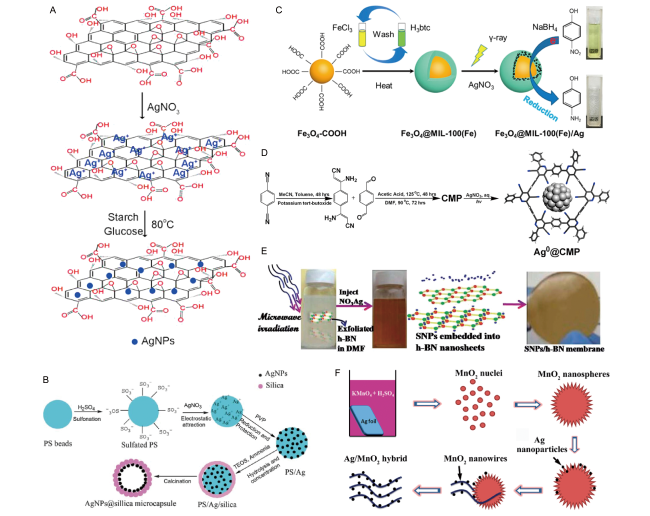
图7 碳材料、二氧化硅、金属有机框架材料(MOFs)、聚合物、金属氧化物和氮化硼为载体的银纳米复合材料合成示意图。(A)氧化石墨烯-银纳米复合材料的制备过程示意图[174];(B)AgNPs@SiO2微胶囊合成示意图[175];(C)形成Fe3O4@MIL-100(Fe)/Ag纳米复合材料示意图[176];(D)银纳米颗粒负载于共轭微孔聚合物(CMP)复合材料(Ag0@CMP)的合成示意图[177];(E)微波辅助合成六方氮化硼负载银纳米颗粒(SNP/h-BN)复合纳米材料示意图[178];(F)银纳米颗粒修饰MnO2纳米线层次化异质结构的形成示意图[179]

Fig.7 Schematic diagram of silver nanocomposites synthesis of carbon materials, silica, metal-organic framework materials (MOFs), polymers, metal oxides and boron nitride as substrates. (A) Schematic of the procedure for preparing GO-Ag nanocomposite[174], Copyright 2015, American Chemical Society. (B) Schematic illustration of the AgNPs@silica microcapsule[175], Copyright 2012, The Royal Society of Chemistry. (C) Fabrication strategy of Fe3O4@MIL-100(Fe)/Ag nanocomposites[176], Copyright 2020, American Chemical Society. (D) Illustration of synthetic pathway and pore structure of CMP for silver nanoparticle immobilization[177], Copyright 2017, American Chemical Society. (E) Schematic synthesis process of SNP/h-BN nanohybrids via a microwave-assisted method[178], Copyright 2014, The Royal Society of Chemistry. (F) Schematic illustration of the formation of the hierarchical heterostructures of AgNPs-decorated MnO2 nanowires[179], Copyright 2015, The Royal Society of Chemistry

表3 不同载体银纳米材料的种类及合成方法总结

Table 3 The types and synthesis methods of silver nanomaterials with different carriers

Method Specie ref
Incipient-wetness impregnation method Zirconia-supported Ag particles 235
Mix silver glue and PVA and evaporation of the solvent Silver-polyvinyl alcohol (Ag-PVA) nanocomposites 214
Calcination Silver/carbon composites 237
Microwave-assisted one-step synthesis Polyacrylamide-metal (M=Ag, Pt, Cu) nanocomposites 215
The Ar+ sputtering in UHV followed by Annealing in air Silver nanoparticles supported on highly oriented pyrolytic graphite (Ag/HOPG) 238
Calcination Ag Nanoparticles supported on Alumina (Ag/Al2O3) materials 211
Incipient-wetness impregnation Silica supported silver nanoparticles (Ag/SiO2) 204
Citrate-protecting method Carbon-supported Ag nanoparticles (Ag/C) 239
One-pot facile synthesis Ag/TiO2-xNx 227
In situ reduction of adsorbed Ag+ by hydroquinone in a citrate buffer solution Silver nanoparticle and graphene oxide nanosheet composites (AgNP/GO) 186
Chemical assembly Silver nanoparticles supported on TiO2 nanotubes (Ag-TiO2) 228
Adsorption Silver nanoparticles supported on reduced graphene oxide (AgNP/rGO) 180
Carbon radical reaction procedure and a chemical reduction method Silver nanoparticles on functionalized graphene with uniform carboxylic sodium groups (AgNPs/CS-G) 182
Chemical reduction Silver nanoparticles loaded the pores of mesoporous silica SBA-15 (Ag@SBA-15) 205
Adenine functionalization Template the growth of silver nanoparticles on the surface of multi-walled carbon nanotubes (Ag/MWCNTs) 190
One-step simultaneous reduction Graphene-Ag nanocomposite 183
In situ assembly Carbon nanofibers/silver nanoparticles (CNFs/AgNPs) composite nanofibers 200
Solvothermal-assisted heat treatment and photoreduction method Nanostructured Ag nanoparticles (Ag-NPs)/nanoporous ZnO micrometer-rods (n-ZnO MRs) 229
Chemical reduction Carbon-Supported Ag Nanoparticles (Ag/C) 240
Chemical reduction Silver nanoparticle-decorated boron nitride nanosheets (Ag-BNNS nanohybrid) 220
Dispersing silica powder in the suspension of destabilized silver nanoparticles Silica-supported silver nanoparticles (Ag/SiO2) 206
Nano-assembly Mesoporous silica microcapsule-supported Ag nanoparticles (AgNPs@silica microcap-sule) 175
Chemical reduction Poly (N-heterocyclic carbene)-supported silver nanoparticles (poly-NHC-Ag nano-composite) 216
One-pot photochemical synthesis Silver nanoparticles supported on graphene composites 184
Biogenic synthesis Ag-ZnO nanocomposite 230
Green synthesis Silver nanoparticles supported on the surface of graphene oxide nanosheets functionalized with mussel-inspired dopamine (Ag/GO-Dop) 188
Assembly Au@Ag core-shell nanoparticle 2D arrays on protein-coated graphene oxide (GO@Au@Ag) 241
In situ hydrolysis Porous TiO2-Ag core-shell nanocomposite 52
Reduced graphene oxide-silver nanoparticle composite (rGO-Ag) 181
Surfactant mediated route ZnO/Ag nanoparticles 242
Microwave assisted one-pot approach Two-dimensional chemically exfoliated layered hexagonal boron nitride (h-BN) and embedded silver nanoparticles (SNP/h-BN) 178
Chemical reduction AgNP-impregnated silica 207
Incipient wetness impregnation Silver nanoparticles supported on alumina (Ag/Al2O3) 225
Successive ion layer adsorption and reaction Silver nanoparticles supported on alumina (Ag/Al2O) 226
Modified solution phase-based nanocapsule method Carbon supported Ag nanoparticles 243
Annealing reduction Silver nanoparticles supported on diamond nanoparticles (Ag/D3) 194
Heated at 500 ℃ Silver nanoparticles supported on nanostructured tungsten oxide (Ag/WO3) 236
Reduction and carbonization Macro-tube/meso-pore carbon frame with decorated mono-dispersed silver nanoparticles (Ag/C) 201
Bottom-up self-assembly method Silver nanoparticles on carbon nitride sheets 224
Solid-state synthetic route Ag/graphene oxide nanocomposites 187
Green approach Silver nanoparticle-decorated graphene oxide (GO-Ag) nanocomposite 174
Etch, precipitate, dry in vacuum Ag nanoparticles-decorated MnO2 nanowires (Ag/MnO2) 179
Impregnation method Three-dimensional ordered mesoporous MnO2-supported Ag nanoparticles (Ag/MnO2) 231
Chemical reduction ilver nanoparticles deposited on mesoporous silica (Ag-MCM-41) 208
Polyol reduction Copper nanoparticles supported on diamond nanoparticles (Cu/D) 195
Chemical reduction Ag and Cu Monometallic and Ag/Cu bimetallic nanoparticle-graphene composites (Ag-Graphene, Cu-raphene, Ag/Cu-graphene) 185
Electron-assisted reduction Silver nanoparticles supported on aminated-carbon nanotubes (Ag/A-CNTs) 191
Classic volumetric impregnation Silver nanoparticles confined in carbon nanotubes (Ag-in/hCNT) 192
Liquid impregnation and light-induced reduction Silver nanoparticles supported on a conjugated microporous polymer (Ag@CMP) 177
Wet impregnation followed by reduction, in situ deposition/reduction Mesoporous silica supported silver nanoparticles (Ag/HMS) 54
Galvanic replacement reaction Bimetallic porous CuO microspheres decorated with Ag nanoparticles (μCuO/nAg) 232
Chemical reduction Crosslinked PVA/PVP supported silver nanoparticles (PVA/PVP/Ag) 217
Evaporate under vacuum and dry Ag nanoparticles supported on activated carbon (Ag/C) 245
Ion exchange, reduce in situ Ag nanoparticles supported on multifunctional Tb-MOF (Ag@CTGU-1) 53
Liquid impregnation method Ag@MOF (Ag@MIL-100(Fe) and Ag@UIO-66(Zr)) 212
Microwave plasma-enhanced chemical vapor deposition Diamond-Ag-Diamond (D-Ag-D) 196
One-pot pyrolysis method Silver nanoparticle-decorated boron nitride (Ag-BN) 221
Microwave-assisted synthesis Carbon nitride-supported silver nanoparticles 197
Thermal condensation, chemical reduction Silver nanoparticles decorated on porous ultrathin two dimensional (2D) graphitic carbon nitride nanosheets (AgNPs@g-CN) 198
Chemical reduction Carbon nanotubes decorated with silver nanoparticles (CNT-AgNP) 193
Activated, suspended, irradiated Fe3O4 nanoparticles coated with Ag-nanoparticle-embedded metal-organic framework MIL-100(Fe) (Fe3O4@MIL-100(Fe)/Ag) 176
Chemical reduction Silver nanoparticles (nAg), chitosan-poly(3-hydroxybutyrate) polymer conjugate (Chit-PHB) (nAg-Chit-PHB) 218

2.3.1 Ynthesis method of silver nano material using carbon as carrier

There are many kinds of support materials composed of carbon, and graphene has become one of the most promising support materials because of its large surface area, excellent thermal and chemical stability. At present, there are many synthetic strategies for silver nanomaterials with graphene as the carrier, for example, Sun et al. Used benzylamine as a reducing agent and stabilizer to chemically reduce graphene oxide to prepare a stable reduced graphene aqueous solution.Then, a reduced graphene supported silver nanoparticle composite with good catalytic activity was prepared by direct adsorption of negatively charged silver nanoparticles, which was subsequently synthesized by Jana et al[180][181]. In addition, graphene, graphene oxide supported silver nanoparticle composites were also successfully synthesized[182~185][174,186~188].
Carbon nanotubes have also been widely used as a support material, and adenine mixed with carbon nanotubes can promote the formation of size-controlled silver nanoparticles on the surface of nanotubes[189][190]. The combination of electron-assisted reduction (EAR) and thermal calcination methods can be used to develop composites of aminated carbon nanotubes loaded with silver nanoparticles[191]. The composite material of carbon nanotube supported rough silver nanowire can be synthesized through space restriction[192]. Silver nanoparticles can also be loaded on the surface of carbon nanotubes by using N, N-dimethylformamide as a reducing agent[193].
Diamond is an allotrope of carbon with stable properties, so, in recent years, it has also been developed as a support material for loading silver nanoparticles[194~196]. In addition, carbon nitride (C3N4), electrospun carbon nanofibers, hierarchical microtubule/mesoporous carbon can also be used as carriers of silver nanoparticles, as shown in Table 3[197~199][200][201].

2.3.2 Ynthesis method of silver nano material using silicon dioxide as carrier

Silica provides an unprecedented platform for the development of functional nanomaterials, and these composites are used in many fields, such as catalysis, optics, sensors, medicine, and electrochemistry. There have been a lot of reports on the preparation of organic and inorganic nanocatalysts by using silica nanoparticles as solid carriers (with outstanding characteristics such as high activity, good selectivity, good stability, high recovery rate and recyclability)[202,203]. Silanol groups present on the surface of silica enhance its hydrophilicity, thus promoting the attachment of metal nanoparticles on the surface of silica. In addition, mesoporous silica has a larger surface area and has been widely used for loading silver nanoparticles 204]. Ghosh et al. Synthesized uniform spherical and rod-like silver nanoparticles in the channels of mesoporous silica by a simple chemical method[205]. Parikh et al. Used silver nitrate as metal precursor, starch as protective agent and sodium borohydride as reducing agent to prepare nano-silver by chemical reduction method. By dispersing silica powder in unstable silver nanoparticle suspension, silver nanoparticles can be loaded on silica[206]. Sheng et al. Prepared mesoporous silica microcapsules by loading silver nanoparticles on the inner wall of mesoporous silica shell[175]. Waite et al. Synthesized silver nanoparticles by chemical reduction and successfully loaded them on silica[207]. When silver nitrate and polyvinylpyrrolidone (PVP) were added to ethylene glycol at a mass ratio of 1 ∶ 20 and stirred with mesoporous silica (MCM-41) for 1 H, silver nanoparticles could grow on the surface and in the pores of mesoporous silica[208]. Lykakis et al. Successfully prepared composites of silver nanoparticles with different sizes supported on mesoporous silica (Ag/HMS) by wet impregnation method using sodium borohydride as a mild reducing agent[54].

2.3.3 Synthesis of silver nanomaterials supported by metal-organic frameworks (MOFs)

Over the past few decades, the design and preparation of metal-organic frameworks (MOFs) have made rapid progress[209]. MOFs are a kind of coordination polymers with three-dimensional pore structure, which are generally connected by metal ions and supported by organic ligands to form a new class of important porous materials with three-dimensional extension, and have been widely used in catalysis, energy storage and other fields[210][211]. MOFs have been intensively studied as support materials for immobilizing metal nanoparticles due to their many characteristics, such as high porosity, high crystallinity, and ultra-high specific surface area. For example, Zhang et al. successfully prepared nanometal-particle @ Ln-MOF composites with uniform size and distribution by reducing Ag (I) to silver nanoparticles in situ and embedding them into the anion framework through room temperature ion exchange method[53]; Yang et al. Prepared two metal-organic framework (Ag @ MOF) composites (Ag @ MIL-100 (Fe) and Ag @ UIO-66 (Zr)), which are environmentally friendly, easy to synthesize and have good reusability[212]; Natarajan et al. Synthesized a novel three-dimensional porphyrin compound, in which the —NH2 group of the ligand was used to load silver nanoparticles (3.83 nm), so that the silver nanoparticles were uniformly distributed in metal-organic frameworks (MOFs)[213]; Later, Chang et al. Prepared silver nanoparticle-metal organic framework MIL-100 (Fe) -coated Fe3O4 nanoparticle composite[176].

2.3.4 Ynthesis method of silver nano material with polymer and boron nitride as carrier

The directional design and synthesis of advanced functional nanomaterials have attracted increasing attention from researchers, among which polymers are one of the key components for the development of supported composites. Therefore, more and more researchers are committed to the development of various polymer support materials[214]. Zhu et al. Synthesized polyacrylamide-metal (M = Ag, Pt, Cu) nanocomposites in ethylene glycol solution by microwave-assisted one-pot method[215]. Zhang et al. Synthesized a novel composite of poly (N-heterocyclic carbene) (NHC) polymer supported silver nanoparticles[216]. Cao et al. Synthesized a composite of conjugated microporous polymer loaded with ultrafine silver nanoparticles[177]. Mahrous et al. Successfully prepared spherical silver nanoparticles with a size of 15 nm, and used polyvinyl alcohol and polyvinylpyrrolidone as carriers to immobilize silver nanoparticles in polymer carriers in an orderly and stable manner[217]. Varma et al. Used chitosan-poly (3-hydroxybutyrate) polymer to prepare silver nanocomposites[218].
In addition to polymers, boron nitride is also widely used as a carrier material because of its high thermal stability, chemical stability, oxidation resistance, extremely high surface area, and good mechanical strength[219]. For example, Connell et al. Used hydrazine as a reducing agent to easily prepare silver (Ag) nano-modified BNNS in the presence of boron nitride nanosheets (BNNS), and the synthesized Ag-BNNS nanocomposite can still be well dispersed in water[220]; Ajayan et al. Developed a microwave-assisted one-pot method and used it to prepare novel nanocomposites consisting of two-dimensional chemically exfoliated layered hexagonal boron nitride and embedded silver nanoparticles[178]; Pang et al. Prepared silver nanoparticle-boron nitride nanosheet composite in situ by one-pot pyrolysis method[221].

2.3.5 Ynthesis method of silver nano material using metal oxide as carrier

Metal oxide as a support for nanomaterials has been fully recognized by the scientific community. In the process of synthesizing composite materials, the size and shape of metal oxide support can be controlled.And the surface can be directionally modified according to different uses and application ranges, so that the physical and chemical properties and the like of the prepared composite material can be greatly influenced, and therefore, the selection of the metal oxide carrier is particularly important according to different application fields[222,223]. There are many kinds of metal oxides used as carrier materials, such as alumina, titanium oxide, zinc oxide, manganese oxide, copper oxide, iron oxide, zirconia, tungsten oxide and so on[224~226][227,228][229,230][179,231][232,233][234][235][236]. The details are as follows: Yamashita et al. Synthesized the composite material of aluminum oxide supported silver nanoparticles by pH-induced method, Poreddy et al. Prepared the material by impregnation method, and Stamplekoskie et al. Prepared the material by continuous ionic layer adsorption method[224][225][226]. Titanium dioxide has also become a widely used support material because of its excellent photocatalytic properties, such as the new Ag/TiO2-xNx nanocomposite which has been reported to be successfully prepared, and Yang et al. Proposed a simple reduction method to prepare Ag/TiO2 composite by polyol process[227][228]. In addition, silver and zinc oxide (Ag-ZnO) nanocomposites have many synthesis methods and are used in many fields[230]. For example, Wang et al. Reported the synthesis of zinc oxide nanoparticles by solvothermal assisted heat treatment, the preparation of silver nanoparticles by solar photoreduction, and the synthesis of silver nanoparticle-modified ZnO microrod composites[229].

2.4 Types and Synthesis Methods of Silver Oxide, Silver Halide and Other Nanomaterials

Silver oxide nanoparticles can be obtained by rapid conversion of silver nanostructures, and have been used in many fields such as catalysis and sensing. Behpour et al. Reported a simple precipitation technique using AgNO3 as a precursor to synthesize AgO nanoparticles[246]; Santosh et al. Used powders of Centella Asiatica and Tridax procumbens plants to synthesize Ag2O nanoparticles using a combustion-based green synthesis method[247]; In addition, nanomaterials such as silver chloride, silver iodide, silver bromide, silver sulfide and silver phosphate have also been successfully synthesized, as listed in Table 3[57][248][56][41,55][249].

3 Environmental Applications of Silver Nanomaterials

3.1 Application of Silver Nanomaterials in Environmental Pollutant Treatment: Adsorption and Catalytic Degradation

Due to the acceleration of global industrialization in modern times, the problem of environmental pollution is becoming more and more serious, which has become an urgent problem facing mankind. In recent years, the environmental problems caused by dyes, microplastics and emerging pollutants have become increasingly serious, which has attracted strong attention from the scientific research community, while the traditional water treatment process is facing the problem of unsatisfactory removal effect. With the application of environmental functional materials, people are increasingly aware that it may be developed into one of the technologies that can effectively solve environmental problems. Among them, silver nanomaterials have become the key materials for the removal and degradation of pollutants due to their excellent surface properties and catalytic activity[250].

3.1.1 Application of Silver Nanomaterials in Dye Treatment

Dyes are widely used in textile, pharmaceutical, food, cosmetics, plastics, paint, ink, photography and paper industries, and have the characteristics of wide variety, complex structure and toxicity.As an important pollutant in environmental water sources, silver nanomaterials have attracted much attention, especially in the search for better ways to eradicate dyes from the environment, and silver nanomaterials have become the preferred materials in the treatment of dye wastewater[26]. There are many dyes in common use, such as methylene blue, methyl orange, Congo red, crystal violet, malachite green, etc.
Chen et al. Dissolved titanium dioxide (TiO2) powder and silver nitrate (AgNO3) in isopropanol, and then prepared a composite material of silver nanoparticles loaded on TiO2 (Ag/TiO2) that can absorb visible light by laser interaction with the liquid, and then used Ag/TiO2 as a photocatalyst to degrade methylene blue, with a degradation rate of 82.3%[251]. Kim et al. Used titanium oxide nanofibers doped with silver nanoparticles as photocatalyst to degrade methylene blue, and the removal rate was as high as 92%[252]. Brazesh et al. Used activated carbon loaded with silver nanoparticles as an adsorbent to remove methylene blue, and the maximum adsorption capacity was 71. 4 mg/G. The composite was proved to be an ideal adsorbent for low-cost wastewater treatment[253]. Wu et al. Used silver nanoparticles to modify carbon microspheres, and the composite material prepared had both adsorption and visible light response characteristics, so it could be used to remove methylene blue: as an adsorbent, the adsorption capacity reached 100% within 1 min[254]; As a photocatalyst, under visible light irradiation, the plasmon resonance effect on the surface of the material can improve the photocatalytic activity, thus photocatalytic degradation of methylene blue. The composite hydrogel of reduced graphene oxide (RGO)/polyethyleneimine (PEI) and silver nanoparticles also showed good catalytic degradation of methylene blue[255]. The photocatalytic degradation of methylene blue by monodisperse zinc oxide-silver nanocomposite can also achieve 95% removal efficiency[256]. Under light irradiation, both silver nanoparticles and gold-silver bimetallic nanostructures can effectively degrade methylene blue, and the degradation efficiency is more than 95%[257]. In addition,Polyacrylic acid-silver/silver nanoparticle composite, cellulose loaded with hibiscus extract to synthesize silver nanoparticles, silver-coated reduced graphene sheets, tungsten oxide loaded with silver nanoparticles,The composite materials of silver nanoparticles and polystyrene-N-isopropylacrylamide-methacrylic acid can be used to efficiently remove methylene blue dye in environmental water by using their adsorption and photocatalytic properties[258][259][260][261][262].
Fong et al. Developed a new type of photocatalyst with oxime fibers loaded with silver nanoparticles, which can range from tens of nanometers to hundreds of nanometers and have different shapes (cubic, flat and spherical).The composite material has high degradation activity on methyl orange organic dye, and the catalyst can be activated for many times by being treated with tetrahydrofuran[263]. The degradation effect of Ag-ZnO nanocomposite on methyl orange becomes better under visible light irradiation, and the dye degradation removal rate can reach 65% within 1 H[264]. In addition to the composite materials synthesized from chemical reagents, green synthesized silver nanomaterials can also be used for the removal of methyl orange. Silver nanoparticles synthesized from the immature fruit extract of Solanum nigrum were used for photocatalytic degradation of methyl orange, and the removal efficiency was about 90%[265]. The composite with catalytic activity was prepared by loading silver nanoparticles on cellulose extracted from roselle, and was used for photocatalytic degradation of organic dye methyl orange. The results showed that the dye was completely decolorized within 1 H of reaction, and the catalytic activity of the composite did not decrease after repeated use[259]. Moradi-Saadatmand et al. Loaded silver nanoparticles (AgNPs) on Fe3O4/HZSM-5 nanomaterials synthesized from walnut leaf extract, and prepared Ag/Fe3O4/HZSM-5 nanocomposite. Ag/Fe3O4/HZSM-5 was used as a reducing agent to reduce methyl orange in aqueous solution, and the activity of the recovered Ag/Fe3O4/HZSM-5 catalyst was not reduced after three times of recycling[266].
The removal rate of Congo red was 99% by using silica composite material loaded with silver nanoparticles, and the desorption rate of dye was 86% by using acetone. The experiment proved that the new adsorbent could be reused[267]. The polyacrylic acid-silver/silver nanoparticle composite can also be used for catalytic degradation removal of Congo red dye[258]. The composite of silver nanoparticles and calcium borate ore was used for photocatalytic degradation of reactive yellow and reactive red dyes, and the photocatalytic degradation efficiency was more than 95%, while the adsorption removal rate was about 70%[268]. Carbon nanotubes modified by silver nanoparticles were used to remove tartrazine dye from aqueous solution, and the adsorption equilibrium was reached after 60 min, with an adsorption capacity of 84. 04 mg/G[269]. The magnetic chitosan microspheres modified by silver nanomaterials can be used to adsorb and remove cocktail dyes[270]. Silver-soil nanocomposite can be used to adsorb and remove crystal violet dye[271]. Ag/CdS nanoparticles and silver nanoparticles synthesized from coconut flower extract can be used for photocatalytic degradation of azo dyes[272]. Under the irradiation of ultraviolet-visible light, the silver nanoparticle/ferrite nanocomposite can be used for photocatalytic decomposition and removal of malachite green in aqueous solution[273].
In addition to the above dyes, methyl red, eosin yellow, bromophenol blue and brilliant blue, rhodamine B, methyl violet 6B and rose Bengal, eosin Y, orange G and bromophenol blue, indigo and carmine can be removed by adsorption and catalytic degradation with silver nanoparticles or silver nanocomposites[252][267][254,255,258][257][259][260]. A comprehensive and concise summary of the application of silver nanomaterials in dye treatment is shown in Table 4.
表4 银纳米材料在染料处理中的应用总结

Table 4 The applications of silver nanomaterials in dye treatment

Materials Applications ref
Ag/TiO2 Photocatalytic degradation; Methylene blue 251
Silver-doped titanium oxide nanofibers Photocatalytic degradation; Methylene blue dihydrate, methyl red 252
Silver nanoparticles on amidoxime fibers (Ag/AOF fibers) Photocatalytic degradation; Methyl orange 263
Ag-ZnO nanocomposite Visible light-assisted degradation; Methyl orange 264
Silver and palladium nanoparticles loaded on activated carbon (Ag-AC and/or Pd-AC) Adsorption; Methylene blue 253
Nano-silica-AgNPs composite material (NSAgNP) Electrostatic adsorption; Congo red (CR), eosin yellow (EY), bromophenol blue (BPB), brilliant blue (BB) 267
Silver nanoparticle-colemanite ore waste (Ag-COW) Adsorptive and photocatalytic removal; Reactive yellow 86 (RY86) and reactive red2 (RR2) 268
Carbon microspheres decorated with silver nanoparticles (AgNP-CMSs) Adsorption and photocatalytic decomposition; Methylene blue (MB) and rhodamine B (RhB) 254
Multi-walled carbon nanotubes decorated with silver nanoparticles (Ag/CNTs) Adsorption; Tartrazine dye 269
Reduced graphene oxide-based silver nanoparticle-containing composite hydrogels (RGO/PEI/Ag hydrogel) Photocatalytic degradation; Rhodamine B (RhB) and methylene blue (MB) 255
ZnO-Ag nano custard apples Photocatalytic degradation; Methylene blue (MB) 256
Silver nanoparticles decorated magnetic-chitosan microsphere Adsorptive removal; Acid blue 113 (AB-113), bromocresol green (BCG), bromophenol blue (BPB), congo red (CR), eosine yellow (EY), solochrome black (SB), solochrome dark blue (SDB), yellow 5GN (Y-5GN) 270
Au-Ag bimetallic nanostructures Photocatalytic degradation; Rose bengal, methyl violet 6 B and methylene blue 257
Ag/CdS nanoparticles immobilized on a cement bed Photocatalytic degradation; Azo dye, direct red 264 (DR 264) 272
Poly (acrylic acid)-silver/silver nanoparticles hydrogels (PAA-Ag/AgNPs hydrogels) Adsorption, photocatalytic degradation; Congo red (CR), rhodamine B (RhB), methylene blue (MB) 258
Silver nanoparticles Photocatalytic degradation; AZO dye 274
Ag-soil nanocomposite Adsorption; Crystal violet 271
Silver nanoparticles coated with Solanum nigrum (Sn-AgNPs) Photocatalytic degradation; Methyl orange 265
Silver nanoparticles supported on cellulose Photocatalytic degradation; Methylene blue, methyl orange, bromophenol blue, eosin Y and orange G 259
Silver-attached reduced graphene oxide nanocomposite Photocatalytic degradation; Indigo carmine, methylene blue 260
Silver (Ag) loaded tungsten oxide (WO3) nanoparticles Photocatalytic degradation; Methylene blue 261
Silver/poly(styrene-N-isopropylacrylamide-methacrylic acid) (Ag/PSNM) nanocomposite spheres Catalytic degradation; Methylene blue (MB) 262
Ag NPs on the Fe3O4/HZSM-5 surface (Ag/Fe3O4/HZSM-5 nanocomposite) Catalytic reduction; Methyl orange, 4-nitrophenol 266
Ferrites modified with silver nanoparticles (ZnFe2O4/Ag-NPs, MgFe2O4/Ag-NPs, CoFe2O4/Ag-NPs) Photocatalytic degradation; Malachite green 273
Chitin nano-crystals/sodium lignosulfonate/Ag NPs nanocomposites (ChNC@NaLS@AgNPs) Catalytic degradation; Congo Red 275

3.1.2 Application of Silver Nanomaterials in Microplastics Treatment

Microplastics refer to plastic residues with a size of less than 5 mm. In recent years, microplastics, as pollutants, have been found in different environments, and their distribution and accumulation in the environment have attracted great attention. The characteristics of microplastics (such as the proliferation of human activities, small size, ubiquity, large quantity and complex chemicals) have greatly affected the ecological environment: they may directly affect food.It may also indirectly affect the ecosystem that supports the primary producers of the food chain, and can enter the human body through various ways (microplastics have been found in human feces, blood and lung tissue)[276,277]. However, strategic control and effective remedies for microplastics in the aqueous phase are still lacking, and Wang et al. Introduced the removal and conversion technologies of microplastics in depth: biodegradation and new applications of advanced oxidation processes (AOPs) (including photocatalysis, photoreforming, and Fenton-like reactions)[278]. Many materials have been used to treat wastewater, among which metal materials (such as silver and metal oxides) have versatility and flexibility in structure, especially nanomaterials have unique physical and chemical properties, which can be used as high-performance adsorbents and photocatalysts for the efficient removal of microplastics in water[279].

3.1.3 Application of Silver Nanomaterials in the Treatment of Emerging Pollutants

Emerging pollutants refer primarily to unregulated anthropogenic chemicals that occur in minute concentrations in air, soil, water, food, and human/animal tissues. New pollutants are not only persistent in the environment, but also have the ability to disrupt the physiology of target receptors. In recent years, pollutants considered as emerging environmental problems mainly include pharmaceuticals, personal care products, surfactants, plasticizers, pesticides, flame retardants, antibiotics, fungicides, insecticides and nanomaterials. Among them, some EDCs have been recognized to have harmful effects on the endocrine system, and neither natural attenuation nor conventional treatment processes can remove these emerging pollutants[280]. At present, the treatment of new pollutants in the environment mainly includes adsorption, biological processes and advanced oxidation processes[281].
There are many materials for catalytic degradation of new pollutants, especially silver nanomaterials. However, with the development of science and technology, nanomaterials can no longer achieve satisfactory results. Based on this, monatomic silver composites emerge as the times require. As far as the research is concerned, in the past decade, there are only a handful of studies on the catalytic degradation of emerging pollutants by monoatomic silver, and the main relevant research results are described below. Wang et al. Prepared single-atom silver-modified mesoporous graphite carbon nitride composite by co-condensation method, and used it as photocatalyst to degrade bisphenol A in the presence of persulfate and visible light irradiation. Because silver can capture more visible light, the presence of persulfate improves the separation efficiency of photogenerated electron-hole pairs, so the removal rate reaches 100%[282]. Liu et al. Reported a novel ternary photocatalyst (SDAg-CQDs/UCN) formed by co-loading monoatomic silver (SDAg) and carbon quantum dots (CQDs) on ultrathin g-C3N4(UCN), which was used to degrade naproxen (NPX). The results showed that SDAg-CQDs/UCN could respond to ultraviolet, visible and near-infrared light.Fig. 8A shows the photocatalytic mechanism of SDAg-CQDs/UCN under broadband light irradiation: the greatly enhanced photocatalytic activity is related to the surface plasmon resonance effect of silver, the up-conversion fluorescence characteristics of CQDs, the narrowing of the energy gap, and the electron separation and transfer capabilities of Ag and CQDs; Based on the identification of intermediate products and the prediction of reaction sites, the authors proposed the degradation pathway of NPX, as shown in Fig. 8B, which is mainly hydroxylation and decarboxylation, and finally ring-opening reaction occurs, and the generated ring-opening products are finally oxidized to generate CO2 and H2O. This novel photocatalyst with broad spectral response provides a promising method for the remediation of water pollution[32]. In addition, they synthesized a composite material with monoatomic silver dispersed on ultrathin g-C3N4, which was used to degrade sulfamerazine, and the catalytic activity in the visible light band was sharply enhanced in the presence of peroxysulfate[283]. Li et al. Successfully synthesized a highly dispersed Ag-g-C3N4 composite by a simple calcination method, and used it as an active catalyst for photocatalytic ozonation degradation of acetaminophen (ACE), and the degradation mechanism is shown in Fig. 8C: under simulated sunlight, 4%Ag-g-C3N4 is excited to generate photogenerated electrons and holes (g-C3N4+hv→e-+h+),Ag as an electron trap to separate the photogenerated electrons,At the same time, as the effective active site of ozonolysis, the photogenerated electrons are transferred to Ag, and a series of reactions occur with the O3 reaction (e-+O3→·O3-),·O3-, resulting in a large number of ·OH(·O3-+H+→HO3·,HO3·→O2+·OH). At the same time, the consumption of photogenerated electrons leads to more holes on the g-C3N4, which will also oxidize ACE. Finally, the formed · OH and holes are conducive to the degradation of ACE[284]. Fig. 8D shows the degradation pathway of ACE by photocatalytic ozonation, which eventually produces water and carbon dioxide. Tong et al. Developed a new method to construct hollow tubular single-atom silver-induced amorphous graphitic carbon nitride (g-C3N4), and the silver-induced complete amorphization not only enhanced the visible light absorption of g-C3N4, but also accelerated the charge transfer; therefore, the as-prepared photocatalyst exhibited a 52-fold increase in naproxen (NPX) removal activity compared with pure g-C3N4[285].
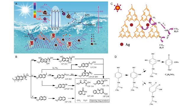
图8 光催化降解机理以及路径图: (A)宽谱光照射下SDAg-CQDs/UCN光催化降解萘普生(NPX)机理示意图;(B)可见光照射下在SDAg-CQDs/UCN水溶液中NPX可能的转化途径[32];(C)4% Ag-g-C3N4光催化臭氧氧化降解对乙酰氨基酚(ACE)的机理;(D)4% Ag-g-C3N4光催化臭氧化降解ACE的途径[284]

Fig.8 Photocatalytic degradation mechanism and pathway. (A) Schematic photocatalytic mechanism for the SDAg-CQDs/UCN under broad-spectrum light irradiation; (B) possible transformation pathways of NPX in the aqueous SDAg-CQDs/UCN solution under visible light irradiation[32], Copyright 2018, Elsevier. (C) Proposed mechanism of photocatalytic ozonation by 4% Ag-g-C3N4 for degrading acetaminophen (ACE); (D) Proposed degradation pathway for photocatalytic ozonation of ACE using 4% Ag-g-C3N4[284], Copyright 2019, Elsevier

3.2 Application of Silver Nanomaterials in Water Purification — — Antibacterial and Antiviral

With the development of science and technology and the prosperity of business, the global clean and fresh water resources have been sharply reduced, and one sixth of the population is facing the problem of insufficient supply of fresh water. There are many factors causing water pollution. According to the United Nations report, as early as 2001, the world produced 2 million tons of industrial and domestic sewage every day, and the polluted water seriously threatened human health. In addition, water can be contaminated by viruses, protozoa, bacteria, fungi, and worms, which spread diseases through water. It is estimated that about 842,000 people (most of them children under 5 years old) die of diarrheal diseases every year due to unsafe drinking water and inadequate sanitation facilities. Therefore, the purification of water resources in the environment, especially drinking water, is particularly important[27,286,287]. Since the first mention of nanotechnology in 1974, nanoparticles have been used in many fields, among which environmental water treatment and purification to obtain freshwater have attracted enough attention of scientists and have been studied in depth[5,288,289].
Over the past decade, there has been an increasing number of studies using silver nanoparticles and silver nanowires for environmental water treatment, with good results in water disinfection, achieving the goal of disinfection without the formation of harmful by-products by conventional chemical disinfectants[289~291]. Fig. 9a shows the whole process of using graphene oxide-silver nanocomposite (GO-Ag) for disinfection, including multiple stages: in the first stage, the AgNPs loaded on the GO sheet are oxidized by dissolved oxygen, and in this stage, the GO-Ag nanocomposite can be dispersed in water to form a stable hydrosol due to the good hydrophilicity and water dispersibility of the GO sheet; In the second stage, the free silver ions are in contact with the bacterial cells, and the negatively charged lipids on the cell membrane capture the positively charged silver ions through electrostatic attraction; In the third stage, silver ions interact with bacteria (by binding to membrane proteins or phospholipid bilayers) and destroy the membrane system of bacteria, and then a large number of free silver ions and even GO-Ag nanocomposites can enter cells. In the last stage, silver ions directly interfere with proteins, lipids, enzymes and DNA, and oxidize them through the generation of reactive oxygen species (ROS). ROS and oxidative stress have strong destructive power. The oxidation of biological macromolecules in cells will cause irreversible damage, eventually leading to cell death. In summary, the antibacterial activity of GO-Ag nanocomposite mainly comes from the cell membrane destruction and oxidative stress synergism[291]. Anshup et al. Reviewed the use of the chemical properties of nanomaterials for water purification and the significant progress that has been made, and pointed out that the application of noble metal nanoparticles in drinking water purification mainly includes the removal of three types of pollutants (halogenated organic compounds (including pesticides), heavy metals and microorganisms)[292]. Among them, silver nanoparticles have good bactericidal activity against various bacteria (such as Escherichia coli, Pseudomonas aeruginosa, Staphylococcus aureus, etc.), which can be used for water disinfection[293~297]. In addition, even under the condition of low concentration of silver ions, silver nanoparticles can more effectively deliver silver ions to the cell membrane or cytoplasm of bacteria, and at the same time, the proton motive force reduces the local pH (as low as pH = 3.0) and enhances the release of Ag+. Finally, the cell membrane and DNA of bacteria are damaged, thus achieving the purpose of sterilization, as shown in Figure 9B[298]. Praveena et al. Discussed in depth the incorporation of silver nanoparticles into a series of low-cost materials (ceramics, polymers, polyurethanes, agricultural wastes, and fibers) to prepare composites, which were then used as antibacterial agents to remove E. coli from water[299]. Kennedy et al pointed out that the provision of safe drinking water is a huge challenge for both developing and developed countries. The increasing demand for water resources and the deterioration of water quality have led to the continuous exploration of new technologies for water purification. Among them, nanotechnology has great prospects in ensuring safe drinking water and has been used in water treatment[300]. Subsequently, Wei et al. Reviewed the synthesis methods of silver nanowires and their applications as antibacterial materials. In addition, other silver nanomaterials with various shapes have also been used for sterilization or antivirus[37][74,81,89,167,301].
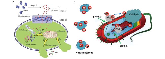
图9 银纳米材料抗菌机理图:(A)GO-Ag的抗菌机理示意图[291];(B)银纳米颗粒(AgNPs),银离子(Ag+)和细胞相互作用示意图[298]

Fig.9 Schematic mechanisms for antibacterial behaviors of silver nanomaterials. (A) Schematic mechanisms for antibacterial behaviors of GO-Ag[291], Copyright 2016, Elsevier. (B) Schematic of AgNPs, Ag+, and cell interactions[298], Copyright 2012, American Chemical Society

3.3 Application of Silver Nanomaterials in Toxic Metal Wastewater Treatment — Sensor

Since the Industrial Revolution, heavy metal pollution has become a major problem affecting the world's environment and human life. The most common toxic heavy metals include lead, mercury, cadmium, chromium, arsenic and so on. The remediation of heavy metals in water has attracted great attention all over the world, especially as the only liquid metal, the detection and remediation of mercury in ecosystems and aqueous media has become a great challenge, and the application of nanomaterials has been widely studied in various remediation strategies[302~304]. Starch-stabilized silver nanoparticles synthesized by reduction method, silver nanoparticles synthesized by using lipid-derived signal molecules and N-steroylethanolamine, and cysteamine-stabilized silver nanoparticles can be used to selectively detect and monitor mercury ions in aqueous solution[305][306][307]. Both silver nanoparticles without capping agent and protected silver nanoparticles (Ag @ MSA, where MSA is mercaptosuccinic acid) can reduce Hg (II) and Hg (I) ions to metallic mercury for the purpose of detection[308]. A colorimetric method for the detection of Hg (Ⅱ) can be established by using water-soluble unmodified silver nanoparticles and the specific binding of functional DNA probes to Hg (Ⅱ), and a highly selective sensing of Hg (II) can also be achieved by using green synthetic unmodified silver nanoparticles[309][310][311]. In addition, based on the linear blue shift of the maximum absorption wavelength of silver nanoparticles and the electrostatic force, mercury ions can be detected with high selectivity[312][313].
In addition to the above silver nanoparticles, silver composites can also be used for sensing toxic metals in the environment. Both the composite of silver nanoparticles loaded in cyclodextrin-silicate and deposited on amine-functionalized silica spheres can be used for colorimetric detection of mercury ions[314,315]. The Ag @ AgCl nanocomposite prepared by Samimi et al. Using green marine crude oil extract showed highly sensitive sensitivity to mercury[324]. In addition, Radhakrishnan et al. Embedded silver nanoparticles into a polyvinyl alcohol (Ag-PVA) film to fabricate a fast, efficient and highly selective universal sensor for mercury (Hg,HgI,HgII)[316].
In addition, Pradeep et al. Supported mercaptosuccinic acid (MSA) protected silver nanoparticles (9 ± 2 and 20 ± 5 nm in diameter) on activated alumina to prepare silver nanocomposites, which were used to remove mercury ions from environmental wastewater[317]. Based on thiol-functionalized silver nanoparticles, Hg (Ⅱ) can also be determined by SERS[318]. Sahoo et al. Prepared a novel p-phenylenediamine (p-PDA) functionalized silver nanoparticles and used them for the detection of heavy metal ions in water. The results showed that the detection limits for Hg2+ and Fe3+ were 0.80 and 1.29 μmol/L, respectively. This method is simple and sensitive[319]. Table 5 succinctly summarizes the application of silver nanomaterials as sensors for toxic metal detection.
表5 银纳米材料作为传感器在有毒金属检测中的应用总结

Table 5 The applications of silver nanomaterials as sensors in the detection of toxic metals

Materials Analyte Detection technique LOD ref
Starch-stabilized AgNPs Hg2+ UV-Vis 5 ppb 305
Silver nanoclusters Hg2+ Fluorescence 10-10 M 320
Silver nanoparticles Hg2+, Hg+ X-ray photoelectron spectroscopic * 308
Silver nanoparticles Hg2+ UV-visible 17 nM 309
Silver nanoparticle-embedded poly (vinyl alcohol) (Ag-PVA) thin film Hg2+, Hg22+, Hg Surface plasmon resonance (SPR) extinction 1 ppb 316
Silver nanoparticle loaded on alumina Hg2+ ICP-OES * 317
Ag25 clusters Hg2+, Pt2+, Au3+ Absorption and fluorescence 1 ppb, ppm 321
Thiol-functionalized silver nanoparticles Hg2+ Surface-enhanced Raman scattering spectroscopy (SERS) 0.0024 μM
Silver nanoparticles Hg2+ UV-vis 2.2×10⁻⁶ M 310
Silver nanoparticles Hg2+ UV-vis 6.6×10⁻⁹ M 313
The p-phenylenediamine (p-PDA) functionalized silver nanoparticles (AgNPs) Hg2+, Fe3+ Surface plasmon resonance (SPR) absorption 0.80 M, 1.29 M 319
Silver nanoparticles embedded in cyclodextrin-silicate composite Hg2+, nitrobenzene Surface plasmon absorption spectra * 314
SiO2/Ag NPs Hg2+ Spectral and colorimetric detection 5 μM 315
Ag nanoparticle-decorated graphene quantum dots ((AgNPs/GQDs) Ag+, Cys, Hcy, GSH Fluorescence 3.5 nM, 6.2 nM, 4.5 nM, 4.1 nM 322
Silver nanoparticles Hg2+ Colorimetric sensing 0.5 nM 312
Silver nanoparticles Hg2+ Colorimetric sensing 0.5 mM 306
Silver nanoparticles Hg2+ Colorimetric sensing 0.273 nM 307
Silver nanoparticles Hg2+ Colorimetric sensing 1.18 nM 311
Silver nanoparticles Hg2+,Cr3+ Colorimetric sensing 0.125 μM, 6.25 μM 323
Ag@AgCl nanomaterial Hg2+ Colorimetric sensing 4.19 nM 324

4 Conclusion and prospect

In this paper, the synthesis methods and types of silver nanomaterials are reviewed, and the chemical and physical synthesis methods and types of silver nanomaterials are classified in detail. Then, the application of silver nanomaterials synthesized by various methods in the environment is summarized in detail, mainly for the treatment of wastewater containing dyes, microplastics, new pollutants, heavy metal ions, bacteria and viruses. Because silver nanoparticles synthesized by biological sources are less toxic than those synthesized by traditional routes, green synthesized nanomaterials have been successful in sensor development, industrial wastewater treatment and environmental remediation, making it possible for nanotechnology to be popularized in analytical chemistry and modern society. However, there are still some challenges to be overcome in the exploration and practical application of silver nanomaterials, in addition to the previous outstanding achievements.
(1) In recent years, the research on low-cost and environmentally friendly materials has increased dramatically, and green synthesis has avoided the traditional use of harmful reagents, but the shortcomings are obvious: the types of silver nanomaterials synthesized by green synthesis are extremely limited, and the particle size is uneven, so the feasibility of green synthesis of various silver nanomaterials needs to be explored urgently.
(2) There are many kinds of silver nanomaterials, and because the reagents or by-products in chemical synthesis are harmful, they may introduce new environmental problems, so the materials that are really effective for the environment are extremely limited. Based on this, the purification and modification technologies of various silver nanomaterials need to be developed urgently to achieve the goal of environmental friendliness and high efficiency, so as to broaden the environmental applications of many existing materials.
(3) As one of the most powerful components of artificial intelligence, machine learning based on computer algorithms can achieve fast and reliable prediction through data mining.It has shown great potential in the exploration of efficient catalysts. In addition, the use of silver nanomaterials in the catalytic degradation and removal of microplastics in the environment is still in its infancy and needs to be further explored and developed.
(4) In recent years, silver nanomaterials have been transformed into monatomic silver composites with catalytic efficiency up to 100%, while the synthesis and application of monatomic silver composites are still in their infancy, and there is still much room for exploration and development.
[1]
Zhang X F, Liu Z G, Shen W, Gurunathan S. Int. J. Mol. Sci., 2016, 17(9): 1534.

[2]
Gunawan C, Marquis C P, Amal R, Sotiriou G A, Rice S A, Harry E J. ACS Nano, 2017, 11(4): 3438.

[3]
Rizzello L, Pompa P P. Chem. Soc. Rev., 2014, 43(5): 1501.

[4]
Fernando A, Dimuthu M Weerawardene K L, Karimova N V, Aikens C M. Chem. Rev., 2015, 115(12): 6112.

[5]
Islam M A, Jacob M V, Antunes E. J. Environ. Manag., 2021, 281: 111918.

[6]
Wiley B, Herricks T, Sun Y G, Xia Y N. Nano Lett., 2004, 4(9): 1733.

[7]
Li H, You Q, Feng X, Zheng C, Zeng X, Xu H. J. Drug Deliv. Sci. Tech., 2023, 80: 104165.

[8]
Steinigeweg D, Schlücker S. Chem. Commun., 2012, 48(69): 8682.

[9]
Personick M L, Langille M R, Zhang J, Wu J S, Li S Y, Mirkin C A. Small, 2013, 9(11): 1947.

[10]
Im S H, Lee Y T, Wiley B, Xia Y N. Angewandte Chemie Int. Ed., 2005, 44(14): 2154.

[11]
Rycenga M, McLellan J M, Xia Y N. Adv. Mater., 2008, 20(12): 2416.

[12]
Jana N R, Gearheart L, Murphy C J. Chem. Commun., 2001(7): 617.

[13]
Wiley B J, Chen Y, McLellan J M, Xiong Y J, Li Z Y, Ginger D, Xia Y N. Nano Lett., 2007, 7(4): 1032.

[14]
Pietrobon B, McEachran M, Kitaev V. ACS Nano, 2009, 3(1): 21.

[15]
Wiley B J, Xiong Y J, Li Z Y, Yin Y D, Xia Y N. Nano Lett., 2006, 6(4): 765.

[16]
Zhu Z X, Wang X L, Yu H Y, Zhou W, Wang Y S, Han J, Guo F. Cryst. Growth Des., 2023, 23(3): 1455.

[17]
Xinling T, Masaharu T, Michiko N, Peng J. Bull. Chem. Soc. Jpn., 2009, 82(10):1304.

[18]
Wijaya Y N, Kim J, Choi W M, Park S H, Kim M H. Nanoscale, 2017, 9(32): 11705.

[19]
Ledwith D M, Whelan A M, Kelly J M. J. Mater. Chem., 2007, 17(23): 2459.

[20]
Zhang Q, Li N, Goebl J, Lu Z D, Yin Y D. J. Am. Chem. Soc., 2011, 133(46): 18931.

[21]
Pastoriza-Santos I, Liz-Marzán L M. Nano Lett., 2002, 2(8): 903.

[22]
Korte K E, Skrabalak S E, Xia Y N. J. Mater. Chem., 2008, 18(4): 437.

[23]
Zhang S H, Xie Z X, Jiang Z Y, Xu X, Xiang J, Huang R B, Zheng L S. Chem. Commun., 2004(9): 1106.

[24]
Wiley B J, Im S H, Li Z Y, McLellan J, Siekkinen A, Xia Y N. J. Phys. Chem. B, 2006, 110(32): 15666.

[25]
Anker J N, Hall W P, Lyandres O, Shah N C, Zhao J, Van Duyne R P. Nat. Mater., 2008, 7(6): 442.

[26]
Marimuthu S, Antonisamy A J, Malayandi S, Rajendran K, Tsai P C, Pugazhendhi A, Ponnusamy V K. J. Photochem. Photobiol. B Biol., 2020, 205: 111823.

[27]
Bahcelioglu E, Unalan H E, Erguder T H. Crit. Rev. Environ. Sci. Technol., 2021, 51(20): 2389.

[28]
Heinemann M G, Rosa C H, Rosa G R, Dias D. Trends Environ. Anal. Chem., 2021, 30: e00129.

[29]
Bu Y R, Lee S. ACS Appl. Mater. Interfaces, 2012, 4(8): 3923.

[30]
Thanh N T K, Green L A W. Nano Today, 2010, 5(3): 213.

[31]
Dong B, Xue N, Mu G H, Wang M J, Xiao Z H, Dai L, Wang Z X, Huang D C, Qian H L, Chen W. Ultrason. Sonochemistry, 2021, 73: 105485.

[32]
Wang F L, Wang Y F, Feng Y P, Zeng Y Q, Xie Z J, Zhang Q X, Su Y H, Chen P, Liu Y, Yao K, Lv W Y, Liu G G. Appl. Catal. B Environ., 2018, 221: 510.

[33]
Nowack B, Krug H F, Height M. Environ. Sci. Technol., 2011, 45(4): 1177.

[34]
Bhattarai B, Zaker Y, Atnagulov A, Yoon B, Landman U, Bigioni T P. Acc. Chem. Res., 2018, 51(12): 3104.

[35]
He D, Garg S, Wang Z M, Li L, Rong H Y, Ma X M, Li G Y, An T C, David Waite T. Environ. Sci.: Nano, 2019, 6(6): 1674.

[36]
Guo Z, Cui K P, Zeng G M, Wang J J, Guo X P. Sci. Total Environ., 2018, 643: 1325.

[37]
Zhang P, Wyman I, Hu J W, Lin S D, Zhong Z W, Tu Y Y, Huang Z Z, Wei Y L. Mater. Sci. Eng. B, 2017, 223: 1.

[38]
Zhang Z, Shen W F, Xue J, Liu Y M, Liu Y W, Yan P P, Liu J X, Tang J G. Nanoscale Res. Lett., 2018, 13(1): 1.

[39]
Nasrollahzadeh M, Mahmoudi-Gom Yek S, Motahharifar N, Ghafori Gorab M. Chem. Rec., 2019, 19(12): 2436.

[40]
Cinelli M, Coles S R, Nadagouda M N, Błaszczyński J, Słowiński R, Varma R S, Kirwan K. Green Chem., 2015, 17(5): 2825.

[41]
Xue J, Liu J X, Mao S, Wang Y, Shen W F, Wang W, Huang L J, Li H L, Tang J G. Mater. Res. Bull., 2018, 106: 113.

[42]
Zhao Q X, Zhao M M, Qiu J Q, Lai W Y, Pang H, Huang W. Small, 2017, 13(38): 1701091.

[43]
Sun D L, Xu X M, Qin Y L, Jiang S P, Shao Z P. ChemSusChem, 2020, 13(1): 39.

[44]
An C H, Wang S T, Sun Y G, Zhang Q H, Zhang J, Wang C Y, Fang J Y. J. Mater. Chem. A, 2016, 4(12): 4336.

[45]
Xue W J, Huang D L, Wen X J, Chen S, Cheng M, Deng R, Li B, Yang Y, Liu X G. J. Hazard. Mater., 2020, 390: 122128.

[46]
Liang C, Lu Z A, Wu J, Chen M X, Zhang Y Y, Zhang B, Gao G L, Li S W, Xu P. ACS Appl. Mater. Interfaces, 2020, 12(49): 54266.

[47]
Zhang J A, Langille M R, Mirkin C A. J. Am. Chem. Soc., 2010, 132(35): 12502.

[48]
Landry M J, GellÉ A, Meng B Y, Barrett C J, Moores A. ACS Catal., 2017, 7(9): 6128.

[49]
Zhang J W, Winget S A, Wu Y R, Su D, Sun X J, Xie Z X, Qin D. ACS Nano, 2016, 10(2): 2607.

[50]
Zhang Y, Ahn J, Liu J Y, Qin D. Chem. Mater., 2018, 30(6): 2151.

[51]
Jia H L, Dou Y R, Yang Y Y, Li F, Zhang C Y. Nanoscale, 2021, 13(47): 20289.

[52]
Zou X X, Silva R, Huang X X, Al-Sharab J F, Asefa T. Chem. Commun., 2013, 49(4): 382.

[53]
Xu G W, Wu Y P, Dong W W, Zhao J, Wu X Q, Li D S, Zhang Q C. Small, 2017, 13(22): 1602996.

[54]
Charistoudi E, Kallitsakis M G, Charisteidis I, Triantafyllidis K S, Lykakis I N. Adv. Synth. Catal., 2017, 359(17): 2949.

[55]
Sadovnikov S I, Gusev A I. J. Mater. Chem. A, 2017, 5(34): 17676.

[56]
Sharma S, Dutta V, Raizada P, Hosseini-Bandegharaei A, Thakur V K, Kalia S, Nguyen V H, Singh P. J. Environ. Chem. Eng., 2021, 9(2): 105157.

[57]
Song W, Querebillo C J, Götz R, Katz S, Kuhlmann U, Gernert U, Weidinger I M, Hildebrandt P. Nanoscale, 2017, 9(24): 8380.

[58]
Jensen T R, Malinsky M D, Haynes C L, Van Duyne R P. J. Phys. Chem. B, 2000, 104(45): 10549.

[59]
Zulkifli N I, Muhamad M, Mohamad Zain N N, Tan W N, Yahaya N, Bustami Y, Abdul Aziz A, Nik Mohamed Kamal N N S. Molecules, 2020, 25(18): 4332.

[60]
Caswell K K, Bender C M, Murphy C J. Nano Lett., 2003, 3(5): 667.

[61]
MÉtraux G S, Mirkin C A. Adv. Mater., 2005, 17(4): 412.

[62]
Raza M, Kanwal Z, Rauf A, Sabri A, Riaz S, Naseem S. Nanomaterials, 2016, 6(4): 74.

[63]
Shameli K, Bin Ahmad M, Davoud Jazayeri S, Sedaghat S, Shabanzadeh P, Jahangirian H, Mahdavi M, Abdollahi Y. Int. J. Mol. Sci., 2012, 13(6): 6639.

[64]
Skrabalak S E, Au L, Li X D, Xia Y N. Nat. Protoc., 2007, 2(9): 2182.

[65]
Sun Y G, Xia Y N. Science, 2002, 298(5601): 2176.

[66]
Banerjee S, Loza K, Meyer-Zaika W, Prymak O, Epple M. Chem. Mater., 2014, 26(2): 951.

[67]
Xia X H, Zeng J, McDearmon B, Zheng Y Q, Li Q G, Xia Y N. Angew. Chem., 2011, 123(52): 12750.

[68]
Liu X X, Li L L, Yang Y D, Yin Y D, Gao C B. Nanoscale, 2014, 6(9): 4513.

[69]
Pan Z Y, Zhou J, Zou H Y, Li Y F, Gao P F, Huang C Z. J. Colloid Interface Sci., 2021, 584: 253.

[70]
Xu H X, Suslick K S. ACS Nano, 2010, 4(6): 3209.

[71]
Chen S L, Wang G H, Sui W J, Parvez A M, Si C L. Green Chem., 2020, 22(9): 2879.

[72]
Pan Z Y, Gao P F, Jing C J, Zhou J, Liang W T, Lei G, Feng W, Li Y F, Huang C Z. Appl. Catal. B Environ., 2021, 291: 120090.

[73]
Adhikari L, Larm N E, Bhawawet N, Baker G A. ACS Sustainable Chem. Eng., 2018, 6(5): 5725.

[74]
Hebbalalu D, Lalley J, Nadagouda M N, Varma R S. ACS Sustainable Chem. Eng., 2013, 1(7): 703.

[75]
Zhang Q, Li W Y, Moran C, Zeng J, Chen J Y, Wen L P, Xia Y N. J. Am. Chem. Soc., 2010, 132(32): 11372.

[76]
Xia Y N, Gilroy K D, Peng H C, Xia X H. Angewandte Chemie Int. Ed., 2017, 56(1): 60.

[77]
Koga N, Tanaka H. Thermochimica Acta, 2002, 388(1/2): 41.

[78]
Hosseinpour-Mashkani S M, Ramezani M. Mater. Lett., 2014, 130: 259.

[79]
Akhtar M S, Panwar J, Yun Y S. ACS Sustainable Chem. Eng., 2013, 1(6): 591.

[80]
Virkutyte J, Varma R S. Chem. Sci., 2011, 2(5): 837.

[81]
Moulton M C, Braydich-Stolle L K, Nadagouda M N, Kunzelman S, Hussain S M, Varma R S. Nanoscale, 2010, 2(5): 763.

[82]
Johnson R W, Hultqvist A, Bent S F. Mater. Today, 2014, 17(5): 236.

[83]
Sharma R K, Yadav S, Dutta S, Kale H B, Warkad I R, Zbořil R, Varma R S, Gawande M B. Chem. Soc. Rev., 2021, 50(20): 11293.

[84]
Gawande M B, Shelke S N, Zboril R, Varma R S. Acc. Chem. Res., 2014, 47(4): 1338.

[85]
Xu H X, Zeiger B W, Suslick K S. Chem. Soc. Rev., 2013, 42(7): 2555.

[86]
Bang J H, Suslick K S. Adv. Mater., 2010, 22(10): 1039.

[87]
Van Hyning D L, Zukoski C F. Langmuir, 1998, 14(24): 7034.

[88]
Wuithschick M, Paul B, Bienert R, Sarfraz A, Vainio U, Sztucki M, Kraehnert R, Strasser P, Rademann K, Emmerling F, Polte J. Chem. Mater., 2013, 25(23): 4679.

[89]
Aziz N, Faraz M, Pandey R, Shakir M, Fatma T, Varma A, Barman I, Prasad R. Langmuir, 2015, 31(42): 11605.

[90]
Dauthal P, Mukhopadhyay M. Ind. Eng. Chem. Res., 2016, 55(36): 9557.

[91]
Nadagouda M N, Varma R S. Green Chem., 2008, 10(8): 859.

[92]
Raveendran P, Fu J, Wallen S L. J. Am. Chem. Soc., 2003, 125(46): 13940.

[93]
Sutradhar P, Saha M. J. Phys. Chem. C, 2016, 120(16): 8941.

[94]
Xue Y Y, Qiu X Q, Liu Z W, Li Y. ACS Sustainable Chem. Eng., 2018, 6(6): 7695.

[95]
Boopathi S, Gopinath S, Boopathi T, Balamurugan V, Rajeshkumar R, Sundararaman M. Ind. Eng. Chem. Res., 2012, 51(17): 5976.

[96]
Kasithevar M, Saravanan M, Prakash P, Kumar H, Ovais M, Barabadi H, Shinwari Z K. J. Interdiscip. Nanomed., 2017, 2(2): 131.

[97]
Nadagouda M N, Iyanna N, Lalley J, Han C, Dionysiou D D, Varma R S. ACS Sustainable Chem. Eng., 2014, 2(7): 1717.

[98]
Nakano M, Fujiwara T, Koga N. J. Phys. Chem. C, 2016, 120(16): 8841.

[99]
Zong R L, Wang X L, Shi S K, Zhu Y F. Phys. Chem. Chem. Phys., 2014, 16(9): 4236.

[100]
Rostami S, Mehdinia A, Jabbari A. Spectrochimica Acta A Mol. Biomol. Spectrosc., 2017, 180: 204.

[101]
Lin X, Lin S, Liu Y L, Gao M M, Zhao H Y, Liu B K, Hasi W, Wang L. Langmuir, 2018, 34(21): 6077.

[102]
Zhang L, Wang Y, Tong L M, Xia Y N. Langmuir, 2013, 29(50): 15719.

[103]
Lin Z W, Tsao Y C, Yang M Y, Huang M H. Chem. A Eur. J., 2016, 22(7): 2326.

[104]
Wiley B, Sun Y G, Mayers B, Xia Y N. Chem. A Eur. J., 2005, 11(2): 454.

[105]
Siekkinen A R, McLellan J M, Chen J Y, Xia Y N. Chem. Phys. Lett., 2006, 432(4/6): 491.

[106]
Sherry L J, Chang S H, Schatz G C, Van Duyne R P, Wiley B J, Xia Y N. Nano Lett., 2005, 5(10): 2034.

[107]
Wang Y, Zou H Y, Huang C Z. Nanoscale, 2015, 7(37): 15209.

[108]
Zhang Q, Li W Y, Wen L P, Chen J Y, Xia Y N. Chem. A Eur. J., 2010, 16(33): 10234.

[109]
Yu D B, Yam V W W. J. Am. Chem. Soc., 2004, 126(41): 13200.

[110]
Jiang L P, Xu S, Zhu J M, Zhang J R, Zhu J J, Chen H Y. Inorg. Chem., 2004, 43(19): 5877.

[111]
Washio I, Xiong Y, Yin Y, Xia Y. Adv. Mater., 2006, 18(13): 1745.

[112]
Aherne D, Ledwith D M, Gara M, Kelly J M. Adv. Funct. Mater., 2008, 18(14): 2005.

[113]
Charles D E, Aherne D, Gara M, Ledwith D M, Gun’ko Y K, Kelly J M, Blau W J, Brennan-Fournet M E. ACS Nano, 2010, 4(1): 55.

[114]
Zhu Y P, Wang X K, Guo W L, Wang J G, Wang C. Ultrason. Sonochemistry, 2010, 17(4): 675.

[115]
Zhang J, Langille M R, Mirkin C A. Nano Lett., 2011, 11(6): 2495.

[116]
Sun Y G, Yin Y D, Mayers B T, Herricks T, Xia Y N. Chem. Mater., 2002, 14(11): 4736.

[117]
Sun Y G, Gates B, Mayers B, Xia Y N. Nano Lett., 2002, 2(2): 165.

[118]
Sun Y G, Mayers B, Herricks T, Xia Y N. Nano Lett., 2003, 3(7): 955.

[119]
Kou J H, Varma R S. Chem. Commun., 2013, 49(7): 692.

[120]
Tang S C, Vongehr S, Wan N, Meng X K. Mater. Chem. Phys., 2013, 142(1): 17.

[121]
Langille M., Personick M., Mirkin C. Angewandte Chemie Int. Ed., 2013, 52(52): 13910-13940.

[122]
Sherry L J, Jin R C, Mirkin C A, Schatz G C, Van Duyne R P. Nano Lett., 2006, 6(9): 2060.

[123]
Jin R C, Cao Y W, Mirkin C A, Kelly K L, Schatz G C, Zheng J G. Science, 2001, 294(5548): 1901.

[124]
Zhang M F, Zhao A W, Sun H H, Guo H Y, Wang D P, Li D, Gan Z B, Tao W Y. J. Mater. Chem., 2011, 21(46): 18817.

[125]
Zhang B, Xu P, Xie X M, Wei H, Li Z P, Mack N H, Han X J, Xu H X, Wang H L. J. Mater. Chem., 2011, 21(8): 2495.

[126]
Kang L L, Xu P, Zhang B, Tsai H, Han X J, Wang H L. Chem. Commun., 2013, 49(33): 3389.

[127]
Tang X H, Cai W Y, Yang L B, Liu J H. Nanoscale, 2014, 6(15): 8612.

[128]
Chen S H, Fan Z Y, Carroll D L. J. Phys. Chem. B, 2002, 106(42): 10777.

[129]
Zhang D B, Qi L M, Yang J H, Ma J M, Cheng H M, Huang L. Chem. Mater., 2004, 16(5): 872.

[130]
Xia X H, Zeng J, Oetjen L K, Li Q G, Xia Y N. J. Am. Chem. Soc., 2012, 134(3): 1793.

[131]
Wang Y, Wan D H, Xie S F, Xia X H, Huang C Z, Xia Y N. ACS Nano, 2013, 7(5): 4586.

[132]
Yang Y, Matsubara S, Xiong L M, Hayakawa T, Nogami M. J. Phys. Chem. C, 2007, 111(26): 9095.

[133]
Burda C, Chen X B, Narayanan R, El-Sayed M A. Chem. Rev., 2005, 105(4): 1025.

[134]
Henglein A. Chem. Mater., 1998, 10(1): 444.

[135]
Tao A, Sinsermsuksakul P, Yang P D. Angewandte Chemie Int. Ed., 2006, 45(28): 4597.

[136]
Rebello Sousa Dias M, Leite M S. Acc. Chem. Res., 2019, 52(10): 2881.

[137]
Liu Y, Huang C Z. ACS Nano, 2013, 7(12): 11026.

[138]
Wu W, Lei M, Yang S L, Zhou L, Liu L, Xiao X H, Jiang C Z, Roy V A L. J. Mater. Chem. A, 2015, 3(7): 3450.

[139]
Chen L M, Deming C P, Peng Y, Hu P G, Stofan J, Chen S W. Nanoscale, 2016, 8(30): 14565.

[140]
Wu Y R, Sun X J, Yang Y, Li J M, Zhang Y, Qin D. Acc. Chem. Res., 2017, 50(7): 1774.

[141]
Karam T E, Smith H T, Haber L H. J. Phys. Chem. C, 2015, 119(32): 18573.

[142]
Peng Z Q, Spliethoff B, Tesche B, Walther T, Kleinermanns K. J. Phys. Chem. B, 2006, 110(6): 2549.

[143]
Sharma M, Pudasaini P R, Ruiz-Zepeda F, Vinogradova E, Ayon A A. ACS Appl. Mater. Interfaces, 2014, 6(17): 15472.

[144]
Wang P Y, Bai Y J, Yao C, Li X M, Zhou L, Wang W X, El-Toni A M, Zi J, Zhao D Y, Shi L, Zhang F. Anal. Chem., 2017, 89(4): 2583.

[145]
AbdelHamid A A, Al-Ghobashy M A, Fawzy M, Mohamed M B, Abdel-Mottaleb M M S A. ACS Sustainable Chem. Eng., 2013, 1(12): 1520.

[146]
Xia B H, He F, Li L D. Langmuir, 2013, 29(15): 4901.

[147]
Chen J Y, Wiley B, McLellan J, Xiong Y J, Li Z Y, Xia Y N. Nano Lett., 2005, 5(10): 2058.

[148]
Li J M, Sun X J, Qin D. ChemNanoMat, 2016, 2(6): 494.

[149]
Peiris S, Sarina S, Han C H, Xiao Q, Zhu H Y. Dalton Trans., 2017, 46(32): 10665.

[150]
Zhang W Q, Yang J Z, Lu X M. ACS Nano, 2012, 6(8): 7397.

[151]
Zhang Z Y, Nenoff T M, Leung K, Ferreira S R, Huang J Y, Berry D T, Provencio P P, Stumpf R. J. Phys. Chem. C, 2010, 114(34): 14309.

[152]
Ghosh Chaudhuri R, Paria S. Chem. Rev. 2012, 112(4): 2373.

[153]
Tsuji M, Miyamae N, Lim S, Kimura K, Zhang X, Hikino S, Nishio M. Cryst. Growth Des., 2006, 6(8): 1801.

[154]
Anandan S, Grieser F, Ashokkumar M. J. Phys. Chem. C, 2008, 112(39): 15102.

[155]
Samal A K, Polavarapu L, Rodal-Cedeira S, Liz-Marzán L M, PÉrez-Juste J, Pastoriza-Santos I. Langmuir, 2013, 29(48): 15076.

[156]
Yang Y, Liu J Y, Fu Z W, Qin D. J. Am. Chem. Soc., 2014, 136(23): 8153.

[157]
Zhuo X L, Zhu X Z, Li Q, Yang Z, Wang J F. ACS Nano, 2015, 9(7): 7523.

[158]
Ye Z J, Weng R, Ma Y H, Wang F Y, Liu H, Wei L, Xiao L H. Anal. Chem., 2018, 90(21): 13044.

[159]
Sun X J, Qin D. J. Mater. Chem. C, 2015, 3(45): 11833.

[160]
Carroll K J, Hudgins D M, Spurgeon S, Kemner K M, Mishra B, Boyanov M I, Brown L W III, Taheri M L, Carpenter E E. Chem. Mater., 2010, 22(23): 6291.

[161]
Li J M, Liu J Y, Yang Y, Qin D. J. Am. Chem. Soc., 2015, 137(22): 7039.

[162]
Kim C, Suh B L, Yun H, Kim J, Lee H. ACS Catal., 2017, 7(4): 2294.

[163]
Khatami M, Alijani H, Nejad M, Varma R. Appl. Sci., 2018, 8(3): 411.

[164]
Poggi E, Gohy J F. Colloid Polym. Sci., 2017, 295(11): 2083.

[165]
Jiang F R, Tian Q W, Tang M H, Chen Z G, Yang J M, Hu J Q. CrystEngComm, 2011, 13(24): 7189.

[166]
Song Y, Liu K, Chen S W. Langmuir, 2012, 28(49): 17143.

[167]
Chang Z M, Wang Z, Lu M M, Shao D, Yue J, Yang D, Li M Q, Dong W F. Colloids Surf. B Biointerfaces, 2017, 157: 199.

[168]
Wang Z, Chang Z M, Lu M M, Shao D, Yue J, Yang D, Li M Q, Dong W F. ACS Appl. Mater. Interfaces, 2017, 9(36): 30306.

[169]
Kirillova A, Schliebe C, Stoychev G, Jakob A, Lang H, Synytska A. ACS Appl. Mater. Interfaces, 2015, 7(38): 21218.

[170]
Buccolieri A, Bettini S, Salvatore L, Baldassarre F, Ciccarella G, Giancane G. Sens. Actuat. B Chem., 2018, 267: 265.

[171]
Guo Y C, Khan A U, Cao K, Liu G L. ACS Appl. Nano Mater., 2018, 1(10): 5377.

[172]
Levard C, Hotze E M, Lowry G V, Brown G E Jr. Environ. Sci. Technol., 2012, 46(13): 6900.

[173]
Sokolov S V, Tschulik K, Batchelor-McAuley C, Jurkschat K, Compton R G. Anal. Chem., 2015, 87(19): 10033.

[174]
Shao W, Liu X F, Min H H, Dong G H, Feng Q Y, Zuo S L. ACS Appl. Mater. Interfaces, 2015, 7(12): 6966.

[175]
Yang H, Liu Y, Shen Q H, Chen L F, You W H, Wang X M, Sheng J S. J. Mater. Chem., 2012, 22(45): 24132.

[176]
Chang S Q, Liu C C, Sun Y F, Yan Z F, Zhang X H, Hu X D, Zhang H Q. ACS Appl. Nano Mater., 2020, 3(3): 2302.

[177]
Cao H L, Huang H B, Chen Z, Karadeniz B, J, Cao R. ACS Appl. Mater. Interfaces, 2017, 9(6): 5231.

[178]
Gao G H, Mathkar A, Martins E P, Galvão D S, Gao D Y, Alves da Silva Autreto P, Sun C J, Cai L T, Ajayan P M. J. Mater. Chem. A, 2014, 2(9): 3148.

[179]
Xia H, Hong C Y, Shi X Q, Li B, Yuan G L, Yao Q F, Xie J P. J. Mater. Chem. A, 2015, 3(3): 1216.

[180]
Liu S, Tian J Q, Wang L, Sun X P. Carbon, 2011, 49(10): 3158.

[181]
Bhunia S K, Jana N R. ACS Appl. Mater. Interfaces, 2014, 6(22): 20085.

[182]
Lu X Q, Qi H T, Zhang X F, Xue Z H, Jin J, Zhou X B, Liu X H. Chem. Commun., 2011, 47(46): 12494.

[183]
Tang X Z, Cao Z W, Zhang H B, Liu J, Yu Z Z. Chem. Commun., 2011, 47(11): 3084.

[184]
Zhang Y M, Yuan X, Wang Y, Chen Y. J. Mater. Chem., 2012, 22(15): 7245.

[185]
Perdikaki A, Galeou A, Pilatos G, Karatasios I, Kanellopoulos N K, Prombona A, Karanikolos G N. ACS Appl. Mater. Interfaces, 2016, 8(41): 27498.

[186]
Bao Q, Zhang D, Qi P. J. Colloid Interface Sci., 2011, 360(2): 463.

[187]
Li Y Z, Cao Y L, Xie J, Jia D Z, Qin H Y, Liang Z T. Catal. Commun., 2015, 58: 21.

[188]
Jeon E K, Seo E, Lee E, Lee W, Um M K, Kim B S. Chem. Commun., 2013, 49(33): 3392.

[189]
Prasek J, Drbohlavova J, Chomoucka J, Hubalek J, Jasek O, Adam V, Kizek R. J. Mater. Chem., 2011, 21(40): 15872.

[190]
Singh P, Lamanna G, MÉnard-Moyon C, Toma F M, Magnano E, Bondino F, Prato M, Verma S, Bianco A. Angewandte Chemie Int. Ed., 2011, 50(42): 9893.

[191]
Yan X L, Li S, Bao J H, Zhang N, Fan B B, Li R F, Liu X G, Pan Y X. ACS Appl. Mater. Interfaces, 2016, 8(27): 17060.

[192]
Zheng J W, Duan X P, Lin H Q, Gu Z Q, Fang H H, Li J H, Yuan Y Z. Nanoscale, 2016, 8(11): 5959.

[193]
Krainoi A, Kummerlöwe C, Vennemann N, Nakaramontri Y, Pichaiyut S, Nakason C. J. Appl. Polym. Sci., 2019, 136(13): 47281.

[194]
Espinosa J C, NavalÓn S, Álvaro M, García H. ChemCatChem, 2015, 7(17): 2682.

[195]
Espinosa J C, NavalÓn S, Álvaro M, García H. Catal. Sci. Technol., 2016, 6(19): 7077.

[196]
Li S, Bandy J A, Hamers R J. ACS Appl. Mater. Interfaces, 2018, 10(6): 5395.

[197]
Patel S B, Vasava D V. ChemistrySelect, 2018, 3(2): 471.

[198]
Veerakumar P, Rajkumar C, Chen S M, Thirumalraj B, Lin K C. J. Electroanal. Chem., 2018, 826: 207.

[199]
Liu J, Wang H Q, Antonietti M. Chem. Soc. Rev., 2016, 45(8): 2308.

[200]
Zhang P, Shao C L, Zhang Z Y, Zhang M Y, Mu J B, Guo Z C, Liu Y C. Nanoscale, 2011, 3(8): 3357.

[201]
Ji T, Chen L, Schmitz M, Bao F S, Zhu J H. Green Chem., 2015, 17(4): 2515.

[202]
Caltagirone C, Bettoschi A, Garau A, Montis R. Chem. Soc. Rev., 2015, 44(14): 4645.

[203]
Sharma R K, Sharma S, Dutta S, Zboril R, Gawande M B. Green Chem., 2015, 17(6): 3207.

[204]
Steffan M, Jakob A, Claus P, Lang H. Catal. Commun., 2009, 10(5): 437.

[205]
Naik B, Hazra S, Prasad V S, Ghosh N N. Catal. Commun., 2011, 12(12): 1104.

[206]
Raji V, Chakraborty M, Parikh P A. Ind. Eng. Chem. Res., 2012, 51(16): 5691.

[207]
He D, Kacopieros M, Ikeda-Ohno A, David Waite T. Environ. Sci. Technol., 2014, 48(20): 12320.

[208]
Chen Z C, Luck R L. Green Chem., 2016, 18(11): 3354.

[209]
Guillerm V, Kim D, Eubank J F, Luebke R, Liu X F, Adil K, Lah M S, Eddaoudi M. Chem. Soc. Rev., 2014, 43(16): 6141.

[210]
Zhu Q L, Xu Q. Chem. Soc. Rev., 2014, 43(16): 5468.

[211]
Czaja A U, Trukhan N, Müller U. Chem. Soc. Rev., 2009, 38(5): 1284.

[212]
Zhu N N, Liu X H, Li T, Ma J G, Cheng P, Yang G M. Inorg. Chem., 2017, 56(6): 3414.

[213]
Dutta G, Jana A K, Singh D K, Eswaramoorthy M, Natarajan S. Chem. Asian J., 2018, 13(18): 2677.

[214]
Mbhele Z H, Salemane M G, van Sittert C G C E, Nedeljković J M, Djoković V, Luyt A S. Chem. Mater., 2003, 15(26): 5019.

[215]
Zhu J F, Zhu Y J. J. Phys. Chem. B, 2006, 110(17): 8593.

[216]
Yu D Y, Tan M X, Zhang Y G. Adv. Synth. Catal., 2012, 354(6): 969.

[217]
Eisa W H, Abdel-Baset T A, Mohamed E M A, Mahrous S. J. Inorg. Organomet. Polym. Mater., 2017, 27(6): 1703.

[218]
Silvestri D, Wacławek S, Venkateshaiah A, Krawczyk K, Sobel B, Padil V V T, Černík M, Varma R S. Carbohydr. Polym., 2020, 232: 115806.

[219]
Sun W L, Meng Y, Fu Q R, Wang F, Wang G J, Gao W H, Huang X C, Lu F S. ACS Appl. Mater. Interfaces, 2016, 8(15): 9881.

[220]
Lin Y, Bunker C E, Fernando K A S, Connell J W. ACS Appl. Mater. Interfaces, 2012, 4(2): 1110.

[221]
Pang J Y, Chao Y H, Chang H H, Li H P, Xiong J, Zhang Q, Chen G Y, Qian J C, Zhu W S, Li H M. ACS Sustainable Chem. Eng., 2018, 6(4): 4948.

[222]
Zheng N F, Stucky G D. J. Am. Chem. Soc., 2006, 128(44): 14278.

[223]
Gawande M B, Pandey R K, Jayaram R V. Catal. Sci. Technol., 2012, 2(6): 1113.

[224]
Mori K, Kumami A, Tomonari M, Yamashita H. J. Phys. Chem. C, 2009, 113(39): 16850.

[225]
Poreddy R, García-Suárez E J, Riisager A, Kegnæs S. Dalton Trans., 2014, 43(11): 4255.

[226]
Stamplecoskie K G, Manser J S. ACS Appl. Mater. Interfaces, 2014, 6(20): 17489.

[227]
Virkutyte J, Varma R S. New J. Chem., 2010, 34(6): 1094.

[228]
Liang Y Q, Cui Z D, Zhu S L, Liu Y, Yang X J. J. Catal., 2011, 278(2): 276.

[229]
Deng Q, Duan X W, Ng D H L, Tang H B, Yang Y, Kong M G, Wu Z K, Cai W P, Wang G Z. ACS Appl. Mater. Interfaces, 2012, 4(11): 6030.

[230]
Ali Ansari S, Khan M M, Ansari M O, Lee J, Cho M H. J. Phys. Chem. C, 2013, 117(51): 27023.

[231]
Bai B Y, Qiao Q, Arandiyan H, Li J H, Hao J M. Environ. Sci. Technol., 2016, 50(5): 2635.

[232]
Chen X M, Ku S, Weibel J A, Ximenes E, Liu X Y, Ladisch M, Garimella S V. ACS Appl. Mater. Interfaces, 2017, 9(45): 39165.

[233]
Bouazizi N, Vieillard J, Thebault P, Desriac F, Clamens T, Bargougui R, Couvrat N, Thoumire O, Brun N, Ladam G, Morin S, Mofaddel N, Lesouhaitier O, Azzouz A, Le Derf F. Dalton Trans., 2018, 47(27): 9143.

[234]
Tang D P, Yuan R, Chai Y Q. J. Phys. Chem. B, 2006, 110(24): 11640.

[235]
Kundakovic L, Flytzani-Stephanopoulos M. Appl. Catal. A Gen., 1999, 183(1): 35.

[236]
Ghosh S, Acharyya S S, Sasaki T, Bal R. Green Chem., 2015, 17(3): 1867.

[237]
Wang Y, Liu Z M, Han B X, Huang Y, Yang G Y. Langmuir, 2005, 21(23): 10846.

[238]
Zhang H, Fu Q, Yao Y X, Zhang Z, Ma T, Tan D L, Bao X H. Langmuir, 2008, 24(19): 10874.

[239]
Guo J S, Hsu A, Chu D, Chen R R. J. Phys. Chem. C, 2010, 114(10): 4324.

[240]
Garcia A C, Gasparotto L H S, Gomes J F, Tremiliosi-Filho G. Electrocatalysis, 2012, 3(2): 147.

[241]
Wang H Q, Liu J B, Wu X, Tong Z H, Deng Z X. Nanotechnology, 2013, 24(20): 205102.

[242]
Fageria P, Gangopadhyay S, Pande S. RSC Adv., 2014, 4(48): 24962.

[243]
Wang Z C, Xin L, Zhao X S, Qiu Y, Zhang Z Y, Baturina O A, Li W Z. Renew. Energy, 2014, 62: 556.

[244]
Kundu M K, Sadhukhan M, Barman S. J. Mater. Chem. B, 2015, 3(7): 1289.

[245]
Guo S X, Zhang Q, Li H B, Guo H F, He W. Nano Res., 2017, 10(9): 3261.

[246]
Sobhani-Nasab A, Behpour M. J. Mater. Sci. Mater. Electron., 2016, 27(2): 1191.

[247]
Rashmi B N, Harlapur S F, Avinash B, Ravikumar C R, Nagaswarupa H P, Anil Kumar M R, Gurushantha K, Santosh M S. Inorg. Chem. Commun., 2020, 111: 107580.

[248]
Wen X J, Shen C H, Fei Z H, Fang D, Liu Z T, Dai J T, Niu C G. Chem. Eng. J., 2020, 383: 123083.

[249]
Li X P, Xu P, Chen M, Zeng G M, Wang D B, Chen F, Tang W W, Chen C Y, Zhang C, Tan X F. Chem. Eng. J., 2019, 366: 339.

[250]
Dargo Beyene H, Werkneh A A, Bezabh H K, Ambaye T G. Sustain. Mater. Technol., 2017, 13: 18.

[251]
Whang T J, Huang H Y, Hsieh M T, Chen J J. Int. J. Mol. Sci., 2009, 10(11): 4707.

[252]
Kanjwal M A, Barakat N A M, Sheikh F A, Khil M S, Kim H Y. Int. J. Appl. Ceram. Technol., 2010, 7: E54.

[253]
Ghaedi M, Heidarpour S, Nasiri Kokhdan S, Sahraie R, Daneshfar A, Brazesh B. Powder Technol., 2012, 228: 18.

[254]
Chen Q C, Wu Q S. J. Hazard. Mater., 2015, 283: 193.

[255]
Jiao T F, Guo H Y, Zhang Q R, Peng Q M, Tang Y F, Yan X H, Li B B. Sci. Rep., 2015, 5: 11873.

[256]
Kaviya S, Prasad E. RSC Adv., 2015, 5(32): 25532.

[257]
Sinha T, Ahmaruzzaman M. J. Colloid Interface Sci., 2015, 453: 115.

[258]
Hou C L, Ma K, Jiao T F, Xing R R, Li K K, Zhou J X, Zhang L X. RSC Adv., 2016, 6(112): 110799.

[259]
Goswami M, Baruah D, Das A M. New J. Chem., 2018, 42(13): 10868.

[260]
Jose P P A, Kala M S, Kalarikkal N, Thomas S. Res. Chem. Intermed., 2018, 44(9): 5597.

[261]
Khan M A M, Kumar S, Ahamad T, Alhazaa A N. J. Alloys Compd., 2018, 743: 485.

[262]
Liao G F, Li Q, Zhao W Z, Pang Q H, Gao H Y, Xu Z S. Appl. Catal. A Gen., 2018, 549: 102.

[263]
Wu Z C, Zhang Y, Tao T X, Zhang L F, Fong H. Appl. Surf. Sci., 2010, 257(3): 1092.

[264]
Zhang D F, Zeng F B. Res. Chem. Intermed., 2010, 36(9): 1055.

[265]
Malaikozhundan B, Vijayakumar S, Vaseeharan B, Jenifer A A, Chitra P, Prabhu N M, Kannapiran E. Microb. Pathog., 2017, 111: 316.

[266]
Rostami-Vartooni A, Moradi-Saadatmand A. IET Nanobiotechnology, 2019, 13(4): 407.

[267]
Das S K, Khan M M R, Parandhaman T, Laffir F, Guha A K, Sekaran G, Mandal A B. Nanoscale, 2013, 5(12): 5549.

[268]
Yola M L, Eren T J, Atar N, Wang S B. Chem. Eng. J., 2014, 242: 333.

[269]
Goscianska J, Pietrzak R. Catal. Today, 2015, 249: 259.

[270]
Ramalingam B, Khan M M R, Mondal B, Mandal A B, Das S K. ACS Sustainable Chem. Eng., 2015, 3(9): 2291.

[271]
Satapathy M K, Das P. Desalination Water Treat., 2016, 57(9): 4073.

[272]
Elmi Fard N, Fazaeli R. Int. J. Chem. Kinet., 2016, 48(11): 691.

[273]
Tsvetkov M, Zaharieva J, Milanova M. Catal. Today, 2020, 357: 453.

[274]
Mariselvam R, Ranjitsingh A J A, Mosae Selvakumar P, Alarfaj A A, Munusamy M A. Bioinorganic Chem. Appl., 2016, 2016: 8629178.

[275]
Yang J, An X Y, Liu L Q, Seta F T, Zhang H, Nie S X, Yao S Q, Cao H B, Xu Q L, Liu H B, Ni Y H. Cellulose, 2020, 27(9): 5071.

[276]
Okeke E S, Okoye C O, Atakpa E O, Ita R E, Nyaruaba R, Mgbechidinma C L, Donald Akan O. Resour. Conserv. Recycl., 2022, 177: 105961.

[277]
Tirkey A, Upadhyay L S B. Mar. Pollut. Bull., 2021, 170: 112604.

[278]
Hu K S, Tian W J, Yang Y Y, Nie G, Zhou P, Wang Y X, Duan X G, Wang S B. Water Res., 2021, 198: 117144.

[279]
Goh P S, Kang H S, Ismail A F, Khor W H, Quen L K, Higgins D. Chemosphere, 2022, 299: 134418.

[280]
Rout P R, Zhang T C, Bhunia P, Surampalli R Y. Sci. Total Environ., 2021, 753: 141990.

[281]
Rodriguez-Narvaez O M, Peralta-Hernandez J M, Goonetilleke A, Bandala E R. Chem. Eng. J., 2017, 323: 361.

[282]
Wang Y B, Zhao X, Cao D, Wang Y, Zhu Y F. Appl. Catal. B Environ., 2017, 211: 79.

[283]
Wang F L, Wang Y F, Li Y Y, Cui X H, Zhang Q X, Xie Z J, Liu H J, Feng Y P, Lv W Y, Liu G G. Dalton Trans., 2018, 47(20): 6924.

[284]
Ling Y, Liao G Z, Xu P, Li L S. Sep. Purif. Technol., 2019, 216: 1.

[285]
Zhao Z W, Zhang W, Liu W, Li Y Y, Ye J Y, Liang J L, Tong M P. Sci. Total Environ., 2020, 742: 140642.

[286]
Chithra A, Sekar R, Senthil Kumar P, Padmalaya G. Chemosphere, 2022, 295: 133915.

[287]
Dayana Priyadharshini S, Manikandan S, Kiruthiga R, Rednam U, Babu P S, Subbaiya R, Karmegam N, Kim W, Govarthanan M. Environ. Pollut., 2022, 306: 119377.

[288]
Ferraris M, Perero S, Miola M, Ferraris S, VernÉ E, Morgiel J. Mater. Chem. Phys., 2010, 120(1): 123.

[289]
Hossain F, Perales-Perez O J, Hwang S, Román F. Sci. Total Environ., 2014, 466/467: 1047.

[290]
Li Q L, Mahendra S, Lyon D Y, Brunet L, Liga M V, Li D, Alvarez P J J. Water Res., 2008, 42(18): 4591.

[291]
Song B, Zhang C, Zeng G M, Gong J L, Chang Y N, Jiang Y. Arch. Biochem. Biophys., 2016, 604: 167.

[292]
Pradeep T, Anshup. Thin Solid Films, 2009, 517(24): 6441.

[293]
Rai M, Yadav A, Gade A. Biotechnol. Adv., 2009, 27(1): 76.

[294]
Guzman M, Dille J, Godet S. Nanomed. Nanotechnol. Biol. Med., 2012, 8(1): 37.

[295]
Maillard J Y, Hartemann P. Crit. Rev. Microbiol., 2013, 39(4): 373.

[296]
Le Ouay B, Stellacci F. Nano Today, 2015, 10(3): 339.

[297]
Durán N, Durán M, de Jesus M B, Seabra A B, Fávaro W J, Nakazato G. Nanomed. Nanotechnol. Biol. Med., 2016, 12(3): 789.

[298]
Xiu Z M, Zhang Q B, Puppala H L, Colvin V L, Alvarez P J J. Nano Lett., 2012, 12(8): 4271.

[299]
Praveena S M, Aris A Z. Water Qual. Expo. Health, 2015, 7(4): 617.

[300]
Prathna T C, Sharma S K, Kennedy M. Sep. Purif. Technol., 2018, 199: 260.

[301]
Jeevanandam J, Krishnan S, Hii Y S, Pan S, Chan Y S, Acquah C, Danquah M K, Rodrigues J. J. Nanostructure Chem., 2022, 12(5): 809.

[302]
Lu F, Astruc D. Coord. Chem. Rev., 2020, 408: 213180.

[303]
Lu F, Astruc D. Coord. Chem. Rev., 2018, 356: 147.

[304]
Driscoll C T, Mason R P, Chan H M, Jacob D J, Pirrone N. Environ. Sci. Technol., 2013, 47(10): 4967.

[305]
Fan Y J, Liu Z, Wang L, Zhan J H. Nanoscale Res. Lett., 2009, 4(10): 1230.

[306]
Anbazhagan V, Ahmed K B A, Janani S. Sens. Actuat. B Chem., 2014, 200: 92.

[307]
Bhattacharjee Y, Chakraborty A. ACS Sustainable Chem. Eng., 2014, 2(9): 2149.

[308]
Bootharaju M S, Pradeep T. J. Phys. Chem. C, 2010, 114(18): 8328.

[309]
Wang Y, Yang F, Yang X R. ACS Appl. Mater. Interfaces, 2010, 2(2): 339.

[310]
Farhadi K, Forough M, Molaei R, Hajizadeh S, Rafipour A. Sens. Actuat. B Chem., 2012, 161(1): 880.

[311]
Nanda Kumar D, Rajeshwari A, Alex S A, Chandrasekaran N, Mukherjee A. New J. Chem., 2015, 39(2): 1172.

[312]
Tan H L, Liu B X, Chen Y. Plasmonics, 2013, 8(2): 705.

[313]
Wu L P, Zhao H W, Qin Z H, Zhao X Y, Pu W D J. Anal. Methods Chem., 2012, 2012: 856947.

[314]
Manivannan S, Ramaraj R. Analyst, 2013, 138(6): 1733.

[315]
Rameshkumar P, Manivannan S, Ramaraj R. J. Nanoparticle Res., 2013, 15(5): 1639.

[316]
Ramesh G V, Radhakrishnan T P. ACS Appl. Mater. Interfaces, 2011, 3(4): 988.

[317]
Sumesh E, Bootharaju M S, Anshup, Pradeep T. J. Hazard. Mater., 2011, 189(1/2): 450.

[318]
Chen Y, Wu L H, Chen Y H, Bi N, Zheng X, Qi H B, Qin M H, Liao X, Zhang H Q, Tian Y. Microchimica Acta, 2012, 177(3/4): 341.

[319]
Bothra S, Solanki J N, Sahoo S K. Sens. Actuat. B Chem., 2013, 188: 937.

[320]
Adhikari B, Banerjee A. Chem. Mater., 2010, 22(15): 4364.

[321]
Chakraborty I, Udayabhaskararao T, Pradeep T. J. Hazard. Mater., 2012, 211/212: 396.

[322]
Ran X, Sun H J, Pu F, Ren J S, Qu X G. Chem. Commun., 2013, 49(11): 1079.

[323]
Zhan L, Yang T, Zhen S J, Huang C Z. Microchimica Acta, 2017, 184(9): 3171.

[324]
Karimi S, Samimi T. Spectrochimica Acta A Mol. Biomol. Spectrosc., 2019, 222: 117216.

Outlines

/