
Synthesis of Traditional Chinese Medicines-Derived Carbon Dots for Bioimaging and Therapeutics
Received date: 2022-11-01
Revised date: 2022-12-01
Online published: 2023-02-20
Supported by
CAS “Light of West China” Program(xbzg-zdsys-202008)
Youth Innovation Promotion Association CAS(2021420)
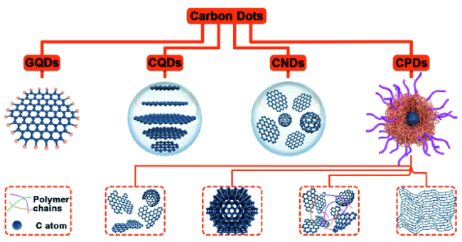
Carbon dots (CDs), with particle size less than 10 nm, are a new type of zero-dimensional photoluminescence nanomaterials. Due to the obvious advantages of adjustable fluorescence emission and excitation wavelength, light stability, low toxicity, good water solubility and biocompatibility, etc., CDs have been widely researched in recent years. As a treasure of ancient Chinese science, Traditional Chinese medicine (TCM) is rich in various active ingredients and plays a variety of pharmacodynamic effects, which has been used for thousands of years. TCM-CDs prepared with TCM as carbon source can create some special functions, and then may play a greater medicinal value. In this paper, the synthesis of TCM-CDs and its application in biological imaging and medical therapy are reviewed. Firstly, different synthetic methods of TCM-CDs (including hydrothermal, pyrolysis, solvothermal and microwave assisted method) are introduced in detail, and their advantages and disadvantages are compared. Subsequently, the latest research on TCM-CDs in biological imaging and medical treatment is comprehensively analyzed. This paper focuses on the application of imaging different types of cells in vitro and the distribution and uptake of TCM-CDs guided by imaging in vivo (mice, zebrafish, etc.). In addition, the intrinsic pharmacological activities of these TCM-CDs (including antibacterial, anti-inflammatory, hemostatic, antioxidant and anticancer, etc.) and their mechanisms are also discussed in order to improve and promote their clinical application. Finally, the importance of TCM-CDs research, the main problems and challenges in this fields and the future development direction are summarized and outlooked.
1 Introduction
2 Synthetic method of TCM-CDs
2.1 Hydrothermal method
2.2 Pyrolysis method
2.3 Solvothermal method
2.4 Microwave assisted method
3 Application of TCM-CDs in bioimaging
3.1 In vitro imaging
3.2 In vivo imaging
4 Application of TCM-CDs in therapeutics
4.1 Anti-bacterial
4.2 Anti-inflammatory
4.3 Hemostasis
4.4 Anti-oxidation
4.5 Anti-cancer
4.6 Other therapeutic effects
5 Conclusion and outlook

Jing He , Jia Chen , Hongdeng Qiu . Synthesis of Traditional Chinese Medicines-Derived Carbon Dots for Bioimaging and Therapeutics[J]. Progress in Chemistry, 2023 , 35(5) : 655 -682 . DOI: 10.7536/PC221024
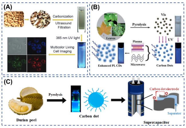
图4 (A)从花生壳合成荧光CDs及其应用于多色活细胞成像的示意图[54];(B)从植物叶片热解合成CDs,等离子体和微波辐射增强PL的示意图[55];(C)榴莲皮CDs的合成与应用示意图[56]Fig. 4 (A)Schematic illustration of the synthesis of fluorescent C-dots from peanut shells and their application in multicolor living cell imaging[54]; (B)Schematic synthesis of CDs from pyrolysis of plant leaves and the PL enhancement by plasma and microwave irradiation[55]; (C)Schematic representation of synthesis and applications of Durian Peel-based CDs[56] |
表1 不同中药源及合成方法及条件制得的CDs的尺寸、荧光性质和荧光QY比较Table 1 Comparison of size, fluorescence properties and fluorescence QY of CDs prepared from different TCM sources, synthesis methods and conditions |
| Source | Synthesis method | Reaction temperature ( ℃/W) | Reaction time (h) | Exciation (nm) | Emission (nm) | Average particle size (nm) | Quantum yield (%) | Surface modification | ref |
|---|---|---|---|---|---|---|---|---|---|
| Reynoutria japonica Houtt. | Hydrothermal | 200 | 3 | 320 | 400 | 35 | 11.5 | - | 24 |
| Citrus Bergamot | Hydrothermal | 200 | 5 | 330 | 440 | 10 | 50.78 | - | 25 |
| Perilla Frutescens(L.)Britt | Hydrothermal | 260 | 5 | 360 | 450 | 2.8 | 9.01 | - | 26 |
| Lycium chinense Miller | Hydrothermal | 180 | 24 | 427 | 550 | 4.5 | 21.8 | - | 28 |
| Lycium chinense Miller | Hydrothermal | 200 | 5 | 350 | 430 | 3.3 | 17.2 | NH3·H2O | 29 |
| Zingiber officinale Roscoe | Hydrothermal | 300 | 2 | 325 | 400 | 4.3 | 13.4 | - | 38 |
| Trapa bispinosa Roxb. | Hydrothermal | 90 | 2 | 450 | 520 | 7.5 | 1.2 | - | 39 |
| Allium sativum L. | Hydrothermal | 200 | 3 | 360 | 442 | 10.7 | 17.5 | - | 40 |
| Salvia miltiorrhiza Bunge | Hydrothermal | 100~180 | 6 | 400 | 490 | 1.53~16.94 | 30~40 | - | 44 |
| Mentha haplocalyx Briq. | Hydrothermal | 200 | 5 | 360 | 450 | 7 | 7.64 | - | 46 |
| Mentha haplocalyx Briq. | Hydrothermal | 180 | 8 | 363 | 441 | 5 | 4.5 | - | 47 |
| Aloe vera | Hydrothermal | 180 | 11 | 441 | 503 | 5 | 10.3 | - | 48 |
| Brassica oleracea Linnaeus var. capitata Linnaeus | Hydrothermal | 140 | 5 | 345 | 432 | 4 | 12.5 | - | 49 |
| Lilium brownii F. E. Brown var. Colchesteri Wils. | Hydrothermal | 240 | 12 | 340 | 405 | 4 | 11 | - | 50 |
| Poria cocos (Schw.) Wolf | Hydrothermal | 200 | 5 | 376 | 450 | 4 | 4.8 | - | 51 |
| Ginkgo biloba Linn. | Hydrothermal | 160~200 | 8 | 420 | 520 | 3.81 | 3.33 | - | 69 |
| Salvia miltiorrhiza Bunge | Hydrothermal | 150 | 6 | 420 | 526 | 3.32 | - | - | 74 |
| Charred Triplet | Hydrothermal | 100 | 2 | 340 | 447 | 5.1 | 7.95 | - | 131 |
| Litchi chinensis Sonn. | Pyrolysis | 300 | 2 | 365 | 440 | 1.12 | 10.6 | - | 53 |
| Peanut Shells | Pyrolysis | 250 | 2 | 320 | 440 | 1.6 | 9.91 | - | 54 |
| Durian Peel | Pyrolysis | 250 | 5 | 368 | 480 | 10 | 11 | - | 56 |
| Gynostemma pentaphyllum (Thunb.) Makino | Pyrolysis | 400 | 4 | 320 | 400 | 2.49 | 5.7 | - | 57 |
| Papaya | Solvothermal | 200 | 5 | 370 | 450 | 3.4/10.8 | 18.98/18.39 | ethanol | 60 |
| Saccharum sinensis Roxb. | Solvothermal | 250 | 6 | 350 | 430 | 1 | 10.7 | ethanol | 61 |
| Saccharum sinensis Roxb. | Solvothermal | 120 | 8 | 360 | 460 | 5 | - | Urea and ethanol | 62 |
| Codonopsis pilosula | Solvothermal | 25 | 4 | 390 | 456 | 11.54 | 12.8 | methanol | 63 |
| Mentha canadensis Linnaeus | Microwave | 960 | 0.07~0.17 | 340 | 436 | 2.43 | 17 | - | 66 |
| Bombyx mori L. | Microwave | 210 | 0.75 | 350 | 440 | 19 | 46 | - | 67 |
| Zingiberis rhizome and Alpinia officinarum | Microwave | 450 | 0.08~0.67 | - | - | 10 | - | - | 68 |
| Ginkgo biloba Linn. | Microwave | 800 | 0.08~0.25 | 440 | 550 | 2.82 | 0.65 | - | 69 |
| Panax ginseng | Microwave | 700 | 0.5 | 380 | 500 | 2 | 8 | AgNPs | 70 |
| Talinum paniculatum (Jacq.) Gaertn. | Microwave | 700 | 0.5 | 380 | 470 | 2 | - | Rutin | 107 |
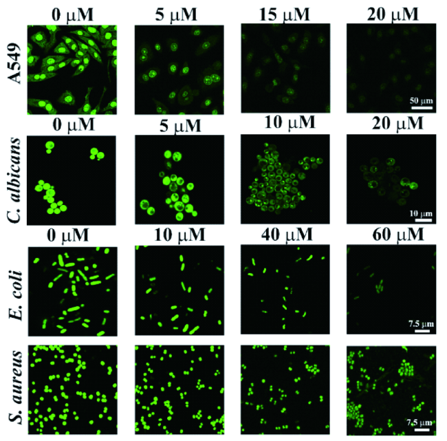
图10 基于Mis-mPD-CDs的细胞内CR成像与传感:A549、C. albicans、E. coli和S. aureus的CLSM图像,用不同浓度的CR处理,然后与Mis-mPD-CDs孵育30 min[93]Fig. 10 Imaging and sensing of intracellular CR based on Mis-mPD-CDs. CLSM images of A549, C. albicans, E. coli, and S. aureus, and treated with CR of different concentrations and then incubated with Mis-mPD-CDs for 30 min[93] |
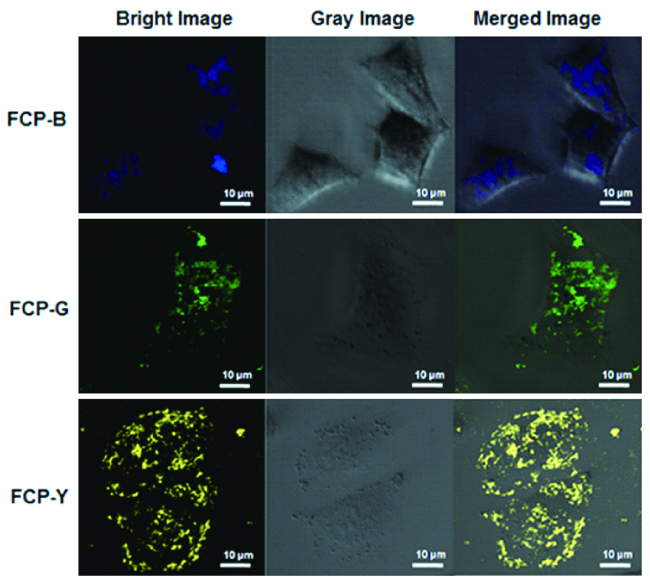
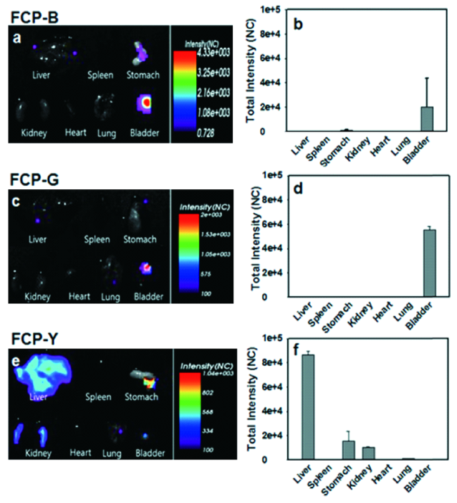
图11 杧果CDs的小鼠体内成像图。尾静脉注射5 mg·kg-1体重后FCP-B、FCP-G和FCP-Y在BALB/c裸鼠体内的生物分布。FCP-B(a,b)、FCP-G(c,d)和FCP-Y(e,f)的体内分布及相应强度[79]Fig. 11 In vivo biodistribution of FCP-B, FCP-G and FCP-Y in balb/c nude mice after tail vein injection of 5 mg·kg-1 of body weight. The in vivo biodistribution and corresponding intensities of FCP-B (a, b), FCP-G (c, d) and FCP-Y (e, f) respectively |
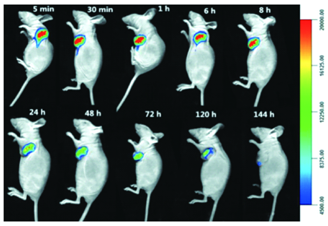
图12 CDs的单光子和双光子生物成像。(A)在不同时间点静脉注射CDs的仰卧裸鼠的体内成像(Ⅰ:胸部区域,Ⅱ:肝脏区域,Ⅲ:小肠区域,Ⅳ:大肠区域,Ⅴ:膀胱区域);(B)在不同时间点静脉注射CDs的裸鼠的实时离体成像[97]Fig. 12 One-photon and two-photon bioimaging of CDs. (A) In vivo imaging of supine nude mice with intravenous injection of CDs at different time points (Ⅰ: thoracic region, Ⅱ: area of liver, Ⅲ: area of small intestine, Ⅳ: area of large intestine, Ⅴ: bladder region). (B)Real-time ex vivo imaging of nude mice with intravenous injection of CDs at different time points |
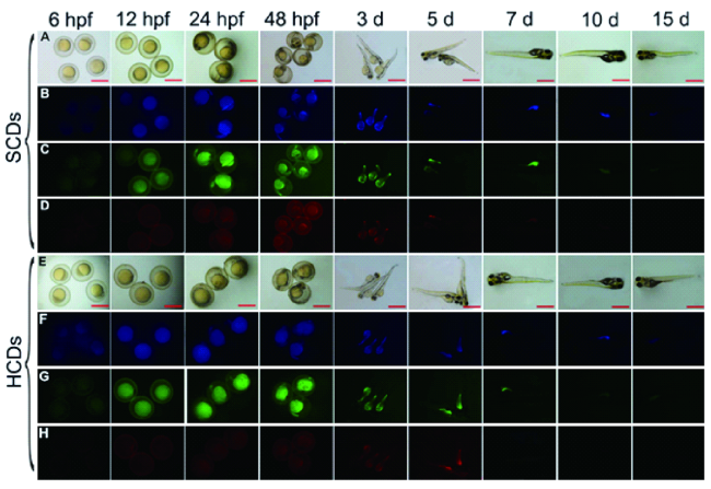
图15 CDs在斑马鱼体内的光致发光衰减。0.4 mg/mL HCDs和SCDs溶液在不同时间点暴露2 d后斑马鱼胚胎的荧光图像[100]Fig. 15 The photoluminescence decay of CDs in zebrafish. The fluorescent microscopic images of bright field and fluorescent field of zebrafish embryos after exposure to 0.4 mg/mL HCDs and SCDs solutions for 2 days at different time points[100] |
表2 中药CDs在生物成像中的应用Table 2 The application of TCM-CDs in bioimaging |
| Source | FL color | Applied Ex/Em (nm) | Application | Biotarget | ref |
|---|---|---|---|---|---|
| Citrus junos Tanada | blue, green | 405, 488/- | Imaging of cells | MG-63 | 74 |
| Mentha haplocalyx Briq | blue, green, red | 360, 470, 530/447, 525, 593 | Imaging of cells | MCF-7 | 75 |
| Prunus persica | blue | - | Imaging of cells | MDA-MB-231 | 76 |
| Prunus mume | blue, green | 365/- | Imaging of cells | MDA-MB-231 | 77 |
| Curcuma Longa | blue, yellow, red | 405, 488, 543/- | Imaging of cells | KB | 78 |
| Mangifera indica L. | blue, green, yellow | 488, 488, 513/505, 530, 560 | Imaging of cells | A549 | 79 |
| Allium sativum L. | blue, green, yellow | 385, 480,550/- | Imaging of cells | A549 | 40 |
| Coriandrum sativum Linn. | green | 470/525 | Imaging of cells | L-132 | 80 |
| Dendranthema morifolium | blue, green | 405, 488/- | Imaging of cells | HeLa | 81 |
| Mentha haplocalyx Briq. | blue, yellow, red | 380, 480, 590/- | Imaging of cells | HeLa | 82 |
| Brassica compestris L.var.purpurea Bailey | blue, yellow, red | 405, 488, 559/- | Imaging of cells | HeLa | 83 |
| Bombyx mori L. | blue, yellow, red | 340, 495, 550/- | Imaging of cells | HeLa | 67 |
| Abelmoschus esculentus (Linn.) Moench | blue | 340/410 | Imaging of cells | HeLa | 84 |
| Lycium chinense Miller | blue, glaucous, green | 400, 415, 485/- | Imaging of cells | HeLa | 29 |
| Alisma plantago-aquatica Linn. | blue, yellow, red | 340, 460, 520/- | Imaging of cells | HeLa | 85 |
| Ginkgo biloba Linn. | blue, green | 405, 488/- | Imaging of cells | HeLa, KYSE-410 | 69 |
| Benincasa hispida (Thunb.) Cogn. | blue | 365/- | Imaging of cells | HepG2 | 86 |
| Prunus cerasifera Ehrhart f. atropurpurea (Jacq.) Rehd. | blue, green | 405, 488/- | Imaging of cells | HepG2 | 87 |
| Litchi chinensis Sonn | blue | 405/- | Imaging of cells | HepG2 | 88 |
| Ginsenoside Re | blue to red | 360-530/- | Imaging of cells | A375 | 89 |
| Brassica oleracea Linnaeus var. capitata Linnaeus | blue, green, red | 405, 488, 543/- | Imaging of cells | HaCaT | 49 |
| Phyllanthus acidus (L.) Skeels | blue, green, red | 405, 488, 555/- | Imaging of cells | Clone9 | 90 |
| Cymbopogon citratus (D. C.) Stapf | blue, yellow, red | - | Imaging of bacteria | BY4742 | 91 |
| Ocimum sanctum Linn. | blue, yellow, red | 405, 488, 561/- | Imaging of bacteria | B. subtilis, E. coli | 92 |
| Salvadora persica | green | 488/550 | Imaging of bacteria | C. albicans,E. coli, S. aureus | 93 |
| Allium cepa Linn. | green | - | Imaging of bacteria | E.coli, S.aureus | 94 |
| Mangifera indica L. | blue to yellow | - | In vivo imaging | mice | 79 |
| Taxus chinensis (Pilger) Rehd. | red | 640/705 | In vivo imaging of tumor | mice | 97 |
| Litchi chinensis Sonn. | blue to red | - | In vivo imaging of tumor | mice | 88 |
| Panax notoginseng | blue to red | - | In vivo imaging of tumor | BALB/c mice | 98 |
| Pisum sativum Linn. | blue | - | In vivo imaging | mice | 99 |
| Crinis Carbonisatus | blue, green, red | 385, 480, 550/- | In vivo imaging | Zebrafish | 100 |
| Gynostemma pentaphyllum (Thunb.) Makino | blue, green, red | - | In vivo imaging | Zebrafish | 57 |
| Salvadora persica | green | 488/550 | In vivo imaging | Zebrafish | 93 |
| Panax notoginseng | blue, yellow, red | 405, 488, 543/- | In vivo imaging | P. caudatum | 98 |
图16 CDs的一般杀菌作用机制。(a)CDs和细菌细胞壁之间最初静电相互作用。(b)CDs内化,在细菌膜中插入,以及细胞质物质泄漏的不可逆破坏。(c)CDs促进细菌光动力灭活,产生ROS和DNA损伤[105]Fig. 16 General bactericidal mechanisms of action of CDs. (A) Schematic representation of the initial electrostatic interaction between CDs and the bacterial cell wall. (B) CDs internalization, intercalation in the bacterial membrane, and irreversible disruption with a leak of cytoplasmatic material. (C) CD-promoted bacterial photodynamic inactivation with ROS production and DNA damage[105] |
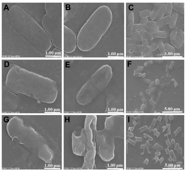
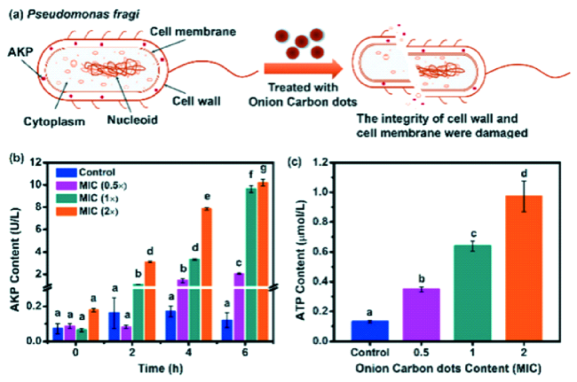
图18 (a)洋葱CDs对细菌细胞壁和细胞膜完整性影响的示意图;洋葱CDs对脆弱假单胞菌胞外(b)AKP和(c)ATP释放的影响[94]Fig. 18 (a) Schematic illustration of the effect of the onion carbon dots on the integrity of the bacterial cell walls and membranes. Effect of onion carbon dots on the release of extracellular (b) AKP and (c) ATP content in Pseudomonas fragi[94] |
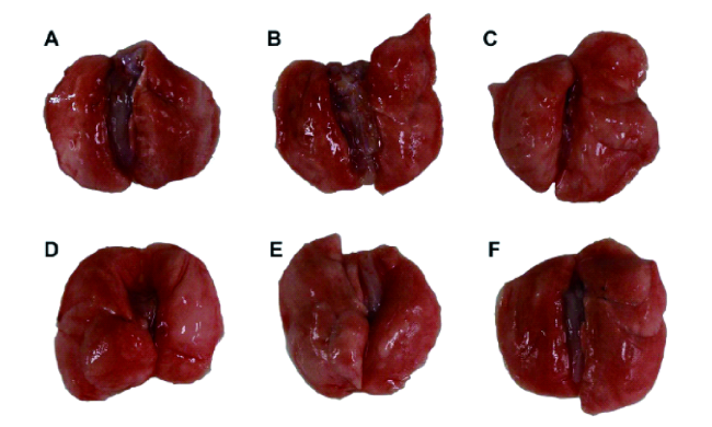
图20 ASAC-CDs改善LPS诱导的大鼠急性肺损伤(ALI)的宏观图像:(A)生理盐水组;(B)示范组;(C)阳性对照组;(D)大剂量ASAC-CDs组;(E)中剂量ASAC-CDs组;低剂量ASAC-CDs组[112]Fig. 20 Macroscopic images of ASAC-CDs ameliorating an LPS-induced acute lung injury (ALI) in rats. (A) Normal saline group; (B) model group; (C) positive control group; (D) high-dose ASAC-CDs group; (E) medium-dose ASAC-CDs group; and (F) low-dose ASAC-CDs group[112] |
| [1] |
|
| [2] |
|
| [3] |
|
| [4] |
(唐志姣, 李攻科, 胡玉玲. 化学进展, 2016, 28: 1455.).
|
| [5] |
(李程浩, 刘亚敏, 卢彬, 萨拉乌拉, 任先艳, 孙亚平. 化学进展, 2022, 34(3): 499.).
|
| [6] |
|
| [7] |
|
| [8] |
(卫迎迎, 陈琳, 王军丽, 于世平, 刘旭光, 杨永珍. 化学进展, 2020, 32: 381.).
|
| [9] |
|
| [10] |
(熊威, 赵琰, 成金俊, 张美龄, 孙紫薇, 罗娟, 朱雅凡, 张亚雪, 孔慧, 屈会化. 中草药, 2019, 50: 1388.).
|
| [11] |
(何闯, 鄂爽, 闫鸿浩, 李晓杰. 化学进展, 2022, 34: 356.).
|
| [12] |
|
| [13] |
(王军丽, 王亚玲, 郑静霞, 于世平, 杨永珍, 刘旭光. 化学进展, 2018, 30: 1186.).
|
| [14] |
|
| [15] |
|
| [16] |
|
| [17] |
|
| [18] |
|
| [19] |
|
| [20] |
|
| [21] |
|
| [22] |
|
| [23] |
|
| [24] |
|
| [25] |
|
| [26] |
|
| [27] |
|
| [28] |
|
| [29] |
|
| [30] |
|
| [31] |
|
| [32] |
|
| [33] |
|
| [34] |
(吴星辰, 梁文慧, 蔡称心. 化学进展, 2021, 33: 1059.).
|
| [35] |
(颜范勇, 邹宇, 王猛, 代林枫, 周旭光, 陈莉. 化学进展, 2014, 26: 61.).
|
| [36] |
(刘禹杉, 李伟, 吴鹏, 刘守新. 化学进展, 2018, 30: 349.).
|
| [37] |
|
| [38] |
|
| [39] |
|
| [40] |
|
| [41] |
|
| [42] |
|
| [43] |
|
| [44] |
|
| [45] |
|
| [46] |
|
| [47] |
|
| [48] |
|
| [49] |
|
| [50] |
|
| [51] |
|
| [52] |
|
| [53] |
|
| [54] |
|
| [55] |
|
| [56] |
|
| [57] |
|
| [58] |
|
| [59] |
|
| [60] |
|
| [61] |
|
| [62] |
|
| [63] |
|
| [64] |
|
| [65] |
|
| [66] |
|
| [67] |
|
| [68] |
Isnaeni,
|
| [69] |
|
| [70] |
|
| [71] |
|
| [72] |
|
| [73] |
|
| [74] |
|
| [75] |
|
| [76] |
|
| [77] |
|
| [78] |
|
| [79] |
|
| [80] |
|
| [81] |
|
| [82] |
|
| [83] |
|
| [84] |
|
| [85] |
|
| [86] |
|
| [87] |
|
| [88] |
|
| [89] |
|
| [90] |
|
| [91] |
|
| [92] |
|
| [93] |
|
| [94] |
|
| [95] |
|
| [96] |
|
| [97] |
|
| [98] |
|
| [99] |
|
| [100] |
|
| [101] |
|
| [102] |
|
| [103] |
|
| [104] |
|
| [105] |
|
| [106] |
|
| [107] |
|
| [108] |
|
| [109] |
|
| [110] |
|
| [111] |
|
| [112] |
|
| [113] |
|
| [114] |
(赵玉升, 李伟洋, 曹天佑, 赵琰, 屈会化. 中医药导报, 2021, 27: 56.).
|
| [115] |
|
| [116] |
|
| [117] |
|
| [118] |
|
| [119] |
|
| [120] |
|
| [121] |
|
| [122] |
|
| [123] |
|
| [124] |
|
| [125] |
|
| [126] |
|
| [127] |
|
| [128] |
|
| [129] |
|
| [130] |
|
| [131] |
|
| [132] |
|
| [133] |
|
| [134] |
|
/
| 〈 |
|
〉 |


