Home Journals Progress in Chemistry
Progress in Chemistry

Abbreviation (ISO4): Prog Chem      Editor in chief: Jincai ZHAO

About  /  Aim & scope  /  Editorial board  /  Indexed  /  Contact  / 
Review

Stimuli-Responsive Polymer Microneedle System for Transdermal Drug Delivery

  • Wanping Zhang ,
  • Ningning Liu ,
  • Qianjie Zhang ,
  • Wen Jiang ,
  • Zixin Wang ,
  • Dongmei Zhang , *
Expand
  • School of Perfume and Aroma Technology, Shanghai Institute of Technology,Shanghai 201418, China
* Corresponding author e-mail:

Received date: 2022-07-11

  Revised date: 2023-03-22

  Online published: 2023-04-25

Supported by

Provincial and ministerial collaborative innovation Center project(XTCXC-202101)

Collaborative Innovation Fund of Shanghai Institute of Technology(XTCX2022-30)

Abstract

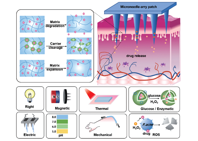
Compared with oral administration and injection administration, the microneedle transdermal delivery system has the characteristics of high efficiency, safety and painless administration. In particular, the stimuli-responsive polymer microneedle systems exhibit good biocompatibility and can be realized according to the micro-changes in the environment. The function of percutaneous local and systemic intelligent drug delivery in time and space is currently an international frontier research topic. This paper focuses on the research of stimulus-responsive polymer microneedles at home and abroad in the past ten years, and focuses on the evolution of polymer microneedles, the types of internal and external environmental stimulus response and its response structure-activity mechanism. In addition, the preparation and characterization of microneedles and the application of microneedle system in the fields of biomedicine delivery, tissue and organs, dermatology and medical beauty are described in detail. The stimulation-responsive polymer microneedle system has the advantages of simple use, adjustable mechanical properties and precise targeted drug delivery, which has great research significance in the field of percutaneous targeted drug delivery. In the future, the biological in vivo load and industrial application of standardization are the direction of continuous efforts and progress of researchers.

Contents

1 Introduction

2 Preparation process and characterization methods of stimuli-responsive polymer microneedles

2.1 Preparation

2.2 Methods for characterizing the properties of polymer microneedle systems

3 Classification of stimulus-responsive polymer microneedles

3.1 Polymer microneedle system triggered by external environmental stimuli

3.2 Polymer microneedle system triggered by in vivo physiological signal stimuli

4 Stimuli-responsive polymer microneedles for transdermal delivery

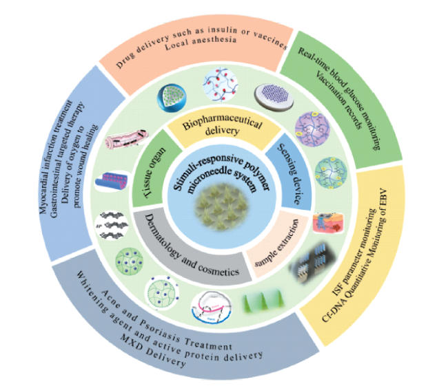
4.1 Biopharmaceutical delivery

4.2 Tissue organ therapy

4.3 Detection and sensing device

4.4 Extraction of samples

4.5 Dermatology and cosmetics

5 Conclusion and outlook

Cite this article

Wanping Zhang , Ningning Liu , Qianjie Zhang , Wen Jiang , Zixin Wang , Dongmei Zhang . Stimuli-Responsive Polymer Microneedle System for Transdermal Drug Delivery[J]. Progress in Chemistry, 2023 , 35(5) : 735 -756 . DOI: 10.7536/PC220710

1 Introduction

In the field of biomedicine, the common ways of drug delivery are oral administration, intravenous injection, pulmonary inhalation, mucosal drug delivery and Transdermal drug delivery (TDD). Oral administration will bring bitter taste to patients, and first-pass metabolic effect will occur through gastrointestinal tract and liver. Subcutaneous needle injection can cause pain, bring great fear and discomfort to patients, and produce a large amount of medical waste[1]. Different from the traditional direct route of drug delivery, TDD means that the drug reaches all layers of the skin and the capillaries at the bottom of the skin through the surface of the skin to achieve local and systemic drug delivery[2]. TDD is non-invasive, non-allergenic, and has the ability to deliver sustainable and controlled doses to achieve uniform drug delivery at a defined rate[3,4]. However, transdermal drug delivery remains controversial. On the one hand, the skin maintains the homeostasis of the human body, and the stratum corneum has a good barrier effect, resulting in many macromolecular drugs can not enter the skin through the stratum corneum, greatly reducing the efficiency of local drug delivery; On the other hand, the dense and thickened ordered vascular endothelium inhibits drug delivery and hinders systemic drug delivery[5]. In order to facilitate the effective delivery of drugs, various active or passive TDDs have been widely studied, such as device-facilitated transdermal drug delivery, microneedles, liposomes, polymeric nanoparticles, and micro-nano emulsions[3][6,7][8][9][10]. Compared with other traditional drug delivery methods, the drug delivery mechanism of microneedle transdermal drug delivery system is to achieve drug delivery performance in time and space through microneedle (patch array composed of needles with length of 10 ~ 2000 μm and width of 10 ~ 50 μm).It efficiently breaks through the barrier of the stratum corneum of the skin, delivers drugs to the skin tissue, and then reaches the target site through cells and vascular tissues to achieve precise drug delivery[2,11][2]. Therefore, the microneedle system has developed into a frontier research hotspot in the field of transdermal drug delivery. In 1976, Gerstel and Place assembled hollow microneedles into arrays and simultaneously encapsulated drugs for delivery through the stratum corneum of the skin. Therefore, "Microneedle array patch (MNAP)" was conceptualized for the first time. In the 1970s, the Food and Drug Administration (FDA) approved a variety of transdermal drugs for marketing. Among them, the first scopolamine patch was approved for marketing in 1979, which laid the foundation for the application of microneedle system in transdermal drug delivery[12]. Due to the limitation of preparation conditions, the rapid development of micro-nano processing technology in the 1990s promoted the emergence of microneedle array patches made of different materials. In 1998, Henry et al. First proposed a research report on transdermal drug delivery of microneedles, which confirmed the skin penetration enhancement ability of microneedles[13]. In recent years, the materials used to prepare microneedles have gradually expanded from silicon, metal to polymer materials[4,14]. Polymer materials have good biocompatibility, degradability and structural controllability, which promote the design of new microneedle structures. In 2005, Miyano et al. Developed maltose-base microneedles with biodegradability, which is a milestone in the birth of soluble microneedles[15]. In 2012, stimulus-responsive microneedles were proposed to overcome the limitations of transdermal delivery performance of traditional microneedles[16]. In 2015, the phenomenon that microneedles showed drug delivery characteristics in response to physiological signals was widely concerned by researchers, and the stimuli-responsive polymer microneedle system was continuously proposed and became an international research hotspot (Fig. 1)[17,18]. Based on this, this paper mainly focuses on the research results of stimuli-responsive polymeric microneedles in transdermal drug delivery in the past 10 years at home and abroad. Firstly, the preparation methods and performance characterization of stimuli-responsive polymeric microneedles are summarized. Secondly, the environmental effects of polymer microneedles are reviewed, including physiological stimuli (such as pH, redox potential, glucose) and external environmental stimuli (such as temperature, electric field, light, and mechanical stress). The structure-activity relationship between the chemical structure of polymer microneedles and the response conditions was further summarized, including the influence of the swelling, degradation or cracking of the aggregate matrix structure, and the reversible dissociation of the molecular structure of the material on the drug delivery performance[6]. Finally, the application of polymer microneedles in transdermal drug delivery was introduced, and the challenges and solutions in cosmetic medicine and skin disease treatment were prospected in combination with the research field of partners.
图1 聚合物微针发展历程示意图

Fig. 1 Schematic diagram of polymer microneedles development

2 Preparation and characterization of stimuli-responsive polymer microneedles

2.1 Preparation method

In that proces of preparing the stimuli-responsive polymer microneedle, in order to promote the drug delivery efficiency, the relationship between the responsive material and the active drug is often considered, and the stimuli-responsive polymer microneedles are efficiently and rapidly prepare. Common polymer microneedle preparation methods are as follows.

2.1.1 Reduced pressure casting method

The vacuum casting method mainly uses a polydimethylsiloxane (PDMS) mold for preparation. The pouring operation conditions are mild, the polymer solution is injected into the mold, the bubbles are discharged by centrifugation or vacuum pumping, and then the polymer solution is solidified and demoulded, which is a low-cost preparation method[19]. However, the controllability of centrifugation is poor, which is easy to cause damage, the size of the centrifuge limits the expansion of microneedle production, and the viscosity of the polymer solution is large, which is subject to air resistance in the process of injecting into the mold, and the microneedle is prone to a large number of bubbles or uneven distribution of drugs. In order to avoid centrifugation and vacuum operation, Vrdoljak et al. proposed to add water into the mold to remove air, then mix 238 μg/mL vaccine with sodium hyaluronate (HA) solution, and drop it into the mold to drive the components to diffuse evenly by using the concentration difference of the solution[20]. Finally, the microneedles were formed after the solvent was volatilized at room temperature (fig. 2A). Chen et al. Designed a waterproof and breathable double penetration female mold (DPFM), which was centrifuged at 3500 rpm for 10 min and dried at room temperature for 12 H. A vacuum condition is formed inside the mold cavity, which reduces gas resistance and promotes the removal of bubbles from the solution (Fig. 2B)[21].
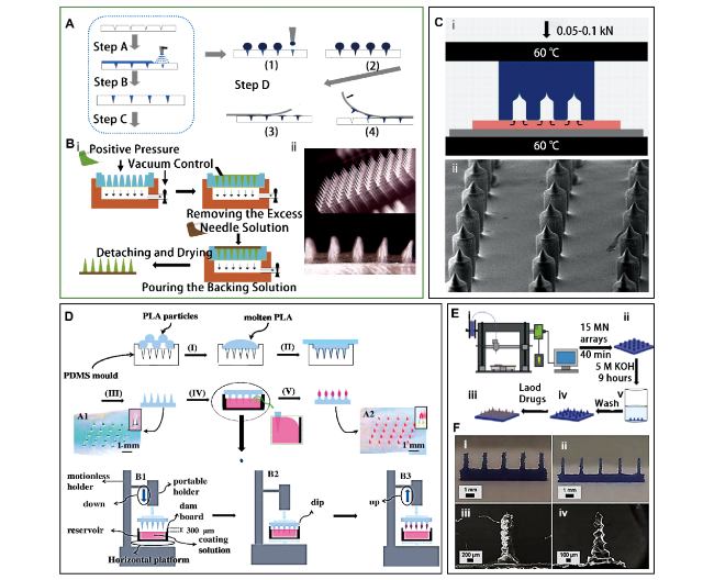
图2 微针的制备方法:(A,B) 减压浇筑法,(A) 利用浓度差制备微针的流程示意图[20]; 使用DPFM制造微针的流程示意图 (B-i); 微针的电子显微图像 (B-ii)[21]; (C) 热压花法[22],热压花制作示意图 (C-i); 微针的扫描电镜图像 (C-ii); (D) 浸渍包覆法,涂层微针制作工艺的示意图[24]; (E,F) 3D打印法[26],(E) 利用FDM3D打印和化学刻蚀技术制作微针的示意图; 制造微针 (F-i) 和在KOH溶液中腐蚀之后 (F-ii) 的光学图像; 制造的微针 (F-iii) 和在KOH溶液中腐蚀之后 (F-iv) 的扫描电子显微镜图像

Fig. 2 Preparation method of microneedles. (A,B) Decompression pouring method. (A) Schematic diagram of the process for preparation of microneedles using concentration differences[20]; Schematic diagram of the process for manufacturing microneedles using DPFM (B-i); Electron microscopic images of microneedles (B-ii)[21]; (C) Hot embossing[22]. Schematic diagram of hot embossing (C-i); Scanning electron microscopic images of microneedles (C-ii); (D) Impregnation and coating method. Schematic diagram of the coating microneedle fabrication process[24]; (E,F) 3D printing method[26]. (E) The schematic illustration of the fabrication of MNs by FDM 3D printing and chemical etching; Optical images of MNs as FDM-fabricated (F-i) and after etching in KOH solution (Fii); SEM images of MNs as fabricated (F-iii) and after etching in KOH solution (F-iv)

The decompression pouring method is mainly an early microneedle preparation method, and continuous improvement on the basis of this method will be the key to promote the development of soluble microneedles from laboratory research to industrialization.

2.1.2 Hot embossing

Hot embossing is a process for preparing polymer microneedle structures. After the preparation of the drug-coated polymer film, the temperature and pressure values are controlled by a bonding press to change the dissolution state of the polymer, so as to achieve the purpose of rapid preparation of the microneedles. The equipment used in this process is simple and the preparation time is short, but at higher processing temperatures, it is easy to cause the loss of drug activity.
In order to effectively preserve and prolong the activity of drugs, it is necessary to select polymer materials with low melting point and good biocompatibility, such as polycaprolactone (PCL), which has a melting temperature of 59 ~ 64 ℃ and is mainly used to load protein drugs that are not resistant to high temperature. Andersen et al. Prepared a PCL film with a thickness of 115 mm by layer-by-layer method to load small molecule drugs. The temperature, pressure and time of embossing are the key factors affecting the quality of microneedles. The PCL film was laid on the PDMS mold with a bonding press equipped with a force gauge and a thermal controller, heated to 60 ℃ and pressurized to 1. 4 MPa, and then placed in the bonding press for 3 min, and the film was completely fused and filled in the stamp. Finally, the pressure was released and the temperature was reduced to 24 ℃, and the microneedle was separated from the stamp after solidification (Fig. 2C)[22]. Abubaker et al. Used poly (methyl methacrylate) (PMMA) with a melting temperature of 105 ℃ as the substrate, used a hot embossing machine at 130 ℃ and 11 MPa, separated the mold when the temperature of the steel platen was reduced to 100 ℃, and repeated the operation after quenching[23]. Hot embossing method mainly relies on hot embossing machine and low melting point polymer, which is suitable for the delivery of high temperature resistant drugs.

2.1.3 Dip coating method

The dip-coating method is a common and rapid preparation method to form coated polymer particles in a short time, which is suitable for both coated and soluble microneedles. Firstly, the microneedles were prepared by PDMS mold, and the microneedles were fixed on the bottom of the horizontal plate of the portable bracket in reverse, and the coating solution was placed under the bracket. Among them, the height and thickness of impregnation are the key to control the length of microneedles and drug dosage. The microneedle was moved downward at a constant speed until it touched the coating solution. Drying and curing the coated microneedles at normal temperature, and molding in the process of complete volatilization of the solvent. Chen et al. Proposed poly (vinyl alcohol) (PVA, cross-linking agent) loaded sulforhodamine B (RhB) and sucrose (drug stabilizer) polylactic acid (PLA) microneedles (height: 650 μm). The viscosity of the coating solution composed of 21% PVA and 26% sucrose was increased to 2850 mPa · s (Fig. 2D)[24]. Due to the large coating angle of the microneedle, the penetration performance is weakened. Chen and He et al. Proposed a PVA-coated insulin-coated PLA microneedle, which can effectively avoid the problem of needle penetration[25].
The drug loading ranged from 4.8 to 13.5 nL/needle after the microneedles of different sizes contacted the coating solution vertically downward at a speed of 0.5 mm/min. Under 80% high humidity environment, the inverted microneedle promotes the coating to dissolve and slide downward, exposing the needle tip to achieve efficient penetration effect.
The polymer microneedle combines the impregnation coating method with the soluble carrier to realize drug coating, and the method has the advantages of low cost and easy operation, and can be applied to the large-scale production of the polymer microneedle. However, the impregnation coating method can not accurately control the amount of coating and the uniformity of coating, and there is a certain error.

2.1.4 3D printing method

The microneedle geometry and length are key parameters that determine transdermal drug delivery. In recent years, the research of bionics has been widely concerned, and the structural design of microneedle body is closely related to bionics. Because traditional preparation methods cannot diversify the microneedle structure, 3D printing is a customizable model manufacturing technology that superimposes polymer materials layer by layer through a 3D printer, with versatility and high resolution. Among them, height, width aspect ratio and tip are the key to effectively control drug volume and stimulus response. Among the 3D printing methods, Fused deposition modeling (FDM) is a low-cost and easy-to-operate method. In order to protect the activity of drugs, FDM requires high temperature resistance of materials, and heat-sensitive polymers should be selected. Commonly used thermoplastic polymers are polylactic acid, acrylonitrile butadiene styrene, nylon, thermoplastic polyurethane, polyetherketone and polyetherimide. Detamornrat et al. Proposed an FDM microneedle preparation technique based on thermoplastic polymer powder. The microneedle model was constructed by vertical fusion of 2D cross-sectional images layer by layer, mainly by heating the powder to make it adhere (Fig. 2 E, F)[26]. Luzuriaga et al. Constructed a PLA microneedle based on 3D printing technology and KOH. The PLA cylinder was mainly fabricated by 3D printing, and then immersed in KOH solution for chemical corrosion to reduce the width of the cylinder, forming a microneedle structure[27].

2.2 Performance characterization method of polymer microneedle system

At present, the stimuli-responsive polymer microneedle system uses polymer materials with good biocompatibility, but the microneedles need to have good mechanical properties and toughness, and the selection of microneedle materials and preparation process are strictly required. In order to judge whether the prepared polymer microneedles can effectively deliver drugs, it is necessary to evaluate their mechanical properties, solubility, drug release properties and other parameters[28].

2.2.1 Mechanical properties of microneedle system

In the stimuli-responsive polymer microneedle system, the mechanical properties are the key factors affecting the microneedle penetration into the skin tissue and the effective drug release, and the mechanical strength and skin penetration performance of the microneedles are mainly characterized. Among them, the structural design and material selection of microneedles can directly affect the mechanical properties. In order to test the mechanical strength of the microneedle, the length of the microneedle after extrusion is measured and compared with the length of the initial microneeedle tip by using a texture analyzer and a microscope, and the maximum compressive limit of the micron needle under different polymer matrices can be compared intuitively according to the relationship between the mechanical force and the length difference, so as to find the optimal ratio. Based on the micromanipulation technique, Du et al. Placed the microneedle patch on a platform with a mechanical sensor, and visualized it by confocal laser scanning microscopy (CLSM) and scanning electron microscopy (SEM)[28]. The compression force can be obtained by compressing a single microneedle with a glass probe at a speed of 2 μm/s. The normal stress was calculated by the compressive force and volume change (formulas 1 ∼ 5) (Fig. 3A, B).
V = 4 3 r 1 2 H + H ' - 4 3 r 2 2 H '
H ' r 2 = ( H + H ' ) r 1
r 2 = 1 2 - r 1 + 3 V H - 3 r 1 2
r δ = 1 2 - r 1 + 3 V H - δ - 3 r 1 2
F = F 4 r δ 2
(H: initial height of microneedle; H ': height of the missing tip of the microneedle; A half side length of a r1: quadrilateral microneedle base; r2: the initial half side length of the tip of the quadrilateral microneedle; δ: displacement of microneedle; Half side length of the contact surface when the compression displacement is δ rδ:)
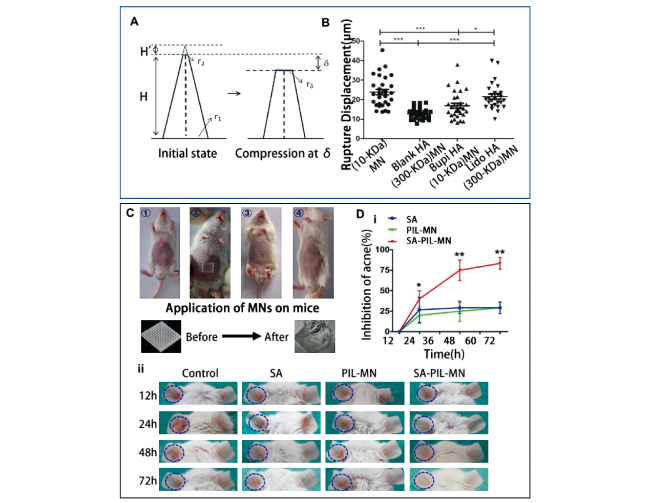
图3 微针系统性能表征方法。(A,B) 微针系统的力学性能[28]。(A) 应力建模示意图 (H: 微针的初始高度; H': 微针缺失尖端的高度; r1: 四边形微针底座的一半边长; r2: 四边形微针尖端的初始半侧长度; δ: 微针的位移; rδ: 压缩位移为δ时,接触面的半边长); (B) 不同类型的微针下位移变化 (*: p < 0.05;**: p < 0.01;***: p < 0.001); (C,D) 微针系统的药物递送性能。(C) 微针给药方式[31]; 不同治疗模式下的抑菌率(D-i); SA、PIL-MN和SA-PIL-MN在小鼠模型中的皮肤痤疮治疗的示意图(D-ii)[32]

Fig. 3 Characterization of polymer microneedle systems. (A,B) Mechanical properties of the microneedle system[28]. (A) Schematic diagram of stress modeling (H: initial height of microneedles; H': height of microneedle missing tip; r1: half the side length of the quadrilateral microneedle base; r2: initial half length of the tip of the quadrilateral micro pin; δ: displacement of microneedle; rδ: when the compression displacement is δ, the half length of the contact surface); (B) Displacement changes under different types of microneedles (* : p < 0.05;** : p < 0.01;*** : p < 0.001); (C,D) Drug delivery performance of the microneedle system. (C) microneedle administration method[31]; Bacteriostatic rate under different treatment modes (D-i); Schematic representation of SA, PIL-MN and SA-PIL-Mn for skin acne treatment in a mouse model (D-ii)[32]

In addition, both the molecular weight of the polymer and the amount of drug loaded affect the fracture behavior and mechanical properties of the microneedles. The mechanical properties of microneedles were evaluated by analyzing the stress-deformation curves of microneedles according to the average compression force, fracture displacement and molecular weight.
The puncture performance of the microneedle can also be judged by the insertion depth of the needle. The spacing, length, thickness and aspect ratio of the microneedle are the important factors affecting the mechanical properties and insertion amount of the microneedle. In order to test the performance of microneedle puncture, Tas et al. Used fresh pigskin as a skin model, because its structure, thickness, hair density, pigmentation, collagen and fat are similar to human skin, and used gentian violet dye for staining[29]. After the transdermal drug delivery, the skin penetration efficiency was calculated by the depth of the skin penetration hole through the digital microscope to determine the puncture performance of the microneedle. The common polymer materials are polyvinylpyrrolidone (PVP), HA and PVA. Among them, PVA microneedles have good hardness but poor toughness, PVP microneedles have good flexibility but easy to deform, and HA microneedle has good hygroscopicity, stability and ability to preserve drugs, which is widely used in mechanical force-responsive drug delivery systems[30].

2.2.2 Drug release performance of microneedle system

The difference of the stimulus-responsive polymer microneedle system is that the microneedle can be swelled or dissolved by the polymer after being inserted into the skin to achieve effective drug delivery. The selection of stimuli-responsive polymer materials is the key to improve the efficiency of transdermal drug delivery, which determines the degree of swelling. At present, the characterization methods include in vitro detection and in vivo detection.
The in vitro detection method has the characteristics of low cost, simple operation, short time consumption and the like. Fresh pigskin is usually used as a skin model, and microneedles are inserted into the pigskin. The microneedles were taken out at intervals, and the dissolution of the microneedles was preliminarily judged by observing the length change of the needle tip through a digital microscope, which further determined the drug release performance of the microneedle[19]. In order to accurately determine the dissolution value, Chen and Liu et al. Proposed an in vitro drug release experiment based on polyethylene glycol-400 (PEG400) and PBS. PEG was used as a water-soluble matrix to simulate the skin environment, and PBS was used to simulate the skin tissue fluid. The pretreated fresh mouse abdominal skin was used as the skin model, and the mouse skin was placed in the receptor chamber of pH 6.8 PBS/PEG mixed solution (7:3 V/V), and the microneedles prepared by different preparation processes and polymer matrices were applied to the skin surface as the blank control group. Collect 1 mL of solution from the receptor compartment at regular intervals at 32 ° C and immediately top up with fresh receptor solution. After 5 hours, the administration process was completed, and the drug content in the receptor solution was detected by high performance liquid chromatography (HPLC) or ultraviolet fluorescence photometer. Drug delivery efficiency (DDE) was calculated according to formula (6). The dissolution and drug release performance of the microneedles can be analyzed by comparing the DDE values and SEM images of different groups (fig. 3C)[31].
D D E ( % ) = m ( + ) m ( ) × 100 %
In vivo detection is a method to detect the drug release of microneedles in living animals. By constructing a microbial model and inducing a mouse skin infection or disease model, different treatments are applied to achieve the purpose of qualitative and quantitative treatment. Therefore, the in vivo model can more accurately determine the delivery performance of microneedles, but the biological model is difficult to construct, and has strict requirements on the culture environment, which takes a long time and costs a lot.
Zhang et al. Constructed a mouse left ear skin infection model by injecting 20 μL of Propionibacterium suspension (108CFUs) for evaluating the drug delivery performance of salicylic acid (SA, non-steroidal anti-inflammatory drug) -loaded microneedles as an effective anti-acne patch (Fig. 3D)[32]. Each mouse received four treatments: 40 μL of PBS, 0.2 mg of 0.5% SA solution, without SA microneedles, and with SA microneedles. The left ear thickness of each group was compared, and the acne treatment performance was analyzed. In addition, Gram-negative Escherichia coli, Gram-positive Staphylococcus aureus and Propionibacterium acnes were used as microbial models, which were cultured with 0.2% SA solution, SA-free microneedles and SA-containing microneedles, respectively. The antibacterial activity of the microneedles containing SA was 100%, and the acne inhibition rate was 80%. Peng et al. Induced obesity in mice by high-fat feeding, and applied drug-free and drug-containing microneedles to the inguinal adipose tissue and testis of mice, respectively. After 30 days, the body weight of the mice was compared, and the mice were killed, and the adipose tissue and testis were removed and weighed. The body weight of the mice was reduced by 10%, the adipose tissue weight was reduced by 50%, and the testis weight was reduced by 80%[33].

3 Stimuli-responsive polymer microneedle classification

In recent years, the study of stimuli-responsive polymer microneedles has attracted wide attention at home and abroad. On the basis of the nanochannel system, the stimulus-responsive polymer microneedle can realize drug delivery in a certain time and space according to the actual situation of different patients[34][6]. For example, the tumor microenvironment is different from the normal tissue environment, and there will be a series of phenomena such as acidity, hypoxia, excessive enzyme production, and high levels of reactive oxygen species, which can be used as physiological signal stimulation for specific treatment of pathological environment. The polymer microneedle diversifies the types of stimulus-responsive polymer microneedles according to the response signals from different sources (Fig. 4).
图4 不同类型刺激响应性聚合物微针系统的触发释放机制

Fig. 4 Trigger release mechanisms of different types of stimulation-responsive polymer microneedles

3.1 Polymer microneedle system triggered by external environmental stimuli

The responsiveness to external environmental stimuli is mainly based on the physical and chemical properties of the polymer to achieve the purpose of the application of the polymer microneedle system in transdermal drug delivery. At present, the most common polymer microneedle systems are single stimuli-responsive polymer microneedle systems, dual-responsive or multi-responsive polymer microneneedles systems. In the process of transdermal drug delivery, the former is stimulated by the external environment, mainly involving polymer molecular interactions, structural changes or bond breakage between polymer molecular chains, and dissolution between polymer molecular chains and microneedle solvents, so as to achieve the purpose of targeted transdermal drug delivery and improve drug efficacy[35]. The known external environmental stimuli mainly include light, electric field, magnetic field, temperature, mechanical stress, etc. Microneedles can be divided into light-responsive microneedles, electricity-responsive microneedles, magnetism-responsive microneEDles, thermal-responsive micro- needles, and mechanical-force-responsive micro- needles[6].

3.1.1 Photoresponsive polymer microneedle system

Depending on the frequency of light, common light response waves are visible light, ultraviolet light, and infrared light. Ultraviolet light (100 ~ 400 nm) has a short wavelength, strong tissue penetration ability and great tissue damage, so it can not be widely used in light-responsive microneedle system. In the light-responsive polymer microneedle system, the common illumination stimulus is near-infrared light (NIR), which has a deep tissue penetration depth and minimal light damage to the tissue[36]. The NIR-responsive microneedle system needs to select a photosensitive substance to encapsulate the drug, and the photosensitive substance is activated to trigger the change of the polymer structure, thereby delivering the drug[37]. NIR materials are endowed by a Photoremovable protecting group (PPG) covalently linked to its own molecular structure, which absorbs photons and converts them into heat energy to trigger the breakage of molecular bonds or the melting of the carrier. By controlling the irradiation time of NIR light, the microneedle structure is controllably cracked to achieve a certain dose of active substance release. It is mainly used in the treatment of skin diseases and skin cancer[38]. Chen et al. Overcame the hydrolytic degradation characteristics of traditional polymer microneedles, realized on-demand drug delivery under different physiological environment changes, and used doxorubicin hydrochloride (DOX, antibiotics) or rhodamine 6G (R6G,Fluorescent dye as a model drug) and silica (SO2) were mixed to load lanthanum hexaboride (LaB6,NIR absorber) to form drug-loaded LaB6@SO2 nanostructures combined with PCL microneedles with low melting point, and a near-infrared photoresponsive PCL microneedle patch was proposed for the treatment of tumors and leukemia[18]. High-resolution thermal imager was used to observe the morphological changes of microneedles, and LaB6@SO2 nanostructures were used as photothermal response conversion agents. When an 808 nm NIR laser (5 or 7 W/cm2) is applied, the LaB6@SO2 nanostructures absorb light and rapidly heat up to 50 ° C, dissolving the low-melting PCL matrix; When the NIR was turned off, the temperature dropped and drug administration was suspended. After 1 day of administration, the nanostructures were cleared by the lymphatic system, and there was no cytotoxicity. In order to avoid the serious side effects of skin cancer treatment, Hao et al. Combined with the melanin vaccine patch technology of Ye et al., used indocyanine green (ICG) as the conversion agent of near-infrared photothermal response, and 5-fluorouracil (5-FU).An active drug for the treatment of skin cancer) and ICG were mixed and loaded with biocompatible monomethoxypolyethylene glycol polycaprolactone (mpeg-PCL) to construct a near-infrared photoresponsive HA polymer microneedle for the treatment of skin cancer[40][39]. When the microneedle opened the skin channel, the nanoparticle (5-Fu-ICG-MPEG-PCL) quickly entered the skin structure. External application of 808 nm laser (1 W/cm2) irradiation made the tumor site reach 65 ° C and 1.5 cm depth within 5 min, and the responsive dissociation of nanoparticles was controlled, and then the drug was released. Decline in activity of epidermoid carcinoma cell line (A431) and human melanoma cell line (A375) was achieved with gradual reduction in tumor volume (Fig. 5A).
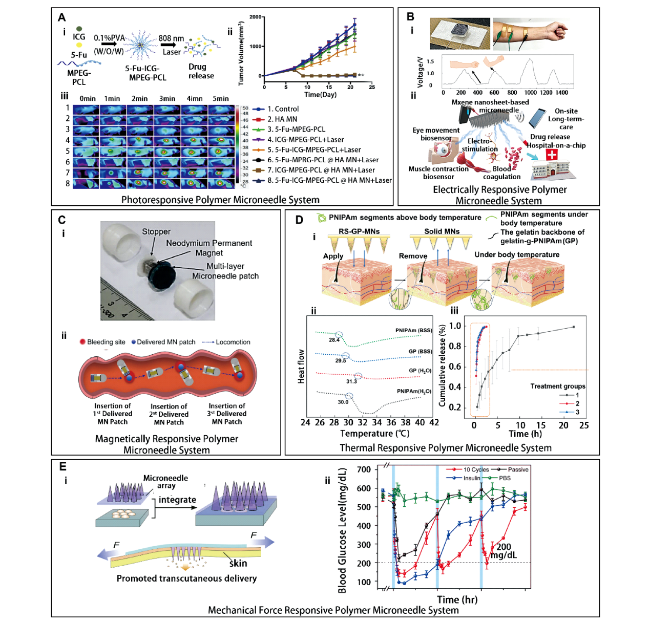
图5 外界环境刺激响应性聚合物微针。(A) 光响应性聚合物微针系统:5-Fu-ICG-MPEG-PCL的制备示意图 (A-i); 各组A431荷瘤小鼠的生长曲线 (平均值±标准偏差 (n=5) “**” 表示P<0.01) (A-ii); 1 W/cm2 808nm激光照射5 min后A431荷瘤小鼠的近红外光热效应 (A-iii)[40]; (B) 电响应性聚合物微针系统:MXene微针生物传感器测量的方式和信号处理后的电位峰值 (B-i); MXene微针生物传感器应用 (B-ii)[45]; (C) 磁响应性聚合物微针系统:带有多层MN贴片的磁性驱动胶囊 (C-i); 使用磁力驱动胶囊将微针贴片递送至靶病变的过程 (C-ii)[50]; (D) 热响应性聚合物微针系统。用于控制透皮给药的明胶-PNIPAm微针意图 (D-i); 不同浓度的H2O2中药物释放速率图 (D-ii); 在37 ℃ RS-GP微针(1)、室温下RS-GP微针 (2)、 37 ℃ 普通明胶微针(3) 下的体外释放速率图 (D-iii)[52]; (E) 机械力响应性聚合物微针系统:装有药物的可穿戴设备与微针阵列贴集成 (E-i); 在不同治疗方式下小鼠血糖水平变化 (E-ii)[57]

Fig. 5 Environmentally responsive polymer microneedles. (A) Photoresponsive polymer microneedle system. Schematic diagram of the preparation of 5-Fu-ICG-MPEG-PCL (A-i); Growth curves of A431 tumor-bearing mice in each group (mean ± standard deviation (n=5) “**” denotes P < 0.01) (A-ii); Near-infrared thermal effect of A431 tumor-bearing mice irradiated with 1 W/cm2 808nm laser for 5 min (A-iii)[40]; (B) Electrically responsive polymer microneedle system. MXene microneedle biosensor measurement method and signal processing potential peak (B-i); MXene microneedle biosensor application (B-ii)[45] ; (C) Magnetically responsive polymer microneedle system. magnetically driven capsules with multi-layer MN patches (C-i); The process of using a magnetically driven capsule to deliver a microneedle patch to a target lesion (C-ii)[50]; (D) Thermal responsive polymer microneedle system. Intent of gelatin-PNIPam microneedles for control of transdermal drug delivery (D-i); Drug release rates in different concentrations of H2O2 (D-ii); In vitro release rates of RS-GP microneedles at 37 ℃ (1), room temperature (2) and common gelatin microneedles at 37 ℃ (3) (D-iii)[52]; (E) Mechanical force responsive polymer microneedle system. Integration of medicine-equipped wearable devices with microneedle array patches (E-i); Changes in blood glucose levels of mice under different treatment methods (E-ii)[57]

In the application of the photoresponsive microneedle system, in addition to the use of photothermal conversion agents to convert light energy into heat energy, which in turn melts the matrix, inactive substances that are cleaved by light can also be used. By adjusting the total energy applied, the cleavage of covalent bonds between molecules can be effectively controlled to achieve the purpose of releasing active drugs. Fan et al. Constructed a near-infrared light-responsive PVA microneedle patch for the treatment of myocardial infarction by loading vascular endothelial growth factor (VEGF) on graphene oxide (GO), a photosensitive substance that endows the microneedle patch with shape memory ability[41]. Among them, PVA has good biocompatibility and degradability, and has good near-infrared light absorption ability. The polymer complex can prolong the activity of the drug. Under the irradiation of NIR (beam diameter =0.5 cm,1.5 W/cm2), the oxygen group of GO absorbs NIR and undergoes reduction reaction. The hydrogen bond between GO and PVA is broken, so that the patch is completely unfolded and the drug is released. The left ventricular ejection fraction (LVEF) and left ventricular fractional shortening (LVFS) are significantly increased, which achieves the purpose of cardiac targeting drug delivery. In the research work of Chen et al. And Hao et al., photosensitive substances with photothermal conversion ability were used to absorb NIR and rapidly heat up, melt the matrix and release the drug. Repeated switching of NIR can achieve multiple cycles of drug administration, thus achieving the precise therapeutic effect of on-demand drug administration and greatly avoiding the side effects of tumor treatment. Fan et al. broke through the common NIR-responsive microneedle technology, using the heat generated by photosensitive substances to destroy covalent bonds and deform the microneedle patch, which can achieve the purpose of targeted drug delivery with less damage, thus reducing the risk and simplifying the operation procedure and time. However, there are few research data to support the clinical application of this technology.

3.1.2 Lectrically responsive polymer microneedle system

Electrical Stimulation (ES) is a signal factor that is easy to regulate, easy to monitor, and can be applied remotely. It can load drugs into the Electrical response carrier by combining with sensors or chips, effectively regulate drug dosage and monitor drug administration wirelessly in real time. However, ES has strict requirements on the voltage and current applied by the equipment[42]. At present, the most common conductive polymer materials are polyelectrolyte gel, polypyrrole (PPy), polyaniline (Pan) and graphene. In the electro-responsive polymer microneedle system, electrical stimulation mainly uses electric field or current to drive drug release. In 1995, in order to overcome the limitation and dependence of drug delivery, Guiseppi-Ellie et al. Selected Conducting Polymers (CPs) as the carrier of active drugs, which responded to the ionic environment of skin structure, and proposed a research idea of combining PPy with UV-absorbing and curable poly (2-hydroxyethyl methacrylate) (p-HEMA) -based hydrogel to form a conductive polymer hydrogel composite[43]. The potential of the gel electrode decreased from 0. 80 V to 0. 68 V when the redox of the CPs network occurred, which was characterized by the microsensor electrode (IME), and the ions could diffuse through the porous structure of the hydrogel to release growth factors, proteins, anti-inflammatory agents and other drugs. Based on the research of conductive hydrogel, Qu et al. Proposed an electrically and pH-responsive hydrogel composed of chitosan (CS) -grafted polyaniline (CP) copolymer (with good conductivity and antibacterial effect) and oxidized dextran (OD, pH-sensitive substance), which was mainly loaded with amoxicillin and ibuprofen. 69% or 82% of amoxicillin was released after repeated application of 1 or 3 V potential for 1 H. When the potential of 3 V was applied for more than 2 H, 35% ibuprofen was released[44]. Yang et al. Used MXene nanosheets (with voltage sensing capability) combined with polylactic acid (PLA) microneedles to form a wearable 2D layered microbial sensor. The sensing effect is achieved by sensing the weak potential difference generated by human eyeball or muscle contraction, which is applied to myasthenia detection, transcutaneous nerve electrical stimulation therapy, and electrical response drug delivery system (Fig. 5B)[45]. In combination with iontophoresis (ITP), Seeni et al. Constructed a new conductive HA microneedle for local anesthesia by loading lidocaine hydrochloride on the conductive polymer of poly (3,4-ethylenedioxythiophene) (PEDOT) and polystyrene sulfonate (PSS). The PEDOT/PSS polymer increased the conductivity of microneedles by two times, reduced the resistance of mouse skin by 30% and the mucosal resistance, and increased the drug penetration by four times after the 3 mA/cm2 current amplitude lasted for 2 to 4 min, which immediately achieved the surface anesthesia effect[46]. Yang et al. And Seeni et al. Combined micro-electronic devices and microneedles to form a biosensor, which effectively avoided the inconvenience of patient examination and the untimely feedback of drug administration, and provided a broad prospect for clinical treatment. However, the number of studies on electroresponsive polymer microneedles is low, and accurate and long-term on-demand drug release through conductive polymer hydrogels remains a challenge.

3.1.3 Magnetically responsive polymer microneedle system

Magnetic wave stimulation system is a kind of signal factor which can be regulated remotely and has no biological toxicity. It is mainly released by alternating magnetic field or fixed magnetic field. In the magnetic response drug delivery system, magnetic response nanoparticles were first used to control the magnetic properties of nanoparticles by changing the application time and total energy of the magnetic field, enhance the conductivity of the matrix, and then regulate the release of drugs in time and space. In the magnetically responsive polymer microneedle system, a safe and stable Magnetic polymer composites (MPC) is constructed by combining polymer materials with Magnetic nanoparticles[47]. Because magnetic nanoparticles contain metal components and have certain biological toxicity to skin tissue, polymer materials with good biocompatibility (such as lignin, polyacid, dextran, etc.) Should be used as carriers of magnetic nanoparticles. Justin et al. Constructed a detachable magnetic-responsive PEG (with good water solubility) composite microneedle based on degradable CS and iron oxide-loaded magnetic graphene quantum dots ( (GO-IO) QD, with superparamagnetism) for the release of lidocaine hydrochloride, bovine serum albumin (BSA)[48]. The (GO-IO) QD magnetically responded to the current when the iontophoretic device applied an electrical stimulus of 9 V, 280 mA, and the microneedle released 1.9 mg or 40.7% BSA in 9 H and 4.5 mg or 96.4% BSA in 24 H.
In addition, inspired by the Lego building block stack, the magnetically responsive polymer microneedle system can be combined by a microneedle patch and a magnetically responsive micro-robot, which mainly includes three components of a magnetically responsive substrate, a detachable connector and a microneeedle, and is widely used in the treatment of gastrointestinal diseases[49]. It has high magnetic saturation and targeted release potential for cancer therapy. Many drugs can cause strong discomfort to cancer patients. By using nanoparticles to coat drugs and applying external magnetic fields, drugs can target specific tissues and organs, alleviating the pain of patients and increasing the efficacy of drugs. Lee et al. Constructed a magnetic-responsive multilayer drug-loaded microneedle patch for the treatment of gastrointestinal diseases by replacing magnetic-responsive micro-components with capsules, capsules wrapped with permanent magnets, and PDMS loaded with LGB (fluorescent dye)[50]. Driven by external magnetic force, the capsule delivered the microneedle patch to the focus of the gastrointestinal tract, and the OD value was reduced to a quarter within 10 minutes, achieving a significant hemostatic effect. In addition, visual observation of microneedle administration and detection of mechanical strength can be performed by confocal microscopy (fig. 5C).
Justin et al. Combined electrical stimulation technology, polymer microneedles and magnetic particles to achieve wearable intelligent transdermal drug delivery and biosensing devices. Compared with magnetic particles, Lee and others have broken through the traditional microneedle drug delivery method, stacking multi-layer microneedles through magnetic capsule endoscopy to achieve organ targeting delivery, but this method is not convenient and safe enough, and there are few studies on polymer microneedles in magnetic responsive transdermal drug delivery, and the data support is insufficient, so there are more prospects for development.

3.1.4 Thermally responsive polymer microneedle system

In the stimuli-responsive polymer microneedle system, the thermal stimulation system is a widely studied and efficient trigger mode, which mainly controls the stimulation temperature, stimulation site, duration and thermal responsive materials to achieve transdermal drug delivery. Among them, thermal responsive materials are the key factor, which can be classified into low critical solution temperature polymers, high critical solution temperature polymers and thermally induced shape memory polymers, and can undergo reversible phase change with temperature change. Poly (N, N-dimethylaminoethyl methacrylate) (PDMAEMA), poly (isopropylacrylamide) (PNIPAAM) and PMMA are the most popular thermoresponsive materials at present. These polymers are amphiphilic block polymers composed of hydrophobic core and hydrophilic end group. By controlling the chain length of the diblock copolymer, the morphological transformation of the diblock copolymer can be effectively regulated[51]. Because the human body temperature is always kept in the range of 35 ~ 37 ℃, the temperature response point of polymer materials is the key to the design of thermoresponsive polymer microneedles.
In the thermoresponsive polymer microneedle system, the mechanism of thermoresponsive polymer is similar to thermodynamics. With the increase of temperature, the hydrophilicity and hydrophobicity of polymer change, which leads to the folding and aggregation of chain segments. Polymers sensitive to lower critical solution temperature, such as cellulose, xyloglucan, CS, protein-like gelatin, elastin-like polypeptide, PNIPAAM and other materials, are biocompatible and degradable, and can realize the conversion between sol and gel above the critical solution temperature, which are widely used in drug delivery, monitoring and other aspects. Li et al. Constructed a heat-sensitive microneedle with strong mechanical properties based on gelatin grafted hydroxyl as the matrix and combined with PLA microneedle, which is mainly used for the delivery of protein drugs such as insulin. The normal body temperature was higher than the low critical phase transition temperature (31.3 ° C) of gelatin-poly (N-isopropylacrylamide) (PNIPAm), and the hydrophobicity affected the polymer crosslinking, and the system rapidly changed from gel to sol within 2 H, with complete drug release (Fig. 5D)[52]. Lee et al. Proposed a heat-responsive wearable polyvinylpyrrolidone (PVP) microneedle for percutaneous diabetes monitoring and treatment based on tridecanoic acid (thermosensitive substance) loaded with metformin (DMBG), a drug for the treatment of diabetes[53]. When the control temperature is higher than the (Tc)41~42℃ of the transition temperature, the PCM can be melted, 45% to 80% of the drug can be released, and the blood glucose concentration can be reduced by one third. The PVP microneedle is combined with the glucose sensor, and the H2O2 generated by the reduction reaction of glucose oxidase (GOx) is used as an electrochemical signal responding to the pH change of sweat, so that the pH and glucose multi-responsive drug delivery effect is realized. In addition, a small number of thermoresponsive microneedles use photothermal conversion agents to melt low critical temperature polymers, thus achieving transdermal drug delivery. Common photothermal conversion agents are black phosphorus (BP), lanthanum hexaboride (LaB6), GO carbon nanomaterials, etc. Lee and Kim et al. Overcame the one-time drug delivery problem of traditional microneedles and proposed a poly (lactic-co-glycolic acid) (PLGA, hydrophobic polymer) loaded RhB thermoresponsive HA microneedle, which was mainly divided into three layers of HA-PLGA-HA[54]. At 37 ℃, 90% of the drug could be released continuously within 26 days. In the above studies, thermosensitive materials dissolved by heat, photothermal conversion agents, and thermal melting materials and technologies were used respectively. Kim et al. Made full use of the hot melting of PLGA to effectively avoid the use of organic reagents, but PLGA needed to be kept at 100 ℃ during the injection process, and the microneedle could only deliver heat-resistant drugs.

3.1.5 Mechanically responsive polymer microneedle system

According to different force directions, mechanical force can be divided into compression, tension and shear, which are triggered remotely mainly through the combination of hand and musculoskeletal movements, ultrasonic waves and magnetic fields. Compression and stretching forces can cause the carrier or mechanoresponsive material to deform, resulting in the destruction of chemical bonds and the release of drugs. Shear stress not only destroys the structure, but also degrades related enzymes. In the process of wound healing, mechanical force is easy to cause secondary damage to the wound, so mechano-responsive drug delivery system is also a hot research topic in wound healing, which mainly applies mechanical force from the outside world to cause biological response, and then achieves drug delivery in time and space[55].
In the mechanoresponsive drug delivery system, polymer materials with good tissue elasticity and viscosity are mainly selected, including natural hydrogel polymers such as sodium alginate, CS and HA, and synthetic hydrogel polymers such as poly (hydroxyethyl methacrylate), polyacrylamide (PAM), poly (ethylene glycol), etc[56]. Di et al. Proposed an insulin-loaded HA microneedle with high tensile properties in combination with a sodium alginate patch composed of PLGA nanoparticles. When 50% tensile strain was applied for 10 cycles, HA was prolonged and better adsorbed on the wound or action site, the nanoparticles were compressed, and the blood glucose level of mice decreased rapidly to normal blood glucose state (< 200 mg/dL) within 30 min, and then increased gradually (Fig. 5E)[57]. Jun et al. Constructed a detachable mechanically responsive microneedle patch with a Single-walled stand (SWS) based on a drug-loaded HA tip and a PCL base array. When a lateral force of 0.04 ~ 0.14 N or a compressive force of 0.56 N was applied, the HA tip was rapidly separated within 1 s[58]. Zhang et al. Proposed a silk fibroin (SF, adjustable stiffness and strength) drug-loaded puncture-responsive PVA microneedle for wound traceless healing. When the microneedle was inserted, the continuous mechanical force inhibited the activation of fibroblasts and promoted the secretion of collagen and fibronectin, and the scar was faded within 30 days[59].
Di et al.'S research is widely used in medicine for protein drug delivery, diagnostics, etc. Jun et al. Used the adhesion between the tip and the base layer and the square pyramid needle to achieve the purpose of needle separation, which overcame the inconvenience and interference problems during the delivery of drugs to skin tissue by microneedles. Zhang et al. Used the continuous stimulation of microneedles to regulate the ultrastructure and biomechanics of tissues, but lacked periodic data.

3.2 Polymer microneedle system triggered by in vivo physiological signal stimulation

In vivo physiological signal stimulus-response system, also known as closed-loop drug delivery system, is a biological signal that achieves drug release through homeostatic regulation. This system is different from the external environment stimulus-responsive system, which can efficiently control the dosage and speed of drug release according to the biological signals related to the pathological process of patients, and is widely used in polymer microneedle systems. In the polymer microneedle system triggered by physiological signal stimulation in vivo, it consists of a sensor and a brake to trigger drug delivery[60]. In order to promote the effective delivery of drugs, polymer materials with good biocompatibility should be selected to produce physical or chemical reactions, such as the expansion of aggregate matrix structure, membrane fusion, dissociation of material molecular structure and bond breakage, so as to achieve effective drug delivery. According to the known physiological signal stimulating factors in vivo, such as pH, glucose, redox potential and enzyme, microneedles can be divided into pH-responsive microneedles, glucose-sensitive microneedle, reactive oxygen species-responsive microneEDles and enzyme-responsive microneEDles[61].

3.2.1 pH-responsive polymer microneedle system

The pH-responsive polymer microneedle system is characterized by the reaction of protonatable cationic groups (such as —NH2) and deprotonated carboxyl groups (such as — COOH) and hydrogen bonds within or between molecules under the change of acid-base environment according to different pKa values, thus changing the structure and solubility of pH-responsive polymers, which are widely used in cancer, vaccines, insulin drug delivery and sensors[62]. According to the different dissociation groups of pH-responsive polymers, it can be divided into anionic pH-responsive polymer microneedles, cationic pH-responsive polymer microneedles and zwitterionic pH-sensitive polymer microneneedles. Anionic pH-responsive polymer microneedles contain a large number of acidic groups, mainly — COOH. At low pH level, the —COO- is protonated, and the generated hydrogen bond leads to the decrease of the degree of dissociation, and the microneedle structure shrinks to release the drug; On the contrary, the solubility of microneedles increases, resulting in electrostatic repulsion to destroy the balance of hydrogen bonds, resulting in the elongation and volume expansion of the polymer chain segment of the microneedle structure, thereby controlling drug delivery. In cationic pH-responsive polymer microneedles, the common groups are —NH2, — NHR, —NR2, all of which are prone to deprotonation. At low pH level, the group exists in the form of —NH 3 +, which produces a large amount of electrostatic repulsion, resulting in the expansion of the microneedle structure. At high pH levels, —NH2 hydrogen bonds with H2O, resulting in microneedle shrinkage. In addition, the drug release can be controlled by adjusting the number of amino groups. Zwitterionic pH-responsive polymer microneedles contain acid-base groups (carboxyl and amino). At low pH levels, the amino group undergoes protonation, and at high pH levels, the carboxyl group undergoes deprotonation. Therefore, the zwitterionic pH-responsive polymer microneedles will swell in all pH ranges, changing the rheology of the microneedles[63].
Li et al. Constructed a pH-responsive PCL microneedle for cancer therapy based on dimethyl maleic anhydride modified polylysine (PLL-DMA, an acid-sensitive charge-reversible polymer) and p53 expression plasmid (negatively charged cancer cell suppressor gene)/polyethylenimine loading model gene (fluorescent DNA model, which can track the effect of feedback gene delivery)[64]. The PLL-DMA transition layer and the gene carrier layer constitute the polyelectrolyte multilayer membrane (PEM). In the simulated cancer environment of pH = 5.5, the chemical bond between PLL and DMA was broken, and the layer changed from negative to positive, which destroyed the transition layer structure, delivered 33% of genes, inhibited the cancer rate of mice by about 90.1%, and then promoted gene delivery. Hu et al. Proposed a pH-responsive peach gum polysaccharide (GPs) microneedle based on acid-sensitive nanoparticle NP(CaCO3), which was synthesized by hybridization of PLGA and CaCO3, loaded with tetrandrine (Tet, anti-arthritic active ingredient). In the physiological environment of pH = 5.5, the CaCO3 responded to the H+, destroyed the particle structure, and released three times the drug[65]. Pu et al. Constructed a triple-response HA microneedle based on DOX-loaded nanoparticles composed of (N-isopropylacrylamide-acrylic acid) (PNA, with thermal/pH responsiveness) and polydopamine (PDA, photothermal conversion agent). In the tumor environment of pH = 5.5, NIR was applied externally, and the π-π conjugate bond between DOX and nanoparticles was destroyed by high temperature within 48 H. Too much negative charge leads to the weakening of the electrostatic interaction between DOX and PNA, which in turn releases 80% of the drug[66]. Jung et al. Proposed a PVP microneedle based on zeolitic imidazole-8 (ZIF-8) acid-sensitive nanoparticles loaded with curcumin (CCM, a drug for cancer and inflammation). In the inflammatory environment of pH = 5.0, the imidazole ion of ZIF-8 structure was protonated, which promoted the degradation of the structure and released 80% of the drug within 6 H (Fig. 6A)[67].
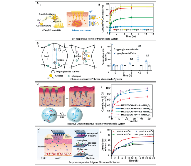
图6 体内生理信号刺激响应性聚合物微针。(A) pH响应性聚合物微针系统:ZIF-8包封微针的示意图 (A-i); 不同pH条件下CCM释放曲线 (A-ii)[67]; (B) 葡萄糖响应性聚合物微针系统:GRS胰高血糖素递送系统的机制和体外性能 (B-i); 用贴剂治疗后糖尿病小鼠的血浆胰高血糖素浓度变化 (B-ii)[70]; (C) 活性氧响应性聚合物微针系统:微针响应H2O2递释MTX治疗银屑病机理图 (C-i); 在不同浓度H2O2下微针体外释放EGCG速率图 (C-ii)[74]; (D) 酶响应性聚合物微针系统:微针溶解感染伤口清除治疗生物膜的机理示意图 (D-i); 不同pH值和温度下的左旋释放曲线 (D-ii)[82]

Fig. 6 Stimulus-responsive polymer microneedles for physiological signals in vivo. (A) pH-responsive polymer microneedle system. Schematic diagram of ZIF-8 encapsulated microneedles (A-i); CCM release curves under different pH conditions (A-ii)[67]; (B) Glucose-responsive polymer microneedle system. The mechanism and in vitro performance of GRS glucagon delivery system (B-i); Changes in plasma glucagon concentrations in diabetic mice treated with the patch (B-ii)[70]; (C) Reactive oxygen reactive polymer microneedle system. Mechanism diagram of microneedles in response to H2O2 delivered MTX in the treatment of psoriasis (C-i); EGCG release rate of microneedles in vitro at different concentrations of H2O2 (C-ii)[74]; (D) Enzyme-responsive polymer microneedle system. Schematic diagram of the mechanism of microneedle lysis of biofilms in infected wound removal (D-i); left-handed release curves at different pH values and temperatures (D-ii)[82]

Because the pH of human skin structure, wound inflammation, cancer and other parts is acidic, it can be used as a stimulus for acid-sensitive drug delivery and applied to pH-responsive polymer microneedles. In the above studies, different acid-sensitive materials and preparation methods were used to induce the structural change of polymers and achieve pH-sensitive drug delivery. Among them, Li et al. Used layer-by-layer assembly technology and acid-sensitive materials to construct gene-loaded PEM. Pu et al. 'S research combined with photothermal therapy and chemical coordination method can effectively regulate drug delivery while stabilizing the structure.

3.2.2 Glucose-sensitive polymer microneedle system

Glucose is an important substance to control the blood sugar level of the human body. At present, the most common treatment for type I and type II diabetes is subcutaneous insulin injection, but this treatment can not accurately control the insulin dosage according to the physiological blood sugar level of the patient, which may cause side effects such as hypoglycemia, blindness, and even renal failure[68]. The response of enzymes to glucose levels is a hot research topic, which has attracted the attention of many scholars. Luo et al. Constructed a glucose- and pH-responsive microneedle for diabetes therapy based on poly (hexamethylenimino) ethyl methacrylate (PHMEMA, pH-responsive material) nanoparticles (SNP-I) loaded with insulin and pH-insensitive nanoparticles (iSNPG + C) loaded with GOx (enzymes that oxidize glucose to produce gluconic acid and H2O2) and catalase (CAT, enzymes that reduce H2O2)[68]. Under the condition of 500 mg/dL blood glucose, GOx in the microneedles induced the production of gluconic acid and H2O2, which led to the decrease of pH value, and the tertiary amino group in the structure of PHMEMA rapidly converted to hydrophobic and hydrophilic, releasing insulin and reducing blood glucose to 100 mg/dL within 14 H. In addition, CAT can reduce the produced H2O2 and avoid the skin damage caused by H2O2. Because H2O2 leads to pH reduction in physiological environment, sodium bicarbonate (NaHCO3) contains ionic groups and has good hydrophilicity, which can be used as glucose and pH multi-responsive materials. When blood glucose levels rise in diabetic patients, GOx induces the production of gluconic acid and H2O2, resulting in a decrease in pH and the release of large amounts of CO2 in response to NaHCO3, which in turn effectively releases insulin. In addition, due to the acidic pH environment of human skin, H+ reacts with NaHCO3 to produce a large amount of carbon dioxide, which increases the pressure of polymer and releases drugs[7].
In addition, in addition to the use of glucose-responsive materials, the ionic bond of phenylboronic acid (PBA) can also be condensed with the hydroxyl group of glucose. PBA generally exists in a hydrophobic form. After combining with glucose to form 1,2-glucose phenyl borate or 1,2,3,5-diglucose phenyl borate, the hydrophilicity of PBA increases. PBA can be used as a carrier for loading insulin or glucagon by using the change of hydrophilic groups of PBA, and is the main material of glucose-responsive polymers. Therefore, a glucose-responsive polymer microneedle system can be constructed using PBA and PBEM[69]. Wang et al. Proposed a microneedle based on a glucagon-loaded polyacrylamide (CPAM, cation-containing)/3- (acrylamido) phenylboronic acid (APBA, glucose-responsive) copolymer. At 50 mg/dL blood glucose, the matrix undergoes charge transfer from cationic to neutral, which disrupts the matrix structure and releases about 100 pg/dL glucagon; At the 400 mg/dL blood glucose level, the negative charge increased to promote electrostatic attraction, and the matrix structure stabilized (Figure 6 B)[70]. Ye et al. Proposed a glucose-responsive microneedle based on the dynamic covalent bonding between insulin-loaded PBA and a polyvalent diol cross-linker. At a blood glucose level of 420 mg/dL, the microneedles could reduce blood glucose to a normal blood glucose level (180 to 200 mg/dL) within 4 H, and remained unchanged for 8 H[71].
Most studies have achieved a glucose smart release model. Among them, PBA is a popular glucose-responsive material. Ye et al. Made full use of the reversible binding property of PBA and glucose and the in situ covalent polymerization technology to form a phenyl borate ester bond. The reaction of free glucose and PBA competed with the diol group, resulting in the weakening of the hydrophobic correlation of the hydrogel and promoting drug release.

3.2.3 Active oxygen responsive polymer microneedle system

Reactive oxygen species (ROS) are a class of oxygen-derived chemicals produced mainly by cellular metabolism of mitochondria and peroxidases and by various lysosomal systems, mainly including Reactive oxygen species, singlet oxygen (1O2), superoxide ( O 2 -), hydroxyl radical (OH ·). Excessive production of ROS in cells can lead to oxidative stress, resulting in inflammation, aging, cancer and other diseases. In the ROS-responsive polymer microneedle system, ROS stimulation can not only promote drug penetration, but also coexist with other stimulation factors in the multi-responsive microneedle[72]. At present, the known ROS-responsive materials of microneedles include thioether, selenium, tellurium, thione, polysaccharide, aminoacrylate, borate, peroxyoxalate and polyproline. Among them, borate ester is the research focus of drug carrier[73]. Bi et al. Constructed methotrexate-loaded HA microneedles for the treatment of psoriasis based on the dynamic borate bond formed between EGCG and PBA. Among the H2O2 concentrations (0, 0.1, 0.5, 1.0 mM), the drug release rate of MTX/EGCG microneedles was the highest at 1.0 mM, reaching 100% within 48 H (Fig. 6 C)[74]. Broaders et al. Constructed an oxidation-responsive dextran nanoparticle (Oxi-DEX) for inflammation therapy based on dextran/arylborate loaded ovalbumin (OVA, protein antigen). At the level of 1 mM H2O2, macrophages produce ROS during immunization, and the borate ester of Oxi-DEX is oxidized to phenol, which promotes particle lysis and releases 27-fold antigen[75]. Zhang et al. Constructed a ROS-responsive HA microneedle for acne treatment based on polyvinyl alcohol (RR-PVA, containing bisphenylboronic acid) loaded with antibiotics (CDM, drug for acne treatment) and diatomaceous earth (DE, cross-linking agent)[76]. Acne is a case of inflammation. When the level of ROS in inflamed tissue is more than 500 μm, the boric acid bond of RR-PVA will be oxidized and hydrolyzed, and about 90% of the drug will be released within 24 H.
In addition, common ROS-responsive materials are chalcogens (such as S, Se, and Te). Their valence state is changed mainly by oxidation, oxygen can bind to chalcogen, while the polarizing group provides hydrogen to bind to water, which in turn changes its hydrophilicity[6]. Wang et al. Constructed a ROS-responsive polymer complex based on selenyltellurium polymer bis (6-hydroxyhexyl) 3,3 ′ -selenodipropionate (C6-C3Se), which can be used as a ROS-responsive carrier. At 1 mM H2O2 level, C6-C3Se was oxidized to bis (6-hydroxyhexyl) 3,3 '-selenyldipropionate (C6-C3SeO), which was then eliminated to 6-hydroxyhexyl acrylate (C6-ole), and finally 90% of the polymer was eliminated after 24 H[77]. Broaders et al. And Wang et al. Studied the use of PBA and selenium-containing compounds to achieve ROS-responsive drug delivery, which can be used as the development prospect of ROS-responsive polymer microneedles. Zhang et al.'s Research is devoted to inhibiting Propionibacterium acnes and achieving acne removal effect, which is a hot spot in the field of cosmetics, but the research background in the field of microneedle acne removal is less, and too much data support is needed[76].

3.2.4 Enzyme-responsive polymer microneedle system

Enzymes are catalytic organic compounds produced by living cells, which are highly efficient and specific to substrates. Because the enzyme is sensitive to temperature, it usually catalyzes in a low temperature and weak acidic water environment. Therefore, enzymes are widely used in stimulus-responsive polymer microneedle systems, mainly through enzyme-responsive polymer materials or enzyme-reactive biomaterials as targeting substrates, and the change of enzyme expression level triggers the chemical structure change of the substrate to achieve drug delivery effect[78].
The International Union of Biochemistry (IUB) classifies enzymes into seven types: oxidoreductase, transferase, hydrolase, lyase, isomerase, synthetase and translocase[79]. Among them, the application of oxidoreductase and hydrolase has received more attention. Ye et al. Formed an enzyme-responsive polymer vesicle (PV) HA microneedle system based on tryptophan (1-MT, indoleamine 2,3-dioxygenase (IDO, immunosuppressive enzyme) inhibitor), loaded antibody (aPD1, antibody against programmed cell death protein (PD1, immunosuppressive receptor for tumor lymphocytes)) and hyaluronidase (HAase) for melanoma therapy. When the microneedle was applied, HAase catalyzed the enzymatic hydrolysis of HA in response to the tumor environment, releasing 5 times of 1-MT within 3 days. In addition, IDO catalyzes the degradation of tryptophan, which in turn inhibits the immune function of T cells[80]. Hu et al. Constructed a glucose oxidase, H2O2 multi-responsive microneedle system based on PBA/PEG/glucose-binding protein (GBP) loaded with GOx and insulin, which has the ability to compete with glucose, for the treatment of type I diabetes.
In the physiological environment of 600 mg/dL blood glucose, acidity and H2O2, when the microneedles were applied to the skin surface, GOx catalyzed glucose to cause a decrease in blood glucose and pH levels, which rapidly decreased to 100 mg/dL blood glucose within 1 H and remained unchanged for 5 H. In addition, the thioether of PEG can be oxidized to sulfone by H2O2, and then release insulin with high efficiency[81]. Yu et al. Used α-amylase (degrading polysaccharide in biofilm) nanoparticle microneedles loaded with levofloxacin (Levo, antibiotic), PDA (with high photothermal conversion efficiency and wound healing promoting ability) for wound healing. In the biofilm (pH = 5.4) on the wound surface, the drug cooperated with α-amylase to degrade the exopolysaccharide of the biofilm, eliminating 80% of the bacteria in wound sealing (Fig. 6 D)[82]. Most enzyme-responsive drug delivery systems need to cooperate with other responsive materials to achieve drug delivery and therapeutic effects. The different types of stimuli-responsive polymer microneedle systems and applications are summarized in Table 1.
表1 不同类型的刺激响应性聚合物微针系统及应用

Table 1 Different types of stimulus-responsive polymer microneedle system and applications

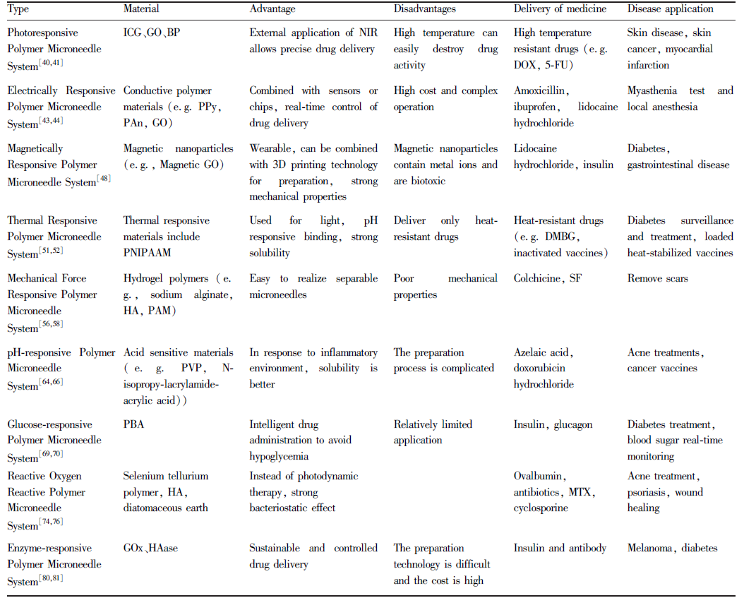
NoteICG: Indocyanine Green; GO: Graphene Oxide; BP: Black Phosphorus; PPy: Polypyrrole; PAn: Polyaniline; PNIPAAM: Poly (N-Isopropylacrylamide) Polymer; HA: Hualuronic Acid; PAM: Polyacrylamids; PVP: Polyvinyl Pyrrolidone; PBA: PhB(OH)2; GOx: Glucose Oxidase; HAase: Hyaluronidase; DOX: Doxorubicin Hydrochloride; 5-FU: 5-Fluorouraci, C4H3FN2O2; DMBG: Metformin; SF: Silk Fibroin; MTX: Methotrexate

4 Stimulus-responsive polymer microneedle transdermal delivery application

Stimulus-responsive polymer microneedles have higher drug loading and physiological environment responsiveness than other microneedles. In the preparation process of polymer microneedles, polymer materials with specific responsiveness are selected, which can not only efficiently and uniformly encapsulate active drugs, but also enhance the stability of microneedles in the process of transportation and use, so as to achieve specific and sustainable release. In addition, the good biocompatibility and biodegradability of polymer materials enhance the safety of drug delivery process, reduce the biological toxicity of drugs, effectively solve the problem of medical waste recycling of needles, and reduce the cost of transdermal drug delivery. Therefore, the stimuli-responsive polymer microneedle has beneficial applications in the fields of biomedical delivery, tissue and organ, sensing, sample extraction, dermatology and medical beauty (Fig. 7).
图7 刺激响应性聚合物微针系统在不同方面的应用

Fig. 7 Application of Stimulus-responsive polymer microneedle systems

4.1 Biomedical Release

At present, most vaccines are administered by subcutaneous injection and intramuscular injection, but because the number of dendritic cells (DCs) in human subcutaneous fat and muscle tissue is small, the vaccine can not fully induce humoral and cellular immune responses, which inhibits the ability of antigen targeting. In order to promote antigen-induced immune response, Choi et al. Used HA microneedles based on PCL-loaded canine influenza virus vaccine for percutaneous rabies vaccination. This is a highly permeable and low-cost vaccine delivery system, which can not only double the ability to induce antibodies, but also preserve the activity of the vaccine for a long time by encapsulating the target vaccine through microneedles[83]. In recent years, many diabetic patients can not accurately control the dosage in the process of self-administration, which can easily trigger complications such as hypoglycemia and even amputation. In order to realize the smart delivery of insulin, Ghavami Nejad et al. Designed a glucose-responsive photocrosslinked methacrylated hyaluronic acid (MeHA) smart microneedle patch based on 4-acrylamide-3-fluorophenylboronic acid (AFBA)/zwitterionic sulfobetaine (SB, superhydrophilic, which can effectively stabilize proteins)/cationic carboxybetaine (CB) polymer microgel loaded with glucagon. At a low glucose level of 50 mg/dL, AFBA forms a double complex with glucose, causing the microgel to shrink, releasing 0.22 mg/dL glucagon, which is widely used in hypoglycemia treatment[84]. Anesthetics, as an indispensable drug in clinical medicine, mainly block nerve impulse transmission by inhibiting voltage-gated sodium channels (VGSCs). Currently known modes of administration are subcutaneous injection and intramuscular injection, but the diffusion rate of anesthetic drugs is slow. In order to promote the penetration of anesthetic and achieve rapid anesthetic effect, Lee et al. Used carboxymethylcellulose (CMC) -loaded lidocaine hydrochloride microneedle patch for local anesthesia. In the process of anesthetic diffusion, lidocaine hydrochloride has a high degree of lipophilicity, which can promote its penetration through the cell membrane and into the cytoplasm, block the transmission of nerve impulses by inhibiting the VGSCs of trigeminal neurons in mice, and increase the threshold of 80 G paw withdrawal, thus producing anesthetic effect[85].

4.2 Tissue and organ therapy

In recent years, the stimuli-responsive polymer microneedle system has been widely used in the treatment of tissues and organs, mainly for the heart, kidney, wound healing and so on. With the continuous development of the country, people's living standards have been greatly improved, while the pressure of work and the pace of life have also increased rapidly, leading to myocardial infarction and heart failure have become frequent diseases. Treatment of heart disease requires the use of growth factors (GFs) to restore the structural and functional integrity of the heart. Because of the metabolism of the internal environment, GFs are easily and rapidly cleared in the process of penetration in vivo, which weakens the effect of cardiac therapy. In order to promote the penetration rate of drugs, Lim et al. Constructed a microneedle bandage with strong adhesion based on adhesive protein loaded with vascular endothelial growth factor (VEGF) and regenerated silk fibroin (SF)[86]. Under the action of VEGF, it can induce the formation and organization of capillaries, achieve 75% wound closure rate, thicken the ventricular wall and increase the mechanical stress, achieve the regenerative effect of protecting the myocardium and remodeling the heart, and is widely used in the treatment of myocardium.
In the physiological environment of kidney, intestines and stomach, the stimulus-responsive polymer microneedle system can precisely regulate drug delivery through the pH change of the focus environment. At the same time, acid-base balance is a biological parameter for disease diagnosis, and microneedles combined with fluorescence sensors can monitor pH changes in real time through optical path length, temperature changes, excitation intensity and other factors. Zhou et al. Prepared an acid-sensitive microneedle based on molybdenum disulfide (MoS2, layered material) nanosheets and polyaniline (PAn). In that physiological condition that the pH value is between 3.0 and 9.0, the PAn/MoS2 has high-efficiency pH responsiveness, the PAn with conductivity is protonate and deprotonated, and the proton is permeate into a MoS2 to cause potential change, so that pH monitoring of brain and kidney is realized[87].
Stimulus-responsive polymer microneedle system can not only meet the delivery of liquid drugs, but also can deliver gas. For example, the disorder of blood sugar and lipid metabolism in diabetic patients leads to insufficient blood supply and protein synthesis, which will inhibit the healing ability of wounds. In order to promote the healing rate of the patient's wound, sufficient oxygen is a prerequisite, which has the ability to promote cell proliferation and tissue reorganization. Zhang et al. Constructed an NIR-responsive composite microneedle composed of a PVA backing layer and a GelMA tip based on BP and hemoglobin (Hb, oxygen carrying protein) loading oxygen. After 2 min of 1.56 W/cm2NIR cycle irradiation, the microneedle temperature was increased to 40 ℃, which inhibited the oxygen binding capacity of Hb by rapidly increasing the local skin temperature, and 48 mmHg oxygen was released within 24 H, which could be rapidly delivered to the deep skin and widely used for wound healing (Fig. 8 A)[88].
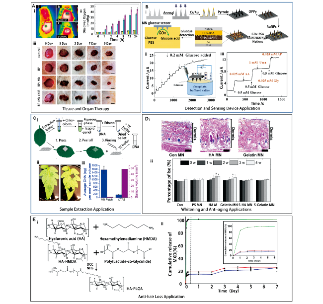
图8 刺激响应性聚合物微针经皮递释应用。(A) 组织器官治疗应用:1 W/cm2 NIR照射2 min前后, MN施加到大鼠背部皮肤的热图像 (A-i); 不同组 (对照组、BP组、BP + Hb组和BP + Hb + NIR组) 的氧释放情况 (A-ii); 第0、3、5、7和9 d不同组皮肤伤口的代表性照片(A-iii)[88]; (B) 检测及传感装置应用:葡萄糖传感器的制造工艺和检测机理图 (B-i); 在0.75 V下连续添加0.2 mM葡萄糖的PBS中的I (A)-t响应曲线 (B-ii); 0.75 V下连续添加0.5 mM葡萄糖、0.025 mM AA、0.5 mM葡萄糖、1 mM尿素和0.025 mM Gly、0.5 mM葡萄糖、0.025 mM AP (B-iii)[91]; (C) 样品提取应用:常规CTAB提取和MN提取示意图 (C-i); 微针贴片穿刺和切断后番茄叶的图像 (C-ii); 微针提取方法和CTAB提取方法作用下的DNA提取量 (C-iii)[93]; (D) 美白及抗衰应用:在对照组、HA微针、明胶微针作用下的皮肤发组织学图像 (D-i); 使用微针前后4周内皮肤弹性变化率 (P<0.05) (D-ii)[99]; (E) 防脱发应用:聚乳酸-乙醇酸接枝透明质酸 (HA-PLGA) 的合成 (E-i); 累积释放曲线(MXD溶液(●)、PLGA/MXD-NP(■)、HA-PLGA/MXD-NP(▲) ) (E-ii)[100]

Fig. 8 Transdermal delivery of stimulus-responsive polymer microneedles. (A) Tissue and organ therapy. Thermal images of MN applied to the back skin of rats before and after 1 W/cm2 NIR irradiation for 2 min (A-i); Oxygen release in different groups (control group, BP group, BP + Hb group and BP + Hb + NIR group) (A-ii);Representative photographs of different groups of skin wounds on days 0, 3, 5, 7 and 9 (A-iii)[88](B) Detection and sensing device application[91]. Diagram of the manufacturing process and detection mechanism of the glucose sensor (B-i); I(A)-t response curve in stirred PBS continuously supplemented with 0.2 mM glucose at 0.75 V (B-ii); 0.5 mM glucose, 0.025 mM AA, 0.5 mM glucose, 1 mM urea, 0.025 mM Gly, 0.5 mM glucose and 0.025 mM AP were added continuously at 0.75 V (B-iii); (C) Sample extraction application. Schematic diagram of conventional CTAB extraction and MN extraction (C-i); Image of tomato leaves after puncture and cut of microneedle patch (C-ii); The amount of DNA extracted by microneedle extraction method and CTAB extraction method (C-iii)[93]; (D) Whitening and anti-aging applications. Histological images of skin hair treated with control, HA and gelatin microneedles (D-i); Change rate of skin elasticity within 4 weeks before and after microneedle application (P<0.05) (D-ii)[99]; (E) Anti-hair loss application. synthesis of polylactic acid-glycolate grafted hyaluronic acid (HA-PLGA) (E-i)[100]; the cumulative release curve (MXD solution (●), PLGA/MXD-NP (■), the HA-PLGA/MXD-NP (▲)) (E-ii)

4.3 Detection and sensing device

At present, the commonly used detection methods are blood test, relying on large-scale instruments for inspection, etc. These methods have strict requirements on users, instruments and places, and can not achieve real-time sensing. Considering that many patients who can not take care of themselves or have difficulty in walking are not convenient to go to the hospital for examination, the research of real-time monitoring technology has been widely discussed. In recent years, a large number of microneedles have been used for rapid detection and diagnosis. Among them, the hollow microneedle made of metal has the ability to extract samples, which can avoid the discomfort caused by blood drawing. However, the preparation process of hollow microneedles is complex and the cost is too high, and it depends on the analytical instruments and spectral technology in the laboratory, so the test results can not be obtained quickly. Compared with hollow microneedles, stimuli-responsive polymer microneedles have more advantages.
The stimuli-responsive polymer microneedle has good biocompatibility and biodegradability, and the preparation cost is low. At the same time, it can also be distinguished by naked eyes through color-changing materials, and is widely used in blood sugar monitoring and other aspects. Zeng et al. Constructed a reversible glucose-responsive colorimetric microneedle patch system for blood glucose monitoring based on FPBA loaded with SiO2 nanoparticles to form glucose-responsive colloidal crystals (GCC)[89]. Among them, the concentration and spacing of SiO2 nanoparticles can change the spectral color of GCC. With the increase of the distance between the SiO2 nanoparticles, the emission spectrum of the colloidal crystal was red-shifted, which eventually led to the reddening of the microneedle patch, and then the change of blood glucose was judged. In addition to vaccine delivery, stimuli-responsive polymer microneedles can also be used in vaccine recording and testing. Antigen-induced immune response usually takes a certain amount of time, and multiple vaccinations are often carried out. In order to effectively ensure the immune situation of the vaccine and the timeliness of vaccination,McHugh et al. Proposed a low-cost near-infrared responsive vaccination recording method using colloidal quantum dots (QDs, fluorescent probes) formed with copper indium selenide as the core and aluminum-doped zinc sulfide as the shell[90]. When 780 nm NIR is applied, QDs and vaccine enter the dermis together, realizing the ability of QDs to encode information. In addition, the microneedle patch can be connected to a smartphone and paired with an 850 nm long-range colored glass filter and an 850 nm long-range dielectric filter for vaccine recording. All the above sensing methods can be combined with electronic biosensors on the back of the microneedle, and real-time monitoring and computer analysis can be carried out through wireless connection to automatically obtain the best treatment plan. However, this kind of microneedle is made of stainless steel, which is easy to cause infection in the long-term use process, and the feeling of use is poor. In order to avoid side effects, improve the feeling of use and the interference of tissue fluid and sweat on blood glucose monitoring, Zhang et al. Proposed a swellable PLA microneedle based on overoxidized polypyrrole (OPPy)/Au-Cr metal film loaded with GOx, which is mainly used for glucose sensors. Since the pH of the skin interstitial fluid (ISF) of diabetic patients is 7.35 ~ 7.45, the H2O2 generated by the reaction of GOx and glucose at 0.75 V potential and 37 ℃ provides better conductivity for the electrode and achieves the highest sensitivity of glucose response (Fig. 8B)[91].

4.4 Sample extraction

At present, the known method of sample extraction is syringe blood drawing, which is mainly used to extract ISF. ISF includes ions, cells, proteins, nucleic acids and other substances, which can be used as biological parameters for diagnosis. Because syringe extraction depends on laboratory chemical testing, there is a certain time error. In order to effectively ensure the real-time monitoring of biological parameters, stimulus-responsive polymer microneedles are a good alternative. In the dermis of the skin, the polymer microneedle can expand the needle body by using strong hydrophilicity, and the manufacturing cost is very low. Yang et al. Proposed a technique combining reverse iontophoresis (R-IP) and hydrogel microneedles for real-time monitoring of Cf-DNA in Epstein-Barr virus (EBV), a biomarker of nasopharyngeal carcinoma[92]. When the microneedle encounters the dermal interstitial solution, the composition of the microneedle is changed to increase the osmotic pressure of the tissue and promote the swelling ability of the hydrogel. Because human skin carries a large number of negative charges, the R-IP method mainly controls the direction of ion penetration by applying current, so that the Cf-DNA of EBV is separated and concentrated on the anode, and then accurately located and extracted. In addition, an electrochemical flexible microfluidic biosensor (POCT) was installed on the patch to achieve quantitative real-time monitoring of Cf-DNA in EBV by reducing the redox current to 0.5 V. Paul et al. Made a breakthrough in the cetyltrimethylammonium bromide (CTAB) technology, which lyses tissues and cells to release nucleic acid substances, and proposed to use swellable PVA microneedles to achieve minimally invasive penetration into plant cells for plant DNA extraction for the diagnosis of various plant diseases[93]. The highly swelling property of PVA drives capillary flow, prompting intracellular DNA molecules to accumulate at the microneedle tip. Since the absorption wavelength of DNA is 260 nm, the DNA extraction rate was analyzed by ultraviolet absorption spectrum, and 1600 ng of DNA could be extracted by the microneedle extraction method at the same sample volume, which was significantly higher than that by the CTAB method (Fig. 8C).

4.5 Field of dermatology and cosmetics

Application in skin disease. In the skin inflammation application of the stimuli-responsive polymer microneedle system, it is mainly used to treat acne, vitiligo, psoriasis, etc. Propionibacterium acnes is the key factor in causing acne, and the inflammation of acne is caused by the chemokines secreted by Propionibacterium acnes, which induce immune cells to produce pro-inflammatory cytokines. Tai et al. Designed an anti-acne HA microneedle loaded with asiaticoside and salicylic acid. After 30 min of microneedling, the transcutaneous water loss (TEWL) of the skin recovered from 15 g/hcm2 to 32 g/hcm2 and the volume of acne decreased. After 7 days of use, the acne volume and red area were reduced by 22.35%, showing skin healing[94].
Vitiligo is an autoimmune disease caused by the destruction of melanocytes, resulting in the disappearance of pigmentation in local skin areas. External application of 5-fluorouracil is usually used for treatment, because the natural barrier effect of the skin inhibits the permeability of the drug. In order to promote the penetration rate of drugs, Mina et al. Proposed microneedle patches as an alternative treatment for vitiligo[95]. They used microneedles applied to the skin surface to release the drug, and the resulting local trauma caused micro-inflammation, which promoted the migration of melanocytes and keratinocytes and stimulated the re-coloration of vitiligo areas. In addition, it transplants melanocytes from pigmented areas to non-pigmented areas and enhances the penetration of topical drugs into the skin. Psoriasis is a chronic inflammation caused by immune abnormalities. Du et al. Constructed a HA microneedle patch based on embedding MTX or cyclosporine, which can be used to deliver vitamins, proteins, DNA and other drugs for the treatment of psoriasis. When the microneedles are applied to the skin surface, HA is rapidly dissolved under the influence of the pH environment to achieve the purpose of transdermal drug delivery[96].
Whitening and anti-aging applications. In recent years, whitening and anti-aging products have been occupying an important position in the skin care market. Common whitening and anti-aging drugs include melatonin, arbutin, nicotinamide, vitamin C and tranexamic acid, which destroy melanin synthesis and release by inhibiting tyrosinase activity, tyrosinase DNA synthesis, and interfering with the interaction between melanocytes and keratinocytes. Stimulus-responsive polymer microneedle system is a popular technology in facial skin care today, which is mainly prepared by combining HA with active drugs. In order to improve the skin absorption of active drugs, Park et al. Constructed a HA microneedle patch loaded with whitening agent. The microneedle opens the micropore and releases the whitening agent into the internal circulation of dermal melanocytes to inhibit pigmentation[97]. In addition, the needle body stimulates the activation of cells in the granular layer, spinous layer, basal layer and dermis, accelerates the metabolic cycle of the skin, and promotes the differentiation of keratinocytes and melanocytes. The results showed that the brightness of skin spots increased from 59.85 to 60.62 and the melanin decreased from 127.52 to 116.16 within 8 weeks of microneedle treatment, which were more effective than essence. Avcil et al. Constructed a HA microneedle for anti-aging based on loading bioactive proteins. Among them, bioactive protein is used as an anti-aging active substance, and HA has good hydrophilicity and viscosity. When the microneedle is applied to the skin surface, the microneedle body rapidly dissolves and releases the drug, and the bioactive protein can catalyze the synthesis of collagen and elastin[98]. Kim et al. Proposed a HA or gelatin microneedle with antioxidant activity. HA can induce fibroblast activity, and gelatin can regulate antioxidant enzymes and degrade metalloproteinases (MMPs) in the cell matrix. By increasing elastin levels and down-regulating the activity of MMP-1 in the dermis, it can avoid collagen degradation and increase the thickness of the dermis, and the skin elasticity can be improved by 17.3% and 12.6% (Fig. 8 D)[99].
Anti-alopecia application. Alopecia disease can be divided into androgenic alopecia and alopecia areata, mainly in men, mostly caused by genetic inheritance and abnormal secretion of male hormones. For example, excessive secretion of dihydrotestosterone hormone reaches the hair follicle, which inhibits the secretion of growth factors in the hair follicle, promotes the increase of apoptotic factors, and leads to hair follicle degeneration. In the application field of hair loss prevention and hair growth, microneedles, as a minimally invasive technology, can induce the formation of collagen, new blood vessels and growth factors in hair follicles by combining with drugs that promote hair growth. Jeong et al. Constructed a hyaluronic acid poly (lactide-co-glycolide) nanoparticle (HA-PLGA/MXD) for the treatment of alopecia based on W/O/W emulsion loading minoxidil (MXD), a drug for the prevention of oily alopecia, which has the ability to dilate blood vessels and accelerate the secretion of growth factors. Taking advantage of the hydrophilicity of HA and the hydrophobicity of PLGA, the nanoparticles have a very small particle size, which makes it easier to deliver drugs into the hair follicle. In addition, RhB was used to stain the nanoparticles, and the drug loading and stability were analyzed. In PBS at 37 ℃, the release rate was 25% (Fig. 8E)[100]. Dhurat et al. Constructed a light-responsive microneedle for hair loss treatment based on methyl 5-aminolevulinic acid (MAL, photosensitizer) loaded with 5% MXD solution. When external stimulation is applied, the transdermal delivery of drugs can be effectively regulated by controlling the total energy applied[101]. In addition, microneedles can penetrate skin structures and cause micro-trauma. In the process of wound healing, platelet-derived growth factor, epidermal growth factor, fibroblast growth factor, Wnt protein and other tissues are produced to activate the stem cells of the bulb and induce the expression of genes related to hair growth, thus realizing the hair regeneration of patients[102].

5 Conclusion and prospect

Stimulus-responsive polymer microneedle system is the result of the continuous progress and development of drug delivery methods. It is a promising research system in the field of transdermal drug delivery with two kinds of stimulus-responsive transdermal drug delivery systems, including physiological signals in vivo and external environment, and is also an international frontier research hotspot. In vivo physiological stimuli (such as pH, redox potential, glucose, and enzyme) and external environmental stimuli (such as temperature, electric field, light, and mechanical stress) are the key factors for the research direction of responsive polymer microneedles. As an efficient and stable stimulator, physiological signals can be connected to intelligent sensing systems to reflect the physiological environment and trigger drug release in real time, which can be widely used in disease treatment, immunobiological drug delivery, disease diagnosis, dermatology, cosmetology and other research fields, and has a high biomedical transformation value. However, there are still some problems to be solved urgently. It is mainly manifested in four aspects. (1) a simple, low energy consumption and efficient preparation process. At present, the main methods used to prepare microneedle patches are pressure casting, hot embossing, impregnation coating and 3D printing. From the perspective of industrial transformation, a preparation process with low time consumption, low energy consumption and simplicity is needed. The pressure casting method and the hot embossing method are more time-consuming, and the impregnation coating method requires a two-step complex process, both of which show inconvenience. 3D printing is currently a faster and more accurate method for the preparation of microneedle patches, which can achieve the preparation of complex and submicron resolution three-dimensional structures, but the selection of biomaterials that can be rapidly crosslinked and melted is a difficulty in this method[103]. (2) that relationship between stimulus-responsive polymer microneedle and percutaneous mechanical penetration. The key feature of microneedles to achieve better intervention and efficacy is that the needle body easily penetrates the stratum corneum without breaking or bending, prompting the polymer microneedle patch to release the active compound at the predetermined time and place[6]. Stimulus-responsive polymer microneedle matrices often have low crosslinking density and poor mechanical strength when ensuring stimulus-responsive characteristics. Therefore, the simultaneous consideration of the response characteristics and mechanical strength of microneedles is a key problem to be solved in the current research. The development of organic-inorganic hybrid polymers with response characteristics may be a better research direction to solve the mechanical strength and stimulus responsiveness. (3) Stimulus-responsive polymer microneedles have the ability to precisely target and regulate local lesions and systemic diseases. The development of precision medicine has benefited from technologies such as molecular analysis, genomic analysis and optimized drug design, which in turn tailor-made treatment for individual patients. Although precision drug delivery has achieved some clinical success, many of the current microneedles are mostly drug entrapment and delivery systems, which have pharmacological problems such as toxicity and drug resistance. From the perspective of precise targeted drug delivery, microneedle synergistic therapies such as kinase inhibition, nucleic acid loading therapy and immunotherapy can be designed, which is of great significance for precise and individual treatment of diseases[104]. (4) Stimulus-responsive polymer microneedle-loaded organisms and their intelligent biomedical applications. Various organisms have been shown to significantly affect human health in a symbiotic or pathogenic manner, with some capable of continuously producing secretions such as cytokines that trigger focal site protection or repair, while others target specific cells or lesions for antigen or drug delivery[105]. Compared with traditional drug administration and surgery, the use of microneedle transdermal delivery to load organisms can significantly improve human health and cure difficult diseases, which is a potential new treatment for diseases. However, the use of stimuli-responsive polymers to load organisms also has several challenges, such as the survival and safety of organisms in the polymer microneedle system (immune antagonism), and the limited microneedle matrix materials suitable for loading and regulating the behavior of viruses, bacteria and cells.
From a regulatory point of view, microneedles are the second type of medical device, and most applications currently require rigorous clinical trials. Although the number of microneedle systems that have finally reached the market is limited, the different phases of clinical trials have ensured that microneedle arrays have shown great potential in the field of site-specific drug delivery and personalized treatment of specific diseases. In particular to diseases related to immune disorder, infection, tumor, tissue injury and the like. With the further development of biology, materials and engineering technology, more clinical trials of stimulus-responsive microneedles are expected to be initiated, which will provide an ideal alternative for many clinical practices.
[1]
Chen M L, Yang D, Sun Y, Quan G L, Wu C B, Pan X. Progress in Pharmaceutical Sciences, 2020, 44(5): 324.

(陈明龙, 杨丹, 孙颖, 权桂兰, 吴传斌, 潘昕. 药学进展, 2020, 44(5): 324. ).

[2]
Hao Y, Li W, Zhou X L, Yang F, Qian Z Y. J. Biomed. Nanotechnol., 2017, 13(12): 1581.

[3]
Ahmed Saeed AL-Japairai K, Mahmood S, Hamed Almurisi S, Reddy Venugopal J, Rebhi Hilles A, Azmana M, Raman S. Int. J. Pharm., 2020, 587: 119673.

[4]
Chen Y H, Li X F, Xu W J, Wang Y X. Progress in Chemistry, 2021, 33(07): 1152.

(陈永杭, 李欣芳, 余伟江, 王幽香. 化学进展, 2021, 33(07): 1152).

[5]
Jeong W Y, Kwon M, Choi H E, Kim K S. Biomater. Res., 2021, 25(1): 24.

[6]
Makvandi P, Jamaledin R, Chen G J, Baghbantaraghdari Z, Zare E N, Di Natale C, Onesto V, Vecchione R, Lee J, Tay F R, Netti P, Mattoli V, Jaklenec A, Gu Z, Langer R. Mater. Today, 2021, 47: 206.

[7]
Ullah, Khan, Choi, Kim. Polymers, 2019, 11(11): 1834.

[8]
Wang W X, Feng S S, Zheng C H. Int. J. Pharm., 2016, 513(1/2): 387.

[9]
Tierney T, Bodnár K, Rasmuson Å, Hudson S. Int. J. Pharm., 2017, 518(1/2): 111.

[10]
Pang B, Liu H, Zhang K. Adv. Colloid Interface Sci., 2021, 296: 102522.

[11]
Chen Y, Song T, Chen W L, Li H L, Lu A Y, Zhang J Y. Progress in Pharmaceutical Sciences, 2021, 40(11): 744.

(陈怡, 宋婷, 陈雯琳, 李海梁, 卢爱玉, 张峻颖. 药学研究, 2021, 40(11): 744.).

[12]
Zhang D D, Wu Y L, Ju D H, Liu M J, Hao B H. China Ind. Econ., 2014, 29(8): 2559.

(张朵朵, 吴艳丽, 鞠大宏, 刘梅洁, 郝保华. 中华中医药杂志, 2014, 29(8): 2559.).

[13]
Henry S, McAllister D V, Allen M G, Prausnitz M R. J. Pharm. Sci., 1998, 87(8): 922.

[14]
Zhao X, Li X F, Zhang P, Wang Y X. Progress in Chemistry, 2017, 29(12): 1518.

(赵笑, 李欣芳, 张鹏, 王幽香. 化学进展, 2017, 29(12): 1518.).

[15]
Miyano T, Tobinaga Y, Kanno T, Matsuzaki Y, Takeda H, Wakui M, Hanada K. Biomed. Microdevices, 2005, 7(3): 185.

[16]
Donnelly R F, Singh T R R, Garland M J, Migalska K, Majithiya R, McCrudden C M, Laxman Kole P, Mahmood T M T, McCarthy H O, David Woolfson A. Adv. Funct. Mater., 2012, 22(23): 4879.

[17]
Yu J, Zhang Y, Ye Y, Disanto R, Sun W, Ranson D, Ligler F S, Buse J B, Gu Z. Proc. Natl. Acad. Sci. U. S. A., 2015, 52(6): 720.

[18]
Chen M C, Ling M H, Wang K W, Lin Z W, Lai B H, Chen D H. Biomacromolecules, 2015, 16(5): 1598.

[19]
Zhang L J, Guo R R, Wang S Q, Yang X T, Ling G X, Zhang P. Int. J. Pharm., 2021, 604: 120749.

[20]
Vrdoljak A, Allen E A, Ferrara F, Temperton N J, Crean A M, Moore A C. J. Control. Release, 2016, 225: 192.

[21]
Chen H, Wu B, Zhang M, Yang P, Yang B, Qin W, Wang Q, Wen X, Chen M, Quan G, Pan X, Wu C. Drug Delivery Transl. Res., 2018, 9( 2019): 240.

[22]
Andersen T E, Andersen A J, Petersen R S, Nielsen L H, Keller S S. Microelectron. Eng., 2018, 195: 57.

[23]
Abubaker S S, Zhang Y J. Int. J. Precis. Eng. Manuf., 2019, 20(4): 631.

[24]
Chen Y, Chen B Z, Wang Q L, Jin X, Guo X D. J. Controlled Release, 2017, 265(2017): 14.

[25]
Chen B Z, He M C, Zhang X P, Fei W M, Cui Y, Guo X D. Drug Deliv. Transl. Res., 2022, 12(11): 2730.

[26]
Detamornrat U, McAlister E, Hutton A R J, Larrañeta E, Donnelly R F. Small, 2022, 18(18): 2106392.

[27]
Luzuriaga M A, Berry D R, Reagan J C, Smaldone R A, Gassensmith J J. Lab a Chip, 2018, 18(8): 1223.

[28]
Du G S, Zhang Z H, He P H, Zhang Z B, Sun X. J. Mech. Behav. Biomed. Mater., 2021, 117: 104384.

[29]
Tas C, Joyce J C, Nguyen H X, Eangoor P, Knaack J S, Banga A K, Prausnitz M R. J. Controlled Release, 2017, 268: 159.

[30]
Zhuang J, Rao F, Wu D M, Huang Y, Xu H, Gao W X, Zhang J R, Sun J Y. Eur. J. Pharm. Biopharm., 2020, 157: 66.

[31]
Chen J M, Liu X Y, Liu S W, He Z M, Yu S J, Ruan Z P, Jin N. Drug Dev. Ind. Pharm., 2021, 47(10): 1578.

[32]
Zhang T K, Sun B, Guo J N, Wang M Y, Cui H Q, Mao H L, Wang B, Yan F. Acta Biomater., 2020, 115: 136.

[33]
Peng H C, Zhang C X, Wang M Y, Zhang W, Xu B, Yan X X, Xin H B, Wang X L. Appl. Mater. Today, 2020, 19: 100577.

[34]
Long Z, Zhan S S, Gao P C, Wang Y Q, Lou X D, Xia F. Anal. Chem., 2018, 90(1): 577.

[35]
Yang Q L, Kang X M, Sun J, Wei L H, Ma Z. Chem. Ind. Eng. Prog., 2015, 34(08): 3075.

(杨倩丽, 康晓明, 孙静, 魏柳荷, 马志. 化工进展, 2015, 34(08): 3075).

[36]
Tang Y, Wang G. J. Photochem. Photobiol., C, 2021, 47(2021): 100420.

[37]
Hardy J G, Larrañeta E, Donnelly R F, McGoldrick N, Migalska K, McCrudden M T C, Irwin N J, Donnelly L, McCoy C P. Mol. Pharmaceutics, 2016, 13(3): 907.

[38]
Liu J Z, Kang W R, Wang W P. Photochem. Photobiol., 2022, 98(2): 288.

[39]
Ye Y Q, Wang C, Zhang X D, Hu Q Y, Zhang Y Q, Liu Q, Wen D, Milligan J, Bellotti A, Huang L, Dotti G, Gu Z. Sci. Immunol., 2017, 2(17): eaan5692.

[40]
Hao Y, Chen Y W, He X L, Yang F, Han R X, Yang C L, Li W, Qian Z Y. Bioact. Mater., 2020, 5(3): 542.

[41]
Fan Z J, Wei Y, Yin Z R, Huang H F, Liao X Z, Sun L Y, Liu B, Liu F Z. ACS Appl. Mater. Interfaces, 2021, 13(34): 40278.

[42]
Xu L L, Yang Y, Mao Y K, Li Z. Adv. Mater. Technol., 2022, 7(2): 2100055.

[43]
Wilson A M, Justin G, Guiseppi-Elie A. Biomedical Applications of Hydrogels Handbook. New York: Springer New York, 2010. 319.

[44]
Qu J, Zhao X, Ma P X, Guo B L. Acta Biomater., 2018, 72: 55.

[45]
Yang Y C, Lin Y T, Yu J, Chang H T, Lu T Y, Huang T Y, Preet A, Hsu Y J, Wang L, Lin T E. ACS Appl. Nano Mater., 2021, 4(8): 7917.

[46]
Seeni R Z, Zheng M, Lio D C S, Wiraja C, Mohd Yusoff M F B, Koh W T Y, Liu Y, Goh B T, Xu C. Adv. Funct. Mater., 2021, 31(47): 2105686.

[47]
Jayaneththi V R, Aw K, Sharma M, Wen J, Svirskis D, McDaid A J. Sens. Actuat. B Chem., 2019, 297: 126708.

[48]
Justin R, Chen B Q. Interface Focus., 2018, 8(3): 20170055.

[49]
Sharifianjazi F, Irani M, Esmaeilkhanian A, Bazli L, Asl M S, Jang H W, Kim S Y, Ramakrishna S, Shokouhimehr M, Varma R S. Mater. Sci. Eng., 2021, 272(2021): 115358.

[50]
Lee J H, Lee H Y, Kim S H, Park S H. Med. Eng. Phys., 2020, 85: 87.

[51]
Hu L, Wang Y, Yin Q, Du K, Yin Q. J. Appl. Polym. Sci., 2019, 136(38): 47972.

[52]
Li J Y, Feng Y H, He Y T, Hu L F, Liang L, Zhao Z Q, Chen B Z, Guo X D. Acta Biomater., 2022, 153: 308.

[53]
Lee H, Choi T K, Lee Y B, Cho H R, Ghaffari R, Wang L, Choi H J, Chung T D, Lu N, Hyeon T, Choi S H, Kim D H. Nat. Nanotechnol., 2016, 11(6): 566.

[54]
Lee C, Kim J, Um D J, Kim Y, Min H S, Shin J, Nam J H, Kang G, Jang M, Yang H, Jung H. Pharmaceutics, 2021, 13(7): 1058.

[55]
Zhang Y, Yu J, Bomba H N, Zhu Y, Gu Z. Chem. Rev., 2016, 116(19): 12536.

[56]
Wang J L, Kaplan J A, Colson Y L, Grinstaff M W. Adv. Drug Deliv. Rev., 2017, 108: 68.

[57]
Di J, Yao S S, Ye Y Q, Cui Z, Yu J C, Ghosh T K, Zhu Y, Gu Z. ACS Nano, 2015, 9(9): 9407.

[58]
Jun H, Ahn M H, Choi I J, Baek S K, Park J H, Choi S O. RSC Adv., 2018, 8(32): 17786.

[59]
Zhang Q, Shi L, He H, Liu X M, Huang Y, Xu D, Yao M Y, Zhang N, Guo Y C, Lu Y F, Li H S, Zhou J Y, Tan J L, Xing M, Luo G X. ACS Nano, 2022, 16(7): 10163.

[60]
Yu J C, Zhang Y Q, Yan J J, Kahkoska A R, Gu Z. Int. J. Pharm., 2018, 544(2): 350.

[61]
Hu L, Shu T, Wan Y, Fang C H, Gao F, Serpe M J. Mol. Syst. Des. Eng., 2021, 6(2): 108.

[62]
Duong H T T, Yin Y, Thambi T, Nguyen T L, Giang Phan V H, Lee min sang, Lee J E, Kim J, Jeong J H, Lee D S. Biomaterials, 2018, 185: 13.

[63]
Cai Z X, Zhang B, Jiang L Y, Li Y Z, Xu G H, Ma J J. Progress in Chemistry, 2019, 31(12): 1653.

(蔡紫煊, 张斌, 姜丽阳, 许国贺, 马晶军. 化学进展, 2019, 31(12): 1653.).

[64]
Li X F, Xu Q N, Zhang P, Zhao X, Wang Y X. J. Control. Release, 2019, 314: 72.

[65]
Hu H M, Ruan H, Ruan S Y, Pei L X, Jing Q, Wu T, Hou X L, Xu H, Wang Y J, Feng N P, Zhang Y T. Chem. Eng. J., 2021, 431(2022): 134196.

[66]
Pu X Q, Ju X J, Liu W Y, Liu Y Q, Li X J, Li Y, Xie R, Wang W, Liu Z, Chu L Y. Ind. Eng. Chem. Res., 2022, 61(23): 7982.

[67]
Jung S, Chang S, Kim N, Choi S O, Song Y, Yuan Y, Kim J. ACS Appl. Nano Mater., 2022, 5(9): 13671.

[68]
Luo F Q, Chen G J, Xu W, Zhou D J, Li J X, Huang Y C, Lin R, Gu Z, Du J Z. Nano Res., 2021, 14(8): 2689.

[69]
Tong Z, Zhou J, Zhong J, Tang Q, Lei Z, Luo H, Ma P, Liu X. ACS Appl. Mater. Interfaces, 2018, 10(23): 20014.

[70]
Wang Z J, Fu R X, Han X, Wen D, Wu Y F, Li S, Gu Z. Adv. Sci., 2022, 9(28): 2203274.

[71]
Ye Z, Xiang Y H, Monroe T, Yu S H, Dong P, Xian S J, Webber M J. Biomacromolecules, 2022, 23(10): 4401.

[72]
Lv X M, Zhu Y Y, Ghandehari H, Yu A, Wang Y J. Chem. Commun., 2019, 55(23): 3383.

[73]
Zhang T H, Xiao C S, Zhuang X L, Chen X S. J. Control. Release, 2017, 259: e126.

[74]
Bi D H, Qu F, Xiao W Y, Wu J X, Liu P, Du H Y, Xie Y W, Liu H M, Zhang L B, Tao J, Liu Y J, Zhu J T. ACS Nano, 2023, 17(5): 4346.

[75]
Broaders K E, Grandhe S, FrÉchet J M J. J. Am. Chem. Soc., 2011, 133(4): 756.

[76]
Zhang Y Q, Feng P J, Yu J C, Yang J, Zhao J C, Wang J Q, Shen Q D, Gu Z. Adv. Ther., 2018, 1(3): 1800035.

[77]
Wang L, Zhu K X, Cao W, Sun C X, Lu C J, Xu H P. Polym. Chem., 2019, 10(16): 2039.

[78]
Chandrawati R. Exp. Biol. Med., 2016, 241(9): 972.

[79]
Ann Benore M. Biochem. Mol. Biol. Educ., 2019, 47(4): 481.

[80]
Ye Y Q, Wang J Q, Hu Q Y, Hochu G M, Xin H L, Wang C, Gu Z. ACS Nano, 2016, 10(9): 8956.

[81]
Hu X L, Yu J C, Qian C G, Lu Y, Kahkoska A R, Xie Z G, Jing X B, Buse J B, Gu Z. ACS Nano, 2017, 11(1): 613.

[82]
Yu X Q, Zhao J, Fan D D. Chem. Eng. J., 2022, 437: 135475.

[83]
Choi I J, Kang A, Ahn M H, Jun H, Baek S K, Park J H, Na W, Choi S O. J. Control. Release, 2018, 286: 460.

[84]
GhavamiNejad A, Li J, Lu B, Zhou L W, Lam L, Giacca A, Wu X Y. Adv. Mater., 2019, 31(30): 1901051.

[85]
Lee B M, Lee C S, Lahiji S F, Jung U W, Chung G, Jung H. Pharmaceutics, 2020, 12(4): 366.

[86]
Lim S, Park T Y, Jeon E Y, Joo K I, Cha H J. Biomaterials, 2021, 278: 121171.

[87]
Zhou J X, Ding F, Tang L N, Li T, Li Y H, Zhang Y J, Gong H Y, Li Y T, Zhang G J. Anal., 2018, 143(18): 4469.

[88]
Zhang X X, Chen G P, Liu Y X, Sun L Y, Sun L Y, Zhao Y J. ACS Nano, 2020, 14(5): 5901.

[89]
Zeng Y, Wang J Q, Wang Z J, Chen G J, Yu J C, Li S, Li Q W, Li H J, Wen D, Gu Z Z, Gu Z. Nano Today, 2020, 35: 100984.

[90]
McHugh K J, Jing L H, Severt S Y, Cruz M, Sarmadi M, Jayawardena H S N, Perkinson C F, Larusson F, Rose S, Tomasic S, Graf T, Tzeng S Y, Sugarman J L, Vlasic D, Peters M, Peterson N, Wood L, Tang W, Yeom J, Collins J, Welkhoff P A, Karchin A, Tse M, Gao M Y, Bawendi M G, Langer R, Jaklenec A. Sci. Transl. Med., 2019, 11(523): eaay7162.

[91]
Zhang B L, Yang Y, Zhao Z Q, Guo X D. Electrochimica Acta, 2020, 358: 136917.

[92]
Yang B, Fang X E, Kong J L. Adv. Funct. Mater., 2020, 30(24): 2000591.

[93]
Paul R, Saville A C, Hansel J C, Ye Y Q, Ball C, Williams A, Chang X Y, Chen G J, Gu Z, Ristaino J B, Wei Q S. ACS Nano, 2019, 13(6): 6540.

[94]
Tai M L, Zhang C G, Ma Y H, Yang J, Mai Z X, Li C G, Leng G. J. Cosmet. Dermatol., 2022, 21(12): 6913.

[95]
Mina M, Elgarhy L, Al-saeid H, Ibrahim Z. J. Cosmet. Dermatol., 2018, 17(5): 744.

[96]
Du H Y, Liu P, Zhu J J, Lan J J, Li Y, Zhang L B, Zhu J T, Tao J. ACS Appl. Mater. Interfaces, 2019, 11(46): 43588.

[97]
Park K Y, Kwon H J, Lee C J, Kim D, Yoon J J, Kim M N, Kim B J. J. Cosmet. Dermatol., 2017, 16(3): 382.

[98]
Avcil M, Akman G, Klokkers J, Jeong D, Çelik A. J. Cosmet. Dermatol., 2020, 19(2): 328.

[99]
Kim D S, Lee H, Kim M J, Seong K, Jeong J S, Kim S Y, Jung E, Yang S Y, An B. J. Ind. Eng. Chem., 2022, 111(2022): 200.

[100]
Jeong W Y, Kim S, Lee S Y, Lee H, Han D W, Yang S Y, Kim K S. Biomater. Res., 2019, 23(1): 16.

[101]
Dhurat R, Sukesh M S, Avhad G, Dandale A, Pal A, Pund P. International Journal of Trichology, 2013, 5(1): 6.

[102]
Faghihi G, Nabavinejad S, Mokhtari F, Fatemi Naeini F, Iraji F. J. Cosmet. Dermatol., 2021, 20(4): 1241.

[103]
Makvandi P, Kirkby M, Hutton A R J, Shabani M, Yiu C K Y, Baghbantaraghdari Z, Jamaledin R, Carlotti M, Mazzolai B, Mattoli V, Donnelly R F. Nano Micro Lett., 2021, 13(1): 1.

[104]
Manzari M T, Shamay Y, Kiguchi H, Rosen N, Scaltriti M, Heller D A. Nat. Rev. Mater., 2021, 6(4): 351.

[105]
Cai B, Gong Y S, Wang Z, Wang L, Chen W. Theranostics, 2021, 11(20): 10012.

Outlines

/