Home Journals Progress in Chemistry
Progress in Chemistry

Abbreviation (ISO4): Prog Chem      Editor in chief: Jincai ZHAO

About  /  Aim & scope  /  Editorial board  /  Indexed  /  Contact  / 
Review

Immunity and Aptamer Biosensors for Cocaine Detection

  • Gehui Chen 1 ,
  • Nan Ma 1 ,
  • Shuaibing Yu 1 ,
  • Jiao Wang 1 ,
  • Jinming Kong , 1, * ,
  • Xueji Zhang 2
Expand
  • 1 School of Environmental and Biological Engineering, Nanjing University of Science and Technology,Nanjing 210094, China
  • 2 School of Biomedical Engineering, Shenzhen University Health Science Center,Shenzhen 518060, China
* Corresponding author e-mail:

Received date: 2022-09-19

  Revised date: 2022-11-13

  Online published: 2023-04-30

Supported by

National Natural Science Foundation of China(21974068)

National Natural Science Foundation of China(21890740)

National Natural Science Foundation of China(21890742)

National Natural Science Foundation of China(9195401)

Abstract

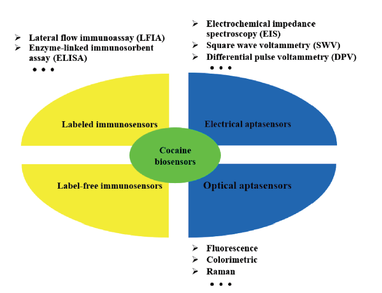
Cocaine has become one of the most dangerous and illicitly abused drugs today due to the adverse effects of long-term cocaine abuse, such as arrhythmia, myocardial infarction, stroke, hypertension and aortic stiffness. Traditional cocaine chromatographic analysis methods have disadvantages such as time-consuming, cumbersome sample processing and complicated operations. Therefore, improving cocaine detection methods has a certain positive impact on crime-fighting and medicine-developing. Due to the accuracy and portability of biosensors, immunological and aptamer technologies for specific capture of targets have become an important direction for cocaine detection. In this review, different types of cocaine biosensors in recent years are mainly described, covering the research progress of cocaine detection based on electrochemical, fluorescence, colorimetric and other methods. The immuno- and aptamer-based biosensors of cocaine are reviewed, the advantages, disadvantages and development directions of cocaine sensors are summarized.

Contents

1 Introduction

2 Immunosensors for cocaine detection

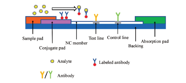
2.1 Labeled immunosensors

2.2 Label-free immunosensors

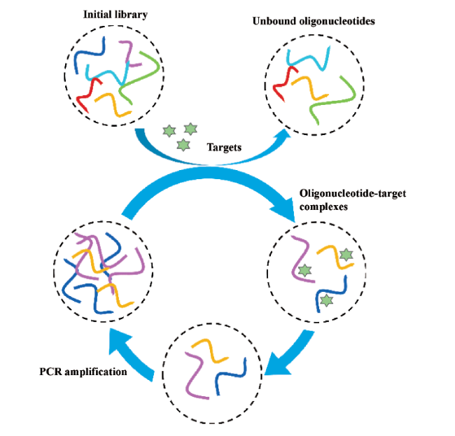
3 Aptasensors for cocaine detection

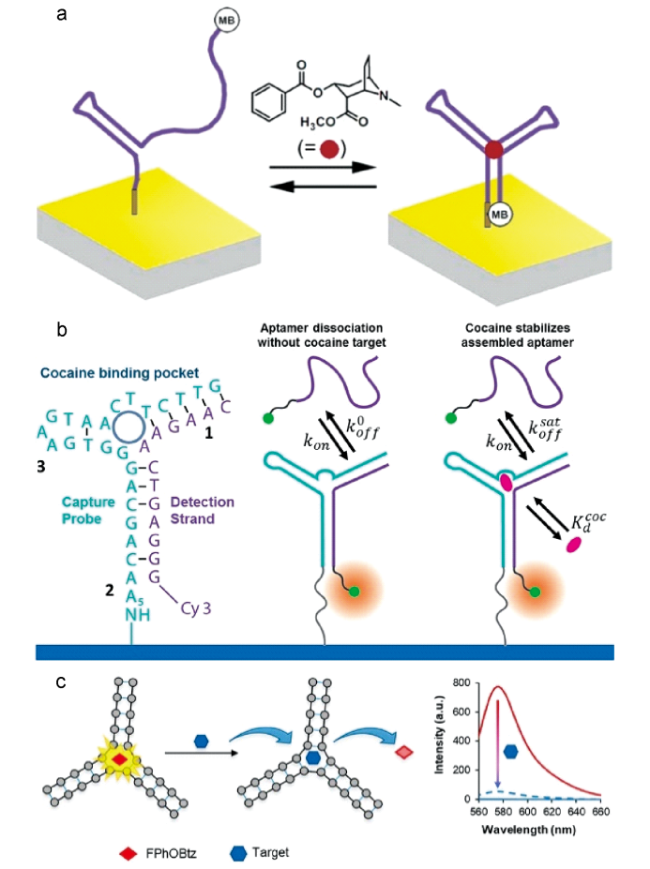
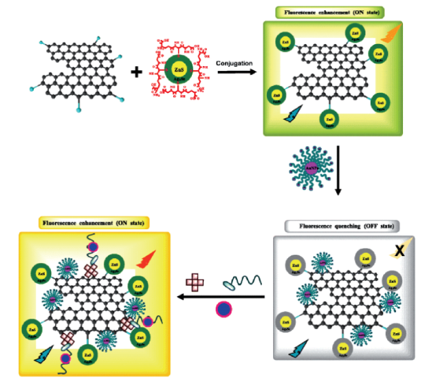
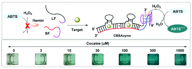
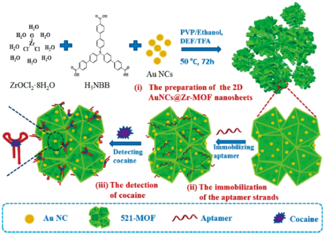
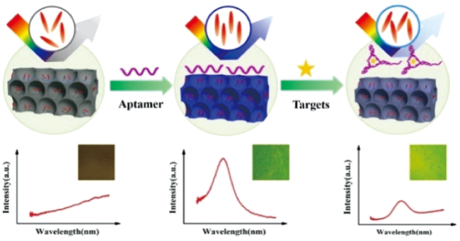
3.1 Fluorescent aptasensors

3.2 Colorimetric aptasensors

3.3 Electrochemical aptasensors

3.4 Other aptasensors

4 Conclusion and outlook

Key words: cocaine; immunity; aptamer; biosensor

Cite this article

Gehui Chen , Nan Ma , Shuaibing Yu , Jiao Wang , Jinming Kong , Xueji Zhang . Immunity and Aptamer Biosensors for Cocaine Detection[J]. Progress in Chemistry, 2023 , 35(5) : 757 -770 . DOI: 10.7536/PC220916

1 Introduction

Cocaine, a naturally occurring alkaloid, is a common addictive stimulant drug that increases dopamine levels in the brain's reward circuitry[1]. In 1860, W. Wöhler and Niemann extracted from Coca leaves a crystalline alkaloid called cocaine, whose stimulant effect on the central nervous system led to the abuse of the drug[2]. Since 1985, cocaine has become one of the major drugs in the world.
Cocaine can lead to a state of deep addiction characterized by compulsion to the use of the drug, which has become one of the most dangerous and illegally abused drugs today[3]. Long-term abuse of these narcotic drugs can have adverse effects on the human body, such as arrhythmia, myocardial infarction, stroke, hypertension, aortic stiffness and so on[4]. According to the United Nations Office on Drugs and Crime's World Drug Report 2022, approximately 21.5 million people have used cocaine at least once, and cocaine production, purity, and seizures have increased in recent years. Therefore, how to detect cocaine conveniently and rapidly in the fields of forensic and clinical medicine is an important task, and the screening technology with good sensitivity and rapid detection for the analysis of cocaine has become a public concern[5].
In previous studies, different chromatographic methods have been used to detect cocaine in samples: Gas chromatography-flame ionisation detection (GC-FID), Liquid chromatography-tandem mass spectrometry (LC-MS/MS), and Gas chromatography-mass spectrometry (GC-MS)[6][7][8]. Chromatographic analysis requires prior extraction of cocaine in organic solvents followed by some form of clean-up, preconcentration, and purification of the extract, and GC-MS also requires sophisticated chemical derivatization procedures for cocaine molecules[9]. Although these methods are highly reliable and accurate for cocaine detection, they require specialized equipment to pretreat the sample and take a long time from sample processing to detection. The detection process of chromatographic analysis is long and the cost is high, and the traditional cocaine analysis method is difficult to meet the needs of field testing in some scenarios[10].
The biosensor, as an analytical device, consists of two basic modules: target recognition and signal transduction. After the biological recognition element recognizes the target, the characteristics of the recognition element change. The transducer then converts the identified change into a signal that can be read[11]. An immunobiosensor uses an antibody as a target recognition element, while an aptamer as a recognition element is called an aptamer sensor. In addition to these two recognition elements, there are also studies using microorganisms and enzymes as recognition elements to detect cocaine (or cocaine metabolites)[12][13]. Compared with immune and aptamer, these biosensors have more stringent reaction conditions, higher cost and are not easy to preserve, so there are few studies on the detection of cocaine.
In recent years, new cocaine biosensors based on immune and aptamer recognition elements have become the mainstream, not only combined with signal amplification technologies such as nanomaterials, but also introduced new signal transducers into cocaine detection. Compared with the traditional cocaine analysis, the new sensor has significant advantages such as high specificity, short analysis time, low cost, and no need for complex sample processing[14]. In this review, we systematically introduce various novel strategies for cocaine detection based on immune biosensors and aptamer biosensors in recent years.

2 Immunosensor for cocaine detection

The working principle of the immunosensor is that the specific recognition between the antibody and the antigen causes a measurable signal change to realize the quantitative detection of cocaine[15]. The immunoaffinity recognition between antibody and antigen facilitates the specificity and sensitivity of the assay, making it ideal for the detection of low concentrations of molecules in complex samples. Immunosensors can be divided into labeled immunosensors and non-labeled immunosensors according to the presence or absence of labels (Table 1).
表1 不同可卡因免疫传感器及其检出限对比

Table 1 A comparison of different cocaine immunosensors and their limits of detection

Approach of detection Used sample Linear detection range (mol/L) Limit of detection (mol/L) ref
Electrochemical-based ELISA Water/Saliva/Urine 4.95×10⁻¹³ 19
Colorimetric Immuno-microarray Oral fluids 3.63×10⁻⁹~9.9×10⁻⁷ 3.63×10⁻⁹ 21
LFIA Urine 1.65×10⁻⁸~1.65×10⁻⁶ 1.65×10⁻⁸ 26
LFIA Saliva 1.65×10⁻⁸~3.30×10⁻⁶ 1.62×10⁻⁹ 27
Electrochemical Urine/Sweat/Saliva/Serum 1.65×10⁻⁸~8.25×10⁻⁷ 1.19×10⁻⁸ 30
Fluorescence PBS buffer 2.30×10⁻¹¹ 31
Electrochemical PBS buffer 0.50×10⁻⁶~2.50×10⁻⁵ 34
SHG PBS buffer 7.5×10⁻¹¹ 35

2.1 Labeled immunosensor

Techniques such as Enzyme-linked immunosorbent assay (ELISA) and nanoparticle-based Lateral flow immunoassay (LFIA) have been widely used in immunoassays for the detection of cocaine.

2.1.1 Enzyme-linked immunosorbent assay

ELISA is a typical laboratory assay that can achieve high-throughput specific detection of small molecules. The traditional ELISA method not only requires a complex operation process and expensive equipment, but also has a nanomolar detection limit, which limits its application in different fields[16]. ELISA usually consists of four main parts, namely, a solid substrate, a recognition element, a signal amplification part, and a readout mode. The characteristics of these parts determine the performance of the ELISA, and all emerging ELISA-derived technologies improve at least one of these four ELISA parts[17].
Abdelshafi et al. Used antibody IP3G2 as the recognition element of cocaine, and developed an analytical strategy for the detection of cocaine on Euro banknotes by competitive ELISA based on the principle that hapten-horseradish peroxidase conjugate competes with cocaine for binding to enzyme-labeled antibody[18]. Through cross-reactivity studies, they obtained that antibody IP3G2 has a higher affinity for cocaine than cocaine metabolites. Sixty-five euro banknotes obtained from different parts of Berlin were analyzed for cocaine, and the frequency of cocaine contamination detected was 100%.
Due to the high affinity of antibody IP3G2 for cocaine, Abdelshafi et al. Utilized antibody IP3G2-grafted magnetic beads (Ab-MBs) to capture cocaine and integrated competitive ELISA into a microfluidic electrochemical sensor[19]. The use of MBs as a solid substrate for ELISA facilitates the separation of antigen-antibody complexes and avoids the repeated elution steps in the traditional ELISA process. Therefore, MBs-based ELISA is more sensitive and easier to automate than standard plate-based ELISA[20].
Zhang et al. Designed an immune microchip for the quantitative detection of a variety of illicit drugs (cocaine, morphine, amphetamine) in body fluids[21]. Firstly, the cocaine-bovine serum albumin complex is immobilized on the chip through the amidation reaction of amino and carboxyl, and the complex can compete with the analyte for binding to the labeled antibody in the solution. Drug identification was performed "at once" by using three different colorimetric reactions, observing the different colors of the drug as well as the location. The colorimetric immunoassay microchip can analyze multiple analytes at the same time, which enhances the potential of ELISA in practical detection and field analysis.

2.1.2 Lateral flow immunoassay

In the late 1960s, LFIA emerged as a low-cost, rapid, and simple assay for the detection of small molecules in Point-of-care (POCT) Point of care testing[22]. Compared with ELISA, LFIA is more suitable for field detection outside the laboratory because of its small size, easy to carry and convenient signal reading.
As shown in Fig. 1, the LFIA test strip usually consists of a back pad, a nitrocellulose membrane (NC membrane), a sample pad, a conjugate pad, and an absorbent pad. Wherein the back pad provides a certain mechanical support for the detection device. The NC membrane provides a platform for capture and recognition in the process of target detection. The capture antibody can be immobilized on the NC membrane through electrostatic interaction, hydrogen bonding or hydrophobic force to form a test line (T line) and a control line (C line). The binding pad is where the labeled antibody attaches. The absorbent pad provides a driving force based on the capillary effect, which helps to maintain the flow of liquid on the membrane, and can also suck away excess reagent and prevent the sample from flowing backwards. LFIA is divided into two forms: sandwich form and competition form. In that sandwich form, the label antibody is released immediately upon contact of the bin pad with the target, forming an antigen-antibody complex. When the complex flows through the T line modified with the capture antibody, the analyte is sandwiched between the two antibodies to form a sandwich structure, and the label on the T line is aggregated, which is a positive result. Labeled antibodies that are not bound to the target cross the T line and are recognized and bound by another type of capture molecule on the C line[23]. On the contrary, if there is no target in the sample, the antibody on the T line can not capture the marker, and the result is negative. In the competitive form of LFIA, the target competes with the hapten immobilized on the T-line for binding to the binding site of the labeled antibody. When the target analyte does not have multiple antigenic sites, a competitive form of LFIA detection is used[24]. Based on the high binding efficiency of antigen and antibody, LFIA can provide qualitative or quantitative detection of a small number of samples under limited conditions[25].
图1 夹心式LFIA测试条的示意图

Fig. 1 Schematic representation of sandwich-type LFIA test strip

Because the traditional LFIA method is not easy to provide quantitative detection of analytes, Wu et al. Combined the competitive form of LFIA with smartphone image processing technology to develop a low-cost and convenient quantitative analysis strategy for cocaine[26]. The modified carboxyl groups on the Magnetic beads are activated and then covalently bind to the amino groups of the cocaine antibody to form a Magnetic bead-antibody (MBs-Ab) complex. The free cocaine (CC) in the sample competes with the immobilized Bovine serum album in-cocaine (BSA-CC) on the T line for binding to the recognition site of MBs-Ab. The less the cocaine content in the test solution, the more MBs-Ab accumulated on the T line, showing a darker color. When no cocaine was present in the sample solution, MBs-Ab reacted completely with BSA-CC on the T line and was the darkest. After the sample flows through the T line, excess MBs-Ab or MBs-Ab-CC can react with goat anti-mouse IgG immobilized on the C line to form a brown band. The quantitative detection of cocaine was achieved by converting the color intensity on the T and C lines combined with MBs into digital signals through the image capture software installed in the smartphone. Ghorbanizamani et al. Chose rhodamine B coated with amphoteric polymer hollow spheres as the marker of antibody, and used smartphone-assisted imaging to achieve competitive LFIA quantitative analysis of cocaine in saliva[27]. Due to the amphiphilic nature of the polymer membrane, it can be used to capture a variety of hydrophilic or hydrophobic marker molecules. The use of the polymer not only stabilizes the marker molecules, but also promotes the binding and functionalization between molecules.

2.1.3 Other immunosensors

Most voltammetric measurement devices use a three-electrode system: working electrode, reference electrode, and counter electrode. Screen-printed electrode (SPE) can be used to print a three-electrode system on the same substrate. By adding the solution to be detected on the surface of SPE, multiple electrodes can be covered to directly detect the analyte. It is usually suitable for the detection in trace solutions[28]. SPE technology can be used for low-cost, one-time, large-scale production of volt-ampere sensors, and has the advantages of fast response, low power, convenience and so on[29]. Sanli et al. Used cobalt oxide as the electroactive label of antibody and SPE as the detection platform, and proposed a biosensor for cocaine detection by Differential pulse voltammetry (DPV), which realized the quantitative detection of cocaine in the Differential pulse voltammetry range[30].
Paul et al. Combined the fluorescently labeled antibody IP3G2 with a competitive form of immunomicrofluidics[31]. The mixture of the labeled antibody and the sample to be tested is injected into the affinity column, on which a large amount of hapten (benzoylecgonine) is immobilized. In the presence of cocaine, the binding sites of some labeled antibodies are blocked, and the amount of labeled antibodies captured on the affinity column is reduced. Finally, the content of cocaine is analyzed by the change of fluorescence signal in the fluid.

2.2 Unlabeled immunosensor

It is considered that a high mass labeled antibody may affect the recognition reaction kinetics with the target, resulting in inaccurate quantification of the analyte[32]. In addition to immobilizing the label on the antibody and indicating the detection signal by labeling the antibody to recognize the antigen, the immune biosensor can also detect the target by label-free method[33]. Sengel et al. Used a glassy carbon electrode modified with electroactive monomer as a detection platform to detect the change of electrical signal before and after the addition of sample in [Fe(CN)6]3-/4- solution[34]. This signal change is produced because the binding of cocaine to the antibody prevents electron transfer through the sensor. The conjugated polymer technology added in the sensor design makes the immobilization of antibodies more advantageous and also improves the sensitivity of the sensor. Tran et al. Realized a label-free immunoassay for the direct detection of cocaine based on optical Second harmonic (SHG) technology, which reached the detection limit of 7.5×10⁻¹¹mol/L (in the presence of interference), and the method has higher sensitivity and selectivity compared with the traditional competitive ELISA (detection limit of 1×10⁻⁹mol/L)[35].

3 Aptamer sensor for detection of cocaine

Aptamers are non-naturally occurring structured oligonucleotides that have similar specificity and affinity for their targets as antibodies. Aptamers spontaneously fold into unique binding structures (including secondary and tertiary structures) through intrastrand or interstrand base pairing, base stacking interactions, van der Waals forces, etc. Under the action of these forces, aptamers form secondary structures such as hairpins and convex rings as the basis of binding structures, or further fold into compact tertiary structures, which have the ability to bind to targets[36]. These nucleic acid molecules are screened in vitro from a random oligonucleotide library in a process called Systematic evolution of ligands by exponential enrichment (SELEX)[37]. The process of SELEX includes iterative steps of oligonucleotide library construction, target binding to the nucleic acid strand, elution of the nucleic acid strand, and amplification into the library (fig. 2). The initial oligonucleotide library contains 1014~1016 single-stranded DNA or RNA molecules, which contain a central random sequence and two constant primer binding sites at the 5 'and 3' ends for subsequent strand amplification. Targets were incubated with the random library under certain conditions, and sequences not bound to the target were discarded. The target-bound nucleic acid strand is eluted from the target, amplified, and a single-stranded pool is constructed for the next round of affinity screening. In the final round, the final selected nucleic acid strand is sequenced and characterized for avidity of binding to the target.
图2 SELEX流程的示意图

Fig. 2 Schematic illustration of SELEX procedure

Since the first aptamer was discovered in 1990, aptamers have been used in the detection of proteins, ATP, viruses, illegal drugs and other substances, because aptamers can usually provide a clear three-dimensional structure in the process of recognizing target molecules[38][39][40][41][42]. Compared with antibodies, aptamers have the advantages of larger binding target range, more flexible structure, lower synthesis cost, easier chemical modification or labeling of nanomaterials[43]. Based on these characteristics, aptamer sensors have become a rapidly developing direction in the field of biochemical analysis[44].
In general, the interaction of aptamers with cocaine involves an unfolding-folding mechanism: aptamers form a Three-way junction (3 WJ) only in the presence of cocaine[45]. Cocaine aptamers can be roughly divided into three categories according to the number of aptamer fragments. The first class of individual aptamer chains, called Monolithic aptamers (MAs), whose detection relies primarily on conformational changes of the aptamer (Figure 3A)[46]. The second type is Double-fragment aptamers (DFAs), in which MAs are generally cut into two pieces, a capture probe and a reporter probe[47]. The increased flexibility of probe modification due to the physical separation of the two strands opens up new detection pathways for aptamer sensors (Figure 3B). The third type is Triple-fragment aptamers (TFAs), which can form a three-way aptamer-cocaine complex only when the three-fragment aptamers are present with cocaine[48]. Compared with DFAs, TFAs have more open terminals available for labeling, which provides more options for probe design and optimization (Figure 3C). To improve the detection system, aptamers can be chemically modified and labeled with a reporter molecule without affecting the affinity of the target molecule[49]. These three types of probes are used to specifically capture cocaine, and then the aptamer is labeled by metal nanoparticles, enzymes, electroactive molecules, fluorophores, semiconductor quantum dots, etc., to convert the recognition reaction between cocaine and aptamer into a detectable signal response[50~53].
图3 (a)亚甲基蓝标记的MAs可卡因传感器[46];(b)DFAs与可卡因的组装过程[47];(c)基于TFAs的荧光传感器[48]

Fig. 3 (a) Methylene blue-labeled MAs cocaine biosensor[46]; (b) DFAs and cocaine assembly process[47]; (c) Fluorescent biosensor based on TFAs[48]

According to the type of label and detection method, cocaine aptamer sensors are mainly divided into two categories: optical and electrochemical detection of cocaine. Commonly used optical methods include fluorescence, colorimetry, electrochemiluminescence, Surface-enhanced raman spectroscopy (SERS) and other detection methods[54~57]. The conformational change of the aptamer before and after cocaine binding leads to different emission characteristics of the reaction system, so the content of cocaine can be analyzed according to the change of light signal. Electrochemical methods include Electrochemical impedance spectroscopy (Electrochemical impedance spectroscopy, EIS), Square wave Voltammetry (Square wave Voltammetry, SWV), DPV, etc., and usually the aptamer is coupled with a signal conversion element (electrode)[58]. Table 2 provides a comparison of the detection limits of cocaine sensors based on different aptamers, depending on the detection method.
表2 不同可卡因适配体传感器及其检出限对比

Table 2 A comparison of different cocaine aptasensors and their detection limits

Method Linear range (mol/L) Detection limit (mol/L) ref
Fluorescence 5×10⁻⁶ 62
Fluorescence anisotropy 63
Fluorescence 0~1×10⁻⁵ 5×10⁻⁸ (in 10% saliva) 64
Fluorescence 5×10⁻¹⁰~8×10⁻⁸ 8.4×10⁻¹¹ 65
Fluorescence 0~1×10⁻¹⁰ 5.4×10⁻¹³ 66
Cas-12a based fluorescence 4.7×10⁻⁷~1.5×10⁻² 3.4×10⁻⁷ 67
EWF-based fluorescence 1×10⁻⁵~5×10⁻³ 1.05×10⁻⁵ 68
Fluorescence 1×10⁻⁶~5×10⁻⁴ 2.5×10⁻⁷ 69
Fluorescence 1×10⁻⁷~1×10⁻⁴ 4.6×10⁻⁹ 72
Fluorescence 1×10⁻⁸~1×10⁻⁴ 8×10⁻¹⁰ 73
Colorimetric 8.25×10⁻⁹ mol (visual)
7.79×10⁻⁹ mol (camera)
75
Colorimetric 2×10⁻¹⁰~2.5×10⁻⁸ 9.7×10⁻¹⁰ 76
Colorimetric 1.32×10⁻⁸ mol (visual)
1.17×10⁻⁸ mol (camera)
77
Colorimetric 0~1×10⁻⁶ 7.49×10⁻⁹ 78
Colorimetric 1×10⁻⁹~1.5×10⁻⁷ 5×10⁻¹⁰ 79
Colorimetric 1×10⁻⁸~1.5×10⁻⁷ 3.3×10⁻⁹ 80
Colorimetric 2×10⁻⁹~1×10⁻⁷ 4.4×10⁻¹⁰ 81
Colorimetric 1×10⁻⁵ 82
Colorimetric 1×10⁻⁵~5×10⁻³ 5×10⁻⁵ (in urine)
2×10⁻⁴ (in sweat)
83
SWV 5×10⁻⁸~1×10⁻⁶ and 1×10⁻⁶~3.5×10⁻⁵ 2.1×10⁻⁸ 86
SWV 87
EIS/DPV 3.3×10⁻¹²~3.3×10⁻⁹ 1.29×10⁻¹² (EIS)
2.22×10⁻¹² (DPV)
89
EIS 1×10-15~1×10-12 and 1×10-12~1×10⁻⁷ 3.33×10⁻¹⁶ 90
EIS 9×10-11~8.5×10⁻⁸ 2.9×10⁻¹¹ 91
DPV 3.3×10⁻¹⁰~3.3×10⁻⁵ 1×10⁻¹⁰ 92
SWV 3.3×10⁻¹¹~3.3×10⁻⁶ 9×10-12 93
DPV 1×10-11~7×10-11 2.6×10⁻¹³ 94
DPV 4×10-11~1.5×10⁻⁷ 1.5×10⁻¹¹ 95
EMPAS 2×10⁻⁶~5×10⁻⁵ 9×10⁻⁷ 96
EMPAS 5×10⁻⁷~5×10⁻⁶ 3×10⁻⁷ 97
Interfacial capacitance sensing 1.45×10⁻¹⁴~1.45×10⁻¹¹ 7.8×10⁻¹⁵ 98
FET 1×10⁻⁹ 99
Conductance change 1×10⁻⁹~1×10⁻⁵ 1×10⁻⁹ 102
α-HL nanopore 5×10⁻⁸~1×10⁻⁴ 5×10⁻⁸ 103
Personal glucometer 1×10⁻⁸~6×10⁻⁷ 5.2×10⁻⁹ 104
LC optical sensor 1×10⁻⁹~1×10⁻⁵ 1×10⁻⁹ 106
LC optical sensor 1×10⁻¹⁰~1×10⁻⁵ 108
LPFG 2.5×10⁻⁵~7.5×10⁻⁵ 2.5×10⁻⁵ 109
PIERS 5×10⁻⁹~1×10⁻⁵ 5×10⁻⁹ 110
ECL 1×10⁻¹⁰~1×10⁻⁷ 6×10-11 111

3.1 Fluorescent aptamer sensor

Fluorescence method has the advantages of flexible quantitative analysis method and wide reaction range. Since Stokes introduced the concept of "fluorescence" in 1852, many properties of fluorescent materials have been developed and applied to the detection of chemical or biological molecules[59].
The rotation rate of a fluorescent molecule in solution is affected by the intrinsic properties of the molecule (such as molecular volume and shape) and environmental factors (such as temperature and viscosity of the solution)[60]. Since MAs undergo conformational changes upon binding cocaine, this change in aptamer conformation has been implemented into cocaine sensors based on altering the local environment of the fluorophore to affect fluorescence emission.
When a Fluorescence anisotropy (FA) labeled fluorophore binds to a ligand to form a complex, the overall size of the fluorescent molecule increases, the rotation rate decreases, the polarization of the fluorophore increases, and the fluorescence emission characteristics change[61]. Liu et al. Compared the conformational changes of TMR molecular modification on different binding sites of MAs and the FA response of different binding sites to TMR by using the characteristic that the binding of Tetramethylrhodamine (TMR) to G base will restrict the local rotation of TMR[62]. The cocaine sensor designed by Billet et al., in the presence of cocaine, the anionic fluorescent dye repels the negatively charged DNA, making the dye rotate almost independently of the entire DNA, showing sensitivity to changes in local electrostatic potential[63]. Cocaine can be quantified according to the readout of dye anisotropy.
Since cocaine can induce a combination of DFAs fragments, a strategy for the detection of cocaine by DFAs in combination with fluorescence-quenching pairs has been developed. Yu et al. Integrated two target binding domains in series and designed a Cooperative binding split aptamer (CBSA) for cocaine, that is, one CBSA contains two cocaine binding domains[64]. Targeted binding on one domain may stabilize the structure of CBSA, facilitating the binding of the second binding domain to cocaine, exhibiting higher target binding affinity. They proposed two fluorescence quenching methods and CBSA in combination for the detection of cocaine. One is to insert a base empty site that can bind to the fluorophore 2-Amino-5,6,7-trimethyl-1,8-naphthyridine (ATMND) at the junction between the two binding domains of the aptamer. After cocaine induces the combination of two aptamers, ATMND binds to the empty site, and the fluorescence of ATMND is quenched. The other is to modify the fluorophore and quencher at the end of the two aptamer chains, respectively. After the aptamer binds to cocaine, the fluorophore is close to the quencher, and fluorescence quenching occurs. The sensor can detect 10% of saliva in 15 minutes. Compared with DFAs with a single binding domain (with a detection limit of 30 nM in 0.5% saliva), the thermal stability and selectivity of the aptamer in this strategy have been greatly improved. Abnous et al. Modified two pairs of fluorescence-quenching groups at the end of cocaine DFAs respectively, and detected cocaine according to the change of the relative fluorescence intensity of the two fluorescent labels[65].
Gao et al. Used molybdenum disulfide (MoS2@AuNPs) composite modified by gold nanoparticles as a substrate, and the capture probe of DFAs was anchored on the substrate through the interaction of bis-thiol and gold[66]. Because MoS2 not only has good biocompatibility, but also has high fluorescence quenching ability, the introduction of MoS2 can well solve the problems of non-specific adsorption of fluorescent reporter probe and false positive signal in fluorescent sensors. Moreover, the dithiol group improves the binding capacity of the capture probe and the MoS2@AuNPs, and realizes the high-sensitivity detection of cocaine with a detection limit of 5.4×10⁻¹³mol/L.
Because the binding avidity of cocaine to MAs is greater than that of MAs to its complementary sequence, a cocaine fluorescent sensor based on cocaine competing with complementary DNA for binding to MAs was developed. Zhao et al. Used the single-stranded DNA (ssDNA) -cleaving enzyme activity of CRISPR-Cas12a (Clustered regularly interspaced short palindromic repeat-associated protein) to achieve a wide range of cocaine detection within the Clustered regularly interspaced short palindromic repeat[67]. In the presence of cocaine, the activity of CRISPR-Cas12a was activated, the ssDNA between the fluorophore and the quencher was cleaved, and the fluorescence signal was significantly enhanced. The sensor was used to detect cocaine in human serum and urine, and the results were consistent with those of High-performance liquid chromatography (HPLC). Qiu et al. Designed an Evanescent wave fiber-optic (EWF) sensor based on MAs for cocaine detection[68]. MAs is anchored to the magnetic beads via biotin-streptavidin interaction, and in the absence of cocaine, a fluorophore-labeled short DNA reporter probe base-paired with MAs is bound to the magnetic beads. After the introduction of cocaine, the short DNA strands are released by competition, and the fluorescently labeled short DNA strands released in the supernatant are specifically detected by the evanescent wave platform.
Wu et al. Used Thioflavin T (ThT) as a fluorescent indicator to compete with cocaine for the binding domain of MAs[69]. When ThT is bound in the cavity provided by the aptamer, a significant fluorescence signal is generated. The presence of cocaine reduces the amount of aptamer-binding fluorescent indicator and the intensity of the fluorescent signal diminishes. The method does not require modification of the aptamer, and the detection is fast, with the mixing and detection steps completed within seconds.
Nanomaterials have unique chemical, optical and mechanical properties, and can be coupled with different biomolecules through electrostatic binding, physical adsorption, biological recognition and covalent binding[70]. Based ON the ability of Quantum dots (QDs) to provide fluorescence signals, this strategy of using Localised surface plasmon resonance (LSPR) -mediated QDs fluorescence intensity enhancement to achieve fluorescence detection of targets has become a powerful tool for the fabrication of highly sensitive fluorescent OFF-on nanoprobes[71]. Using this strategy, Adegoke et al. Synthesized a nanoassembly of cetyltrimethylammonium bromide-AuNPs-graphene-QDs, and the fluorescence of QDs was quenched[72]. As cocaine was added to the reaction system, the nanoassembly of AuNPs and QDs was opened, the LSPR-induced signal of AuNPs was excited, and the fluorescence signal of QDs was enhanced (Fig. 4). Burton et al. Also used the principle of target-induced fluorescence enhancement by the combination of plasmonic nanoparticles and QDs to adsorb thiolated MAs on the surface of QDs-AuNPs to achieve the quantitative detection of cocaine[73].
图4 基于AuNPs-QD组合体荧光检测可卡因的示意图[72]

Fig. 4 Schematic diagram of fluorescence detection of cocaine based on AuNPs-QD combination[72]

3.2 Colorimetric aptamer sensor

In various analysis methods, the colorimetric sensor has a visual or spectral change depending on the color before and after the addition of the target. The strategy of colorimetric analysis of cocaine has attracted attention because the results are easy to operate and can even be observed directly by the naked eye[74].
The distance change of metal nanoparticles can affect the optical properties of nanoparticles, which is an ideal marker for colorimetry. Because the high concentration of salt will destroy the charge distribution on the surface of gold nanoparticles, it can act as an aggregation inducer of AuNPs, so that the gold nanoparticles are in an aggregation situation, and the color changes from red to blue-black. At present, new biosensors based on AuNPs and MAs have been used in the colorimetric detection of cocaine. Wang et al. And Sanli et al. Designed cocaine colorimetric sensors with salt-induced AuNPs aggregation on paper microfluidic devices and 96-well plates, respectively[75][76]. Due to the protection of cocaine MAs adsorbed on the surface of AuNPs, AuNPs did not aggregate in high concentration salt solution. However, when the aptamer was targeted to bind cocaine, the protection of the aptamer on the surface of AuNPs was reduced, and the AuNPs aggregated and showed color changes. A four-channel paper microfluidic device was then developed to simultaneously detect cocaine, codeine, and methamphetamine in a sample[77]. Gao et al. Improved the sensitivity of the AuNPs-MAs colorimetric sensing system for cocaine detection by taking advantage of the large specific surface area and good conductivity of MoS2[78]. In the absence of cocaine, the aptamer was temporarily stored on MoS2, and only part of the AuNPs did not aggregate. After MAs combined with cocaine, they left the MoS2, enhanced the salt tolerance of AuNPs, and effectively prevented the aggregation of AuNPs.
Mao et al. Synthesized a silver-coated gold nanoparticle (Au @ AgNPs) as a colorimetric marker for the sensor[79]. In the absence of cocaine, cocaine MAs linked Au @ AgNPs with magnetic beads through base complementary pairing with complementary strands, forming a magnetic bead-MAs-Au @ AgNPs conjugate. After magnetic separation of the solution, the complex was separated, and the content of Au @ AgNPs in the supernatant was also reduced. In contrast, when cocaine was present, the MAs were disconnected from the complementary strand, and part of the Au @ AgNPs were no longer coupled to the magnetic beads, resulting in a darker supernatant after magnetic separation than without the target. The content of cocaine can be obtained by measuring the absorbance, and the detection limit is 5×10⁻¹⁰mol/L. On this basis, they designed colorimetric sensors that can simultaneously detect two analytes, methamphetamine and cocaine, using AuNPs and Au @ AgNPs as tags, respectively[80]. The average recoveries of methamphetamine and cocaine in spiked wastewater samples were 85. 5% and 83. 9%, respectively.
Abnous et al. Anchored the cocaine capture probe on the surface of AuNPs through the assembly of Au and sulfhydryl groups, and used the catalytic activity of AuNPs to detect cocaine[81]. The AuNPs lost catalytic activity under the protection of TFAs-cocaine complex. In the absence of cocaine, AuNPs catalyzed the reduction of 4-nitrophenol, and the color of the sample changed from yellow to colorless. This strategy uses nanomaterials instead of enzyme catalysis, which not only avoids the high cost of enzyme purification, preparation and storage, but also maintains a relatively stable catalytic activity. This is particularly important for the long-distance transportation of sensors and the detection of targets in harsh environments. Similarly, instead of enzyme catalysis, Luo et al. Used a new multi-modular split DNA structure (called CBSAzymes) to mimic catalase catalysis (Fig. 5)[82]. CBSAzymes are composed of two blocks, which are separated in the absence of a target and can be effectively combined in the presence of the target to form a complex that can catalyze the oxidation of 2,2 '-azinobis (3-ethylbenzthiazoline-6-sulfonic acid) diammonium salt. The colorimetric sensor is based on the detection method of CBSAzymes, which has the advantages of simplicity, no label, rapidity and no instrument, and has great potential in the field detection of cocaine.
图5 基于CBSAzymes的检测可卡因的原理图[82]

Fig. 5 Schematic diagram of cocaine detection based on CBSAzymes[82]

Jing et al. Used AuNPs as a colorimetric marker to detect cocaine by lateral flow test paper[83]. The spontaneous assembly of the capture probe-cocaine-recognition probe and the specific recognition between streptavidin and biotin were used to immobilize the AuNPs-DNA complex in the T and C regions of the test strip, showing a red line. This sandwich lateral flow strip method can provide immediate detection of cocaine in urine and sweat samples within 15 min, with a naked eye detection limit as low as 1×10⁻⁵mol/L.

3.3 Electrochemical aptamer sensor

The electrochemical aptamer sensor consists of three parts: (1) a conductive platform, which not only serves as an interface for electrochemical reactions, but also provides a suitable platform for the immobilization of recognition elements; (2) recognition element, electrochemical aptamer sensor is an electroanalytical biosensor with aptamer as the biological recognition element; (3) Signal readout device. After the analyte is combined with the recognition element, the corresponding electrochemical signal changes on the sensor can be recorded by EIS, SWV, DPV and other technologies. Compared with optical aptamer sensors, electrochemical aptamer sensors stand out in the rapid detection of small molecules because of their advantages of low cost, reusability and high sensitivity[84].
Many studies have been based on the electrochemical strategy of changing the electrical signal caused by the conformational change of MAs after binding to cocaine. Specifically, when MAs are not combined with cocaine, they are in a partially folded, more extended state. After binding to cocaine, the aptamer is in a fully folded compact state, which makes the electroactive label at the end of the aptamer closer to the electrode surface, thus promoting the electron transfer between the electroactive label and the electrode and causing changes in the electrochemical signal[85]. However, this signal change is relatively weak, and the electrochemical detection technology using various nanomaterials and nanostructures (such as metal nanoparticles, conjugated polymers, etc.) To modify the conductive platform to achieve signal amplification can further enhance the sensitivity of the sensing platform. Tavakkoli et al. Prepared a nanoporous gold electrode by anodizing a gold electrode and using ascorbic acid as a reducing agent. MAs with electroactive tags can be immobilized on the electrode surface through Au-S bonds[86]. In the presence of cocaine, the conformational change of the aptamer shortens the distance between the electroactive label and the electrode surface, the electron transfer efficiency of the electroactive label increases, and the electrical signal changes. The gold nanoparticles coated on the electrode surface increase the overall electrode surface area and provide a reaction plane for aptamer immobilization, reduce the overall electrode impedance, and improve the electrode conductivity and sensor sensitivity. Similarly, Taylor et al. Used Methylene blue (MB) as an electroactive marker and an implantable silicon-based nerve recording probe to achieve real-time detection of cocaine in vivo[87]. The area of available gold on the electrode surface was increased by electrodepositing dendritic gold on the silicon-based electrode. The sensor can measure direct local cocaine injection in mice with high time resolution within 2 H after implantation into the mouse brain.
In order to explore whether the anchoring direction of the aptamer affects the sensor performance, Chamorro-Garcia et al. Used MB as the electrical signal tag to study the effect of different MAs aptamer coupling directions on the sensor electrical signal based on the conformational change of MAs[88]. The detection performance of the sensor with the 5 ′ end of the aptamer and the 3 ′ end anchored on the electrode was compared, and it was found that the former showed greater signal gain. DNA strands anchored to the electrode surface using the 5 ′ end are more vertical than those anchored at the 3 ′ end, resulting in faster electron transfer for the 5 ′ -end anchored sensor, possibly due to subtle differences in the specific geometric connections of the 3 ′ and 5 ′ -end hydroxyl groups. Although the conclusion is not universal, which may be related to the particularity of the aptamer structure in the study, it provides a new idea for optimizing the performance of electrochemical sensors and improving the sensitivity.
In addition to MAs terminal modification and electroactive labeling, $\left[\mathrm{Fe}(\mathrm{CN})_{6}^{3-/4-}\right]$ can also be used as a probe, and the binding of the aptamer and cocaine increases the impedance of the electrode surface, so that the obstruction of charge transfer can be observed in the $\left[\mathrm{Fe}(\mathrm{CN})_{6}^{3-/4-}\right]$ solution, thereby realizing the quantitative detection of cocaine. Using this principle, Su et al. Prepared zirconium-based metal-organic framework nanosheets embedded with gold nanoclusters (AuNCs @ Zr-MOF) as a sensor carrier, and a large number of cocaine MAs chains were immobilized on the nanosheets through the combination of Au and sulfhydryl groups (Fig. 6)[89]. After cocaine binding to MAs, the charge transfer resistance of the electroactive probe $\left[\mathrm{Fe}(\mathrm{CN})_{6}^{3-/4-}\right]$ on the electrode surface in solution changed. Roushani et al. Modified the surface of SPE with dendrimers and silver nanoparticles (AgNPs)[90]. Then AgNPs-MAs were attached to the electrode surface through the interaction with the amino group of the dendrimer, and the change of the charge transfer resistance was studied by recording the impedance response of the sensor. Hashemi et al. Combined magnetic nanoparticles, conductive polymers and gold nanoparticles as a sensor platform for anchoring aptamers[91]. The MAs were immobilized on the magnetic reduced graphene oxide/polyaniline/gold nanoparticle composite via Au — S bonds, and the external magnet could capture the magnetic composite on the working electrode. With the formation of target-aptamer complex, the structure of MAs changes, and the charge transfer resistance of the $\left[\mathrm{Fe}(\mathrm{CN})_{6}^{3-/4-}\right]$ in the electrolyte on the electrode surface increases, which can be used to analyze the content of cocaine through impedance response.
图6 AuNCs@Zr-MOF作为基底检测可卡因的示意图[89]

Fig. 6 Schematic diagram of AuNCs@Zr-MOF as a substrate to detect cocaine[89]

Indium tin oxide (ITO) glass is often used in electrochemical sensors due to its excellent conductivity, wide electrochemical working window and low cost. Wang et al. Modified the ITO electrode surface step by step: amino groups were introduced to the ITO surface by 3-aminopropyltriethoxysilane, and then the capture probes in DFAs were anchored to the electrode by amino crosslinking agents[92,93]. These two sensors introduced the living polymerization signal amplification technique into the detection of cocaine, which achieved signal amplification, and the detection limits reached 1.1×10⁻¹⁰ and 9.9×10⁻¹²mol/L, respectively. Azizi et al. Used physical vapor deposition to modify gold nanoparticles on ITO electrodes[94]. The AuNPs modified ITO electrode has a faster electron transport rate and a larger current response than the bare ITO electrode. The capture probe in DFAs was immobilized on the electrode surface by Au-S, and the thionine-modified carbon dots were used as the label of the reporter probe to provide redox signals. Under optimal conditions, this cocaine biosensor showed a dynamic range of 1×10-11~7×10-11mol/L with a detection limit as low as 2.6×10⁻¹³mol/L.
CRISPR-Cas has ultra-high specificity for target nucleic acid sequences, and Abnous et al. Have achieved highly sensitive and selective detection of cocaine by using the strategy of Terminal deoxynucleotidyl transferase (Terminal deoxynucleotidyl transferase, TdT) and CRISPR-Cas12a binding. When cocaine is absent, the 3 'end of the complementary strand of the aptamer is extended by TdT, which activates the trans-cleaving enzyme activity of CRISPR-Cas12a[95]. The target nucleic acid chain on the electrode surface was cleaved, and the charge transfer of $\left[\mathrm{Fe}(\mathrm{CN})_{6}^{3-/4-}\right]$ was not hindered, resulting in a significant enhancement of the electrochemical signal.

3.4 Other cocaine aptamer sensors

In addition to electrochemistry, fluorescence and colorimetry, various aptamer sensors for cocaine detection based on different detection platforms and signal generation principles have been developed, which are divided into electrical and optical parts.

3.4.1 Electricity

Electromagnetic piezoelectric acoustic sensor (EMPAS) provides a label-free sensing platform that can detect changes in mass loading and viscoelasticity at the liquid/sensor interface in real time. After cocaine binds to the aptamer, the structural change of the MAs aptamer causes a physical change on the sensor surface, and the quantitative detection of cocaine can be achieved without a label. Neves et al. Successively reported the detection of cocaine by cocaine aptamers MN4 and MN6 on the EMPAS platform[96,97]. Due to the difference in the binding mechanism of the two aptamers, it was found that the EMPAS sensor assay performance of MN6 (which undergoes a structural switch when bound to cocaine) was better than that of MN4. Oueslati et al. Designed a sensor to reflect the binding state of cocaine and aptamer by detecting the capacitance change of the MAs modified electrode interface[98]. The time from the determination of the sample to the detection result was 30 s, and the detection limit was 1.34×10⁻¹⁴mol/L in serum. Chen et al. Observed the structural changes of the aptamer in the process of binding to the target by real-time electrical signal recording using a Field-effect transistor (FET), and found that only the tertiary structure of the aptamer changed during the binding to cocaine[99]. Among them, the use of graphene provides excellent advantages in terms of high conductivity, stable sensing interface, good biocompatibility and easy device integration.
Inspired by biological ion channels, sensors based on nanochannel technology have been developed for the detection of small molecules[100,101]. The aptamer technology is combined with the nanochannel, and when the aptamer binds to the target under the applied potential, the aptamer will cause the transient change of the ionic current and the change of the electrically recorded current. Wang et al. Nanochannel as a platform for a cocaine resistive pulse sensor and immobilized DFAs on the nanochannel wall[102]. In the cocaine-bound state, the channel is partially or completely blocked, shielding the charge on the surface of the channel, and the transmembrane ionic current of the nanochannel is significantly blocked. Similar is the nanopore technique, where charged molecules induce a current change when they pass through a protein nanopore at an applied potential. Rauf et al. Designed a cocaine biosensor based on α-hemolysin nanopore and target-induced chain release strategy[103].
In addition, Li et al. Proposed a strategy for the detection of cocaine by a personal glucometer[104]. The sensor has two complementary strands of cocaine MAs grafted on TiO2 nanotubes (DNA1-TiNTA) and gold nanoparticles (DNA2-AuNP), respectively. The aptamer connects the complex formed by the DNA1-TiNTA and the DNA2-AuNP through complementary base pairing, and encapsulates the glucose molecule into the nanotube. When cocaine is present, the cocaine reacts with the aptamer and the DNA2-AuNP dissociates from the DNA1-TiNTA, thereby causing the glucose molecules in the nanotube to be released. The amount of glucose molecules released depends on the concentration of cocaine in the sample, enabling quantitative detection of cocaine.

3.4.2 Optics

Liquid crystals (LC) are a kind of optical materials with the mobility of Liquid and the order of solid at the same time, and have optical anisotropy similar to crystals[105]. Wang et al. Used amphiphilic 3-morpholinopropanesulfonic acid to establish a recognition site at the water/LC interface for the detection of cocaine[106]. The response mechanism of DNA-mediated water/LC biosensor is mainly caused by the hybridization reaction between DNA and its complementary sequence or the change of DNA conformation[107]. This change induces an anchoring transition of the LC molecule from the homeotropic orientation to the planar orientation. The transition of the LC interface from black to bright light signal can be observed by polarization optical microscopy. After that, Wang et al. Combined LC with the microporous structure of inverse opal photonic crystal to construct an LC microarray film for cocaine detection (Fig. 7)[108]. The change of aptamer structure affected the orientation of LC molecules in the microarray, which was then observed by the change of reflection peak intensity of the inverse opal photonic crystal. Aqueous/LC biosensors based on significant conformational changes of aptamers have the advantages of label-free, visualization, and inexpensive instrumentation, making them a potential means of real-time visual detection. Celebanska et al. combined MN6 with a Long-period fiber rating (LPFG) to detect cocaine[109]. Cocaine produces a shift in the transmission spectrum by causing conformational rearrangement of the aptamer, thereby altering the refractive index change of the LPFG interfacial aptamer layer.
图7 LC微阵列薄膜检测可卡因的原理图[108]

Fig. 7 Schematic diagram of LC microarray film for detection of cocaine[108]

Man et al. Modified AuNPs on the end of cocaine DFAs to construct a cocaine detection platform based on Photo-induced enhanced Raman spectroscopy (PIERS) effect[110]. Through the formation of aptamer-cocaine-aptamer complex, the distance between the plasmonic nanoparticle AuNPs and the photoactivated substrate (TiO2@AgNP) is shortened, so that the analyte can obtain higher surface Raman enhancement effect under UV light induction.
Wang et al. Used ruthenium derivatives as labels for cocaine Electrochemiluminescence (ECL) sensors[111]. In the presence of cocaine, the structure of MAs combined with cocaine folds, and the distance between the ECL signal and the glassy carbon electrode decreases, showing stronger ECL characteristics.

4 Conclusion and prospect

In this review, various immunological and aptamer biosensors for the detection of cocaine are described, both of which have advantages and disadvantages. Antibodies, as recognition elements, have strong binding affinity for specific analytes, and naturally selected antibodies make them perfectly suited for a variety of biomedical uses, maintaining high sensitivity in complex biological samples. However, for the detection of small molecules, the immunosensor may be affected by similar non-target interferers. In contrast, aptamers are cheaper to synthesize, easier to obtain, and have better binding specificity. However, due to the flexibility and structural instability of the nucleic acid chain, the aptamer sensor is sensitive to environmental changes in complex environments, and the experimental repeatability is poor.
In addition to biometrics, advances in biosensing technology are closely related to emerging technologies, including nanotechnology. The large specific surface area of nanomaterials provides a good detection platform for cocaine sensors, which has significant advantages in immobilization, as a marker molecule and transducer material. However, new challenges arise with the integration of nanomaterials: nanomaterials used for sensor detection need to be stable and functional under physiological conditions, withstand high ionic strength buffers, and perform well at room or body temperature, in aqueous solutions, or in ambient air. Therefore, the challenge is to synthesize and functionalize these nanomaterials. It is necessary to ensure that the recognition reaction of the sensor is not affected under the condition of maintaining the functionality of the nanomaterial. Therefore, the growth strategy focusing on the thin shell and polymer of nanomaterials will have a greater development space in the future cocaine sensor analysis.
In recent years, with the development of biomimetic concept, chemical sensors based on biomimetic affinity ligands and molecular imprinting technology have been proposed for the detection of cocaine[112~115]. These cocaine capture molecules determine the target analyte by mimicking natural recognition, and they are stable, non-denaturing, and highly tolerant of the environment, making them ideal for detecting the target under harsh conditions. However, at present, chemical sensors based on molecular probes are limited to laboratory-level operation and research. The binding affinity and analyte selectivity of sensors based on artificial capture probes still do not meet the requirements of practical detection. Before establishing a complete artificial molecular probe, the structure of the biomimetic and the binding mechanism between the biomimetic and the target should be accurately analyzed. At present, the research and development of chemical sensors for artificial molecular probes is still in its infancy. Due to the problems of sensors based on artificial molecular probes, databases on the thermodynamics and kinetics between sensors and analytes have been established and need to be expanded.
There is still a lot of work to be done to design a cocaine sensor that is stable, specific, simple, immune to interference, and can be produced on a large scale. In view of the advantages of combining the specific recognition elements of the two kinds of cocaine with highly sensitive detection platforms and nanomaterials: simplicity, portability and rapid analysis, we believe that with the continuous intersection and progress of various disciplines, commercial products that can be used for field detection of cocaine can be produced in the future.
[1]
Roushani M, Shahdost-Fard F. Anal. Chim. Acta, 2015, 853: 214.

[2]
Gay G R, Inaba D S, Sheppard C W, Newmeyer J A. Clin. Toxicol., 1975, 8(2): 149.

[3]
Johanson C E, Fischman M W. Pharmacol. Rev., 1989, 41(1): 3.

[4]
Mu S, Fantegrossi W E, Rusch N J. Hypertension, 2018, 71(4): 561.

[5]
Shahdost-Fard F, Roushani M. Talanta, 2016, 154: 7.

[6]
Yüksel B, Şen N. J. Res. Pharm., 2018, 22(1): 181.

[7]
Ambach L, Menzies E, Parkin M C, Kicman A, Archer J R H, Wood D M, Dargan P I, Stove C. Drug Test. Anal., 2019, 11(5): 709.

[8]
Concheiro M, Lendoiro E, de Castro A, GÓnzalez-Colmenero E, Concheiro-Guisan A, Peñas-Silva P, Macias-Cortiña M, Cruz-Landeira A, LÓpez-Rivadulla M. Drug Test. Anal., 2017, 9(6): 898.

[9]
Janicka M, Kot-Wasik A, Namienśik J. Trac-Trend. Anal. Chem., 2010, 29(3): 209.

[10]
Rycke E D, Stove C, Dubruel P, Saeger S D, Beloglazova N. Biosens. Bioelectron., 2020, 169: 112579.

[11]
Kulkarni M B, Ayachit N H, Aminabhavi T M. Biosensors, 2022, 12(7): 543.

[12]
Catal T, Kul A, Atalay V E, Bermek H, Ozilhan S, Tarhan N. J. Power Sources, 2019, 414: 1.

[13]
Asturias-Arribas L, Alonso-Lomillo M A, Domínguez-Renedo O, Arcos-Martínez M J. Talanta, 2013, 105: 131.

[14]
Tappura K, Vikholm-Lundin I, Albers W M. Biosens. Bioelectron., 2007, 22(6): 912.

[15]
Song C K, Oh E, Kang M S, Shin B S, Han S Y, Jung M, Lee E S, Yoon S Y, Sung M M, Ng W B, Cho N J, Lee H. Anal. Chim. Acta, 2018, 1027: 101.

[16]
Kirschbaum K M, Musshoff F, Wilbert A, Röhrich J, Madea B. Forensic Sci. Int., 2011, 207(1/3): 66.

[17]
Peng P, Liu C, Li Z, Xue Z, Mao P, Hu J, Xu F, Yao C, You M. Trac-Trend. Anal. Chem., 2022, 152: 116605.

[18]
Abdelshafi N A, Panne U, Schneider R J. Talanta, 2017, 165: 619.

[19]
Abdelshafi N A, Bell J, Rurack K, Schneider R J. Drug Test. Anal., 2019, 11(3): 492.

[20]
Campbell J, Pollock N R, Sharon A, Sauer-Budge A F. Anal. Methods, 2015, 7(19): 8472.

[21]
Zhang L, Li X, Li Y, Yu H. Analyst, 2021, 146(2): 538.

[22]
Guo J, Chen S, Guo J, Ma X. J. Mater. Sci. Technol., 2021, 60: 90.

[23]
Koczula K M, Gallotta A. Essays Biochem., 2016, 60(1): 111.

[24]
Hu J, Wang S Q, Wang L, Li F, Pingguan-Murphy B, Lu T, Xu F. Biosens. Bioelectron., 2014, 54: 585.

[25]
Mirica A C, Stan D, Chelcea I C, Mihailescu C M, Ofiteru A, Bocancia-Mateescu L A. Front. Bioeng. Biotech., 2022, 10: 922772.

[26]
Wu J, Dong M, Zhang C, Wang Y, Xie M, Chen Y. Sensors, 2017, 17(6): 1286.

[27]
Ghorbanizamani F, Moulahoum H, Timur S. Ieee Sens. J., 2022, 22(2): 1146.

[28]
Finšgar M, Majer D, Maver U, Maver T. Sensors, 2018, 18(11): 3976.

[29]
Ahamed A, Ge L, Zhao K, Veksha A, Bobacka J, Lisak G. Chemosphere, 2021, 278: 130462.

[30]
Sanli S, Moulahoum H, Ugurlu O, Ghorbanizamani F, Gumus Z P, Evran S, Coskunol H, Timur S. Talanta, 2020, 217: 121111.

[31]
Paul M, Tannenberg R, Tscheuschner G, Ponader M, Weller M G. Biosensors, 2021, 11(9): 313.

[32]
Sun Y S, Landry J P, Fei Y Y, Zhu X D, Luo J T, Wang X B, Lam K S. Langmuir, 2008, 24(23): 13399.

[33]
Juzgado A, Soldà A, Ostric A, Criado A, Valenti G, Rapino S, Conti G, Fracasso G, Paolucci F, Prato M. J. Mater. Chem. B, 2017, 5(32): 6681.

[34]
Sengel T Y, Guler E, Gumus Z P, Aldemir E, Coskunol H, Akbulut H, Goen Colak D, Cianga I, Yamada S, Timur S, Endo T, Yagci Y. Sensor. Actuat. B-Chem., 2017, 246: 310.

[35]
Tran R J, Sly K L, Conboy J C. Anal. Chem., 2020, 92(19): 13163.

[36]
Qi S, Duan N, Khan I M, Dong X, Zhang Y, Wu S, Wang Z. Biotechnol. Adv., 2022, 55: 107902.

[37]
Ellington A D, Szostak J W. Nature, 1990, 346(6287): 818.

[38]
Jiang B, Wang M, Chen Y, Xie J, Xiang Y. Biosens. Bioelectron., 2012, 32(1): 305.

[39]
Qing M, Sun Z, Wang L, Du S, Zhou J, Tang Q, Luo H, Li N. Sensor. Actuat. B-Chem., 2021, 348: 130713.

[40]
Chen X, Feng Y, Chen H, Zhang Y, Wang X, Zhou N. Sensors, 2022, 22(7): 2425.

[41]
Lou B, Liu Y, Shi M, Chen J, Li K, Tan Y, Chen L, Wu Y, Wang T, Liu X, Jiang T, Peng D, Liu Z. Trac-Trend. Anal. Chem., 2022, 157: 116738.

[42]
Soni S, Jain U, Burke D H, Chauhan N. J. Electroanal. Chem., 2022, 910: 116128.

[43]
Celikbas E, Balaban S, Evran S, Coskunol H, Timur S. Biosensors, 2019, 9(4): 118.

[44]
Bunka D, Stockley P. Nat. Rev. Microbiol., 2006, 4: 588.

[45]
Moradi R, Khalili N P, Septiani N L W, Liu C, Doustkhah E, Yamauchi Y, Rotkin S V. Small, 2022, 18(10): 2104847.

[46]
Baker B R, Lai R Y, Wood M S, Doctor E H, Heeger A J, Plaxco K W. J. Am. Chem. Soc., 2006, 128(10): 3138.

[47]
Morris F D, Peterson E M, Heemstra J M, Harris J M. Anal. Chem., 2018, 90(21): 12964.

[48]
Van Riesen A J, Le J, Slavkovic S, Churcher Z R, Shoara A A, Johnson P E, Manderville R A. ACS Appl. Bio Mater., 2021, 4(9): 6732.

[49]
Ebrahimi M, Hamzeiy H, Barar J, Barzegari A, Omidi Y. Sensor Lett., 2013, 11(3): 566.

[50]
Stojanovic M N, de Prada P, Landry D W. J. Am. Chem. Soc., 2001, 123(21): 4928.

[51]
Freeman R, Sharon E, Tel-Vered R, Willner I. J. Am. Chem. Soc., 2009, 131(14): 5028.

[52]
Zhang D, Sun C J, Zhang F T, Xu L, Zhou Y L, Zhang X X. Biosens. Bioelectron., 2012, 31(1): 363.

[53]
Li X, Qi H, Shen L, Gao Q, Zhang C. Electroanal., 2008, 20(13): 1475.

[54]
He J, Wu Z, Zhou H, Wang H, Jiang J, Shen G, Yu R. Anal. Chem., 2010, 82(4): 1358.

[55]
Zhang J, Wang L, Pan D, Song S, Boey F Y, Zhang H, Fan C. Small, 2008, 4(8): 1196.

[56]
Li Y, Ji X, Liu B. Anal. Bioanal. Chem., 2011, 401: 213.

[57]
Chen J, Jiang J, Gao X, Liu G, Shen G, Yu R. Chem-Eur. J., 2008, 14(27): 8374.

[58]
Bilge S, Dogan-Topal B, Gürbüz M M, Yücel A, Sınağ A, Ozkan S A. Trac-Trend Anal. Chem., 2022, 157: 116768.

[59]
Stokes G G. Philos. T. R. Soc. B, 1852, 142: 463.

[60]
Zhao Q, Tao J, Uppal J S, Peng H, Wang H, Le X C. Trac-Trend. Anal. Chem., 2019, 110: 401.

[61]
Jameson D M, Ross J A. Chem. Rev., 2010, 110(5): 2685.

[62]
Liu Y, Zhao Q. Anal. Bioanal. Chem., 2017, 409(16): 3993.

[63]
Billet B, Chovelon B, Fiore E, Oukacine F, Petrillo M A, Faure P, Ravelet C, Peyrin E. Angew. Chem., 2021, 133(22): 12454.

[64]
Yu H, Canoura J, Guntupalli B, Lou X, Xiao Y. Chem. Sci., 2017, 8(1): 131.

[65]
Abnous K, Danesh N M, Ramezani M, Taghdisi S M, Emrani A S. Anal. Methods, 2018, 10(26): 3232.

[66]
Gao L, Wang H, Deng Z, Xiang W, Shi H, Xie B, Shi H. New J. Chem., 2020, 44(6): 2571.

[67]
Zhao X, Li S, Liu G, Wang Z, Yang Z, Zhang Q, Liang M, Liu J, Li Z, Tong Y, Zhu G, Wang X, Jiang L, Wang W, Tan G, Zhang L. Sci. Bull., 2021, 66(1): 69.

[68]
Qiu Y, Tang Y, Li B, He M. Roy. Soc. Open Sci., 2018, 5(10): 180821.

[69]
Wu Z, Zhou H, Han Q, Lin X, Han D, Li X. Analyst, 2020, 145(13): 4664.

[70]
Liu M, Qiu J G, Ma F, Zhang C Y. Wires Nanomed. Nanobi., 2021, 13(5): 1716.

[71]
Adegoke O, Daeid N N. J. Photoch. Photobio. A., 2022, 426: 113755.

[72]
Adegoke O, Pereira-Barros M A, Zolotovskaya S, Abdolvand A, Daeid N N. Microchim. Acta, 2020, 187(2): 104.

[73]
Burton K, Daeid N N, Adegoke O. J. Photoch. Photobio. A., 2022, 433: 114131.

[74]
Ajay Piriya V S, Joseph P, Kiruba Daniel S C G, Lakshmanan S, Kinoshita T, Muthusamy S. Mater. Sci. Eng. C, 2017, 78: 1231.

[75]
Wang L, Musile G, McCord B R. Electrophoresis, 2017, 39(3): 470.

[76]
Sanli S, Moulahoum H, Ghorbanizamani F, Celik E G, Timur S. Biomed. Microdevices, 2020, 22(3): 51.

[77]
Wang L, McCord B. Anal. Biochem., 2020, 595: 113619.

[78]
Gao L, Xiang W, Deng Z, Shi K, Wang H, Shi H. Nanomedicine, 2020, 15(4): 325.

[79]
Mao K, Yang Z, Li J, Zhou X, Li X, Hu J. Talanta, 2017, 175: 338.

[80]
Mao K, Ma J, Li X, Yang Z. Sci. Total Environ., 2019, 688: 771.

[81]
Abnous K, Danesh N M, Ramezani M, Taghdisi S M, Emrani A S. Anal. Chim Acta, 2018, 1020: 110.

[82]
Luo Y, Yu H, Alkhamis O, Liu Y, Lou X, Yu B, Xiao Y. Anal. Chem., 2019, 91(11): 7199.

[83]
Jing L, Xie C, Li Q, Yao H, Yang M, Li H, Xia F, Li S. J. Anal. Test., 2022, 6(2): 120.

[84]
Yan X, Cao Z, Lau C, Lu J. Analyst, 2010, 135(9): 2400.

[85]
Sharon E, Freeman R, Tel-Vered R, Willner I. Electroanalysis, 2009, 21(11): 1291.

[86]
Tavakkoli N, Soltani N, Mohammadi F. RSC Adv., 2019, 9(25): 14296.

[87]
Taylor I M, Du Z, Bigelow E T, Eles J R, Horner A R, Catt K A, Weber S G, Jamieson B G, Cui X T. J. Mater. Chem. B, 2017, 5(13): 2445.

[88]
Chamorro-Garcia A, Ortega G, Mariottini D, Green J, Ricci F, Plaxco K W. Chem. Commun., 2021, 57(88): 11693.

[89]
Su F, Zhang S, Ji H, Zhao H, Tian J, Liu C, Zhang Z, Fang S, Zhu X, Du M. ACS Sens., 2017, 2(7): 998.

[90]
Roushani M, Shahdost-Fard F. Microchim. Acta, 2018, 185(4): 214.

[91]
Hashemi P, Bagheri H, Afkhami A, Ardakani Y H, Madrakian T. Anal. Chim. Acta, 2017, 996: 10.

[92]
Wang J, Liu J, Wang M, Qiu Y, Kong J, Zhang X. Anal. Chim. Acta, 2021, 1184: 339041.

[93]
Wang J, Qiu Y, Li L, Qi X, An B, Ma K, Kong J, Zhang X. Microchem. J., 2022, 181: 107714.

[94]
Azizi S, Gholivand M B, Amiri M, Manouchehri I, Moradian R. J. Electroanal. Chem., 2022, 907: 116062.

[95]
Abnous K, Abdolabadi A K, Ramezani M, Alibolandi M, Alinezhad Nameghi M, Zavvar T, Khoshbin Z, Lavaee P, Taghdisi S M, Danesh N M. Talanta, 2022, 241: 123276.

[96]
Neves M A D, Blaszykowski C, Bokhari S, Thompson M. Biosens. Bioelectron., 2015, 72: 383.

[97]
Neves M A D, Blaszykowski C, Thompson M. Anal. Chem., 2016, 88(6): 3098.

[98]
Oueslati R, Cheng C, Wu J, Chen J. Biosens. Bioelectron., 2018, 108: 103.

[99]
Chen X, Zhou C, Guo X. Chin. J. Chem., 2019, 37(9): 897.

[100]
Xie Z, Yang M, Luo L, Lv Y, Song K, Liu S, Chen D, Wang J. Talanta, 2020, 219: 121213.

[101]
Laucirica G, Toum Terrones Y, CayÓn V, Cortez M L, Toimil-Molares M E, Trautmann C, MarmisollÉ W, Azzaroni O. Trac-Trend. Anal. Chem., 2021, 144: 116425.

[102]
Wang J, Hou J, Zhang H, Tian Y, Jiang L. ACS Appl. Mater. Interfaces, 2018, 10(2): 2033.

[103]
Rauf S, Zhang L, Ali A, Liu Y, Li J. ACS Sens., 2017, 2(2): 227.

[104]
Li G, Tang D. J. Mater. Chem. B, 2017, 5(28): 5573.

[105]
Luan C, Luan H, Luo D. Micromachines, 2020, 11(2): 176.

[106]
Wang S, Zhang G, Chen Q, Zhou J, Wu Z. Microchim. Acta, 2019, 186(11): 724.

[107]
Xiao F, Tan H, Wu Y, Liao S, Wu Z, Shen G, Yu R. Analyst, 2016, 141(10): 2870.

[108]
Wang S, Qi Y, Chen Q, Zhang G, Liu B, Xiao F, Zhou J, Wu Z, Yu R. Anal. Chem., 2021, 93(34): 11887.

[109]
Celebanska A, Chiniforooshan Y, Janik M, Mikulic P, Sellamuthu B, Walsh R, Perreault J, Bock W J. Opt. Lett., 2019, 44(10): 2482.

[110]
Man T, Lai W, Xiao M, Wang X, Chandrasekaran A R, Pei H, Li L. Biosens. Bioelectron., 2020, 147: 111742.

[111]
Wang X, Zhang T, Wang B, Qi H, Zhang C. J. Electrochem., 2019, 25(2): 223.

[112]
Oliveira N C L, El Khoury G, Versnel J M, Moghaddam G K, Leite L S, Lima-Filho J L, Lowe C R. Sensor. Actuat. B-Chem., 2018, 270: 216.

[113]
Chantada-Vazquez M P, de-Becerra-Sánchez C, Fernández-del-Río A, Sánchez-González J, Bermejo A M, Bermejo-Barrera P, Moreda-Piñeiro A. Talanta, 2018, 181: 232-238.

[114]
Florea A, Cowen T, Piletsky S, De Wael K. Talanta, 2018, 186: 362.

[115]
Florea A, Cowen T, Piletsky S, De Wael K. Analyst, 2019, 144(15): 4639.

Outlines

/