Home Journals Progress in Chemistry
Progress in Chemistry

Abbreviation (ISO4): Prog Chem      Editor in chief: Jincai ZHAO

About  /  Aim & scope  /  Editorial board  /  Indexed  /  Contact  / 
Microplastics Special Issue

Neurotoxicity and Mechanisms of Microplastics and Nanoplastics Exposure in Terrestrial Mammals

  • Yuji Huang 1 ,
  • Kailin Xu 1 ,
  • Boxuan Liang 1 ,
  • Yizhou Zhong 1 ,
  • Maoyong Song 2 ,
  • Zhenlie Huang , 1, *
Expand
  • 1 National Medical Products Administration (NMPA) Key Laboratory for Safety Evaluation of Cosmetics, Guangdong Provincial Key Laboratory of Tropical Disease Research, Department of Toxicology, School of Public Health, Southern Medical University, Guangzhou 510515, China
  • 2 Key Laboratory of Environmental Nanotechnology and Health Effects, Research Center for Eco-Environmental Sciences, Chinese Academy of Sciences, Beijing 100085, China

†These authors contributed equally to this work.

Received date: 2024-07-10

  Revised date: 2024-12-18

  Online published: 2025-01-07

Supported by

National Natural Science Foundation of China(82273656)

National Natural Science Foundation of China(82304177)

National Natural Science Foundation of China(22241604)

National Natural Science Foundation of China(82073519)

Guangdong Province University Student Innovation and Entrepreneurship Training Program projects(S202312121124)

Abstract

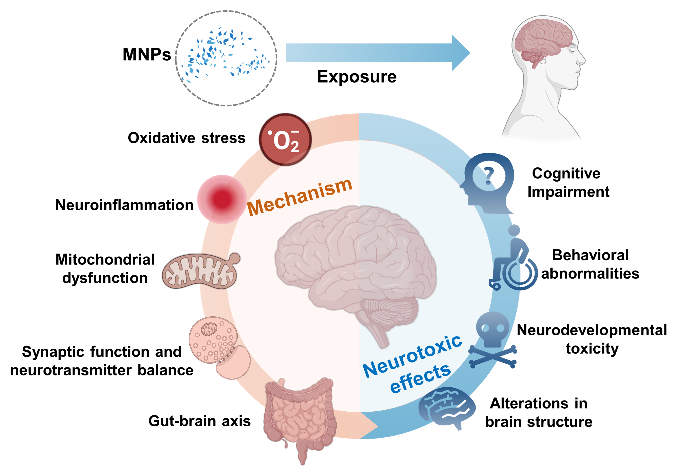
Microplastics and nanoplastics (MNPs) pollution has become a serious environmental issue. MNPs can enter the human body through inhalation, ingestion, and skin contact, raising significant concerns about their potential risks to the nervous system. This paper reviews the studies on the neurotoxic effects of MNPs in terrestrial mammals, focusing on their possible toxic mechanisms. Studies have shown that MNPs can cause damage to the nervous system by inducing oxidative stress, inflammatory responses, and mitochondrial dysfunction. Additionally, the impact of MNPs on the gut-brain axis is considered a crucial mechanism leading to neurotoxicity. Despite current progress, there are still insufficient data and incomplete understanding of the neurotoxic mechanisms involved. Future research should enhance epidemiological studies on MNP exposure, develop more suitable experimental models, investigate the health effects of different types of MNPs, explore their mechanisms in greater depth, and comprehensively assess various influencing factors. These efforts will provide essential insights for a more thorough understanding of the impact of MNPs on human health.

Contents

1 Introduction

2 Human exposure to MNPs

2.1 Routes of human exposure to MNPs

2.2 Detection of MNPs in human tissues and organs

3 Neurotoxic effects of MNPs

3.1 Cognitive impairment

3.2 Behavioral abnormalities

3.3 Neurodevelopmental toxicity

3.4 Alterations in brain structure

3.5 Combined neurotoxic effects of MNPs and other environmental pollutants

4 Mechanisms of toxicity

4.1 Oxidative stress

4.2 Neuroinflammation

4.3 Mitochondrial dysfunction

4.4 Synaptic function and neurotransmitter balance

4.5 Gut-brain axis mechanism

5 Conclusion and outlook

Cite this article

Yuji Huang , Kailin Xu , Boxuan Liang , Yizhou Zhong , Maoyong Song , Zhenlie Huang . Neurotoxicity and Mechanisms of Microplastics and Nanoplastics Exposure in Terrestrial Mammals[J]. Progress in Chemistry, 2025 , 37(1) : 76 -88 . DOI: 10.7536/PC240709

1 introduction

With the wide use of plastic products, the problem of plastic pollution has become increasingly serious. According to the data, the global plastic production has reached 368million metric tons in 2019, and it is expected to double by 2039[1]During the pandemic of new coronavirus infection, the use of a large number of disposable plastic protective products (such as masks, gloves, etc.) further exacerbated the global plastic pollution[2]
Waste plastics are difficult to degrade in the natural environment, but under the action of photochemical reaction, physical shear, biological action and other environmental factors, they will fragment and form plastic particles[3]Microplastics (MPS) refers to plastic particles with particle size less than 5 mm, while nanoplastics (NPS) refers to plastic particles with particle size less than 100 nm[4]Microplastics and nano plastics (MNPs) have been widely found in water, soil and air. In the offshore waters of China, the maximum concentration of MNPs can reach 9575.50/m ³; In the fresh water system, the detectable concentration is between tens of thousands to tens of thousands/m ³[5]In soil, especially farmland soil, the concentration of MNPs can reach 1430~3410/kg[6]The concentration of MNPs in the air can not be ignored. The number of MNPs per cubic meter in the air of coastal cities in eastern China is (224 ± 70)[7]Human beings have been inevitably exposed to MNPs, which poses a potential threat to health[8-9]
With the increasing attention to the human health risks of MNPs, the focus of toxicity research is gradually shifting from aquatic organisms to higher-level terrestrial mammal models to explore their potential hazards to human health. At present, the target organs of MNPs toxicity effect research mainly include gastrointestinal tract, nervous system, cardiovascular system, liver, kidney, reproductive development system and immune system[10]However, the toxic effects of MNPs on the nervous system of terrestrial mammals and their molecular mechanisms are limited. Previous published reviews of MNPs' neurotoxicity mainly focused on non mammalian neurotoxicity[11]Therefore, it is urgent to systematically sort out the research on the neurotoxicity of MNPs to terrestrial mammals. In this paper, combined with the exposure of MNPs to human body, the neurotoxic effects and toxic mechanism of MNPs on terrestrial mammals were reviewed, aiming to provide reference for further research on the neurotoxic effects of MNPs on human body, and put forward the scientific issues that need to be further focused in future research.

2 Exposure of MNPs to human body

2.1 Exposure mode of MNPs to human body

Human exposure to MNPs mainly includes inhalation, oral intake and skin contact[12]Although the proportion of skin exposure to human total MNPs is relatively low, skin exposure will still occur when human skin contacts personal care products or water containing MNPs. In personal care products, the particle size of MPS usually detected is about 10~100 μ M[13-14]The pore size of human skin is about 40~80 μ M[15]Although the skin has the protective effect of barrier, components in personal care products such as oleic acid and ethanol can increase the lipid fluidity between cells, thus promoting the transdermal transmission of MNPs[16]An Iranian survey showed that the skin of the face and hands adsorbed more than 4000 MNPs particles in one day[17]
Oral intake and inhalation are considered to be the main exposure routes of MNPs. Humans may be directly exposed to MNPs in drinking water, sea salt and air, or indirectly through the food chain[18-19]MNPs in the air were detected in all regions, no matter in urban areas with frequent human activities[20-21]Or other inaccessible areas (such as the Arctic, atmospheric boundary layer, etc.)[22-23]It is estimated that adults inhale 1.9 × 10 per year through indoor and outdoor air respectively3~1.0 × 10⁵And 0~3.0 × 10⁷MNPs[24]It should be noted that MNPs in the air not only enter the human body through inhalation, but also may be swallowed by depositing on the food surface[25]MNPs have been found in a large number of meat and processed foods, including shellfish, fish, salt, beverages, dairy products and meat[26-30]MNPs were even detected in many agricultural products such as carrots, apples, lettuce, celery and broad beans[31-33]Foods of marine origin need to be particularly vigilant, because MNPs will increase their concentration in foods through the enrichment of the food chain[34]Another major source of oral intake is drinking water, in which the concentration of MPS is 0-104The particle count may be higher for NPs with smaller size[35]
At present, due to the difficulties in sampling and analyzing MNPs, especially NPs, it is still challenging to accurately assess the actual exposure of humans or organisms. According to the researchers' estimate, the annual exposure of MPS per person is about 74000-121000. However, this estimate does not consider NPs, so the actual exposure may be higher[18]Another higher estimate is that the average weekly intake of MNPs by humans is 0.1-5 G[36]

2.2 Detection of MNPs in human tissues and organs

Because of the small size of MNPs, they can pass through the physiological barrier of human body and enter the body. Once MNPs enter the blood circulation system, they may spread and transfer in various tissues and organs[37-38]Figure 1). MNPs have been detected in human blood samples with a concentration of about 1.6 μ g/ml, which confirms that MNPs can diffuse in the blood circulation[39]The study also found MNPs in liver, spleen and kidney, which also provided evidence for the accumulation of MNPs in peripheral organs through blood circulation[40]MNPs concentrations in hair, colon, lung, placenta and other parts of the human body have also been reported[17,41-43]In addition, MNPs were also detected in the closed inner environment of human eyeball vitreous[44]Currently, MNPs detected in human body include polypropylene (PP), polystyrene (PS), polyvinyl chloride (PVC), nylon-66 (PA66), polyethylene terephthalate (PET), polymethylmethacrylate (PMMA), etc(Table 1)。
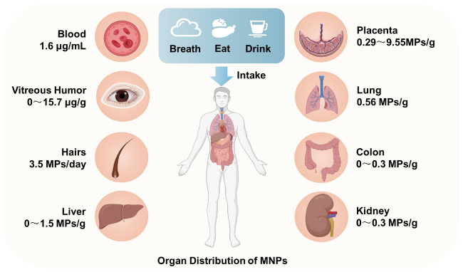
Figure 1 Distribution of micro nano plastics in human tissues and organs

Fig. 1 Distribution of MNPs in the human body

Table 1 Distribution of MNPs in human tissues and organs

Table 1 MNPs were detected in various parts of the human body

Human tissue types MNPs types MNPs content range Ref
Blood PET, PVC, PP, PMMA 1.6 μg/mL 39
Vitreous humor PA66, PVC, PS 0~15.7 μg/g 44
Hairs PP, PET, PS 3.5 MPs/day 17
Liver PS, PVC, PET 0~1.5 MPs/g 40
Kidney PS, PVC, PET 0~0.3 MPs/g 40
Spleen PS, PVC, PET 0.4~2.2 MPs/g 40
Colon PS, PVC, PET 0~0.3 MPs/g 41
Lung PP, PET, PS 0.56 MPs/g 42
Placenta PVC, PP, PBC 0.29~9.55 MPs/g 43
Due to the difficulty in obtaining human brain tissue samples, there is no research report to detect the distribution of MNPs in human brain tissue. However, a large number of animal experimental data have proved that MNPs can enter animal brain tissue. An in vivo experiment showed that the accumulation of MNPs in brain tissue could be detected only 2 hours after oral intake of MNPs in mice[45]This suggests that MNPs (especially NPs) can enter the blood circulation through gastrointestinal absorption and enter the brain across the blood-brain barrier. In addition, MNPs can also enter the brain directly through respiratory pathway (without blood circulation). A study showed that compared with MNPs injected into the blood through the tail vein, MNPs inhaled tended to accumulate in the brain rather than in the liver[46]Therefore, based on the results of in vitro fluorescence images, researchers speculated that MNPs could be transported to the olfactory bulb and cerebral cortex of mice through the olfactory nerve pathway, and also to the cerebellum and pons through the trigeminal nerve pathway[46]

3 Neurotoxic effects of MNPs

The neurotoxic effect of MNPs is an important part of evaluating its risk to human health. Early studies focused on lower organisms and aquatic organisms[11]In recent years, the effects of MNPs on the nervous system of terrestrial mammals have gradually attracted extensive attention. In the first study on the neurotoxicity assessment of MNPs to mammals, researchers failed to observe significant differences in the behavioral tests of the divine Scriptures after 5 weeks of oral administration of 1, 3, 6 and 10 mg/kg polystyrene nanoplastics (PS NPs) (25 and 50 nm) in adult male Wistar rats[47]The researchers speculate that this may be due to the small sample size or low exposure dose[47]With the in-depth study, more and more evidence shows that MNPs have neurotoxic effects on land mammals, including cognitive dysfunction, behavioral abnormalities, nerve development retardation and brain structural changes.

3.1 Cognitive impairment

Cognitive function is the ability of the brain to process, store and extract information, mainly for learning and memory. Long term exposure to MNPs can significantly affect the cognitive function of mammals, leading to the decline of learning and memory ability. After male BALB/c mice were fed with aqueous solutions containing 100 and 1000 μ g/L polystyrene microplastics (PS MPS) for 180 days, compared with the control group, the escape latency of mice in the water maze test was significantly increased, and the discrimination rate in the new object recognition test was significantly reduced, showing cognitive deficits, which was dose-dependent with the exposure concentration[48]Although PS MPs with different sizes of 0.5, 4 and 10 μ m were used in this study, no significant difference in particle size dependence was observed[48]In another 180 day chronic exposure experiment, PS NPs with smaller size (25 nm) were used. The results showed that with the increase of PS NPs exposure dose (0, 10, 25 and 50 mg/(kg · d)) to male c57blg6j mice, the spatial learning and memory ability of mice decreased in a dose-dependent manner[49]In the study of short exposure time, it was also found that MNPs had an effect on the cognitive function of mouse brain. After 6 weeks of oral administration of 80 nm PS NPs (60 (μ g · d)) in male C57BL/6J mice, the damage of neurons in the hippocampus resulted in the decline of learning and memory ability in mice[50]Similarly, another study found that male C57BL/6J mice developed cognitive impairment after oral administration of 30-50 nm PS NPs (10 and 20 mg/kg) for 7 weeks, but their motor function and social behavior were not significantly affected[51]In addition, in the short-term exposure experiment with only 3 days, male Swiss mice also showed a decline in memory ability in the new object recognition experiment after intraperitoneal injection of 14.6 ng/(kg · d) of 25 nm PS NPs[52]Gender difference is also an important factor affecting the neurotoxicity of MNPs. In one study, male mice showed body dependent learning and memory impairment after oral administration of 2 μ m PS MPS (8 and 16 mg/(kg · d)) for 8 weeks, while female mice were not significantly affected[53]This gender difference may be related to the changes of estradiol, progesterone or testosterone levels. These hormones play an important role in regulating spatial memory and situational memory, and the specific mechanism needs to be further studied.
Cognitive impairment caused by MNPs may be related to some common neurological diseases, such as Alzheimer's disease (AD). In vitro studies showed that 1 μ g/cm ² of ammoniated PS NPs (50 nm) treated undifferentiated SH-SY5Y cells for 48 h resulted in increased secretion of amyloid β (a β) and decreased neuronal differentiation[54]Another study found that at concentrations as low as 100 PM, PS NPs can significantly accelerate the formation of two subtypes of a β 40 and a β 42, promote the accumulation of a β oligomers, and thus cause obvious neurotoxicity[55]However, whether MNPs can promote the occurrence and development of ad still needs more population epidemiological survey data and animal experimental research.

3.2 Abnormal behavior

Behavioral function is another core function of the brain, involving emotion regulation, social behavior and motor behavior. Mammals exposed to MNPs can have a variety of abnormal behaviors, among which anxiety and depression like behavior is one of the most common affective disorders induced by MNPs. Male C57BL/6J mice were exposed to 100 nm and 1 μ m PS MPS (0.5 mg/D) by oral gavage for 30 days and 60 days, respectively, and showed a high degree of anxiety like behavior[56]The specific performance was as follows: the activity distance and time percentage in the central area of the open field experiment were significantly reduced, and the residence time percentage of the open arm in the elevated cross maze test was significantly lower than that in the control group[56]Correlation analysis showed that these anxiety like behaviors were closely related to intestinal flora imbalance and neurotransmitter metabolism in mice[56]Another study showed that male C57BL/6J mice exposed to 480nm PS MPs of 2 mg/(kg · d) by oral gavage showed anxiety like behavior after only one week of exposure, which may be related to the pro-inflammatory reaction caused by microglia activation[57]The causes of anxiety like behavior are various, which may be related to the influence of various biological functions of the body. In a 6-month study, C57BL/6J mice with 25 nm PS NPs orally administered at 50 mg/(kg · d) showed decreased sucrose preference and increased immobility time in tail suspension test, indicating that depression like behavior was induced[58]In addition, a study explored the effects of different surface modified PS NPs (100 nm) on mice. The results showed that these PS NPs could destroy the blood-brain barrier and accumulate in the brain, leading to a series of neurobehavioral changes, including anxiety, depression and social behavior damage[59]However, some studies have shown that adult male Swiss albino mice showed anti anxiety behavior after oral administration of 50 nm PS NPs (0.2 and 1 mg/(kg · d)) for 8 weeks, which may be related to the changes of neurotransmitter system[60]In conclusion, MNPs have an important impact on mammalian emotional function, and long-term exposure may lead to affective disorder related diseases.
In addition to emotional and social behavior, MNPs also have an important impact on sports behavior. After exposure to PS NPs, the motor activity of mice was impaired[60]In an animal experiment of oral exposure, male C57BL/6J mice were given 250 mg/(kg · d) of 50 nm PS NPs by oral gavage, and the exercise ability was significantly impaired after 28 days of exposure. The activity ability of these mice decreased, and the latency and grip strength decreased in the rod rotation test. These changes were related to the damage of dopaminergic neurons[61]It is suggested that the sports injury caused by PS NPs may be a neurodegenerative injury similar to Parkinson's disease (PD). Further studies revealed that its toxic mechanism may be related to cerebral mitochondrial dysfunction. Melatonin intervention can reduce the effect of MNPs on motor function to a certain extent[62]The neuropathological feature of PD is the pathological aggregation of α - synuclein (called Lewy body)[63]These Lewy bodies are closely related to the degeneration of dopaminergic neurons[64]There is a high affinity interaction between anionic NPs and α - synuclein. This interaction can slow down the degradation of α - synuclein, induce the formation of α - synuclein inclusion bodies in dopaminergic neurons, and promote its transmission in various brain regions[65]In another study, the Parkinson's disease model of Caenorhabditis elegans and human neuron cells found that 25 nm PS NPs could increase the formation of α - synuclein aggregates, leading to the degeneration of dopaminergic neurons. At a low concentration of 10 μ g/L, it was observed that the motor ability of Caenorhabditis elegans was significantly affected[66]Although these findings reveal the relationship between MNPs exposure and sports behavior injury, the correlation between MNPs exposure and human PD development still needs further research to confirm.

3.3 Neurodevelopmental toxicity

Neural development is an important basis for the development and normal function of mammalian nervous system, including several key steps such as neuron generation, migration, differentiation, synapse formation and neural network establishment. Abnormalities in any link may lead to serious neurological diseases and behavioral disorders[67]The pregnant SD rats were orally administered with PS NPs with different particle sizes (25 and 50 nm) from the first day to the 18th day of pregnancy, and the exposure doses were 0.5, 2.5, 10 and 50 mggkg, respectively. The placental structure damage in the exposure group was aggravated in a dose-dependent manner[68]PS NPs can penetrate the placental barrier, cause oxidative stress and inflammatory reaction in various brain regions of fetal rats, produce neurotoxicity on them, and affect the growth and development of fetal rats[68]It is worth noting that small particle size and high-dose PS NPs exposure have more significant neurotoxic effects on offspring[68]In addition, PS NPs ingested by mothers can also be transferred to offspring through breast milk. When the mother mice were exposed to 50 nm fluorescent PS NPs, the fluorescent signal of PS NPs could be detected in the brain of the breast fed offspring mice, which led to the dysfunction of neural stem cells, and ultimately led to neurodevelopmental defects[69]Some studies have also found that there are gender specific differences in the brain development abnormalities of offspring caused by MNPs, which may be related to the gender specific synaptic development, function and neurotransmitter activity of the hippocampus[70]By using the 3D model of human forebrain cortex sphere to simulate the early development of human cerebral cortex, it was found that exposure to 1 and 10 μ m PS MPs had an adverse effect on the differentiation of cortical layer. Long term exposure to PS MPs can reduce cell viability and down regulate the expression of markers of neuronal maturation[71]
MNPs exposure may be one of the risk factors inducing autism spectrum disorder (ASD) - like behavior. ASD is a neurodevelopmental disorder that occurs in infants and young children. Its core symptoms are persistent social defects and repetitive stereotyped behaviors and interests[72]C57BL/6J mice were indirectly exposed to 2 μ m PS MPs through their mothers from the embryonic stage to the age of 4 weeks. After weaning, they continued to drink 1 mg/L PS MPs aqueous solution for 24 weeks. The results showed that in the three box social test, they showed the social defect of impaired social novelty preference[73]A study compared the effects of exposure to 10-20 μ m polyethylene (PE) MPs at different life stages on the behavior of C57BL/6J mice. It was found that whether prenatal exposure (10 μ g/d, 2 weeks) or post weaning exposure (10 μ g/d, 2 weeks), mice showed more repetitive and compulsive behavior in nestlet tearing test, and showed social defects with reduced social novelty, revealing the characteristics similar to ASD in mice[74]These results suggest that MNPs exposure may be an important reason for the results of some neurodysplasia. Therefore, in addition to investigating the impact of MNPs on ASD, we can further explore the impact on other neurodevelopmental disorders, such as intellectual disability, attention deficit and hyperactivity disorder.

3.4 Changes in brain structure

Cognitive dysfunction and abnormal behavior are the external manifestations of the internal damage of the nervous system. These changes in brain nerve function suggest that MNPs have caused a certain degree of damage to the brain structure. Among them, the destruction of blood-brain barrier is one of the most frequently reported brain structural damage. Blood brain barrier is a kind of biological barrier, which is composed of brain microvascular endothelial cells, basement membrane and surrounding astrocytes. Its main function is to protect the brain from harmful substances and maintain the stability of the internal environment of the brain[75]MNPs can destroy the blood-brain barrier and increase its permeability[48,61,76-77]NPs can even enter the brain tissue, and the destruction of blood-brain barrier is considered to be an important premise for MNPs to cause neurotoxicity. For example, NPs pass through the blood-brain barrier and accumulate in the brain, triggering the activation of microglia in mice and leading to neuronal degeneration[76]The destruction of blood-brain barrier may make it easier for toxic substances to enter brain tissue, thus affecting the function and health of neurons[78]
MNPs can also affect the structure of brain nerve cells. After mice were exposed to 500 nm PS MPs for 4 weeks, Golgi staining showed that the density of dendrites and dendritic spines in prefrontal cortical neurons was disordered[79]After 4 weeks of oral administration of 5.0-5.9 μ m PS MPs 1 mg/D in Kunming mice, the neurons in the hippocampus showed signs of disorder and relaxation, the pyramidal neurons were scattered, and the main dendritic structure in the brain was reduced or missing[80]MNPs not only affect the morphology of nerve cells in the brain, but also affect their number. There is evidence that PS NPs may cause the loss of dopaminergic neurons by causing mitochondrial dysfunction and energy metabolism disorder in the substantia nigra of the mouse brain[61]In C57BL/6J mice exposed to PS MPs, hematoxylin and eosin (h&e) staining of brain tissue showed that the nucleolus in neurons was stained deeper, and the number of neurons in hippocampus was significantly reduced[48]Similarly, the number of Nissl bodies in the hippocampus of Kunming mice exposed to PS MPs decreased significantly[80]

3.5 Combined neurotoxicity of MNPs and other environmental pollutants

In addition to the direct effect of MNPs, they often coexist with other chemical pollutants in water, soil and air, forming a complex exposure environment. This environment makes MNPs and other pollutants may have joint toxic effects[81]For example, when polystyrene microplastics (PS MPS) and plasticizer di - (2-ethylhexyl) phthalate (DEHP) are exposed together, they will cause more serious damage to the mitochondrial function of mouse neurons, leading to more obvious apoptosis, indicating that they have synergistic toxic effects[82]In addition, the CO exposure of DEHP and PP MPs also showed cumulative or synergistic toxicity, and induced significant neurocognitive deficits in young mice[83]
The joint neurotoxic effects of MNPs and residual antibiotics in the environment are also concerned. The combined exposure of doxycycline (DOX) and PS MPs can disturb the intestinal microflora homeostasis of mice, mediate inflammatory brain damage, and then lead to the decline of learning and memory ability. Although the combined exposure of DOX and PS MPs showed synergistic neurotoxicity, DOX did not aggravate the intestinal injury caused by PS MPS[84]In addition, the joint toxicity of MNPs and heavy metals is also one of the research hotspots. The study found that after 9-month-old male c57blg6j mice drank water containing ferric ammonium citrate (5 g/L) and 5 μ m PS MPS (1000 μ g/L) for 3 months, the presence of PS MPs promoted the accumulation of iron content in the brain of mice, resulting in iron homeostasis disorder, leading to lipid peroxidation and inflammation related to iron death, which further aggravated cognitive impairment[85]
Table 2 Study on the neurotoxicity of micro nano plastic exposure to terrestrial mammals

Table 2 Research on the neurotoxicity of MNP exposure in terrestrial mammals

Species MNPs types Size Method of
administration
Exposure dose Exposure
duration
Neurotoxic effects Ref
Male wistar rats (6~8 weeks) PS-NPs 25 and
50 nm
Oral gavage 1,3,6,and 10 mg/(kg·d) 35 d No significant neurobehavioral effects. Rafiee et al. [47]
Male BALB/c mice (6 weeks) PS-MPs 0.5,4 and 10 μm Drinking water
exposure
100 and
1000 μg/L
180 d Cognitive and memory deficits; disruption of the blood-brain barrier; reduced dendritic spine density; an inflammatory response in the hippocampus; a concentration-dependent trend. Jin et al. [48]
Male C57BL/6 mice (5 weeks) PS-NPs 25 nm Oral gavage 0,10,25,
50 mg/(kg·d)
180 d Cognitive decline; synaptic damage in the prefrontal cortex; increased sensitivity to synaptic dysfunction. Chen et al. [49]
Male C57BL/6 mice (5 weeks) PS-NPs 80 nm Oral gavage 60 μg/day 42 d Caused neuronal damage in the hippocampus,impaired learning and memory abilities; neurotoxicity involved gut-brain axis-mediated pathways related to circadian rhythm. Kang et al. [50]
Male C57BL/6 mice (7 weeks) PS-NPs 30~50 nm Oral gavage 10 and
20 mg/(kg·d)
42 d Induced memory impairment,activated microglia,triggered inflammatory responses,and dysregulated hippocampal neuronal activity. Paing et al. [51]
Male Swiss mice (4~6 weeks) PS-NPs 25 nm Intraperitoneal administration 14.6 ng/(kg·d) 3 d Cognitive impairment; REDOX imbalance; suppressed AChE activity; DNA damage. Estrela et al. [52]
Male or female C57BL/6 mice (6 weeks) PS-MPs 2 μm Oral gavage 8 and
16 mg/(kg·d)
28 and 56 d Learning and memory deficits; decreased synaptic protein levels; induced neuroinflammation. Lee et al. [53]
SH-SY5Y cells Amine-functionaliz- ed PS-NPs 50 nm In culture medium 1, 5, 10, and
50 µg/cm2
24 h and
48 h
Decreased neuronal differentiation and increased Amyloid β secretion. Schroter et al. [54]
SHSY-5Y cells Unmodified and
amino-modified PS-NPs
70 and 150 nm In culture medium 100 pM 24 h and
7 d
Accelerated the aggregation of β-amyloid peptides. Gou et al. [55]
Male C57BL/6 mice (6~8 weeks) PS-MPs and PS-NPs 100 nm and 1.0 µm Oral gavage 0.5 mg/day 30 and 60 d Induced anxiety-like behaviors; changed serum metabolic profiles,particularly in neurotransmitter metabolites. Chen et al. [56]
Male C57BL/6 mice (8 weeks) PS-MPs 480 nm Oral gavage 2 and 10 mg/(kg·d) 180 d Induced anxiety-like behavior; related to inflammation pathways. Li et al. [57]
Male C57BL/6 mice (5 weeks) PS-NPs 25 nm Oral gavage 10, 25 and
50 mg/(kg·d)
180 d Depression-like responses; activation of axon guidance,the neurotrophin signaling pathway,and dopaminergic synapse-related lncRNAs. Liu et al. [58]
Male BALB/c mice (4 weeks) PS-NPs with different surface modifications (PS,PS-COOH,and PS-NH2) ∼100 nm Oral gavage 1 mg/day 28 d Disrupted the tight junctions formed by brain endothelial cells; induced mitochondrial dysfunction in neurons; induced anxiety,depression,and social deficits. Ma et al. [59]
Male Swiss albino mice (10 weeks) PS-NPs 50 nm Oral gavage 0.2 and
1 mg/(kg·d)
56 d Induced anxiogenic behavior and impaired learning; induced oxidative stress and inhibited acetylcholinesterase activity; decreased the number of hippocampal pyramidal cells; affected neuronal architecture of the cortex and hippocampus. Sharma et al. [60]
Male C57BL/6 mice (8 weeks) PS-NPs 50 nm Oral gavage 0.5, 5, 50 and 500 mg/(kg·d) 28 d Induced Parkinson’s disease (PD)-like neurodegeneration; disturbed PD-associated gene expression; inhibited ATP metabolism Liang et al. [61]
Male C57BL/6 mice (8 weeks) and SHSY-5Y cells PS-NPs 50 nm in vivo:Oral gavag; in vitro: in culture medium in vivo: 500 mg/(kg·d) ;
in vitro: 0.25,2.5, 25, 250 μg/mL
in vivo:
28 d
in vitro:
48 h
PS-NPs disrupted mitochondrial function by affecting CI,leading to excessive mitophagy through the AMPK/ULK1 pathway,ultimately causing dopaminergic neuron death. Huang et al. [62]
Primary neuron; CD1 mice
(3 months)
Anionic and cationic PS-NPs 23.2~
115.6 nm
in vivo: intracranial injection; in vitro: in culture medium in vivo: 15 μg;
in vitro: 1 nM
in vivo: 3-d, or two months; in vitro: 24 h Causing mild lysosomal impairment that slows the degradation of aggregated α-synuclein; exacerbates the spread of α-synuclein pathology across interconnected vulnerable brain regions. Liu et al. [65]
C. elegans strains; A53T α-syn-EGFP SH-SY5Y cells PS-NPs 25 nm in vivo: in culture medium; in vitro: in culture medium in vivo: 10,100,or 1000 μg/L;
in vitro: 15 mg/L
in vivo: 24 h; in vitro: 24 h Low concentrations of nanoplastics inhibit the growth and movement of nematodes; increase α-synuclein aggregation. Jeong et al. [66]
SD pregnant rats PS-NPs 25 and 50 nm Oral gavage 0.5, 2.5, 10 and 50 mg/(kg·d) 1st to the 18th day of pregnancy Affect the growth and development of fetal rats; damage the placental barrier; cause oxidative stress and inflammatory responses. Zhang et al. [68]
Pregnant female C57BL/6 mice PS-NPs 50 and 500 nm Oral administration 0.5~1000 μg/day embryonic day 8 until 2 weeks after birth Induced neurodevelopmental defects; caused NSC dysfunction; led to the dysregulation of brain functions in progeny. Jeong et al. [69]
3D model of human forebrain cortical spheroids PS-MPs 1 and
10 μm
In culture medium 5,50,and
100 µg/mL
6 and 26 d Altered the gene expression related to DNA damage and neural tissue patterning; affected embryonic brain-like tissue development in forebrain cerebral spheroids. Hua et al. [71]
C57BL/6 J mice (8-weeks) PS-MPs 2 μm Drinking water
exposure
1 mg/L embryonic stage to adulthood Impaired offspring social novelty preferences. So et al. [73]
Female C57BL/6 J mice PE-MPs 10~20 μm Oral gavage 10 μg/day prenatal:
14 d; post-weaning period:
14 d; puberty: 14 d; adult: 84 d.
Impaired social interaction and repetitive behaviors; disturbance of metabolites and gene expression in the brain; leads to Autism Spectrum Disorder (ASD)-like traits. Zaheer et al. [74]
Male C57BL/6 mice (8 weeks); hCMEC/D3,murine microglia BV2,hippocampus-derived neuronal
HT22 cells
PS-NPs 42 nm in vivo: Oral gavage;
in vitro: in culture medium
in vivo: 0.5,2.5,10,and
50 mg/(kg·d);
in vitro: 0,25,50,and
100 μg/mL
in vivo: 7 d; in vitro: 48 h Induced microglia activation and neuronal damage in the brain; caused oxidative stress,inflammatory response,and necroptosis in hCMEC/D3 cells; disrupted tight junctions. Shan et al. [76]
Male C57BL/6 mice (3 weeks) PS-MPs 5 and
50 μm
Drinking water
exposure
100 and 1000 μg/L 70 d Induced social disorders in adult mice; decreased oxytocin levels; reduced mucin layers and caused gut microbiota dysbiosis. Wang et al. [77]
Male Swiss albino mice (20~30 g) PS-MPs 500 nm Oral gavage 0.1,1,and
10 ppm
28 d Reduced Nissl bodies in the PFC; affected neuronal cytoarchitecture of PFC neurons; decreased spine density in PFC neurons; down-regulated BDNF gene. Suman et al. [79]
Male KM mice (5 weeks) PS-MPs 5.0~5.9 μm Oral gavage 1 mg/d 28 d Impaired learning and memory functions; induced a reduction in the level of acetylcholine; increased levels of ROS and MDA. Wang et al. [80]
KM mice (18~22 g); NS20Y cells PS-MPs; Di-(2-ethylhexyl) phthalate (DHEP) 1~10 μm Food and water exposure in vivo: food containing 200 mg/kg DEHP and water containing 10 mg/L MPs;
in vitro: 25 μmol/L DEHP and 775 mg/L PS-MPs
in vivo: 30 d; in vitro: 24 h DEHP and MPs caused changes in mitochondrial function; led to apoptosis of neurons; exhibited a combined toxic effect on mouse cerebra. Zhang et al. [82]
Male ICR mice (3 weeks) PP-MPs; DHEP / Oral gavage PP-MPs: 5,20 and 80 mg/(kg·d);
DHEP:
250 mg/(kg·d)
28 d Neurocognitive defects; induced impairments in the hippocampal CA3 region; exhibited additive or synergistic toxic effects. Yang et al. [83]
Male C57BL/6 mice (7~8 weeks) PS-MPs; doxycycline hyclate (Dox) 500 nm Oral gavage PS-MPs:
5 mg/(kg·d);
Dox:
20 mg/(kg·d)
30 d Co-exposure caused brain lesions and inflammation; decreased learning and memory behaviors through the gut-brain axis. Sun et al. [84]
Male C57BL/6 mice (9 months) PS-MPs; Ferric ammonium citrate (FAC) 5 µm Drinking water
exposure
PS-MPs:
1000 μg/L;
Dox: 5 g/L
90 d Co-exposure caused significant iron overload and cognitive deficits; elevated lipid peroxidation and inflammation; aggravated cognitive impairment. Liu et al. [85]
It is worth noting that in a 3-day short-term exposure experiment, male Swiss mice were intraperitoneally injected with 14.6 ng/kg PS NPs (25 nm) and zinc oxide nanoparticles (70 nm), although the single exposure caused cognitive impairment in mice, the combined exposure did not show additive or synergistic neurotoxic effects[52]This increase in joint toxicity may be related to the high specific surface area of MNPs. MNPs adsorb other pollutants by physical or chemical means, thus increasing the potential health risks associated with MNPs[86]Although the joint neurotoxic effects of MNPs and other pollutants have been preliminarily explored, the current research is still limited. Future research should pay more attention to the impact of multiple exposures of MNPs and other pollutants in the actual environment on human nervous system health.

4 Discussion on toxic mechanism

4.1 oxidative stress

Oxidative stress is considered to be one of the important mechanisms by which MNPs exert neurotoxicological effects[11]Oxidative stress refers to the imbalance of oxidation and reduction in the body, leading to the accumulation of reactive oxygen species (ROS) and causing cell and tissue damage. ROS refers to reactive chemical species containing oxygen, including superoxide anion (o2•−)Hydrogen peroxide (H2O2)And hydroxyl radical (OH). MNPs induced ROS production in organisms involves a variety of biomolecular mechanisms. One of the key mechanisms is the activation of NADPH oxidase (NOx), which is responsible for the production of O2•−Complex enzyme[87]Mice exposed to MNPs can activate NADPH oxidase through a variety of ways, including the activation of Toll like receptor 4 (TLR4) and aryl hydrocarbon receptor (AHR)[88]Once NOx is activated, it will generate o2•−And further converted to other ROS, such as H2O2And · oh.
In addition, MNPs can aggravate oxidative damage by destroying antioxidant defense system. Antioxidant enzymes include peroxidase, catalase, superoxide dismutase and glutathione peroxidase. These antioxidant enzymes play an important role in protecting tissues from ROS damage. Their activities are often used as biomarkers of oxidative stress. Studies have shown that PS NPs exposure significantly changes the activities of these antioxidant enzymes in mouse brain tissue, indicating that there is a significant oxidative stress response in brain tissue[60,89]In vitro experiments also showed that MNPs could induce a large number of ROS in mouse hippocampal neurons HT22 cells, leading to oxidative damage[90]
The central nervous system is very sensitive to oxidative stress due to its high oxygen demand and low antioxidant capacity[91]High levels of ROS can directly damage the biological macromolecules of nerve cells, leading to cell death. For example, PS NPs can increase ROS levels, promote lipid peroxidation, and cause iron death in mouse microglial cell line BV2[92]PS MPs can induce apoptosis by increasing ROS production, and blocking ROS can alleviate this kind of apoptosis[93]In addition, oxidative stress can also activate microglia and astrocytes, induce neuroinflammatory reaction, and further aggravate nerve injury[94]The neuroinflammatory response can not only increase the oxidative damage of nerve cells, but also cause the inflammation of blood-brain barrier, which further aggravates the neurotoxic effect caused by MNPs. For example, PS NPs induce inflammatory response in human brain microvascular endothelial cells (hcmec/d3), which is related to oxidative stress of cells[76]Therefore, oxidative stress induced by MNPs may lead to the injury and death of neurons, and then affect the cognitive and behavioral functions of the nervous system.

4.2 Neuroinflammation

Neuroinflammation is an important mechanism of neurotoxicity, which usually leads to neuronal injury and neurological dysfunction. It can trigger a series of pathophysiological processes, including inflammatory cell infiltration, inflammatory cytokine release, and activation of neurons and a variety of glial cells[95]MNPs, as environmental pollutants, are closely related to neuroinflammation and may cause neurotoxicity by triggering inflammatory reaction. A study on PS MPs showed that the mRNA levels of TNF - α and IL-1 β in the hippocampus of mice were significantly increased after exposure, indicating the increase of neuroinflammation in mice[53]The neuroinflammatory response induced by MNPs includes inflammatory cell infiltration around neurons and microglia activation[76]Activated microglia may trigger a series of neuroinflammatory reactions and promote the occurrence of brain injury[96]For example, PS NPs activated microglia release inflammatory factors that inhibit the activity of hippocampal neurons, leading to cognitive dysfunction in mice[51]In addition, PS MPs can further promote the progress of cognitive impairment by inducing microglial cell death and aggravating neuroinflammation[97]
Astrocytes also play an important role in neuroinflammation. Studies have found that PS NPs and PS MPs can induce astrocyte activation in vitro, and up regulate the pathways related to neuroinflammation and immunity[98]Compared with the original MPs, aging MPs significantly increased the activation of immune related pathways[99]In vitro experiments on hmc-3 cells showed that the inflammatory response induced by aging MPS was more serious than that induced by primitive MPs[99]This suggests that aging MNPs in the environment may have higher neuroinflammatory ability. Neuroinflammation may also lead to neuron damage and neurotransmitter disorder, which will lead to neurological dysfunction, such as cognitive impairment, learning and memory ability decline, etc[100]In addition, neuroinflammation may aggravate oxidative stress reaction and further damage nerve cells. Oxidative stress and neuroinflammation promote each other, forming a vicious circle, leading to further deterioration of nervous system damage.

4.3 Mitochondrial dysfunction

Mitochondria have important biological functions in cells, including energy metabolism, redox balance, apoptosis, lipid metabolism and calcium homeostasis. Mitochondrial dysfunction is considered to be an important factor in the mechanism of neurotoxicity. As an environmental pollutant, MNPs have been found to be closely related to mitochondrial dysfunction. Animal experiments show that NPs can enter neurons and accumulate in mitochondria, leading to mitochondrial dysfunction and reduced ATP production. These changes may be related to abnormal animal behavior[59]Similarly, the accumulation of PS NPs in neurons can also lead to mitochondrial dysfunction and increased energy consumption, thus affecting neural development and cognitive function[101]The study also found that PS NPs could induce significant mitochondrial damage in differentiated SH-SY5Y cells, which mainly showed changes in mitochondrial structure, decreased mitochondrial membrane potential and decreased ATP production[62]This reduction in ATP production may lead to excessive mitochondrial autophagy and ultimately the death of dopaminergic neurons[62]After analyzing the brain of exposed mice by single nuclear RNA sequencing, it was found that PS NPs caused mitochondrial dysfunction and energy metabolism disorder, especially the most significant effect on excitatory neurons[102]The existence of MNPs destroys the structural integrity of mitochondria, reduces the mitochondrial membrane potential, and affects the integrity of the mitochondrial genome, leading to the dynamic imbalance between mitochondrial division and fusion[102]These changes directly affect the important functions of mitochondria such as energy production and apoptosis, which may affect the normal structure and function of the nervous system. Therefore, mitochondrial dysfunction is one of the important mechanisms of MNPs induced neurotoxicity. This process may lead to the disorder of cell energy metabolism and the aggravation of oxidative damage, and then affect the normal structure and function of the nervous system.

4.4 Synaptic function and neurotransmitter balance

Synapse is the key structure of signal transmission between neurons, and neurotransmitters play an important role in this process. Neurotransmitters such as glutamate, γ - aminobutyric acid, dopamine and 5-hydroxytryptamine play a key role in regulating nervous system function and behavior. The balance between synaptic function and neurotransmitters is essential for the normal operation of the nervous system. The exposure of MNPs significantly affected the structure and function of synapses. For example, the mouse model of long-term exposure to PS MPs showed that the density of dendritic spines in hippocampal neurons decreased, which was related to memory impairment in mice[48]In cortical neurons, the exposure of MNPs resulted in a significant decrease in the length and number of neurites[88]In addition, MNPs can also induce severe damage to synaptic function by affecting the expression of synaptic related genes in the brain, thereby affecting learning and memory ability[49,53]The change of synaptic function will directly affect the balance of neurotransmitters. Studies have found that water suspended plastic smoke particles can reduce the membrane potential of neurites and the acidification of synaptic vesicles, weaken synaptosome transporters, lead to excessive increase of glutamate concentration in synaptic space, and then over activate ionic glutamate receptors, resulting in neurotoxic effects[103]In addition, the exposure of MNPs can also lead to the decrease of acetylcholinesterase activity in the brain, which is closely related to its accumulation in the brain[52]The inhibition of acetylcholinesterase activity is closely related to the activation of oxidative stress, which has been observed to lead to impaired motor activity in mice[60]In conclusion, the effects of MNPs on synaptic function and neurotransmitter balance may exist in different species, and these effects may lead to neurological dysfunction, such as cognitive impairment, learning and memory decline, etc.

4.5 Gut brain axis mechanism

The gut brain axis plays an important role in maintaining the health and function of the nervous system. It is a complex physiological pathway that connects the digestive system and the central nervous system. It plays an important role in regulating emotion, cognitive function and behavior[104]MNPs exposure may have a negative impact on the gut brain axis, leading to nervous system dysfunction and neurotoxic effects. First, MNPs exposure may cause intestinal microbiome imbalance[105]Studies have shown that MNPs exposure destroys the balance of intestinal microbiome, leading to the reduction of beneficial bacteria and the increase of opportunistic pathogens, thus affecting intestinal function and immune regulation, and then affecting the nervous system through the gut brain axis[56,106]The results of hippocampal transcriptome, 16S rRNA sequencing of intestinal flora and plasma metabonomics showed that the circadian rhythm related pathways mediated by intestinal brain axis were involved in the neurotoxicity of MNPs. Probiotics supplementation could significantly reduce intestinal injury and restore the expression of circadian rhythm related genes and neural plasticity molecules[50]In addition, exposure to 50 μ m MPs could significantly reduce the production of colonic mucin and induce changes in intestinal microbiota. Blocking the vagus nerve pathway improves these behavioral disorders, highlighting the key role of the gut brain axis in mediating neurobehavioral outcomes[77]Secondly, MNPs can also induce intestinal flora imbalance and disrupt the metabolic dysfunction of brain and intestine in mice. For example, PS MPs leads to changes in intestinal Firmicutes and Bacteroides, which disturbs the metabolic process of mouse brain in the way of intestinal brain axis[107]The imbalance of intestinal microbiota can change the level of neurotransmitters (such as 5-hydroxytryptamine) produced by the intestine, thus affecting the neural function and behavior of the brain[105,108-109]In addition, the destruction of intestinal barrier and inflammatory reaction can increase systemic inflammation, and then affect the immune response of the brain and lead to neuroinflammation[110]In conclusion, MNPs exposure may affect the function of the gut brain axis through a variety of ways, thereby causing toxic effects on the nervous system.

5 Conclusion and Prospect

The potential impact of MNPs on human health has attracted extensive attention, but there are still many uncertainties and challenges in the current research. Although some studies have preliminarily explored the toxic effects of MNPs on mammalian nervous system, the following five aspects still need to be focused on and further studied.
(1) Lack and demand of epidemiological research: although some studies have estimated human exposure to MNPs, the impact of long-term exposure on human health has not been clear. More large-scale and long-term epidemiological studies, especially in different exposure levels and populations, are needed to comprehensively assess their potential health effects.
(2) Selection and applicability of animal models: at present, most studies rely on a single animal model, which may not fully represent the real human exposure situation. Future studies should consider the use of animal models closer to human physiological and metabolic characteristics to explore the differences in the toxic reactions of different individuals and populations to MNPs.
(3) Aging of MNPs and environmental risk considerations: at present, most studies use commercial MNPs, while in the actual environment, there are aging or weathering MNPs, and there may be significant differences between the two properties. Future research should strengthen the study of aging MNPs and their behavior and impact in the environment.
(4) Comprehensive evaluation of different types of MNPs: at present, the research mainly focuses on MNPs of common materials such as PS, while the research on other types of MNPs, especially new materials, is relatively lacking. Future research should include the evaluation of various new materials to fully understand the potential effects of different chemical compositions and physical properties on health.
(5) In depth exploration of toxicity mechanism: Currently, the specific mechanism of MNPs' impact on the nervous system through oxidative stress, inflammatory reaction and other ways is not well understood. More mechanism studies are needed in the future to reveal the toxic mechanism of MNPs on the nervous system and other key physiological processes, and provide a scientific basis for the development of prevention and intervention measures.
Comprehensive evaluation of various influencing factors such as particle size, shape, chemical composition, exposure route and dose of MNPs is the key to comprehensively understand their health risks. Therefore, interdisciplinary cooperation and comprehensive utilization of research achievements in materials science, environmental science, toxicology, epidemiology and other fields are important directions and challenges for future research.
[1]
Lebreton L, Andrady A. Palgrave Commun., 2019, 5(1): 6.

[2]
Lee M H, Kim H. Nanomaterials, 2022, 12(5): 851.

[3]
Thompson R C, Olsen Y, Mitchell R P, Davis A, Rowland S J, John A W G, McGonigle D, Russell A E. Science, 2004, 304(5672): 838.

[4]
Benson N U, Agboola O D, Fred-Ahmadu O H, De-la-Torre G E, Oluwalana A, Williams A B. Front. Mar. Sci., 2022, 9: 851281.

[5]
Yang Y J, Wang S B, Ye C B, Shi Y X, Li X R, Yuan W X, Fuo J Q. Advances in Environmental Protection, 2024, 14:51.

(杨壹珺, 王帅兵, 叶长兵, 史应仙, 李雪柔, 原祎昕, 霍佳祺. 环境保护前沿, 2024, 14:51. ).

[6]
Ding L, Zhang S Y, Wang X Y, Yang X M, Zhang C T, Qi Y B, Guo X T. Sci. Total Environ., 2020, 720: 137525.

[7]
Liao Z L, Ji X L, Ma Y, Lv B Q, Huang W, Zhu X, Fang M Z, Wang Q, Wang X D, Dahlgren R, Shang X. J. Hazard. Mater., 2021, 417: 126007.

[8]
Robin R S, Karthik R, Purvaja R, Ganguly D, Anandavelu I, Mugilarasan M, Ramesh R. Sci. Total Environ., 2020, 703: 134947.

[9]
Wang S M, Chen H Z, Zhou X W, Tian Y Q, Lin C, Wang W L, Zhou K W, Zhang Y B, Lin H. Environ. Pollut., 2020, 264: 114125.

[10]
Zhang Q, He Y, Cheng R J, Li Q, Qian Z Y, Lin X H. Environ. Sci. Pollut. Res., 2022, 29(27): 40415.

[11]
Prust M, Meijer J, Westerink R H S. Part. Fibre Toxicol., 2020, 17: 24.

[12]
Prata J C, da Costa J P, Lopes I, Duarte A C, Rocha-Santos T. Sci. Total Environ., 2020, 702: 134455.

[13]
Lei K, Qiao F, Liu Q, Wei Z L, Qi H L, Cui S, Yue X, Deng Y X, An L H. Mar. Pollut. Bull., 2017, 123(1-2): 122.

[14]
Guerranti C, Martellini T, Perra G, Scopetani C, Cincinelli A. Environ. Toxicol. Pharmacol., 2019, 68: 75.

[15]
Flament F, Francois G, Qiu H X, Ye C D, Hanaya T, Batisse D, Cointereau-Chardon S, Seixas M D G, Dal Belo S E, Bazin R. Clin. Cosmet. Investig. Dermatol., 2015: 85.

[16]
Kuo T R, Wu C L, Hsu C T, Lo W, Chiang S J, Lin S J, Dong C Y, Chen C C. Biomaterials, 2009, 30(16): 3002.

[17]
Abbasi S, Turner A. J. Hazard. Mater., 2021, 403: 123799.

[18]
Cox K D, Covernton G A, Davies H L, Dower J F, Juanes F, Dudas S E. Environ. Sci. Technol., 2019, 53(12): 7068.

[19]
Jasso-Salcedo A B, Díaz-Cruz C A, Rivera-Vallejo C C, Jiménez-Regalado E J, Aguirre-Loredo R Y. Water Air Soil Pollut., 2024, 235(2): 139.

[20]
Vianello A, Jensen R L, Liu L, Vollertsen J. Sci. Rep., 2019, 9: 8670.

[21]
Liu K, Wang X H, Fang T, Xu P, Zhu L X, Li D J. Sci. Total Environ., 2019, 675: 462.

[22]
Bergmann M, Mützel S, Primpke S, Tekman M B, Trachsel J, Gerdts G. Sci. Adv., 2019, 5(8): eaax1157.

[23]
Gonzalez-Pleiter M, Edo C, Aguilera A, Viudez-Moreiras D, Pulido-Reyes G, Gonzalez-Toril E, Osuna S, de Diego-Castilla G, Leganes F, Fernandez-Pinas F, Rosal R. Sci. Total Environ., 2021, 761: 143213.

[24]
Zhang Q, Xu E G, Li J N, Chen Q Q, Ma L P, Zeng E Y, Shi H H. Environ. Sci. Technol., 2020, 54(7): 3740.

[25]
Catarino A I, Macchia V, Sanderson W G, Thompson R C, Henry T B. Environ. Pollut., 2018, 237: 675.

[26]
Karami A, Golieskardi A, Choo C K, Larat V, Galloway T S, Salamatinia B. Sci. Rep., 2017, 7: 46838.

[27]
Li J N, Qu X Y, Su L, Zhang W W, Yang D Q, Kolandhasamy P, Li D J, Shi H H. Environ. Pollut., 2016, 214: 177.

[28]
Neves D, Sobral P, Ferreira J L, Pereira T. Mar. Pollut. Bull., 2015, 101(1): 119.

[29]
Diaz-Basantes M F, Conesa J A, Fullana A. Sustainability, 2020, 12(14): 5514.

[30]
Kedzierski M, Lechat B, Sire O, Le Maguer G, Le Tilly V, Bruzaud S. Food Packag. Shelf Life, 2020, 24: 100489.

[31]
Oliveri Conti G, Ferrante M, Banni M, Favara C, Nicolosi I, Cristaldi A, Fiore M, Zuccarello P. Environ. Res., 2020, 187: 109677.

[32]
Jiang X F, Chen H, Liao Y C, Ye Z Q, Li M, Klobučar G. Environ. Pollut., 2019, 250: 831.

[33]
Bosker T, Bouwman L J, Brun N R, Behrens P, Vijver M G. Chemosphere, 2019, 226: 774.

[34]
Toussaint B, Raffael B, Angers-Loustau A, Gilliland D, Kestens V, Petrillo M, Rio-Echevarria I M, Van den Eede G. Food Addit. Contam. Part A, 2019, 36(5): 639.

[35]
Koelmans A A, Mohamed Nor N H, Hermsen E, Kooi M, Mintenig S M, De France J. Water Res., 2019, 155: 410.

[36]
Senathirajah K, Attwood S, Bhagwat G, Carbery M, Wilson S, Palanisami T. J. Hazard. Mater., 2021, 404: 124004.

[37]
Yang Z Y, Wang M X, Feng Z H, Wang Z Q, Lv M X, Chang J H, Chen L Q, Wang C. Curr. Pollut. Rep., 2023, 9(3): 468.

[38]
Delaney S, Rodriguez C, Sarrett S M, Dayts E J, Zeglis B M, Keinänen O. Sci. Total Environ., 2023, 904: 166320.

[39]
Leslie H A, van Velzen M J M, Brandsma S H, Vethaak A D, Garcia-Vallejo J J, Lamoree M H. Environ. Int., 2022, 163: 107199.

[40]
Horvatits T, Tamminga M, Liu B B, Sebode M, Carambia A, Fischer L, Püschel K, Huber S, Fischer E K. eBioMedicine, 2022, 82: 104147.

[41]
Ibrahim Y S, Tuan Anuar S, Azmi A A, Wan Mohd Khalik W M A, Lehata S, Hamzah S R, Ismail D, Ma Z F, Dzulkarnaen A, Zakaria Z, Mustaffa N, Tuan Sharif S E, Lee Y Y. JGH Open, 2021, 5(1): 116.

[42]
Amato-Lourenco L F, Carvalho-Oliveira R, Junior G R, Dos Santos Galvao L, Ando R A, Mauad T. J. Hazard. Mater., 2021, 416:126124.

[43]
Zhu L, Zhu J Y, Zuo R, Xu Q J, Qian Y H, An L H. Sci. Total Environ., 2023, 856: 159060.

[44]
Zhong Y Z, Yang Y H, Zhang L N, Ma D H, Wen K L, Cai J C, Cai Z M, Wang C, Chai X Y, Zhong J W, Liang B X, Huang Y J, Xian H Y, Li Z M, Yang X F, Chen D, Zhang G M, Huang Z L. Sci. Total Environ., 2024, 921: 171109.

[45]
Kopatz V, Wen K, Kovacs T, Keimowitz A S, Pichler V, Widder J, Vethaak A D, Holloczki O, Kenner L. Nanomaterials, 2023, 13(8): 1404.

[46]
Liu X Y, Zhao Y C, Dou J B, Hou Q H, Cheng J X, Jiang X Y. Nano Lett., 2022, 22(3): 1091.

[47]
Rafiee M, Dargahi L, Eslami A, Beirami E, Jahangiri-rad M, Sabour S, Amereh F. Chemosphere, 2018, 193: 745.

[48]
Jin H B, Yang C, Jiang C Y, Li L X, Pan M G, Li D M, Han X D, Ding J. Environ. Health Perspect., 2022, 130(10): 107002.

[49]
Chen C, Zhang Y L, Liu Q P, Pang Y X, Niu Y J, Zhang R. Ecotoxicol. Environ. Saf., 2022, 241: 113785.

[50]
Kang H W, Zhang W, Jing J R, Huang D Y, Zhang L, Wang J Y, Han L, Liu Z Y, Wang Z Y, Gao A. J. Hazard. Mater., 2023, 458: 131949.

[51]
Paing Y M M, Eom Y, Song G B, Kim B, Choi M G, Hong S, Lee S H. Sci. Total Environ., 2024, 924: 171681.

[52]
Estrela F N, Guimaraes A T B, da Costa Araujo A P, Silva F G, da Luz T M, Silva A M, Pereira P S, Malafaia G. Chemosphere, 2021, 271: 129476.

[53]
Lee C W, Hsu L F, Wu I L, Wang Y L, Chen W C, Liu Y J, Yang L T, Tan C L, Luo Y H, Wang C C, Chiu H W, Yang T C, Lin Y Y, Chang H A, Chiang Y C, Chen C H, Lee M H, Peng K T, Huang C C. J. Hazard. Mater., 2022, 430: 128431.

[54]
Schroter L, Jentsch L, Maglioni S, Munoz-Juan A, Wahle T, Limke A, von Mikecz A, Laromaine A, Ventura N. Small, 2024, 20(10): 2302907.

[55]
Gou X L, Fu Y C, Li J, Xiang J, Yang M H, Zhang Y. J. Hazard. Mater., 2024, 465: 133518.

[56]
Chen X B, Xu L Y, Chen Q L, Su S Y, Zhuang J S, Qiao D F. Ecotoxicol. Environ. Saf., 2023, 259: 115000.

[57]
Li G J, Liu X Y, Sun X, Huang L, Kuang W H, Ou J H, Zhang J Z, Zhang Z Y, Li H Y, Tang H, Feng C R, Gu L W, Yang C B, Wang P L, Wang J G. Environ. Int., 2024, 185: 108543.

[58]
Liu Q P, Hu W T, Zhang Y L, Ning J, Pang Y X, Hu H F, Chen M Y, Wu M Q, Wang M R, Yang P H, Bao L, Niu Y J, Zhang R. Toxics, 2023, 11(7): 600.

[59]
Ma Y H, Xu D H, Wan Z C, Wei Z Y, Chen Z N, Wang Y H, Han X D, Chen Y B. Sci. Total Environ., 2024, 919: 170739.

[60]
Sharma A, Kaur M, Sharma K, Bunkar S K, John P, Bhatnagar P. NeuroToxicology, 2023, 99: 139.

[61]
Liang B X, Huang Y J, Zhong Y Z, Li Z M, Ye R Y, Wang B, Zhang B L, Meng H, Lin X, Du J X, Hu M J, Wu Q H, Sui H X, Yang X F, Huang Z L. J. Hazard. Mater., 2022, 430: 128459.

[62]
Huang Y J, Liang B X, Li Z M, Zhong Y Z, Wang B, Zhang B L, Du J X, Ye R Y, Xian H Y, Min W C, Yan X L, Deng Y H, Feng Y, Bai R B, Fan B C, Yang X F, Huang Z L. Part. Fibre Toxicol., 2023, 20: 44.

[63]
Goedert M, Spillantini M G, Del Tredici K, Braak H. Nat. Rev. Neurol., 2013, 9(1): 13.

[64]
Kalia L V, Lang A E. Lancet, 2015, 386(9996): 896.

[65]
Liu Z Y, Sokratian A, Duda A M, Xu E Q, Stanhope C, Fu A, Strader S, Li H Z, Yuan Y, Bobay B G, Sipe J, Bai K, Lundgaard I, Liu N, Hernandez B, Bowes Rickman C, Miller S E, West A B. Sci. Adv., 2023, 9(46): eadi8716.

[66]
Jeong A, Park S J, Lee E J, Kim K W. J. Hazard. Mater., 2024, 465: 133289.

[67]
Brown R E. Overview of CNS Organization and Development. Eds: Eisenstat D D,Goldowitz D,Oberlander T F, Yager J Y. Neurodevelopmental Pediatrics:Genetic and Environmental Influences, Springer International Publishing, 2023. 3-28.

[68]
Zhang Y P, Tian L, Xie X Q, Wang Y T, Lyu P, Xi Z G. Zhongguo Ying Yong Sheng Li Xue Za Zhi, 2022, 38 (6):760.

(张亚萍, 田蕾, 谢晓倩, 王亚婷, 吕鹏, 袭著革. 中国应用生理学杂志, 2022, 38(06):760.)

[69]
Jeong B, Baek J Y, Koo J, Park S, Ryu Y K, Kim K S, Zhang S, Chung C, Dogan R, Choi H S, Um D, Kim T K, Lee W S, Jeong J, Shin W H, Lee J R, Kim N S, Lee D Y. J. Hazard. Mater., 2022, 426: 127815.

[70]
Cahill L. Nat. Rev. Neurosci., 2006, 7(6): 477.

[71]
Hua T, Kiran S, Li Y, Sang Q A. J. Hazard. Mater., 2022, 435: 128884.

[72]
Lord C, Elsabbagh M, Baird G, Veenstra-Vanderweele J. Lancet, 2018, 392(10146): 508.

[73]
So Y H, Shin H S, Lee S H, Moon H J, Jang H J, Lee E H, Jung E M. NeuroToxicology, 2023, 99: 206.

[74]
Zaheer J, Kim H, Ko I O, Jo E K, Choi E J, Lee H J, Shim I, Woo H J, Choi J, Kim G H, Kim J S. Environ. Int., 2022, 161: 107121.

[75]
Kadry H, Noorani B, Cucullo L. Fluids Barriers CNS, 2020, 17(1): 69.

[76]
Shan S, Zhang Y F, Zhao H W, Zeng T, Zhao X L. Chemosphere, 2022, 298: 134261.

[77]
Wang L M, Li S X, Hao Y T, Liu X, Liu Y Q, Zuo L R, Tai F D, Yin L Y, Young L J, Li D M. Sci. Total Environ., 2024, 945: 174026.

[78]
Galea I. Cell. Mol. Immunol., 2021, 18(11): 2489.

[79]
Suman A, Mahapatra A, Gupta P, Ray S S, Singh R K. Chemosphere, 2024, 351: 141165.

[80]
Wang S W, Han Q, Wei Z L, Wang Y Y, Xie J, Chen M Q. Food Chem. Toxicol., 2022, 162: 112904.

[81]
Hu L H, Zhao Y, Xu H Y. J. Hazard. Mater., 2022, 439: 129652.

[82]
Zhang W Y, Sun X Y, Qi X, Liu X J, Zhang Y L, Qiao S Q, Lin H J. J. Agric. Food Chem., 2022, 70(35): 10771.

[83]
Yang G, Gong C Y, Zheng X Y, Hu F, Liu J, Wang T, Chen X Y, Li M, Zhu Z H, Zhang L, Li R. Environ. Pollut., 2023, 336: 122406.

[84]
Sun H, Yang B W, Zhu X K, Li Q, Song E Q, Song Y. J. Hazard. Mater., 2024, 467: 133714.

[85]
Liu X, Yang H K, Yan X Z, Xu S F, Fan Y G, Xu H, Ma Y, Hou W J, Javed R, Zhang Y H. Ecotoxicol. Environ. Saf., 2022, 233: 113342.

[86]
Ullah S, Ahmad S, Guo X L, Ullah S, Ullah S, Nabi G, Wanghe K Y. Front. Endocrinol., 2023, 13: 1084236.

[87]
Han S W, Kim T Y, Bae J S, Choi J, Ryu K Y. Biochem. Biophys. Res. Commun., 2023, 668: 19.

[88]
Vojnits K, de León A, Rathore H, Liao S, Zhao M, Gibon J, Pakpour S. J. Hazard. Mater., 2024, 469: 134017.

[89]
Bas O, Ilhan H, Hanci H, Celikkan H, Ekinci D, Degermenci M, Karapinar B O, Warille A A, Cankaya S, Ozkasapoglu S. J. Chem. Neuroanat., 2023, 132: 102314.

[90]
Liu S J, Li Y W, Shang L W, Yin J H, Qian Z Y, Chen C X, Yang Y M. Chemosphere, 2022, 303: 135280.

[91]
Brown D R. Folia Primatol., 2005, 43(4): 229.

[92]
Sun J Y, Wang Y H, Du Y L, Zhang W X, Liu Z D, Bai J, Cui G Q, Du Z J. Int. J. Mol. Med., 2023, 52: 61.

[93]
Sun R L, Liu M M, Xiong F, Xu K, Huang J W, Liu J Y, Wang D Q, Pu Y P. Sci. Total Environ., 2024, 912: 169228.

[94]
Zuo L, Prather E R, Stetskiv M, Garrison D E, Meade J R, Peace T I, Zhou T Y. Int. J. Mol. Sci., 2019, 20(18): 4472.

[95]
Xanthos D N, Sandkuhler J. Nat. Rev. Neurosci., 2014, 15(1): 43.

[96]
DiSabato D J, Quan N, Godbout J P. J. Neurochem., 2016, 139(S2): 136.

[97]
Wang G H, Lin Y C, Shen H P. Mol. Neurobiol., 2024, 61(2): 900.

[98]
Marcellus K A, Bugiel S, Nunnikhoven A, Curran I, Gill S S. Nanomaterials, 2024, 14(5): 429.

[99]
Kim H Y, Ashim J, Park S, Kim W, Ji S, Lee S W, Jung Y R, Jeong S W, Lee S G, Kim H C, Lee Y J, Kwon M K, Hwang J S, Shin J M, Lee S J, Yu W, Park J K, Choi S K. Environ. Res., 2023, 233: 116411.

[100]
Zhang W F, Xiao D, Mao Q W, Xia H B. Signal Transduct. Target. Ther., 2023, 8: 267.

[101]
Yang S, Lee S, Lee Y, Cho J H, Kim S H, Ha E S, Jung Y S, Chung H Y, Kim M S, Kim H S, Chang S C, Min K J, Lee J. Free. Radic. Biol. Med., 2023, 208: 194.

[102]
Liu T, Hou B L, Wang Z P, Yang Y L. Ecotoxicol. Environ. Saf., 2022, 237: 113520.

[103]
Borysov A, Tarasenko A, Krisanova N, Pozdnyakova N, Pastukhov A, Dudarenko M, Paliienko K, Borisova T. Environ. Pollut., 2020, 263: 114502.

[104]
Fried S, Wemelle E, Cani P D, Knauf C. Neuropharmacology, 2021, 197: 108721.

[105]
Hirt N, Body-Malapel M. Part. Fibre Toxicol., 2020, 17(1): 57.

[106]
Jiang W B, Hu C, Chen Y Y, Li Y, Sun X Y, Wu H Y, Yang R M, Tang Y W, Niu F R, Wei W, Sun C H, Han T S. Sci. Total Environ., 2023, 874: 162101.

[107]
Lee S H, Lin W Y, Cheng T J. Chemosphere, 2024, 350: 141026.

[108]
Dicks L M T. Microorganisms, 2022, 10(9): 1838.

[109]
Chen Y J, Xu J Y, Chen Y. Nutrients, 2021, 13(6): 2099.

[110]
Di Vincenzo F, Del Gaudio A, Petito V, Lopetuso L R, Scaldaferri F. Intern. Emerg. Med., 2024, 19(2): 275.

Outlines

/