Home Journals Progress in Chemistry
Progress in Chemistry

Abbreviation (ISO4): Prog Chem      Editor in chief: Jincai ZHAO

About  /  Aim & scope  /  Editorial board  /  Indexed  /  Contact  / 
Chemistry: A Century of Life-Special Edition

Recent Development of Small Molecule Drugs for the Treatment of Osteoarthritis

  • Xiaofei Zhang ,
  • Chunhao Yang , *
Expand
  • Shanghai Institute of Materia Medica Chinese Academy of Sciences, State Key Laboratory of Drug Research, Shanghai 201203, China
* e-mail:

Received date: 2024-11-07

  Revised date: 2024-12-06

  Online published: 2024-12-21

Abstract

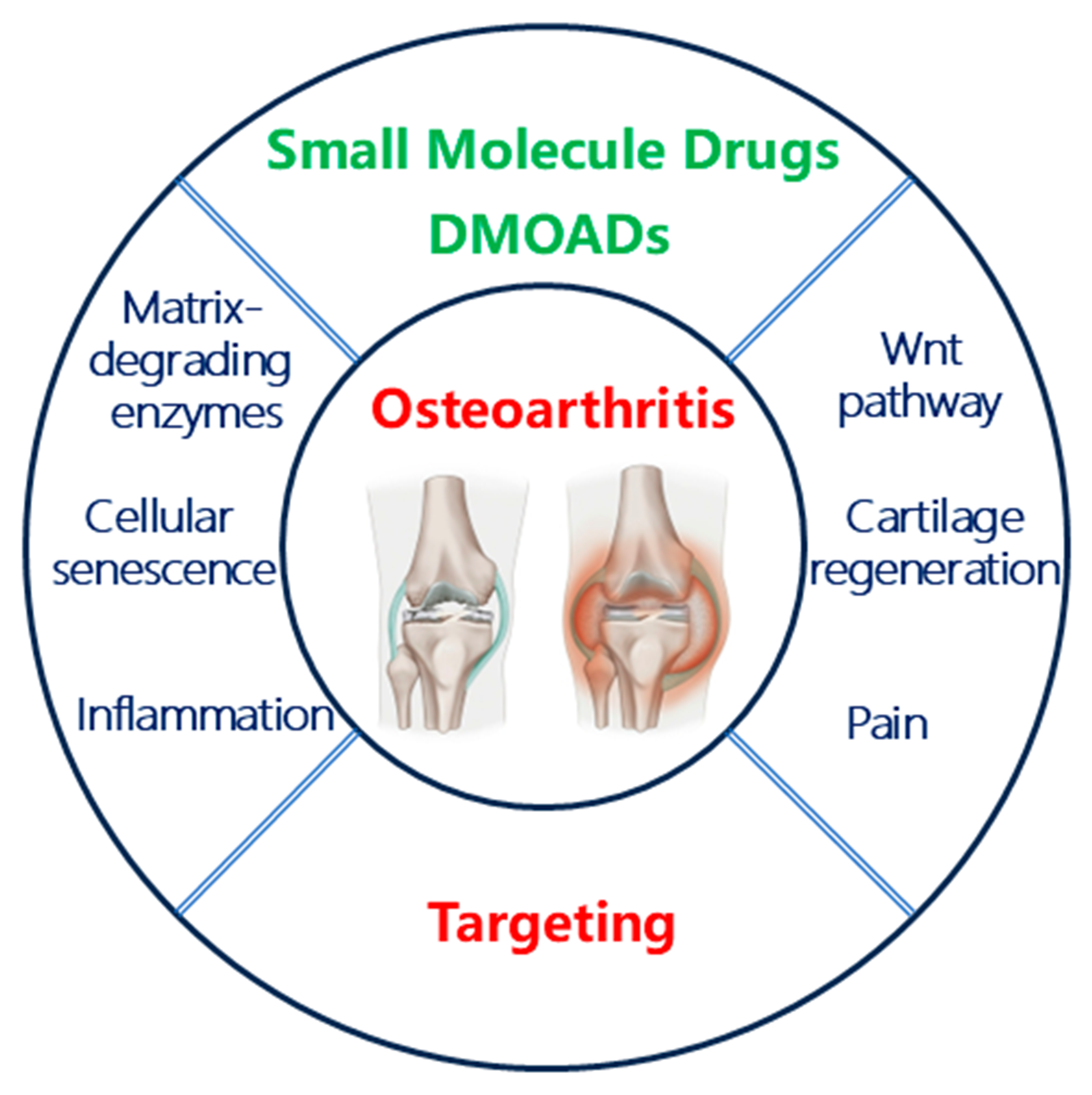
Osteoarthritis is a common degenerative disease in middle-aged and elderly people, and its lesions involve the entire joint, such as cartilage, subchondral bone and synovial membrane. Although there are some launched drugs that can be used to relieve the pain caused by osteoarthritis, the therapeutic effects are insufficient and there are some certain side effects. For now, except for artificial joint replacement for late stage of osteoarthritis, there are no effective drugs for early and middle stage of osteoarthritis to delay its progression. The treatment of osteoarthritis in clinical faces a huge demand for valid drugs, but the development of therapeutic drugs is way behind schedule. This review focuses on the progress of small molecule drugs for the treatment of osteoarthritis, especially the small molecule chemical entities which have entered the clinical trial stage. We hope this article will provide inspiration for the researchers on the drug development of osteoarthritis.

Contents

1 Introduction of osteoarthritis

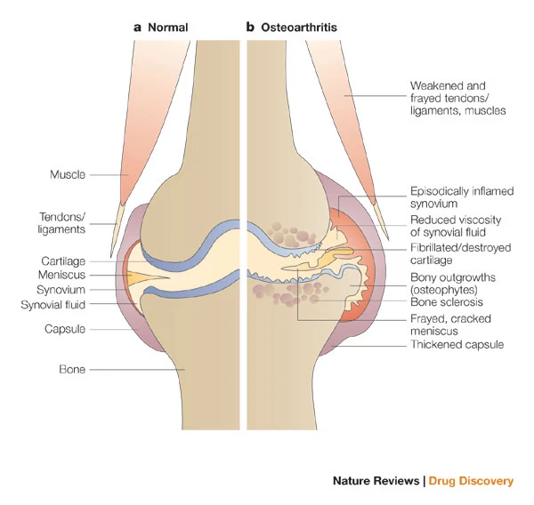
2 Features of osteoarthritis

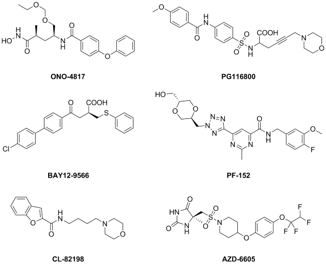
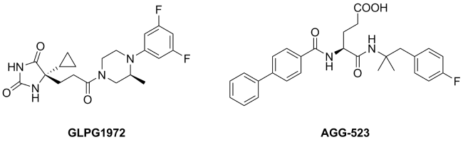
3 Small molecule drugs against osteoarthritis

3.1 Clinical small molecule drugs against osteoarthritis

3.2 The development of small molecule disease- modifying osteoarthritis drugs (DMOADs)

4 Conclusion and outlook

Cite this article

Xiaofei Zhang , Chunhao Yang . Recent Development of Small Molecule Drugs for the Treatment of Osteoarthritis[J]. Progress in Chemistry, 2024 , 36(12) : 1849 -1858 . DOI: 10.7536/PC241115

1 Introduction to Osteoarthritis

Osteoarthritis, also known as degenerative arthritis, osteochondral arthritis, and proliferative osteoarthritis, is a chronic joint disease characterized by the degeneration and destruction of articular cartilage, as well as bone proliferation and synovial inflammation[1]. There are many causes of osteoarthritis, including aging[2], obesity[3], genetics[4], gender[5], and joint injuries[6], all of which can potentially trigger osteoarthritis. Among all joints in the body, the weight-bearing joints such as the spine, hip, and knee are most affected, with the knee being particularly prone to osteoarthritis, having the highest incidence.
Osteoarthritis, as the most common degenerative joint disease causing pain and disability, has seen a yearly increase in the number of patients. The incidence of osteoarthritis rises with age, and epidemiological surveys from multiple countries have shown that the incidence of osteoarthritis in people over 60 years old is as high as 15%[7]. The most common symptom of osteoarthritis is pain, with clinical manifestations also including joint stiffness, swelling, deformity, and limited mobility, directly leading to a decrease in the quality of life for patients. Among them, 80% of osteoarthritis patients experience restricted movement, and 25% of patients are almost unable to work and live normally, with severe cases leading to incapacity and disability[1]. In developed countries, the direct medical costs and indirect socioeconomic losses caused by osteoarthritis amount to 1.0%~2.5% of the gross domestic product[8]. In 2018, the U.S. Food and Drug Administration recognized osteoarthritis as a serious disease with far unmet needs for therapeutic drugs[9]. This article focuses on reviewing the research and development progress of small molecule drugs for the treatment of osteoarthritis, especially those small molecular chemical entities that have entered the clinical research stage, aiming to provide references for researchers developing related small molecule chemical drugs for osteoarthritis.

2 Characteristics of Osteoarthritis

Osteoarthritis is a whole-joint disease with complex causative factors, resulting from an imbalance between joint tissue repair and destruction, leading to dynamic changes that involve structural alterations in articular cartilage, subchondral bone, ligaments, joint capsule, synovium, and periarticular muscles[10](Figure 1). Cartilage, subchondral bone, synovium, and inflammation may all play key roles in the pathogenesis of the disease, which involves factors such as stress, inflammation, and metabolism; these factors further directly or indirectly affect the course of osteoarthritis by activating or inhibiting various signaling pathways through specific mediators and receptors[11]. The following sections will elaborate on several typical pathological characteristics of osteoarthritis.
图1 受骨关节炎影响的关节结构[10]

Fig. 1 Structure of joints affected by osteoarthritis[10]

Cartilage is a special transparent tissue layer on the joint, covering the surface of the bone in the joint, which can reduce the friction generated by joint movement and has an anti-pressure effect. Chondrocytes are the only type of cells present in cartilage, supplied with nutrients from the subchondral bone. The structure and biochemical composition of cartilage are strictly regulated by chondrocytes. Due to the relatively simple structure of cartilage, its natural repair capacity is very poor. The degeneration and loss of articular cartilage leading to joint pain and functional impairment are clinically diagnosed as osteoarthritis, which is one of the most prominent features of osteoarthritis[12].
The lesions of subchondral bone, including bone marrow edema, osteophyte formation and remodeling, and neovascularization, generally develop earlier than cartilage degeneration. Abnormal subchondral bone remodeling and neovascularization can directly or indirectly cause cartilage destruction and pain. Compared with normal joints, the development of osteoarthritis is accompanied by the formation of osteophytes and cysts in the subchondral bone at the joint margins. The gradual thickening of the subchondral plate is another feature of progressive osteoarthritis, intuitively reflecting the damage to articular cartilage and changes in the properties of subchondral bone. Therefore, early changes in the subchondral bone are one of the early diagnostic features of osteoarthritis[13].
Synovitis is also one of the common features of osteoarthritis. In osteoarthritis, synovial cells proliferate significantly, and the synovial tissue becomes hypertrophic with increased vascularity. Synovial cells also release a large amount of inflammatory factors and degradative enzymes, leading to severe synovitis. The joint swelling caused by synovitis is also an important clinical indicator for the diagnosis of osteoarthritis[14].
The production of inflammation (redness, heat, pain, etc.) is a complex pathophysiological process. These inflammatory processes are activated by various signaling factors secreted by mast cells, macrophages, granulocytes, platelets, lymphocytes, nerve endings, and endothelial cells. During the progression of osteoarthritis, chondrocytes are activated by inflammatory signals originating from other joint structures (such as the synovium or subchondral bone) and secrete multiple pro-inflammatory cytokines, such as interleukin-β (Interleukin-β, IL-β), interleukin-6 (Interleukin-6, IL-6), and tumor necrosis factor-α (Tumour necrosis factor-α, TNF-α), leading to an inflammatory response that results in joint redness, heat, and pain. At the same time, to cope with inflammation and stress, osteoarthritic chondrocytes also produce a variety of matrix-degrading enzymes. The dysregulation of these degrading enzymes in osteoarthritic chondrocytes will further lead to cartilage degradation[15].
In osteoarthritis, nuclear factor kappa B (Nuclear factor kappa B, NF-κB) and mitogen-activated protein kinase (Mitogen-activated protein kinase, MAPK) pathways play a major role in the expression of matrix metalloproteinases (Matrix metalloproteinase, MMP) and inflammatory factors. The NF-κB signaling pathway is the main catabolic pathway in cartilage of osteoarthritis, and upregulation of the NF-κB signaling pathway is closely related to cartilage destruction, synovial inflammation, and subchondral bone resorption. This pathway and the associated receptor activator of nuclear factor-κB ligand (Receptor activator of nuclear factor-κB ligand, RANKL)/receptor activator of nuclear factor-κB (Receptor activator of nuclear factor-κB, RANK) pathway[16] are crucial for the differentiation and activation of osteoclasts (Osteoclasts, OC)[17].
The Wnt/β-catenin signaling pathway has been demonstrated in numerous in vitro and in vivo experiments to lead to the catabolism or loss of stable phenotype of articular chondrocytes upon its activation. The Wnt/β-catenin signaling pathway is involved in joint development, homeostasis, and remodeling, and has been shown to play a crucial role in cartilage matrix synthesis and bone formation[18].
Transforming growth factor-β (Transforming growth factor-β, TGF-β) signaling pathway plays a crucial role in cartilage development and maintenance of cartilage homeostasis. The downregulation and over-upregulation of the TGF-β/Smad 2/3 (Small mothers against decapentaplegic 2/3, Smad 2/3) pathway can both exacerbate osteoarthritis and cause abnormal cartilage function[19].
Pro-inflammatory cytokine IL-1β has a variety of biological activities, and the abnormal upregulation of IL-1β can lead to the exacerbation of inflammation and obstruction of chondrocyte metabolic pathways. These processes are, to some extent, involved in the occurrence and development of osteoarthritis. Reducing local IL-1β levels, blocking its specific receptors, or interrupting its signaling pathways holds significant research value in the field of osteoarthritis drug development[20].

3 Small Molecule Drugs for Osteoarthritis Treatment

3.1 Clinical Medications for Osteoarthritis Treatment

Drug therapy remains the primary measure for treating osteoarthritis, and currently, there is no drug that can block or reverse the pathological process of osteoarthritis. The existing clinical drugs can only alleviate some symptoms in the short term.
Common small molecule chemical drugs for osteoarthritis include analgesics, non-steroidal anti-inflammatory drugs (Non-steroidal anti-inflammatory drugs, NSAIDs), and hormonal medications[21]. Acetaminophen and NSAIDs are usually considered first-line treatments for osteoarthritis. Acetaminophen is suitable for patients with early-stage or mild osteoarthritis, has fewer side effects, and is less likely to cause gastrointestinal bleeding, but its pain-relieving effect is weaker. Non-selective NSAIDs simultaneously inhibit cyclooxygenase-1 (Cyclooxygenase-1, COX-1) and cyclooxygenase-2 (Cyclooxygenase-2, COX-2), often accompanied by side effects such as gastric ulcers. Selective NSAIDs act more on COX-2 and have better anti-inflammatory and analgesic effects. The most commonly used selective NSAIDs in clinical practice are celecoxib and meloxicam, with common adverse reactions being gastrointestinal dysfunction and potential cardiovascular disease risks. Patients with gastrointestinal diseases should use NSAIDs, including selective or non-selective NSAIDs with added gastric protectants, cautiously[22]. When other analgesics are ineffective or contraindicated, stronger analgesics such as weak opioids and anesthetic analgesics can be considered, but due to the addictive nature of opioids, the use of such drugs must be handled with great caution[23].
Intra-articular injection of glucocorticoids can effectively suppress the inflammatory response in patients with osteoarthritis, improving symptoms by inhibiting capillary dilation, reducing exudation and edema, and suppressing leukocyte infiltration and phagocytosis. However, excessive use of hormones may lead to cartilage repair disorders and cause severe secondary damage[24].
Oral chondroitin sulfate can also alleviate pain in patients with mild or moderate osteoarthritis, reduce the concentration of pro-inflammatory cytokines and transcription factors associated with inflammation, and reinforce cartilage-specific matrix by inhibiting hydrolases and preventing the oxidation of lipids and proteins, thereby preventing the denaturation of collagen in chondrocytes[25].
Oral glucosamine sulfate can also achieve the purpose of controlling pain and improving joint function, and its mechanism may be to reduce the production of peroxide groups to lower the inflammatory response, thereby controlling pain[26].
Chondroitin sulfate and glucosamine sulfate, although widely used, have been subject to ongoing debates regarding their efficacy and safety because they require long-term administration to achieve a certain therapeutic effect. A large number of clinically relevant studies are still in progress.

3.2 Progress in the Development of Small Molecule Drugs (DMOADs) for Improving Osteoarthritis Conditions

The progression of osteoarthritis includes continuous cartilage loss, subchondral bone remodeling, osteophyte formation, and synovial inflammation, constituting a chronic disability affecting the entire joint. Due to the numerous pathogenic factors of osteoarthritis, the research on related targeted drugs is difficult and progresses slowly; currently, the drugs used in clinical practice can only alleviate symptoms and hardly slow down the disease progression. In recent years, the development of drugs for improving osteoarthritis (Disease-modifying osteoarthritis drugs, DMOADs) has received increasing attention from medicinal chemists, with the aim of discovering drugs that can both slow down the progression of osteoarthritis and alleviate its symptoms[27]. So far, although no DMOADs have been approved for market, some small molecule compounds entering clinical trials still offer us a glimmer of hope for the discovery of new osteoarthritis drugs.
This article classifies drugs based on their targets or mechanisms of action and reviews the recent progress in the development of small molecule drugs for osteoarthritis.

3.2.1 Metalloprotease Inhibitors

The primary structural protein of cartilage is type II collagen, which provides a reticular structure for the cartilage, rich in various proteoglycans. These components increase the tensile and compressive strength of the cartilage[28]. Matrix metalloproteinases (MMPs) promote the degradation and destruction of type II collagen, being associated with various diseases including osteoarthritis. Theoretically, targeting MMPs could effectively protect cartilaginous tissues[29]. Yamada et al.[30] (Ono Pharmaceutical, Ono) reported the broad-spectrum MMP inhibitor ONO-4817 in 2000. This compound effectively inhibited the production of proteoglycans in the synovial fluid of a guinea pig model of osteoarthritis, demonstrating an inhibitory effect on articular cartilage degradation. Compound ONO-4817 was in Phase I clinical trials for treating osteoarthritis, but no further reports have been seen. The broad-spectrum MMP inhibitor PG116800 (Procter & Gamble) advanced to Phase II clinical studies (NCT00041756) for treating osteoarthritis, but it was terminated due to poor therapeutic efficacy and reversible musculoskeletal toxicity. Broad-spectrum MMP inhibitor BAY12-9566 (Bayer) showed better safety in preclinical studies, but further clinical research was discontinued due to insufficient efficacy[31]. Since broad-spectrum MMP inhibitors inhibit the degradation of many other matrix proteins while inhibiting the degradation of type II collagen, there may be certain risks[32]. In recent years, studies[33] have found that MMP-13 has high substrate selectivity for type II collagen. Selective MMP-13 inhibitors are gradually receiving attention from pharmaceutical companies and research institutions. For example, MMP-13 inhibitors such as PF152 (Pfizer), CL-82198 (Pfizer)[34], ALS 1-0635 (Alantos Pharmaceuticals AG, structure undisclosed)[35], and AZD-6605 (AstraZeneca)[36] have all shown excellent chondroprotective effects in animal models of osteoarthritis, but so far, no MMP-13 inhibitor has entered the clinical trial stage (Figure 2).
图2 基质金属蛋白酶抑制剂

Fig. 2 MMP Inhibitors

A disintegrin and metallo-proteinase with thrombospondin motif (ADAMTSs) is a newly discovered family of Zn2+ dependent metalloproteinases. Among them, ADAMTS-4 and ADAMTS-5 are key enzymes in the degradation of cartilage aggrecan. In animal models of osteoarthritis, inhibiting the activity of ADAMTS-4 and ADAMTS-5 can effectively slow down the loss of joint cartilage[37]. The compound GLPG1972 (Galapagos NV) is a highly selective and orally effective small molecule inhibitor of ADAMTS-5. Phase I clinical studies (NCT02851485/NCT03311009) confirmed the safety of this compound, but in a phase II clinical trial involving 938 patients (NCT03595618), the therapeutic effect of the compound did not reach the primary clinical endpoint[38], leading to the termination of further clinical research. Compound AGG-523 (Pfizer) is a dual inhibitor of ADAMTS-4 and ADAMTS-5, which showed excellent cartilage protection effects in rat osteoarthritis models. Subsequently, this compound entered phase I clinical studies[39] (NCT00427687 and NCT00454298), but further clinical research was terminated for unknown reasons (Figure 3).
图3 ADAMTSs抑制剂

Fig. 3 ADAMTSs inhibitors

3.2.2 Inhibitors of the Wnt/β-catenin Pathway

The Wnt signaling pathway is mediated by a series of Wnt family glycoproteins (19 types in mammals), with β-catenin being an important family protein in the canonical Wnt pathway[40]. The Wnt/β-catenin pathway plays a very important role in maintaining the homeostasis of joints, and it is significantly activated in osteoarthritis. The activated Wnt/β-catenin pathway then induces high expression of metalloproteinases, further leading to cartilage loss and inflammation[41].
Compound SM04690 (Biosplice Therapeutics Inc, generic name Lorecivivint) is a Wnt/β-catenin pathway inhibitor obtained through high-throughput screening, which has shown excellent cartilage-protective effects in rat osteoarthritis models[42]. Subsequent studies found that compound SM04690 downregulates the activity of the Wnt/β-catenin pathway by inhibiting intranuclear kinases CLK2 and DYRK1A[43]. In a Phase I clinical trial (NCT02095548) with 61 subjects, intra-articular injection of compound SM04690 effectively avoided systemic toxic side effects, confirming the safety of the compound[44]. In a Phase IIa study (NCT02536833, with 455 patients), compared to placebo, SM04690 did not meet the clinical endpoint set by the WOMAC pain score at 13 weeks. However, at 52 weeks, the 0.07 mg dose group (intra-articular injection) showed a significant improvement in pain compared to the placebo group[45]. In the Phase IIb clinical study (NCT03122860, with 695 patients), compound SM04690 demonstrated statistically significant pain relief in the 0.07 mg and 0.23 mg dose groups (intra-articular injection)[46] (WOMAC score). Currently, this compound is in Phase III clinical trials (NCT03928184, NCT04385303, and NCT04520607) and is one of the promising DMOADs.
In 2017, Lietman et al[47] reported that the compound XAV-939 showed good anti-cartilage damage and synovitis improvement effects in a mouse osteoarthritis model (intra-articular injection). In mechanistic studies, it was found that the compound exerts its effect by inhibiting the Wnt/β-catenin pathway, and this compound is still in the preclinical research stage (see Figure 4).
图4 Wnt/β-catenin通路抑制剂

Fig. 4 Wnt/β-catenin inhibitors

3.2.3 Cathepsin K Inhibitors

Cysteine cathepsins (Cathepsins) include 11 subtypes, among which Cathepsins B, H, K, and S are the four main types that degrade natural collagen[48]. Cathepsin K is significantly overexpressed in the joints of patients with clinical osteoarthritis[49], and multiple Cathepsin K inhibitors have shown excellent effects in inhibiting cartilage degradation and alleviating pain in animal models of osteoarthritis[50]. Among them, the representative compound MIV-711 (Medivir AB) is an orally effective small molecule inhibitor of Cathepsin K, which has demonstrated good safety in healthy individuals in a Phase I clinical study[51] (NCT03443453). However, it failed to meet the primary clinical endpoint (Numeric Rating Scale, NRS) in subsequent Phase IIa trials (NCT02705625 and NCT03037489). Radiographic examinations revealed that MIV-711 could indeed inhibit joint cartilage loss and alleviate the pain associated with osteoarthritis (WOMAC score). Further Phase IIb clinical studies for this compound have not yet commenced.
The compound GSK-462795 (GlaxoSmithKline) is also an orally effective small molecule inhibitor of Cathepsin K, which has shown good anti-bone resorption effects in the osteoarthritis model of cynomolgus monkeys[52]. This compound entered Phase I clinical trials as early as 2002, but no further reports have been seen. Some other small molecule Cathepsin K inhibitors have also entered clinical trial stages, such as AAE-581 (Novartis), which reached Phase II clinical trials for osteoarthritis treatment, but was terminated for unknown reasons; and MK-0822 (Merck Sharp & Dohme), which reached Phase III clinical trials for osteoarthritis treatment, but further research was interrupted due to increased risk of cardiovascular side effects and stroke[53](Figure 5).
图5 半胱氨酸组织蛋白酶K抑制剂

Fig. 5 Cathepsin K inhibitors

3.2.4 Chondrogenic Small Molecule Compounds

Targeting catabolic enzymes, DMOADs have shown some efficacy in slowing down cartilage loss but are insufficient in restoring and reconstructing cartilage tissue. The repair capacity of normal articular cartilage tissue rapidly declines with increasing age and joint damage. The goal of chondrogenic drugs is to restore the normal structure and function of damaged articular cartilage, with the main mechanisms of action being the promotion of stem cell differentiation into chondrocytes and the enhancement of chondrocyte cartilage matrix synthesis[54].
Kartogenin is a small molecule compound that promotes the differentiation of mesenchymal stem cells (Mesenchymal stem cells, MSCs) into chondrocytes. Kartogenin works by binding to filamin A, blocking the interaction between filamin A and the transcription factor core-binding factor β-subunit, thereby upregulating the expression of type II collagen and proteoglycans[54]. Due to poor metabolic stability and other reasons, Kartogenin has not advanced to clinical studies (Figure 6). Compound KA34 (Scripps Research Institute, structure undisclosed), a derivative of Kartogenin, shows better chondrogenic differentiation activity, metabolic stability, and safety in vitro compared to its predecessor[55]. KA34 has completed phase I clinical trials for intra-articular injection treatment of osteoarthritis, but the results have not been disclosed, and phase II clinical trials have not yet begun.
图6 Kartogenin的化学结构

Fig. 6 The chemical structure of Kartogenin

3.2.5 Anti-cellular Senescence Small Molecule Compounds

Cellular senescence refers to the irreversible termination of the normal cell cycle. In cartilage, oxidative stress associated with aging and stress loading leads to the accumulation of senescent chondrocytes, which trigger the formation of an arthritic joint microenvironment by secreting pro-inflammatory cytokines and proteases. These factors, known as senescence-associated secretory phenotype (SASP) factors, collectively accelerate cartilage degeneration and synovial inflammation in osteoarthritis[56].
The compound UBX-0101 (Unity Biotechnology Inc, structure undisclosed) is the first reported small molecule active compound with the activity of eliminating senescent chondrocytes, and it also showed good effects in reducing cartilage damage and pain relief in a mouse osteoarthritis model. The compound UBX-0101 exerts its function by promoting the apoptosis of senescent chondrocytes through inhibiting the binding of p53 to MDM2[55]. In an intra-articular injection treatment for osteoarthritis in a phase I clinical study (NCT03513016, number of subjects 48), UBX-0101 effectively alleviated osteoarthritis-induced pain at doses of 1-4 mg without significant side effects[57]. However, in a phase II clinical study (NCT04129944, number of patients 180), UBX-0101 did not meet the primary clinical endpoint, leading to the termination of further research.
Fisetin is a flavonoid compound (Figure 7), which has been reported to possess in vitro anti-senescence and anti-inflammatory activities, and it also demonstrates good joint-protective effects in a mouse meniscus injury model[58]. Currently, a phase I clinical study of Fisetin for the treatment of osteoarthritis via oral administration is underway (NCT04210986).
图7 Fisetin的化学结构

Fig. 7 The chemical structure of Fisetin

3.2.6 Anti-inflammatory and Analgesic Small Molecule Compounds

Interleukin-1 (IL-1) and tumor necrosis factor (TNF) are the most characteristic pro-inflammatory cytokines, which can stimulate the production of inflammatory mediators in the joint microenvironment, such as prostaglandin E, nitric oxide synthase, chemokines, and other cytokines. In addition, IL-1 and TNF can also directly promote the expression of matrix metalloproteinases (MMPs) and other matrix-degrading enzymes involved in cartilage degeneration, disrupting the structure of articular cartilage[59]. At present, there are no small molecule compounds targeting the release of IL-1 and TNF that have entered clinical trials for osteoarthritis treatment, but some compounds have been reported to have anti-osteoarthritic effects in animal models, such as anemonin[60] (which inhibits IL-1β), necrostatin-1[61] (which inhibits IL-1β), compound TAK-242[62] (which inhibits IL-1), and Cf-02[63] (which inhibits TNF-α) (Figure 8). In addition, inhibitors of some inflammation-related targets have also been reported to have in vivo and in vitro activities against osteoarthritis, such as NLRP3 inflammasome inhibitors[64-65] and nitric oxide synthase inhibitors[66].
图8 具有抗炎活性的小分子化合物

Fig. 8 Bio-active compounds targeting oa-associated inflammation

Chronic pain is one of the prominent symptoms of osteoarthritis, and alleviating pain is also an important goal in the clinical treatment of osteoarthritis. Chronic pain is regulated through the peripheral and central nervous systems, and structures within the joint cavity such as the joint capsule, synovium, subchondral bone, and ligaments are rich in nerve end receptors. Specific receptors at the peripheral nerve endings, such as thermal receptors, chemotactic receptors, and mechanoreceptors, can detect various stimuli in the joint cavity, including cytokines, chemokines, neuropeptides, and prostaglandins, thereby generating pain[67]. Research[68] has shown that the role of nerve growth factor (NGF) in the damaged joint environment is closely related to the pain experienced by patients with osteoarthritis.
NGF is a member of the peripheral and central nervous system trophic factors. In peripheral nociceptors, NGF interacts with its receptor tropomyosin-related kinase A (TrkA) to activate transient receptor potential cation channel subfamily V member 1 (TRPV1), collectively participating in tissue injury-related pain hypersensitivity[69]. The compound ASP7962 (Astellas, structure undisclosed) is an orally effective small molecule inhibitor of TrkA that advanced to Phase II clinical trials for osteoarthritis treatment but was terminated due to not meeting the primary clinical endpoint[70]. The compound GZ389988A (Sanofi) is also a small molecule inhibitor of TrkA, which entered Phase II clinical trials for intra-articular injection in treating osteoarthritis pain but was discontinued due to pipeline adjustments by the company. Trans-capsaicin (CNTX-4975, Centrexion Therapeutics), as an effective small molecule inhibitor of TRPV1, demonstrated a dose-dependent relationship between pain improvement and the compound in a Phase II clinical trial (NCT02558439, 175 patients). A 1.0 mg dose of CNTX-4975 significantly reduced pain over 24 weeks, while a 0.5 mg dose showed significant pain relief at 12 weeks but no obvious effect at 24 weeks[71]. Currently, CNTX-4975 is in Phase III clinical trials for the treatment of osteoarthritis pain (Figure 9).
图9 能减轻骨关节炎疼痛的小分子化合物

Fig. 9 Bio-active compounds targeting OA-associated pain

3.2.7 Academician Xie Yuyuan and Related Work of Our Research Group

There have been numerous studies on the use of anti-osteoporosis drugs for the treatment of osteoarthritis, but no related indications have been approved yet. Academician Xie Yuyuan discovered during the development of radioactive nuclide promoting excretion drugs that geminal diphosphonate compounds, which are also medical chelating agents, can be used for the treatment of bone metabolic diseases such as Paget's disease and osteoporosis[72-73]. At the end of the 1990s, Academician Xie Yuyuan's research group gradually began to study anti-osteoporosis drugs and was one of the earliest groups in China to conduct such research. The research group successfully constructed in vitro and in vivo osteoporosis evaluation models and completed the screening of a large number of active compounds, discovering a batch of compounds with good anti-osteoporosis activity in animals[74-77], among which Y-134 is a selective estrogen receptor modulator, with 121 times selectivity for ERα over ERβ, and its in vivo anti-osteoporosis effect is comparable to that of the marketed drug raloxifene[77-78]. This research group also modified the structure of rhein[79-80] and obtained a dual inhibitor that promotes osteogenesis while inhibiting osteoclasts[80], providing a potential direction for the study of new anti-osteoporosis drugs. RANKL is an important target for anti-osteoporosis drugs, and currently, only the antibody drug denosumab has been marketed, which achieved good results in phase IIa clinical trials for erosive hand osteoarthritis, but so far, no small molecule RANKL inhibitors have entered clinical trials. The research group has made certain progress in the study of small molecule RANKL inhibitors, obtaining non-peptide small molecules with good in vivo activity, such as Y1693[81-82], laying the foundation for further modifications. Given the complexity of the pathogenesis of osteoarthritis, the research group utilized the "multi-target" feature of natural products, modifying betulinic acid and oleanolic acid, and obtained active compounds YCH1919[83] and YCH2886[84] (Figure 10), which significantly improved osteoarthritis symptoms in animals. Related research is still ongoing.
图10 YCH1919和YCH2886的化学结构

Fig. 10 Chemical structures of YCH1919 and YCH2886

4 Conclusions and Prospects

As our understanding of the pathogenic mechanisms of osteoarthritis has deepened, a variety of DMOADs have been discovered and have made some progress in clinical trials. In this paper, we mainly elaborate on small molecule DMOADs with multiple mechanisms of action, such as targeting matrix degradation enzymes, the Wnt/β-catenin pathway, and anti-inflammatory effects to improve cartilage matrix degradation; chondrogenic DMOADs show good prospects in promoting stem cell chondrogenesis and cartilage matrix reconstruction; anti-cell senescence strategies are considered a new direction for DMOADs, capable of modulating previously undruggable targets in joint tissues, etc. Furthermore, given the complex pathogenesis of osteoarthritis, the "one drug, one target" research strategy is often ineffective, hence the "one drug, multiple targets" strategy is also applied to the discovery of anti-osteoarthritis drugs[85], especially natural products from herbs[86] and their modified compounds[83-84], providing new ideas for drug development for this disease. Overall, the development of anti-osteoarthritis drugs still has a long way to go.
Given the complexity of the pathogenesis of osteoarthritis, it is also very necessary to develop personalized osteoarthritis treatment guidelines for clinical guidance. In addition, there is a need to quickly develop biomarkers that can diagnose early-stage osteoarthritis. By detecting these markers, the use of DMOADs can be combined with the early diagnosis of osteoarthritis, allowing the full potential of DMOADs to be realized as early as possible, thereby ensuring therapeutic efficacy and a good prognosis.
[1]
Glyn-Jones S, Palmer A J R, Agricola R, Price A J, Vincent T L, Weinans H, Carr A J. Lancet, 2015, 386(9991): 376.

[2]
Shane Anderson A, Loeser R F. Best Pract. Res. Clin. Rheumatol., 2010, 24(1): 15.

[3]
Kulkarni K, Karssiens T, Kumar V, Pandit H. Maturitas, 2016, 89: 22.

[4]
Valdes A M, Spector T D. Rheum. Dis. Clin. N Am, 2008, 34(3): 581.

[5]
Gignac M A M, Irvin E, Cullen K, Van Eerd D, Beaton D E, Mahood Q, McLeod C, Backman C L. Arthritis Care Res., 2020, 72(3): 378.

[6]
Anderson D D, Chubinskaya S, Guilak F, Martin J A, Oegema T R, Olson S A, Buckwalter J A. J. Orthop. Res., 2011, 29(6): 802.

[7]
Li Y S, Xie W Q, Xiao W F, Dou D. Bone Res., 2022, 10: 41.

[8]
Thysen S, Luyten F P, Lories R J U. Dis. Models Mech., 2015, 8(1): 17.

[9]
Kraus V B, Simon L S, Katz J N, Neogi T, Hunter D, Guermazi A, Karsdal M A. Osteoarthr. Cartil., 2019, 27(4): 571.

[10]
Wieland H A, Michaelis M, Kirschbaum B J, Rudolphi K A. Nat. Rev. Drug Discov., 2005, 4(4): 331.

[11]
Deschaseaux F, Sensébé L, Heymann D. Trends Mol. Med., 2009, 15(9): 417.

[12]
Martel-Pelletier J, Boileau C, Pelletier J P, Roughley P J. Best Pract. Res. Clin. Rheumatol., 2008, 22(2): 351.

[13]
Hu Y, Chen X, Wang S C, Jing Y Y, Su J C. Bone Res., 2021, 9: 20.

[14]
Berenbaum F. Osteoarthr. Cartil., 2013, 21(1): 16.

[15]
Goldring M B, Otero M. Curr. Opin. Rheumatol., 2011, 23(5): 471.

[16]
Wada T, Nakashima T, Hiroshi N, Penninger J M. Trends Mol. Med., 2006, 12(1): 17.

[17]
Rigoglou S, Papavassiliou A G. Int. J. Biochem. Cell Biol., 2013, 45(11): 2580.

[18]
Velasco J, Zarrabeitia M T, Prieto J R, Perez-Castrillon J L, Perez-Aguilar M D, Perez-Nuñez M I, Sañudo C, Hernandez-Elena J, Calvo I, Ortiz F, Gonzalez-Macias J, Riancho J A. Osteoporos. Int., 2010, 21(1): 109.

[19]
Zhai G J, Doré J, Rahman P. Rheumatol. Int., 2015, 35(8): 1283.

[20]
Daheshia M, Yao J Q. J. Rheumatol., 2008, 35(12): 2306.

[21]
Walsh D A, Stocks J. SLAS Discov., 2017, 22(8): 931.

[22]
Roth S H, Anderson S. Phys. Sportsmed., 2011, 39(3): 62.

[23]
Jeffery M M, Hooten W M, Henk H J, Bellolio M F, Hess E P, Meara E, Ross J S, Shah N D. BMJ, 2018, 362: k2833.

[24]
Samuels J, Pillinger M H, Jevsevar D, Felson D, Simon L S. Osteoarthritis Cartilage, 2021, 29(1): 8.

[25]
Bottegoni C, Muzzarelli R A A, Giovannini F, Busilacchi A, Gigante A. Carbohydr. Polym., 2014, 109: 126.

[26]
Kucharz E J, Kovalenko V, Szántó S, Bruyère O, Cooper C, Reginster J. Y. Curr. Med. Res. Opin., 2016, 32(6): 997.

[27]
Oo W M, Little C, Duong V, Hunter D J. Drug Des. Dev. Ther., 2021, 15: 2921.

[28]
Krishnan Y, Grodzinsky A J. Matrix Biol., 2018, 71: 51.

[29]
Aranapakam V, Grosu G T, Davis J M, Hu B H, Ellingboe J, Baker J L, Skotnicki J S, Zask A, DiJoseph J F, Sung A, Sharr M A, Killar L M, Walter T, Jin G X, Cowling R. J. Med. Chem., 2003, 46(12): 2361.

[30]
Yamada A, Uegaki A, Nakamura T, Ogawa K. Inflamm. Res., 2000, 49(4): 144.

[31]
Leff R L, Elias I, Ionescu M, Reiner A, Poole A R. J. Rheumatol., 2003, 30(3): 544.

[32]
Vandenbroucke R E, Libert C. Nat. Rev. Drug Discov., 2014, 13(12): 904.

[33]
Schnute M E, O’Brien P M, Nahra J, Morris M, Howard Roark W, Hanau C E, Ruminski P G, Scholten J A, Fletcher T R, Hamper B C, Carroll J N, Patt W C, Shieh H S, Collins B, Pavlovsky A G, Palmquist K E, Aston K W, Hitchcock J, Rogers M D, McDonald J, Johnson A R, Munie G E, Wittwer A J, Man C F, Settle S L, Nemirovskiy O, Vickery L E, Agawal A, Dyer R D, Sunyer T. Bioorg. Med. Chem. Lett., 2010, 20(2): 576.

[34]
Wang M N, Sampson E R, Jin H T, Li J, Ke Q H, Im H J, Chen D. Arthritis Res. Ther., 2013, 15(1): R5.

[35]
Piecha D, Weik J, Kheil H, Becher G, Timmermann A, Jaworski A, Burger M, Hofmann M W. Inflamm. Res., 2010, 59(5): 379.

[36]
De Savi C, Waterson D, Pape A, Lamont S, Hadley E, Mills M, Page K M, Bowyer J, Maciewicz R A. Bioorg. Med. Chem. Lett., 2013, 23(16): 4705.

[37]
Glasson S S, Askew R, Sheppard B, Carito B, Blanchet T, Ma H L, Flannery C R, Peluso D, Kanki K, Yang Z, Majumdar M K, Morris E A. Nature, 2007, 446(7131): 102.

[38]
van der Aar E M, Desrivot J, Fagard L, Amantini D, Larsson S, Struglics A, Lohmander S, Vanhoutte F, Dupont S. Osteoarthr. Cartil., 2018, 26: S310.

[39]
Chockalingam P S, Sun W, Rivera-Bermudez M A, Zeng W, Dufield D R, Larsson S, Lohmander L S, Flannery C R, Glasson S S, Georgiadis K E, Morris E A. Osteoarthr. Cartil., 2011, 19(3): 315.

[40]
Nusse R, Clevers H. Cell, 2017, 169(6): 985.

[41]
Lambert C, Dubuc J E, Montell E, Vergés J, Munaut C, Noël A, Henrotin Y. Arthritis Rheumatol., 2014, 66(4): 960.

[42]
Kahn M. Nat. Rev. Drug Discov., 2014, 13(7): 513.

[43]
Deshmukh V, O’Green A L, Bossard C, Seo T, Lamangan L, Ibanez M, Ghias A, Lai C, Do L, Cho S, Cahiwat J, Chiu K, Pedraza M, Anderson S, Harris R, Dellamary L, Kc S, Barroga C, Melchior B, Tam B, Kennedy S, Tambiah J, Hood J, Yazici Y. Osteoarthr. Cartil., 2019, 27(9): 1347.

[44]
Yazici Y, McAlindon T E, Fleischmann R, Gibofsky A, Lane N E, Kivitz A J, Skrepnik N, Armas E, Swearingen C J, DiFrancesco A, Tambiah J R S, Hood J, Hochberg M C. Osteoarthr. Cartil., 2017, 25(10): 1598.

[45]
Yazici Y, McAlindon T E, Gibofsky A, Lane N E, Clauw D, Jones M, Bergfeld J, Swearingen C J, DiFrancesco A, Simsek I, Tambiah J, Hochberg M C. Arthritis Rheumatol., 2020, 72(10): 1694.

[46]
Yazici Y, McAlindon T E, Gibofsky A, Lane N E, Latterman C, Skrepnik N, Swearingen C J, DiFrancesco A, Tambiah J R, Hochberg M C. Osteoarthr. Cartil., 2019, 27: S503.

[47]
Lietman C, Wu B, Lechner S, Shinar A, Sehgal M, Rossomacha E, Datta P, Sharma A, Gandhi R, Kapoor M, Young P P. JCI Insight, 2018, 3(2): e96308.

[48]
Patel S, Homaei A, El-Seedi H R, Akhtar N. Biomed. Pharmacother., 2018, 105: 526.

[49]
Ben-Aderet L, Merquiol E, Fahham D, Kumar A, Reich E, Ben-Nun Y, Kandel L, Haze A, Liebergall M, Kosińska M K, Steinmeyer J, Turk B, Blum G, Dvir-Ginzberg M. Arthritis Res. Ther., 2015, 17(1): 69.

[50]
Hayami T, Zhuo Y, Wesolowski G A, Pickarski M, Duong L T. Bone, 2012, 50(6): 1250.

[51]
Lindström E, Rizoska B, Henderson I, Terelius Y, Jerling M, Edenius C, Grabowska U. J. Transl. Med., 2018, 16(1): 125.

[52]
Kumar S, Dare L, Vasko-Moser J A, James I E, Blake S M, Rickard D J, Hwang S M, Tomaszek T, Yamashita D S, Marquis R W, Oh H, Jeong J U, Veber D F, Gowen M, Lark M W, Stroup G. Bone, 2007, 40(1): 122.

[53]
Binkley N, Orwoll E, Chapurlat R, Langdahl B L, Scott B B, Giezek H, Santora A C. Osteoporos. Int., 2021, 32(1): 173.

[54]
Johnson K, Zhu S T, Tremblay M S, Payette J N, Wang J N, Bouchez L C, Meeusen S, Althage A, Cho C Y, Wu X, Schultz P G. Science, 2012, 336(6082): 717.

[55]
Johnson K A, Woods A K, Joseph S B, Chatterjee A K, Zhu S, Wisler J, Klyushnichenko V, Li J, Lotz M K, Tremblay M, Schultz P G. Osteoarthr. Cartil., 2020, 28: S518.

[56]
Jeon O H, Kim C, Laberge R M, Demaria M, Rathod S, Vasserot A P, Chung J W, Kim D H, Poon Y, David N, Baker D J, van Deursen J M, Campisi J, Elisseeff J H. Nat. Med., 2017, 23(6): 775.

[57]
Latourte A, Kloppenburg M, Richette P. Nat. Rev. Rheumatol., 2020, 16(12): 673.

[58]
Zheng W H, Feng Z H, You S B, Zhang H, Tao Z Y, Wang Q, Chen H, Wu Y S. Int. Immunopharmacol., 2017, 45: 135.

[59]
Chow Y Y, Chin K Y. Mediat. Inflamm., 2020, 2020: 8293921.

[60]
Wang Z Q, Huang J L, Zhou S R, Luo F T, Xu W, Wang Q, Tan Q Y chen L, Wang J, Chen H G, Chen L, Xie Y L, Du X L. J. Cell. Mol. Med., 2017, 21(12): 3231.

[61]
Liang S, Lv Z T, Zhang J M, Wang Y T, Dong Y H, Wang Z G, Chen K, Cheng P, Yang Q, Guo F J, Lu W W, Zhu W T, Chen A M. Front. Pharmacol., 2018, 9: 1378.

[62]
Wei Z, Sun X, Xu Q, Zhang Y, Tian F, Sun H, Zheng H, Dai J. Die Pharmazie, 2016, 71(10): 583.

[63]
Liu F C, Lu J W, Chien C Y, Huang H S, Lee C C, Lien S B, Lin L C, Chen L W, Ho Y J, Shen M C, Ho L J, Lai J H. Int. J. Mol. Sci., 2018, 19(5): 1453.

[64]
Pan D Y, Yin P, Li L K, Wu K L, Tong C G, Liu D P. Biochem. Biophys. Res. Commun., 2023, 657: 59.

[65]
Zhang Y, Lin Z, Chen D, He Y. Biochem. Biophys. Res. Commun., 2021, 553: 119.

[66]
Ahmad N, Ansari M Y, Haqqi T M. J. Cell. Physiol., 2020, 235(10): 6366.

[67]
Malfait A M, Schnitzer T J. Nat. Rev. Rheumatol., 2013, 9(11): 654.

[68]
Lane N E, Schnitzer T J, Birbara C A, Mokhtarani M, Shelton D L, Smith M D, Brown M T. N Engl J. Med., 2010, 363(16): 1521.

[69]
Caterina M J, Rosen T A, Tominaga M, Brake A J, Julius D. Nature, 1999, 398(6726): 436.

[70]
Watt F E, Blauwet M B, Fakhoury A, Jacobs H, Smulders R, Lane N E. Osteoarthr. Cartil., 2019, 27(11): 1590.

[71]
Stevens R M, Ervin J, Nezzer J, Nieves Y, Guedes K, Burges R, Hanson P D, Campbell J N. Arthritis Rheumatol., 2019, 71(9): 1524.

[72]
谢毓元, 陈文致. 化学通报, 1980, 5: 43.

[73]
谢毓元. 国外医学药学分册, 1984, 43.

[74]
Wang J B, Yang C H, Yan X M, Wu X H, Xie Y Y. Chin. Chem. Lett., 2005, 16(7): 859.

[75]
Xie Y L, Ding H S, Qian L H, Yan X M, Yang C H, Xie Y Y. Bioorg. Med. Chem. Lett., 2005, 15(13): 3267.

[76]
Ji Q G, Gao J, Wang J B, Yang C H, Hui X, Yan X M, Wu X H, Xie Y Y, Wang M W. Bioorg. Med. Chem. Lett., 2005, 15(11): 2891.

[77]
Yang C H, Xu G Y, Li J, Wu X H, Liu B, Yan X M, Wang M W, Xie Y Y. Bioorg. Med. Chem. Lett., 2005, 15(5): 1505.

[78]
Ning M, Zhou C, Weng J, Zhang S, Chen D, Yang C, Wang H, Ren J, Zhou L, Jin C, Wang M W. Br. J. Pharmacol., 2007, 150(1): 19.

[79]
Xu X, Qi X Y, Yan Y F, Qi J, Qian N D, Guo L, Li C W, Wang F, Huang P, Zhou H B, Jiang M, Yang C H, Deng L F. Eur. J. Med. Chem., 2016, 123: 769.

[80]
Jiang M, Wang T Q, Yan X M, Liu Z C, Yan Y F, Yang K, Qi J, Zhou H B, Qian N D, Zhou Q, Chen B, Xu X, Xi X B, Yang C H, Deng L F. J. Bone Miner. Res., 2019, 34(2): 361.

[81]
Jiang M, Peng L, Yang K, Wang T Q, Yan X M, Jiang T, Xu J R, Qi J, Zhou H B, Qian N D, Zhou Q, Chen B, Xu X, Deng L F, Yang C H. J. Med. Chem., 2019, 62(11): 5370.

[82]
Yang K, Li S Y, Wang T Q, Yan X M, He Q, Ning R N, Xu X, Yao W, Zhang X F, Yang C H, Jiang M, Deng L F. J. Med. Chem., 2022, 65(16): 10992.

[83]
Wang J, Wei W H, Zhang X F, Cao S Q, Hu B T, Ye Y, Jiang M, Wang T Q, Zuo J P, He S J, Yang C H. J. Med. Chem., 2021, 64(18): 13676.

[84]
Lin Z M, Xiang M, Wei W H, Fan S H, Chen L, Wang J, Yang X Q, Yang C H, He S J, Zuo J P. Science China Life Sciences, 2024. https://doi.org/10.1007/s11427-023-2587-2.

[85]
Anzini M, Di Capua A, Valenti S, Brogi S, Rovini M, Giuliani G, Cappelli A, Vomero S, Chiasserini L, Sega A, Poce G, Giorgi G, Calderone V, Martelli A, Testai L, Sautebin L, Rossi A, Pace S, Ghelardini C, Di Cesare Mannelli L, Benetti V, Giordani A, Anzellotti P, Dovizio M, Patrignani P, Biava M. J. Med. Chem., 2013, 56(8): 3191.

[86]
Yang T Y, Cao T T, Yang X G, Wang G L, Li Y L. Gene, 2024, 911: 148351.

Outlines

/