Home Journals Progress in Chemistry
Progress in Chemistry

Abbreviation (ISO4): Prog Chem      Editor in chief: Jincai ZHAO

About  /  Aim & scope  /  Editorial board  /  Indexed  /  Contact  / 
Review

Catechol Hydrogel as Wet Tissue Adhesive

  • Yiming Chen ,
  • Huiying Li ,
  • Peng Ni ,
  • Yan Fang , * ,
  • Haiqing Liu ,
  • Yunxiang Weng , *
Expand
  • College of Chemistry and Materials Science, Fujian Normal University,Fuzhou 350007, China
* Corresponding author, e-mail: (Haiqing Liu);
(Yunxiang Weng)

Received date: 2022-10-26

  Revised date: 2023-01-11

  Online published: 2023-02-16

Supported by

National Natural Science Foundation of China(52103108)

National Natural Science Foundation of China(22175037)

Key Project for Advancing Science and Technology of Fujian Province([2021]415-2021G02005)

Social Development of Instructive Program of Fujian Province(2020Y0020)

Education and research project for young and middle-aged teachers of Fujian Province(JAT210047)

Abstract

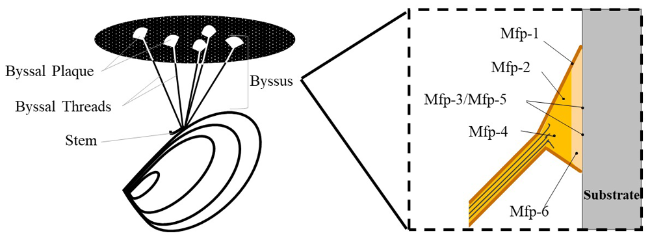
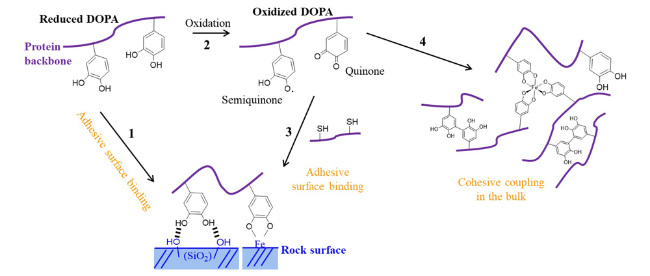
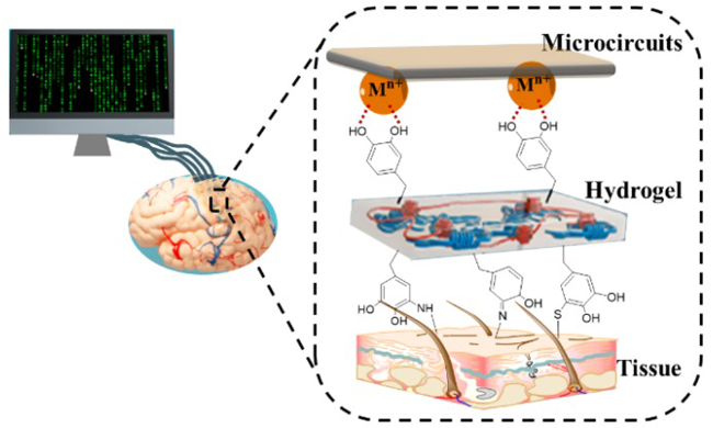
Wet adhesion plays an important role in the gestation and development of life. The research shows that hydrogel is a kind of intelligent material with both solid and liquid properties. They have been widely used in such areas as wound closure and repair, cell engineering and tissue engineering, owing to their noteworthy versatility and bio-compatibility. However, the physiological environment is usually wet, and the hydration layer on wet tissue surface prevents hydrogel from forming interfacial adhesion bonds with tissue surface. Faced with this challenge, inspired by the fact that the catechol group of DOPA is critical group for the underwater adhesion of mussels, the structure and functional unit design of catechol hydrogel have attracted wide attention. This review introduces the structure and wet adhesion mechanism of mussel foot proteins (Mfps), and the main types of catechol derivatives are classified into natural Mfps or Mfps synthesized by genetic engineering, catechol small molecular compounds, natural polymers modified by catechol groups and synthesized functional polymers containing catechol groups. Nextly, the research progress of catechol hydrogel as wet tissue adhesive in the past decade is summarized, such as tissue wound repair materials, biological coating materials, targeted drug delivery materials and bioelectronic equipment materials. Finally, the opportunities and challenges of catechol hydrogel are prospected.

Cite this article

Yiming Chen , Huiying Li , Peng Ni , Yan Fang , Haiqing Liu , Yunxiang Weng . Catechol Hydrogel as Wet Tissue Adhesive[J]. Progress in Chemistry, 2023 , 35(4) : 560 -576 . DOI: 10.7536/PC221016

Contents

1 Introduction
2 Composition and adhesion mechanism of mussel foot protein
3 Catechol derivatives used in wet tissue-adhesion hydrogel
3.1 Natural Mfps or Mfps synthesized by genetic engineering
3.2 Catechol small molecular compounds
3.3 Natural polymers modified by catechol groups
3.4 Synthesized functional polymers containing catechol groups
4 Catechol hydrogel and its functional application
4.1 Tissue wound repair materials
4.2 Biological coating materials
4.3 Targeted drug delivery materials
4.4 Bioelectronic equipment materials
5 Conclusion and outlook

1 Introduction

Wet adhesion behavior is ubiquitous in nature and organisms, and plays an important role in the gestation and development of life. On land, snails and salamanders can climb on wet stones and trees. In the ocean, mussels and barnacles thrive in seawater, firmly adhering to reefs or smooth skin surfaces[1]. In organisms, cell-cell adhesion and cell-extracellular matrix adhesion are involved in cell migration, proliferation, and differentiation. Strong adhesion between tendon, ligament, and cartilage and soft tissue muscle and hard tissue bone is a key factor in the action of organisms[2]. Inspired by these miraculous life phenomena, people have studied the wet adhesion problem in daily life in order to provide feasible solutions for the rapid and effective closure of wet tissue wounds, the development of drug delivery and implantable devices, which are important directions related to people's life and health needs.
As a class of smart materials with both solid-liquid properties, hydrogels have been widely used in the biomedical field due to their versatility and biocompatibility in mimicking the properties of natural tissues[3~6][7~12][13][14,15]. Traditional wound closure methods generally use sutures and staples, which can lead to stress concentration and tissue damage at the suture site of the wound, and subsequent removal may also cause secondary injury. Therefore, there is an urgent need to develop safe and efficient adhesion materials that do not damage tissues in the biomedical field. However, the physiological environment is usually humid, and hydrogels with strong interfacial adhesion in the wet state need to be developed to achieve this goal. Compared with the dry environment, there is a hydration layer on the surface of the wet material, which hinders the formation of an effective interaction force between the adhesive and the surface of the material. Moreover, water droplets trapped at the interface can reduce the contact area of the interface. On the other hand, when the material is exposed to moisture for a long time, water molecules will penetrate into the adhesive, which will cause plasticization, swelling, erosion, degradation or hydrolysis of the adhesive, and eventually lead to adhesive failure. In response to these problems, researchers at home and abroad have reported a variety of wet tissue adhesion hydrogels that mimic marine biological strategies[16].
In nature, some marine organisms can secrete adhesion proteins to make them firmly adhere to various substrate surfaces underwater. For example, mussels rely on byssus and its secretion of Mussel byssus protein (Mussel foot proteins, Mfps) to tightly adhere their bodies to moist and hard reef surfaces[17]. Sandcastle worms protect themselves by assembling sand and stones into tubular shell structures through secreted Pc1 adhesion protein[18]. Barnacles adhere their hard shells to the rock through Cement proteins (CPs)[19]. Studies have shown that the adhesion proteins secreted by these marine organisms contain DOPA, in which the catechol group (also known as catechol) is one of the important groups mediating underwater adhesion[20]. In this paper, the composition and adhesion mechanism of mussel byssus protein were introduced, and the catechol derivatives used in wet viscose were described, and the research status and application of wet tissue adhesion hydrogel containing catechol groups were further reviewed, and finally the development of this kind of material was prospected.

2 Composition and Adhesion Mechanism of Mytilus byssus Protein

Mfps can attach to the surface of any wet medium without being torn, which benefits from the strong adhesion and cohesion of Mfps[21]. At present, studies have found that there are eight kinds of Mfps: Mfp1 ~ 6, preCoID and preCoING, of which Mfp1 ~ 6 have adhesion, and the latter two collagens are the main components of the core skeleton of mussel byssus fibers[22,23].
Mfps play an important role in the adhesion of mussels and the maintenance of mussel byssus morphology, and their distribution is shown in Figure 1[24]. Mfp-1, which exists on the surface of byssus, was first discovered, and it has a protective effect on the core and plaque of byssus. Its molecular weight is 108 kDa, and it is composed of 75-80 repeated polypeptide fragments. There are hydroxylation modifications in the sequence, and the content of DOPA is 10% -15%[25]. Both Mfp-2 and Mfp-6 are distributed in the byssus disc. Mfp-2 has a molecular weight of about 45 kDa, contains 11 consensus sequences for epidermal growth factor to bind to metal ions Ca2+, and can be linked by intramolecular disulfide bonds. It is considered to be a stabilizer of byssus[25,26]. In addition, the content of DOPA is less than 5%. The molecular weight of Mfp-6 is 11.6 kDa, and the content of DOPA is less than 2 mol%. Mfp-6 is rich in free cysteine, which reacts with dopaquinone to form cross-links to prevent DOPA from being oxidized. Moreover, the sulfhydryl group in cysteine can be used as a reducing agent to reduce DOPA quinone to DOPA, thereby enhancing the lasting adhesion ability of mussel protein[26]. The adhesion protein in the byssus disc near the byssus fiber is Mfp-4, which has a molecular weight of 90 kDa and a DOPA content of only 2 mol%. This protein contains a histidine-rich decapeptide sequence repeated 35 times, which is essential for the connection between byssus disc adhesion proteins[26]. Mfp-3 and Mfp-5 play a major role in the adhesion of byssus proteins, and they exist in the adhesion between byssus disc and medium, with very low molecular weights of 5 ~ 7 kDa and 9 kDa, respectively, but with high DOPA content of 10 mol% ~ 20 mol% and 30 mol%, respectively[27]. Despite the large span in molecular weight, these byssus proteins all contain DOPA. Studies have shown that the adhesion ability of mussel will decrease after DOPA is oxidized, so DOPA is the key substance for mussel to achieve strong adhesion and diversification of adhesion on the surface of wet medium[28].
图1 Mfps的分布示意图

Fig.1 Distribution map of Mfps

The catechol group in DOPA is chemically active, because the catechol group has a lone pair of electrons, which can not only accept hydrogen atoms from hydrogen donors, but also provide hydrogen atoms for other active groups.It can interact chemically or physically with a variety of substances, including hydrogen bonding, metal-catechol coordination bonds, π-π interactions, π-cation interactions, oxidative cross-linking, and electrostatic interactions[29,30]. These forces not only exist inside the material to improve its cohesion, but also distribute at the interface between the material and the substrate to increase the adhesion force. In view of this, substances with catechol as the terminal chain or side chain have been designed and developed one after another to prepare wet viscose[26,31]. The interaction of catechol with different substrates also affects the adhesion properties of Mfps. For example, catechol groups can form coordination bonds with metal ions on the surface of the medium and hydrogen bonds with oxygen atoms of SiO2 at the bonding site, resulting in strong wet adhesion (Fig. 2, 1 path).
图2 儿茶酚基团的作用机理图

Fig.2 Action mechanism diagram of catechol group

In addition, the catechol group is easily oxidized to form semiquinone or benzoquinone under the action of oxidants, enzymes, or slightly alkaline seawater (high pH) (Fig. 2, 2 pathway), so catechol is of wide interest in pH-responsive wet adhesives[32]. The sulfhydryl group in Mfp-6 can maintain its redox balance and achieve long-term and lasting adhesion[33]. Based on this, Bian Liming et al. Used thiourea-quinone coupling reaction to synthesize thiourea-functionalized polymers with durable adhesion[34]. The structure of benzoquinone is unstable, and it is easy to self-polymerize and cross-link. It can undergo Michael addition reaction or Schiff base reaction with nucleophilic groups (amino group, sulfhydryl group in cysteine, etc.). This covalent bond is also often used to design organizational interface adhesion and enhance adhesion to the surface of the medium (Fig. 2, 3)[35]. Benzoquinone can also complex with metal ions Fe3+ to improve strength (Figure 2, 4 pathway), which has attracted much attention in the design of self-healing materials[36].
On the other hand, with the cooperation of hydrophobic amino acids, the positively charged lysine is able to disrupt the hydration layer and form a strong electrostatic interaction with the negatively charged rock surface, resulting in strong wet adhesion of Mfps[37]. At the same time, the positively charged lysine can also form cation-π interactions with DOPA and other aromatic amino acid residues, which greatly improves the cohesive strength of Mfps under strong hydrophobic interaction[19]. The study confirmed that the non-polar hydrophobic group can protect catechol from oxidation, which is of great significance in the long-term wet adhesion process[20].

3 Catechol Derivatives Applied to Wet Tissue Adhesive

The Mfps secreted by mussels has the ability of rapid solidification, strong adhesion and diversity of adhesion under water, so that mussels can firmly adhere to the surface of rocks, hulls and other media in humid environment, and withstand long-term scouring by seawater. In recent years, inspired by the strong wet adhesion ability of mussel, scholars at home and abroad have prepared wet tissue adhesion hydrogels using a series of catechol derivatives, which can be roughly divided into four types according to the source of raw materials: (1) natural Mfps or Mfps synthesized by genetic engineering; (2) small molecules containing catechol groups[20]; (3) natural polymer modified by catechol group; (4) Synthetic functional polymers containing catechol groups.

3.1 Natural Mfps or Mfps synthesized by genetic engineering

Mfps has the advantages of diversified adhesion, water resistance, fast curing speed, corrosion resistance, good bioaffinity, no toxic effect, antibacterial activity and no immune reaction in human body, and has important research value in the aspects of organism protective film, tissue wound adhesive and cell attachment coating[38]. In order to obtain this kind of material, it is undoubtedly the most direct and effective method to extract the natural adhesion protein component directly from mussel, and this research system is non-toxic and harmless to the environment[39]. As early as the 1990s, Ohkawa et al. Purified a protein with a molecular mass of 96 kDa and DOPA from the feet of Asian mussels by acid urea extraction, 36 wt% ammonium sulfate precipitation, propylene dextran gel filtration, and reverse-phase high performance liquid chromatography[40]. In 2017, Zhou Yajun et al. Used high-intensity pulsed electric field (PEF) to extract protein from mussels[41]. However, the yield of Mfps in mussels is small and the extraction is limited, which is difficult to meet the needs of practical application.
At the same time, the rise of genetic engineering and molecular biotechnology has made artificial mussel byssus protein possible. These technologies generally produce Mfps through E. coli expression, which has a higher yield than that extracted directly from mussels[42]. However, E. Coli-derived Mfps tend to lack DOPA because it cannot be post-translationally modified by itself, which severely limits underwater adhesion[43]. Therefore, attempts have been made to incorporate DOPA into E. Coli-derived Mfps to obtain bioengineered Mfps (dMfps). For example, Jeong et al. Studied the co-expression of DOPA recognizing tyrosyl-tRNA synthetase, and achieved the expression of dMfp-3 in E. coli with a yield of 72% and a DOPA incorporation rate of more than 90%[44]. dMfps can be processed as solutions, nanofibrils, coacervates, and hydrogels, and have great potential in medical and biotechnological applications, especially as bioadhesives and coating materials[45].
Based on the protein sequence of Mfps, scholars have successively imitated and designed homologous derivatives of mussel byssus protein to meet the research needs of biomedical and marine applications. For example, Wei et al. Designed a peptide analogue consisting of 25 amino acids (mfp3S-pep), which has similar self-aggregation ability to natural Mfps at a given pH and ionic strength, and can be applied to a variety of wet polar surfaces[46]. For example, Fichman et al. Synthesized a new peptide rich in lysine and dopa by self-assembly of Mfp-5, and prepared an antibacterial adhesive hydrogel (Fig. 3), which can be used as a coating or locally injected into the tissue implantation site[47]. It can be seen that the exploration of catechol derivatives of Mfps has been paid more and more attention, and the use of Mfps or the design of its homologous catechol derivatives to prepare tissue adhesion materials has become a research hotspot in the field of biomedicine.
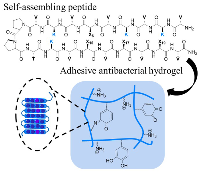
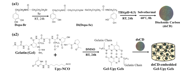
图3 含有多巴、赖氨酸、精氨酸和酪氨酸残基的自组装肽示意图,肽组装成纤维网络,并且这些残基的侧链具有局部纤维区域特异性

Fig.3 Schematic of self-assembling peptide showing sequential positions of DOPA, lysine, arginine and tyrosine residues. Peptides assemble into fibrillar networks displaying the side chains of these residues with local fibril regiospecificity

3.2 Catechol-containing small molecular compound

Natural catechol compounds widely exist in nature, among which DA (dopamine, 3,4-dihydroxyphenethylamine), TA (tannic acid), UH (urushiol) and so on are more common (Fig. 4), and are often used to prepare bioadhesive materials. DA is a kind of brain secretion, which is famous for its good biocompatibility, strong bioadhesion and multiple active sites, and its performance in materials is very similar to that of DOPA. Polydopamine (PDA) with complex covalent structure is synthesized by oxidative polymerization of DA after oxidation, cyclization and isomerization, which has good biocompatibility, hydrophilicity and thermal stability, and is widely used as biomedical materials[48].
图4 (a)TA、(b)DA以及(c)UH的分子结构式

Fig.4 Molecular structure of(a) TA, (b) DA and (c) UH

TA is a kind of plant polyphenolic small molecule, which has the advantages of antioxidant, antibacterial, anti-inflammatory and biodegradable. Due to the existence of polyphenol structure, it has a variety of interaction sites, including hydrogen bonding, ionic bonding, coordination bonding, hydrophobic interaction and Van der Waals force[49]. Therefore, the preparation of excellent tissue adhesion hydrogels by using the various characteristics of TA has become a hot research topic, such as TA/cellulose nanocrystals/polyvinyl alcohol hydrogel, methacrylated gelatin/TA hydrogel, polyacrylic acid/chitosan /TA/Al3+ hydrogel and so on[50][51][52]. TA can not only act as a crosslinking agent to increase physical crosslinking points to induce gelation or enhance the mechanical properties of hydrogels; It can also interact with various substrates (organic, inorganic) through the polyphenolic hydroxyl structure, thereby enhancing the adhesion of the hydrogel[53~55].
UH is a cheap and abundant natural catechol derivative derived from Rhus vernicifera. The long-chain unsaturated alkyl side chain on the catechol group in its structure can be embedded into the polymer network through vinyl copolymerization to avoid the loss of small molecules or dissolution in solvents[56]. For the first time, UH was used to prepare tissue adhesion hydrogel in our research group. Acrylamide (AAm) and UH were used as monomers to prepare freeze-resistant and heat-resistant wet adhesion poly (acrylamide-co-urushiol) hydrogel (PAAm-co-PUH) by free radical polymerization in glycerol/water binary solvent[57]. The UH component endows the hydrogel with hydrophobic association and π-π interactions, which act as reversible physical crosslinking points to enhance the mechanical strength and toughness of the hydrogel; Hydrophobic interaction removes the hydration layer at the interface, and catechol groups contribute to the diversification of adhesion and underwater adhesion.

3.3 Catechol-modified natural polymer

Natural polymers, including peptides, proteins and polysaccharides, have good biocompatibility and biodegradability, and contain a large number of active groups (such as —NH2, -COOH, -OH, etc.) Which are beneficial to chemical modification.Bioadhesive materials prepared by catechol modification can not only maintain the activity of biomacromolecules, but also endow them with wet adhesion, hemostasis and bacteriostasis. In addition, the hydroxyl group of the catechol group can easily lose electrons, and the oxidized catechol group has high chemical activity and can be grafted to the molecular chain of other polymers under the action of oxidants (such as oxygen, hydrogen peroxide, and periodic acid) and enzymes (such as tyrosinase and horseradish peroxidase)[58].
Gelatin is a hydrolysate of collagen and a widely used polypeptide in the field of hemostatic materials. Guo Baolin et al. Covalently crosslinked dopamine-modified gelatin and dopamine at low temperature to prepare a series of biodegradable interpenetrating polymer network (IPN) dry low-temperature gel hemostatic agents (GT/DA), which have wet adhesion and hemostatic properties[59]. In addition, a series of biomacromolecule-catechol gel systems based on proteins (fibrin, bovine serum albumin, etc.) Have been developed and designed[60]. Zhu et al. Used bovine serum albumin (BSA), citric acid (CA) and dopamine to make "BCD" tissue glue[61]. Successful experiments in seroma prevention and immediate hemostasis have demonstrated the potential of BCD tissue glue as a medical tissue adhesive.
Natural polysaccharides are a class of natural polymers composed of aldoses or ketoses linked by glycosidic bonds, which widely exist in organisms.Including chitosan, hyaluronic acid, cellulose, sodium alginate, fructan, etc. Using natural polysaccharide materials as substrates for various functional designs in the biomedical field has become a hot topic in the field of scientific research[62,63]. These polysaccharides contain a large number of hydroxyl groups and amino groups, and have high reaction activity sites and active centers for crosslinking reaction, which can be grafted with catechol groups to synthesize a three-dimensional network structure with polysaccharides as monomers and catechol groups as side chains.For example, catechol conjugated chitosan (CS-C), catechol functionalized quaternized chitosan (QCS-CA), gallic acid modified chitosan (CS-GA), catechol-conjugated aldehyde-modified hyaluronic acid (AH-CA), DA-amidated carboxymethyl cellulose (CMC-DA), sodium alginate-catecholamine (Alg-C), and catechol-conjugated fructan (Levan-Cat) etc. (Fig. 5)[64][65][66][67][68][69][70].
图5 儿茶酚功能化多糖的结构式

Fig.5 Structures of catechol-functionalized polysaccharide

3.4 Synthetic functional polymer containing catechol group

Common synthetic polymers have the characteristics of many candidates, simple preparation and low cost. Common synthetic polymers include polyethylene glycol, polyvinyl alcohol, polyacrylic acid and its derivatives, polyacrylamide, etc. At present, small molecular catechol derivative such as 3, 4-dihydroxyphenylalanine, 3, 4-dihydroxyphenylethylamine, 3, 4- dihydroxyphenylpropionic acid and that like are mainly coupled or embed to the tail end or the side chain of a synthetic macromolecular polymer skeleton,Such as catechol-capped polyethylene glycol (cPEG), DOPA modified polyvinyl alcohol (PVA-DOPA), catechol-acrylic copolymer (P (Cat-AA)), dihydrocaffeic acid modified polyamide (PPCA), catechol functionalized pressure sensitive adhesive (PSA-DMA), etc. (Fig. 6)[71][72][73][74][75][76]. Subsequently, a three-dimensional network polymer material is formed, which can replicate the magical properties of natural mussel byssus protein. Based on this, the introduction of catechol derivatives is expected to endow hydrogels with various excellent properties, including cell affinity, adhesion and high toughness, and different polymer backbones show different excellent properties due to their different polarities[77].
图6 儿茶酚功能化合成高分子的结构式

Fig.6 Structures of catechol-functionalized synthesized polymer

At present, the common research method of mussel-like adhesion materials is to take marine mussels as the research object, explore their main adhesion protein composition and adhesion mechanism, and imitate their adhesion behavior, so as to prepare adhesives with strong wet adhesion performance. For example, Xu Hong and Chi Bo designed an Mfp-5 mimetic polymer (Fig. 7), which is dopamine-modified ε-polylysine, and developed a biomimetic dopamine-modified ε-polylysine-polyethylene glycol-based hydrogel wound dressing (PPD hydrogel) in situ by using horseradish peroxidase crosslinking technology[78]. The PPD polymer provides a catechol-lysine synergistic effect, giving the PPD hydrogel superior wet tissue adhesion properties (up to 147 kPa). In addition, the preparation of tissue adhesion hydrogels by radical polymerization of vinyl-functionalized catechol monomers has attracted much attention. Just like the hydrogel prepared by Lu Xiong et al. Using dopamine modified methacrylamide (MADA), 2- (dimethylamino) ethyl methacrylate, AA and QCS,Another example is the synthesis of methacrylamide dopamine (DMA) and the preparation of doped hydrogels by Zhao et al., in which the catechol groups of MADA and DMA endow these hydrogels based on mussel catechol chemical strategy with long-term effective adhesion ability[79][80].
图7 Mfp-5模拟聚合物示意图

Fig.7 Schematic diagram of Mfp-5 mimetic polymer

4 Catechol-containing wet tissue adhesion hydrogel and its functional application

Wet tissue adhesion hydrogel is a kind of soft material with both adhesion and cohesion, which can firmly adhere to soft and irregular biological tissues in wet environment or underwater. The surface adhesion between hydrogel and biological tissues is mainly due to chemical bonds or non-covalent interactions between hydrogels and active groups in biological tissues[81][82,83]. Many functional groups (such as hydroxyl, carboxyl, or catechol) in wet tissue adhesion hydrogels can react with functional groups (such as carboxyl, amino, and thiol groups) on the tissue surface to form covalent bonds such as imines and amides, or non-covalent interactions such as hydrogen bonds, cation-π interactions, and mechanical interlocking at the interface. Where covalent bonds are generally strong and irreversible, and non-covalent bonds are relatively weak but reversible, reproducible adhesion and peeling. In addition, the tissue wet adhesion hydrogel has good biocompatibility, hemostasis, antibacterial properties and the like. Therefore, such hydrogels are widely used in wound repair, biological coating, targeted drug delivery materials, and bioelectronics.

4.1 Tissue wound repair material

In recent years, there has been an increasing demand for medical tissue adhesives, which can simplify surgical procedures, reduce pain for injured patients, and minimize trauma caused by the use of sutures and staples[84,85]. In addition, the tissue adhesive may be adapted for torsional motion of biological tissue. In summary, medical tissue adhesives require strong adhesion and cohesion, especially for applications where material longevity is a requirement. However, currently approved synthetic adhesives suffer from poor adhesion in the presence of biological fluids, cytotoxicity, and slow degradation[86][87][88]. Therefore, the development of high-performance and easy-to-use adhesives has become a hot research topic. The ideal medical adhesive should be biodegradable, non-durable, safe, non-toxic, non-carcinogenic, non-teratogenic, non-mutagenic and biocompatible.
From the point of view of bionics, the construction of biocompatible hydrogels with a variety of physical, chemical and biological functions should mimic the hydrogel properties, elasticity and mechanical behavior of biological soft tissues and extracellular matrix (ECM) composed of collagen, glycoprotein/proteoglycan and glycosaminoglycan. These biocompatible polymer hydrogels are generally characterized by high water content, injectability, adaptability, excellent mechanical properties, good wound adhesion and healing, and low toxicity risk. In order to enhance wet tissue adhesion, catechol derivatives were used to modify the hydrogel to make it adhere to wet tissue, inspired by Mfps. Inspired by the microstructure and function of ECM and the catechol chemistry of mussel, Dong et al. Grafted catechol (DA) and glucose (Glu) groups onto the backbone of poly (L-lysine) (PLL) to synthesize a glycopeptide hydrogel (PGD) with coordinated and covalently cross-linked network (Fig. 8)[36]. PGD has adjustable tissue adhesion strength (14. 6-83. 9 kPa) and microporous structure (8-18 μm), and its hemostatic speed is only 14 s, and the minimum blood loss is 6%. In this work, a general method for the preparation of glycopeptide hydrogels with tunable adhesion and microporous structure was constructed, which also provided a reference for the design of high-performance hemostatic and excellent healing hydrogels. In addition, our research group simulated the functional group and chemical structure of Mfps, and prepared a super-tough underwater tissue-selective adhesion hydrogel by free radical polymerization of acrylic acid (AA), chitosan (CS) and UH solution, which may replace the traditional tissue suture and has a wide range of applications in surgery[20].
图8 配位和共价糖多肽水凝胶(即R-凝胶和V-凝胶)的示意图;HRP:辣根过氧化物酶

Fig.8 Schematic diagram for coordinated and covalent glycopolyeptide hydrogels (i.e., R-Gels and V-Gels); HRP: Horseradish peroxidase

In order to adapt to the stretching movement of biological tissues, the tissue adhesive used for wound dressing should have the properties of self-repair and mechanical toughness. Adhesion of tissue glue to wound tissue must also have excellent cell affinity and tissue adhesion for practical use. Liu et al. Prepared a hydrogel with excellent cell affinity, strong tissue adhesion, super toughness and self-repair[89]. The self-healing ability of hydrogels is obtained by hydrogen bonding and dynamic Schiff base cross-linking between dopamine-grafted oxidized sodium alginate (OSA-DA) and polyacrylamide (PAM) chains, where covalent cross-linking is responsible for mechanical structural stabilization. It is important to note that open wounds (e.g. Burns and trauma) are susceptible to bacterial infection, leading to necrosis and sepsis[90,91]. Therefore, there is an urgent need to develop new wound adhesives that can prevent bacterial infection and promote healing. Li Peng et al. Developed a new type of open wound healing antibacterial hydrogel (catechol/polyamine chemicals), which was prepared by direct oxidative crosslinking of catechol (CT) and ε-poly-L-lysine (EPL) at room temperature[92]. This easy-to-prepare CT/EPL hydrogel has great medical potential as a wound dressing that can resist infection by drug-resistant bacteria and promote wound healing.
For non-degradable tissue adhesive, it should be easy to remove, environmentally friendly and recyclable. Compared with chitosan, quaternized chitosan (QCS) has good biocompatibility, better water solubility and stronger bactericidal activity[93]. Guo et al. Designed a series of adhesive antioxidant antibacterial self-healing hydrogels (QCS-PA @ Fe) with broad application prospects through Fe3+, protocatechualdehyde (PA) containing catechol and aldehyde groups, and double dynamic bond crosslinking between QCS[94]. Where the addition of an iron chelator determines the pH sensitivity of the hydrogel and the catechol-Fe bond stability, the viscous hydrogel can be dissolved or removed on demand by applying an appropriate stimulus. Meanwhile, combined with the dynamic Schiff base linkage, the reversible cleavage and reformation can improve the mechanical properties and endow the hydrogel with injectability and self-healing properties. In addition, the dynamically cross-linked catechol and aldehyde groups endow the hydrogel with excellent adhesion properties. Guan Fangxia, Yao Minghao et al. Developed a TA-crosslinked multifunctional injectable chitosan hydrogel (QCS/TA), in which the formation of ionic and hydrogen bonds between quaternary ammonium salt of chitosan (QCS) and TA is rapid and reversible, and the gelation time decreases with the increase of TA concentration[95]. In addition, the polyphenol groups in TA produce strong binding force with the thiol groups and amino groups of polypeptides and proteins on the tissue surface, which endows QCS/TA hydrogel with high wet tissue adhesion characteristics. As a new hemostatic material and skin wound healing dressing, the hydrogel can be perfectly integrated into irregular wounds by shear thinning injection or in situ formation. In vivo and in vitro animal experiments show that QCS/TA hydrogel has good hemostatic performance, excellent wound healing rate and tissue regeneration ability, and has great potential application value in the field of biomedicine. Cheng et al., inspired by mussel, chemically coupled gelatin with dopamine motif to prepare GelMA-DOPA hydrogel with strong binding affinity to wound surface, followed by encapsulation of antimicrobial peptide (AMP) and cerium oxide-loaded nanoparticles (CeONs) into GelMA-DOPA hydrogel to make it antibacterial and scavenge reactive oxygen species (ROS)[96]. In summary, the advantages of the hydrogel as a wound dressing are sprayability, tissue adhesion, antibacterial activity, degradability, and ROS scavenging ability and skin remodeling ability.

4.2 Biological coating material

4.2.1 Antibacterial coating

Bacterial attachment to medical devices, especially implantable devices, leads to prolonged hospitalization, increased treatment costs, and increased mortality[97][98]. Currently, the use of antimicrobial coatings on the surface of medical devices is an effective strategy to prevent bacterial infection[99]. Hydrogels with three-dimensional crosslinked network can not only load a large number of antibacterial agents and regulate local release, but also have strong interface toughness, adhesion and environmental response, so they are expected to become antibacterial coatings for medical devices[100,101][102][103]. As early as 2012, Fullenkamp et al. Synthesized Ag-releasable antibacterial hydrogel coatings by redox reaction between silver nitrate and catechol groups of branched PEG polymers[104]. The hydrogel film is both resistant to bacteria and capable of attaching to mammalian cells. Garc García-Fern Fernández et al. Introduced a chlorine atom into the benzene ring of catechol to prevent the formation of bacterial biofilm[105]. In this work, 2-chloro-4,5-dihydroxyphenylalanine (Cl-DOPA) was grafted onto the end of PEG and mixed with DOPA-grafted PEG polymer in a certain proportion to form PEG-ClDop4 based hydrogel. Due to the presence of Cl-DOPA, the PEG-ClDop4 hydrogel was able to prevent bacterial adhesion and was not toxic to the attached cells. Therefore, Cl-DOPA-functionalized biomaterials can be widely used as antimicrobial coatings for various biomedical devices.
MPFS-inspired PDA can be prepared by oxidation and self-polymerization of DA in a weak alkaline solution containing an oxidizing agent. PDA can be attached to various organic and inorganic surfaces, and has been widely used in the construction of functional coatings on the surface of materials[106]. L-dopa, as a chemical precursor of dopamine, is often used to synthesize adhesives and construct biological coatings[107][108]. Compared with DA, L-dopa showed better biocompatibility, higher stability, and lower price. Xu Zhikang et al. Can rapidly prepare biological coatings through the co-deposition strategy of L-dopa and polyethyleneimine (PEI) (Fig. 9)[109]. The oxidative polymerization rate of L-dopa is slow and the carboxyl group charge is repulsive, so it is difficult to form enough aggregates to produce a mussel-like coating on the surface of the material. PEI can accelerate the formation of aggregates, which is attributed to the Michael addition and Schiff base reaction of PEI with oxidized L-dopa intermediates (such as L-dopaquinone, 5,6-dihydroxyindole-2-carboxylic acid, 5,6-dihydroxyindole), thus realizing the rapid construction of L-dopa/PEI coatings. The formation rate of L-dopa/PEI coating was 4 times higher than that of poly L-dopa, PDA, or DA/PEI coating. In addition, the amino-rich L-dopa/PEI coating exhibited excellent antibacterial properties, reducing the survival rate of Staphylococcus aureus and Escherichia coli by 90.59% and 89.84%, respectively. At the same time, this work also provides a new idea for further understanding the molecular oxidative polymerization mechanism and coating formation mechanism of polycatechol coatings.
图9 左旋多巴、左旋多巴/PEI和DA的反应机理以及涂层形成过程的示意图

Fig.9 Schematic illustration of the reaction mechanisms and the coating formation processes of L-dopa, L-dopa/PEI, and DA

4.2.2 Biocompatible coating

At present, the development of a soft material for bridging the interface between body tissue and foreign substances is still a major challenge in the emerging medical field. Although some curing agents, such as cyanoacrylate or silane-based chemicals, have been explored to bridge the hydrogel to the tissue surface and also to achieve stable adhesion under physiological conditions[110][111]. However, most of these chemicals are cytotoxic and the formation of wet adhesion is very slow. To solve these problems, biocompatible hydrogels need to be developed as soft adhesives to achieve immediate, tough, and reversible adhesion to various wet tissues in medical practice. Adhesive properties of catechol-based viscous hydrogels under wet conditions are still insufficient compared to natural marine organisms[110]. The difference is mainly due to the limited water removal and water shielding capacity at the interface between the hydrogel and the matrix, as well as the lack of structure in the internal hydrogel network for energy dissipation[112]. Zeng Hongbo et al. Constructed a soft nail hydrophobic adhesion layer outside the hydrophilic polymer matrix by adjusting metal coordination chemistry, and then produced an instant and powerful wet tissue adhesion hydrogel[113]. The hydrogel was prepared by polydopamine-Fe-sodium dodecyl sulfate (PDA-Fe-SDS) complexation, in which the soft armor-like hydrophobic interface was composed of AAm copolymerized with sodium dodecyl sulfate (SDS)/octadecyl methacrylate (C18) micelles as the outermost layer on the hydrogel matrix (Figure 10 a). This hydrophobic layer (water contact angle > 110 ° in air) is triggered by Fe3+ to the formation of PDA-Fe-SDS complex as well as the reorganization of the hydrogel surface structure (Figure 10B), which can rapidly remove the hydration layer upon contact with the substrate surface. At the same time, the hydrogel matrix and the target surface are tightly connected by different intermolecular interactions (i.e., hydrogen bonding, π-π stacking, cation-π, and hydrophobic interactions) under the action of partial PDA. The wet tissue adhesion hydrogel has high water content (about 86.7 wt%) and good biocompatibility, and shows good impedance matching with biological tissues and excellent imaging quality for in vivo environmental diagnosis, and can be used as a tissue coupling agent for ultrasonic imaging. In addition, in clinical practice, the bridging interface is often stained by blood, so there is an urgent need for surface anticoagulant modification of various biological and clinical devices. Heparin has long been used as an anticoagulant to enhance the hemocompatibility of various biomedical devices, such as catheters, grafts, and stents[114]. Zhao et al. Synthesized sodium alginate (SASS) with different degrees of sulfation, whose chemical structure and biological activity were similar to heparin, and then grafted DOPA onto the surface of SASS to obtain mussel-inspired viscous macromolecule DA-g-SASS[115]. The results showed that the heparin-mimetic coating significantly promoted cell adhesion and proliferation, and inhibited thrombosis and inflammation induced by the material interface. Heparin-mimetic coatings can be widely used in blood contact and surface modification of biomedical materials, as well as in blood purification, tissue implantation, and preparation of other micro/nano-materials.
图10 (a)PAM-SDS-C18-DA预水凝胶表面的软甲状疏水粘附层示意图;(b)铁离子触发SDS胶束重组的示意图

Fig.10 (a) Constructing soft armour-like hydrophobic adhesive layer on the surface of PAM-SDS-C18-DA prehydrogel; (b) Schematic illustration of iron triggered recombination of SDS micelle

Layer-by-layer film technology is also one of the important strategies for the construction of coating materials, which has been widely used to explore the construction of various organic-inorganic thin films[116]. In terms of biomimetic materials, the hierarchical structure of nacre has also attracted wide attention from the scientific community. Nacre is a natural laminated composite made from tiny polygonal tablets of aragonite between sheets of an organic matrix formed from protein polysaccharides (i.e., β-chitin, filamentous proteins, and aspartic acid-rich acid glycoproteins)[117]. In which microscopic bricks (aragonite flakes) are arranged in a specific 3D manner as a skeleton, while the protein-polysaccharide layer acts as a mortar to provide cohesion and energy dissipation[117]. Inspired by the inorganic-organic nanostructure of the nacreous layer, Rego et al. Developed a novel multifunctional mussel-inspired laminated membrane by alternately combining bioactive nanoparticles and biopolymer layers with catechol groups[118]. The laminated film was prepared by combining dopamine-modified hyaluronic acid (HA-C), chitosan (CS) and bioactive glass nanoparticles (BG), in which the catechol group was used as a "biological glue" and the BG layer was used as a "brick". The study verifies that the coating has strong adhesion and bioactivity characteristics, is non-toxic, and can be applied to the field of plastic surgery.

4.3 Targeted drug delivery materials

The hydrogel polymer network is filled with a large amount of water, which endows it with soft, moist surface and tissue affinity, which can reduce irritation to surrounding tissues, making it potentially valuable in drug delivery systems. Meanwhile, catechol-functionalized hydrogels have attracted attention due to their wet adhesive properties and biocompatibility[35]. In recent years, researchers have developed many adhesive polymers to increase the residence time of drugs at specific sites, but it is difficult to obtain satisfactory adhesive properties[119]. Inspired by mussels, Kim et al. Grafted DOPA onto CS to prepare an adhesive for drug delivery (Fig. 11)[120]. DOPA-g-CS has been shown to have a prolonged residence time in the gastrointestinal tract compared to unmodified CS due to the formation of irreversible catechol-mediated covalent cross-links with mucin. The results indicate that mucoadhesive polymers modified with catechol derivatives are promising as a new generation of adhesives for mucosal drug delivery. In addition, taking advantage of the interfacial adhesion, biocompatibility, and biodegradability of dMfps, Lim et al. Fused dMfps with a vascular endothelial growth factor (VEGF) -derived peptide and a fibronectin-derived RGD peptide, which significantly promote the proliferation and migration of endothelial cells in vitro, thereby creating a therapeutic cardiac patch that can promote the regeneration of infarcted myocardium[121]. Due to the prolonged retention of the therapeutic peptide and the strong underwater adhesion between the dMfps-patch and the host myocardium, better myocardial protection and regeneration of cardiac remodeling were demonstrated in the rat model.
图11 多巴接枝的壳聚糖与粘蛋白的胺和半胱氨酸残基形成共价键示意图

Fig.11 DOPA-g-CS form covalent linkages with the amines and cysteine residues of mucin

Given the great need for new therapies for PD, DOPA-crosslinked injectable polysaccharide hydrogels have great potential as topical drug delivery systems. Guo Baolin et al. Used a mixture of QCS, gelatin (Gel) and DA to prepare a DOPA-crosslinked injectable hydrogel, in which DA (as a drug for Parkinson's disease) and metronidazole (as an anti-inflammatory drug) were encapsulated in the hydrogel[122]. The drug release profiles showed that the injectable hydrogel had a large capacity as a carrier for long-term topical delivery system of DA and metronidazole. Meanwhile, its cytocompatibility was confirmed by cell viability and proliferation assay of mouse L929 fibroblasts. In the field of drug delivery, polymer capsules are generally functionalized by adhesion groups and stimulus-responsive groups to achieve drug immobilization and controlled release[123]. Catechol groups can not only respond to intracellular pH changes, but also immobilize pH-responsive anticancer drugs on drug carriers to control drug release. For example, polydopamine capsules containing catechol groups not only have excellent biocompatibility and low cytotoxicity, but also are an underwater adhesive for immobilizing pH-responsive anticancer drugs[124]. Cui et al. Used the chemical reactivity of polydopamine film and the acid-labile groups on the side chain of the polymer to achieve sustained pH-induced drug release[32]. The anticancer drug doxorubicin (Dox) was first conjugated with pH-responsive thiolated poly (methacrylic acid) (PMASH) with cleavable hydrazone bond, and PMASH-Dox was subsequently immobilized in polydopamine capsules via polymer-drug coupling and thiol-catechol reaction between the capsule walls (Fig. 12). The findings showed limited release of loaded Dox at physiological pH, but more than 85% release at endosomal/lysosomal pH. Under the same assay conditions, polydopamine capsules loaded with Dox showed an increased efficiency in eliminating HeLa cancer cells compared with the free drug. Therefore, the pH-triggered response of mussel-inspired catechol derivatives or their analogs can enhance the therapeutic effect of drug delivery and reduce systemic toxicity, which is expected to be applied to stimulus-responsive drug delivery systems.
图12 聚多巴胺胶囊中阿霉素的固定化及pH响应性释放

Fig.12 Immobilization and pH-Responsive Release of Dox from PDA Capsules

Compared with other methods of administration (such as nasal, injection, rectal, etc.), oral administration has the advantages of ease of use, painlessness, lower cost of care, less patient supervision, and higher patient compliance[125]. Nanoparticles (NPs) have recently shown great promise in transmucosal barrier drug delivery due to their controlled or sustained release behavior[126]. Wan Ganbing and Wang Jian et al. Proposed an oral tissue adhesive in the form of a tunable film prepared by the combination of mucoadhesive polymers polyvinyl alcohol (PVA) and DOPA[73]. In vitro porcine and rat in vivo models have shown that the film can form strong adhesion to wet cheek tissue and a good mechanical match. Then, core-shell poly (lactic-co-glycolic acid) (PLGA) nanoparticles with different surface coatings were assembled by using three polymers of PEG, PVA and PDA, and incorporated into PVA-DOPA membrane to form a combined oral drug delivery system (PVA-DOPA @ NPs membrane) (NPs refer to PLGA, PLGA-PEG, PLGA-PVA or PLGA-PDA NPs). The study showed that the PDA-modified NPs exhibited good mucus penetration ability and cellular uptake ability. In addition, PVA-DOPA @ PLGA-PDA NPs film has the best bioavailability and excellent therapeutic effect on oral mucosal inflammation. Therefore, the catechol-modified film has strong mucoadhesion, can prolong the residence time in the shielding film and can precisely control the release of the drug.

4.4 Materials for bioelectronic devices

The emergence of soft conductive materials has greatly contributed to the development of stretchable and flexible electronics over the past few decades[127]. Compared with conventional organic elastomer matrices, hydrogels have physiological and mechanical properties similar to those of human skin and are considered to be a better alternative for body-related electronic applications. There is no doubt that conductive hydrogels are the basis of hydrogel bioelectronics, and their water-rich three-dimensional network is conducive to the transport of electrons and ions[128]. At the same time, it can avoid the diffusion and leakage of ions to the surrounding environment, and has better compatibility with biological tissues, which is widely used in biosensors and tissue engineering bioelectronics. However, the complex application environment exposes a series of stringent requirements, including biocompatibility, biostability, and strong adhesion in physiological fluids. Therefore, introducing self-adhesion into currently available bioelectronics remains a formidable task.
The mussel-inspired viscose strategy provides a new direction for designing self-adhesive bioelectronics, with applications ranging from skin patches to implantable integrated bioelectronics in demand. To date, mussel-inspired tissue-adhesive conductive hydrogels, which form stable and intimate interfacial interactions with biological tissues, have been used as skin-like sensors to sense external stimuli such as strain and pressure, as well as implantable biointegrated devices for electrical stimulation and recording of neural activity[119]. Recently, Lu et al. Designed a bioadhesive ultra-soft brain-computer interface (BMI) based on bioelectronics and high conductivity hydrogel integration of methacrylate-dopamine hybrid poly (3,4-ethylenedioxythiophene) nanoparticles (dPEDOT NP) (Fig. 13)[129]. The hydrogel exhibits strong adhesion, enabling tight integration with metal microcircuits and seamless adhesion to brain tissue. Most importantly, it exhibits brain-level modulus, reducing mechanical differences with brain tissue. At the same time, the hydrogel can effectively avoid immune reaction and prevent the formation of fibrous tissue encapsulation and neuroinflammation after implantation. Based on this, the immune-evasive, bioadhesive, ultra-soft, and conductive hydrogel integrated BMI will perform long-term and accurate EEG signal acquisition and communication with minimal foreign body reaction.
图13 水凝胶集成的生物粘附性超软脑机接口示意图

Fig.13 Schematic illustration of the hydrogel-integrated bio-adhesive ultrasoft BMI

In 2014, Suo Zhigang et al. Proposed a new concept of ionic skin, which uses highly stretchable, transparent and conductive polyacrylamide/potassium chloride (PAAm/KCl) hydrogel as an artificial strain and pressure sensor to convert mechanical deformation into electronic signal output[130]. In sharp contrast to conventional PAAm conductive hydrogels as epidermal sensors, mussel catechol chemistry-inspired conductive hydrogels have some unique advantages, such as self-healing and self-adhesive capabilities and the ability to mimic skin regeneration functions[131]. For example, the conductive polyvinyl alcohol/functionalized single-walled carbon nanotube/polydopamine hydrogel developed by Zhang Liqun et al. Can be tightly adhered to the skin surface and easily peeled off from the skin surface without residue[132]. This self-adhesive strain sensor can be used to monitor human activities such as walking, chewing, pulse, and bending of the fingers. In addition, because of its skin-like self-healing ability, it maintains its inherent sensing ability even when damaged. On the other hand, the development of implantable self-adhesive bioelectronics through tissue-electrode interaction for human health monitoring has attracted the attention of many scholars.
Won et al. Developed a redox-responsive self-healing hydrogel that can detect the cancer environment and distinguish various electronic signals occurring during the treatment process in the redox environment[133]. Se was first introduced into halogenated catechol-containing alkane (DOPA-Br) to obtain bis-catechol composite, followed by self-polymerization and carbonization to prepare diselenide-containing carbon dots (dsCD) (fig. 14a1), which responded to the stimulus, and finally dsCD was bound to ureidopyrimidinone-conjugated gelatin hydrogel (Gel-UPy) (fig. 14a2), which conferred self-healing, adhesion, and electrochemical properties. In vitro and in vivo experiments show that the hydrogel biosensor has high sensitivity and selectivity for the detection of cancer cells, and it has wide applicability in the visual and electrochemical detection of cancer.
图14 (a1)dsCD的合成和化学结构;(a2)Gel-UPy/dsCD水凝胶的合成及化学结构

Fig.14 (a1) Synthesis and chemical structures of dsCD; (a2) Synthesis and chemical structures of Gel-UPy/dsCD hydrogels

5 Conclusion and prospect

First, with the further understanding and development of marine organisms with adhesion ability, the inspiration provided by the adhesion and anchoring mechanism of marine organisms has promoted the research and development of biomimetic catechol chemical strategy adhesives, which can show excellent adhesion in dry and underwater conditions. At the same time, the composite also provides an experimental basis for marine bioadhesive strategies, as well as a simple and effective practical scheme to reproduce many of the features found in natural adhesives, including hydrogen bonding, metal-catechol coordination, electrostatic interactions, cation-π interactions, and π-π aromatic interactions. The various interactions described above have been shown to contribute to the enhancement of the wet adhesion ability of marine shellfish. Based on this, wet tissue adhesives containing catechol groups have potential applications in many fields (tissue repair, drug and cell delivery, flexible electronic materials, etc.), and the reported synthesis process and characterization are conducive to improving the performance of commercial wet tissue adhesives. However, the diversification of tissue adhesion is still a major challenge, and it is expected that new tissue adhesives will not only be able to adhere to different tissues, but also contain a variety of functions, such as multi-purpose therapeutic agents.
Secondly, the design of targeted or stimuli-responsive adhesives to match tissue-specific environments can provide more precise therapeutic effects, and simple and precise control of dopa responsiveness is undoubtedly the best choice. However, the susceptibility of catechols to oxidation inhibits the release of substances as well as the practical use of mussel-like materials. Despite the amount of research that has been done, there are no commercial products that meet the need for efficiency and precision, leaving room for future designs. Currently, research is focused on specifically designed systems (such as mussel byssus proteins and DOPA chemistry), and these cases do not reveal the overall interactions of different mussel species, nor do they provide a common benchmark for understanding the underlying adhesion mechanisms. In addition, due to the great achievements of the combination of polymer and mussel chemistry, the influence of the chemical composition and molecular configuration of the polymer on mussel adhesion is also noteworthy.
In this review, we summarize the sources of catechol derivatives, elaborate the chemical mechanism of mussel catechol, and finally discuss the latest progress of mussel-inspired hydrogels, which provides a reference for mussel catechol-mimicking chemical materials. Over the past decade, mussel-inspired hydrogels have become one of the most popular soft materials in a wide range of fields, such as hemostatic materials, cell engineering, tissue engineering, and electronic biomedicine, due to their unique physical and chemical properties. At present, the understanding of adhesion and cohesion mechanisms is mainly DOPA-mediated, which may be only the tip of the iceberg of mussel adhesion behavior, while the rest of the iceberg remains unilluminated and unexplored, which limits the study of reproducing mussel strong adhesion behavior. It is worth noting that the basis for the study of intrinsic molecules is the future development trend, that is, the interaction between different byssus proteins or compounds.
Finally, there are still some key challenges for large-scale practical applications, such as mechanical strength, long-term stability in air, material cost, biosafety and biodegradation, and sensor sensitivity. In addition, many mussel-inspired hydrogels have developed robust and versatile interfacial adhesion properties, but still cannot non-destructively and intelligently separate electronic/biological functional devices from living organisms, so the development of reversible adhesion is also of great significance[134]. All in all, solving the above problems will greatly promote the development of a new generation of mussel-inspired implantable biological devices, which are more closely integrated with biological systems to achieve real-time monitoring of the health status of organisms.
[1]
Yuk H, Wu J J, Sarrafian T L, Mao X Y, Varela C E, Roche E T, Griffiths L G, Nabzdyk C S, Zhao X H. Nat. Biomed. Eng., 2021, 5(10): 1131.

[2]
Sun Z Q, Guo S S, Fässler R. J. Cell Biol., 2016, 215(4): 445.

[3]
Yuk H, Lin S T, Ma C, Takaffoli M, Fang N X, Zhao X. Nat. Commun., 2016, 8: 14230.

[4]
Zhang Y S, Khademhosseini A. Science, 2017, 356(6337): eaaf3627.

[5]
Sun H, He Y, Wang Z, Liang Q. Adv. Funct. Mater., 2021, 32(6): 2108489.

[6]
Yang C H, Suo Z G. Nat. Rev. Mater., 2018, 3(6): 125.

[7]
Fu F F, Wang J L, Zeng H B, Yu J. ACS Mater. Lett., 2020, 2(10): 1287.

[8]
Xu Y Y, Liu H, Cui X, Shao J, Yao P, Huang J, Qiu X, Huang C. Colloid Surface A, 2020, 593: 124622.

[9]
Shu S D, Li Q, Xu W G, Tu S C, Yan L S, Zhao C W, Ding J X, Chen X S. Adv. Sci. 2018, 5(5): 1700527.

[10]
Zhu H F, Xu G M, He Y Y, Mao H L, Kong D L, Luo K, Tang W B, Liu R, Gu Z W. Adv. Healthc. Mater., 2022, 11(15): 2200874.

[11]
Tavakolizadeh M, Pourjavadi A, Ansari M, Tebyanian H, Seyyed Tabaei S J, Atarod M, Rabiee N, Bagherzadeh M, Varma R S. Green Chem., 2021, 23(3): 1312.

[12]
He X, Li Z K, Li J, Mishra D, Ren Y X, Gates I, Hu J G, Lu Q Y. Small, 2021, 17(49): 2103521.

[13]
Liang Y P, He J H, Guo B L. ACS Nano, 2021, 15(8): 12687.

[14]
Bertsch P, Diba M, Mooney D J, Leeuwenburgh S. C. G. Chem. Rev. 2023, 123(2): 834.

[15]
Ho H. P. B, Yuen Y, Zhao X. Chem. Eng. J. 2022, 431: 133372.

[16]
Su X, Xie W Y, Wang P D, Tian Z L, Wang H, Yuan Z Y, Liu X Z, Huang J Y. Mater. Horiz., 2021, 8(8): 2199.

[17]
Ahn B K, Das S, Linstadt R, Kaufman Y, Martinez-Rodriguez N R, Mirshafian R, Kesselman E, Talmon Y, Lipshutz B H, Israelachvili J N, Waite J H. Nat. Commun., 2015, 6: 8663.

[18]
Stewart R J, Wang C S, Song I T, Jones J P. Adv. Colloid Interface Sci., 2017, 239: 88.

[19]
Fan H L, Wang J H, Gong J P. Adv. Funct. Mater., 2021, 31(11): 2009334.

[20]
Fan X M, Fang Y, Zhou W K, Yan L Y, Xu Y H, Zhu H, Liu H Q. Mater. Horiz., 2021, 8(3): 997.

[21]
Shin M, Shin J Y, Kim K, Yang B, Han J W, Kim N K, Cha H J. J. Colloid Interface Sci., 2020, 563: 168.

[22]
Waite J H. J. Exp. Biol., 2017, 220(4): 517.

[23]
Clancy S K, Sodano A, Cunningham D J, Huang S S, Zalicki P J, Shin S, Ahn B K. Biomacromolecules, 2016, 17(5): 1869.

[24]
Lee H, Scherer N F, Messersmith P B. Proc. Natl. Acad. Sci. U. S. A., 2006, 103(35): 12999.

[25]
Li Y R, Cao Y. Nanoscale Adv., 2019, 1(11): 4246.

[26]
Guo Q, Chen J S, Wang J L, Zeng H B, Yu J. Nanoscale, 2020, 12(3): 1307.

[27]
Aich P, An J, Yang B, Ko Y H, Kim J, Murray J, Cha H J, Roh J H, Park K M, Kim K. Chem. Commun., 2018, 54(89): 12642.

[28]
Zhang W, Yang H, Liu F H, Chen T, Hu G X, Guo D H, Hou Q F, Wu X, Su Y, Wang J B. RSC Adv., 2017, 7(52): 32518.

[29]
Fan H, Gong J P. Adv. Mater. 2021, 2102983.

[30]
Wilker J J. Nat. Chem. Biol., 2011, 7(9): 579.

[31]
Zhang K, Zhang F L, Song Y Y, Fan J B, Wang S T. Chin. J. Chem., 2017, 35(6): 811.

[32]
Cui J W, Yan Y, Such G K, Liang K, Ochs C J, Postma A, Caruso F. Biomacromolecules, 2012, 13(8): 2225.

[33]
Xie C M, Wang X, He H, Ding Y H, Lu X. Adv. Funct. Mater., 2020, 30(25): 1909954.

[34]
Xu Y J, Wei K C, Zhao P C, Feng Q, Choi C K K, Bian L M. Biomater. Sci., 2016, 4(12): 1726.

[35]
Zhang W, Wang R X, Sun Z M, Zhu X W, Zhao Q, Zhang T F, Cholewinski A, Yang F K, Zhao B X, Pinnaratip R, Forooshani P K, Lee B P. Chem. Soc. Rev., 2020, 49(2): 433.

[36]
Teng L, Shao Z W, Bai Q, Zhang X L, He Y S, Lu J Y, Zou D R, Feng C L, Dong C M. Adv. Funct. Mater., 2021, 31(43): 2105628.

[37]
Li Y R, Cheng J, Delparastan P, Wang H Q, Sigg S J, DeFrates K G, Cao Y, Messersmith P B. Nat. Commun., 2020, 11: 3895.

[38]
Ivarsson M, Prenkert M, Cheema A, Wretenberg P, Andjelkov N. CARTILAGE, 2021, 13(2suppl): 663S.

[39]
Hansen D C, Zimlich K R, Bennett B N. Electrochimica Acta, 2019, 301: 411.

[40]
Ohkawa K, Nishida A, Ichimiya K, Matsui Y, Nagaya K, Yuasa A, Yamamoto H. Biofouling, 1999, 14(3): 181.

[41]
Zhou Y J, He Q, Zhou D. J. Food Process. Preserv., 2017, 41(3): e12962.

[42]
Choi Y S, Kang D G, Lim S, Yang Y J, Kim C S, Cha H J. Biofouling, 2011, 27(7): 729.

[43]
Choi Y S, Yang Y J, Yang B, Cha H J. Microb. Cell Factories, 2012, 11(1): 139.

[44]
Jeong Y S, Yang B, Yang B, Shin M, Seong J, Cha H J, Kwon I. Biotechnol. Bioeng., 2020, 117(7): 1961.

[45]
Wang J, Scheibel T. Biotechnol. J., 2018, 13(12): 1800146.

[46]
Wei W, Petrone L, Tan Y, Cai H, Israelachvili J N, Miserez A, Waite J H. Adv. Funct. Mater., 2016, 26(20): 3496.

[47]
Fichman G, Andrews C, Patel N L, Schneider J P. Adv. Mater., 2021, 33(40): 2103677.

[48]
Laura Alfieri M, Weil T, Ng D Y W, Ball V. Adv. Colloid Interface Sci., 2022, 305: 102689.

[49]
Liang H S, Zhou B, Wu D, Li J, Li B. Adv. Colloid Interface Sci., 2019, 272: 102019.

[50]
Lin F C, Wang Z, Chen J S, Lu B L, Tang L R, Chen X R, Lin C S, Huang B, Zeng H B, Chen Y D. J. Mater. Chem. B, 2020, 8(18): 4002.

[51]
Liu B C, Wang Y, Miao Y, Zhang X Y, Fan Z X, Singh G, Zhang X Y, Xu K G, Li B Y, Hu Z Q, Xing M. Biomaterials, 2018, 171: 83.

[52]
Su X, Luo Y, Tian Z L, Yuan Z Y, Han Y M, Dong R F, Xu L, Feng Y T, Liu X Z, Huang J Y. Mater. Horiz., 2020, 7(10): 2651.

[53]
Hao S W, Shao C Y, Meng L, Cui C, Xu F, Yang J. ACS Appl. Mater. Interfaces, 2020, 12(50): 56509.

[54]
Bai Z X, Wang T Y, Zheng X, Huang Y P, Chen Y N, Dan W H. Polym. Eng. Sci., 2021, 61(1): 278.

[55]
Jafari H, Ghaffari-Bohlouli P, Niknezhad S V, Abedi A, Izadifar Z, Mohammadinejad R, Varma R S, Shavandi A. J. Mater. Chem. B, 2022, 10(31): 5873.

[56]
Kord Forooshani P. Lee B P. J. Polym. Sci. A Polym. Chem., 2017, 55(1): 9.

[57]
Fan X M, Zhou W K, Chen Y M, Yan L Y, Fang Y, Liu H Q. ACS Appl. Mater. Interfaces, 2020, 12(28): 32031.

[58]
Quan Hu, Liu Ouyang, Zhang Li, Li Yang. Molecules, 2019, 24(14): 2586.

[59]
Huang Y, Zhao X, Zhang Z Y, Liang Y P, Yin Z H, Chen B J, Bai L, Han Y, Guo B L. Chem. Mater., 2020, 32(15): 6595.

[60]
Geng H M, Cui J W, Hao J C. Acta Chimica Sinica, 2020, 78(2): 105.

耿慧敏, 崔基炜, 郝京诚. 化学进展. 2020, 78(2): 105.).

[61]
Zhu W Z, Peck Y, Iqbal J, Wang D A. Biomaterials, 2017, 147: 99.

[62]
Liu F F, Liu X, Chen F, Fu Q. Prog. Polym. Sci., 2021, 123: 101472.

[63]
Liu K, Dong X Z, Wang Y, Wu X P, Dai H L. Carbohydr. Polym., 2022, 298: 120047.

[64]
Kim K, Ryu J H, Koh M Y, Yun S P, Kim S, Park J P, Jung C W, Lee M S, Seo H I, Kim J H, Lee H. Sci. Adv., 2021, 7(13): eabc9992.

[65]
Zhong Y J, Seidi F, Wang Y L, Zheng L, Jin Y C, Xiao H N. Carbohydr. Polym., 2022, 298: 120103.

[66]
Sun C Y, Zeng X L, Zheng S H, Wang Y L, Li Z Y, Zhang H N, Nie L L, Zhang Y F, Zhao Y B, Yang X L. Chem. Eng. J., 2022, 427: 130843.

[67]
An S, Jeon E J, Han S Y, Jeon J, Lee M J, Kim S, Shin M, Cho S W. Small, 2022, 18(41): 2202729.

[68]
Li Y P, Li L, Zhang Z P, Cheng J R, Fei Y S, Lu L B. Chem. Eng. J., 2021, 420: 129736.

[69]
Kim S, Moon J M, Choi J S, Cho W K, Kang S M. Adv. Funct. Mater., 2016, 26(23): 4099.

[70]
Osman A, Lin E H, Hwang D S. Carbohydr. Polym., 2023, 299: 120172.

[71]
Zhang H, Zhao T Y, Newland B, Liu W G, Wang W, Wang W X. Prog. Polym. Sci., 2018, 78: 47.

[72]
Brubaker C E, Kissler H, Wang L J, Kaufman D B, Messersmith P B. Biomaterials, 2010, 31(3): 420.

[73]
Hu S S, Pei X B, Duan L L, Zhu Z, Liu Y H, Chen J Y, Chen T, Ji P, Wan Q B, Wang J. Nat. Commun., 2021, 12: 1689.

[74]
Mazzotta M G, Putnam A A, North M A, Wilker J J. J. Am. Chem. Soc., 2020, 142(10): 4762.

[75]
Ma C, Pang H W, Cai L P, Huang Z H, Gao Z H, Li J Z, Zhang S F. J. Clean. Prod., 2021, 308: 127309.

[76]
David B Tiu B, Delparastan P, Ney M R, Gerst M, Messersmith P B. ACS Appl. Mater. Interfaces, 2019, 11(31): 28296.

[77]
Mu Y B, Wu X, Pei D F, Wu Z L, Zhang C, Zhou D S, Wan X B. ACS Biomater. Sci. Eng., 2017, 3(12): 3133.

[78]
Wang R, Li J Z, Chen W, Xu T T, Yun S F, Xu Z, Xu Z Q, Sato T, Chi B, Xu H. Adv. Funct. Mater., 2017, 27(8): 1604894.

[79]
Gan D L, Xu T, Xing W S, Ge X, Fang L M, Wang K F, Ren F Z, Lu X. Adv. Funct. Mater., 2019, 29(1): 1805964.

[80]
Zhao N Y, Yuan W Z. Compos. B Eng., 2022, 230: 109525.

[81]
Ma Z W, Bao G Y, Li J Y. Adv. Mater., 2021, 33(24): 2007663.

[82]
Deng K, Yuk H, Guo C F, Zhao X, Wu J, Varela C E, Chen X, Roche E T. Nat. Mater. 2020, 20: 229.

[83]
Luo J, Yang J J, Zheng X R, Ke X, Chen Y T, Tan H, Li J S. Adv. Healthcare Mater., 2020, 9(4): 1901423.

[84]
Peng H T, Shek P N. Expert Rev. Med. Devices, 2010, 7(5): 639.

[85]
Mehdizadeh M, Yang J. Macromol. Biosci., 2013, 13(3): 271.

[86]
Seyednejad H, Imani M, Jamieson T, Seifalian A M. Br. J. Surg., 2008, 95(10): 1197.

[87]
Klimo P, Khalil A, Slotkin J R, Smith E R, Scott R M, Goumnerova L C. Oper. Neurosurg., 2007, 60(4): 305.

[88]
Fortelny R H, Petter-Puchner A H, Walder N, Mittermayr R, Öhlinger W, Heinze A, Redl H. Surg. Endosc., 2007, 21(10): 1781.

[89]
Chen T, Chen Y J, Rehman H U, Chen Z, Yang Z, Wang M, Li H, Liu H Z. ACS Appl. Mater. Interfaces, 2018, 10(39): 33523.

[90]
Annabi N, Rana D, Shirzaei Sani E, Portillo-Lara R, Gifford J L, Fares M M, Mithieux S M, Weiss A S. Biomaterials, 2017, 139: 229.

[91]
Guan T, Li J Y, Chen C Y, Liu Y. Adv. Sci., 2022, 9(10): 2104165.

[92]
Xu M, Khan A, Wang T J, Song Q, Han C M, Wang Q Q, Gao L L, Huang X, Li P, Huang W. ACS Appl. Bio Mater., 2019, 2(8): 3329.

[93]
Zhao X, Guo B L, Wu H, Liang Y P, Ma P X. Nat. Commun., 2018, 9: 2784.

[94]
Liang Y Q, Li Z L, Huang Y, Yu R, Guo B L. ACS Nano, 2021, 15(4): 7078.

[95]
Guo S, Ren Y K, Chang R, He Y M, Zhang D, Guan F X, Yao M H. ACS Appl. Mater. Interfaces, 2022, 14(30): 34455.

[96]
Cheng H, Shi Z, Yue K, Huang X S, Xu Y C, Gao C H, Yao Z Q, Zhang Y S, Wang J. Acta Biomater., 2021, 124: 219.

[97]
Álvarez-Lerma F, Olaechea-Astigarraga P, Palomar-Martínez M, Catalan M, Nuvials X, Gimeno R, Gracia-Arnillas M P, Seijas-Betolaza I. J. Hosp. Infect., 2018, 100(3): e204.

[98]
Arciola C R, Campoccia D, Montanaro L. Nat. Rev. Microbiol., 2018, 16(7): 397.

[99]
Long L, Fan Y Q, Yang X, Ding X K, Hu Y, Zhang G C, Xu F J. Chem. Eng. J., 2022, 444: 135426.

[100]
Lu B Y, Han X, Zou D, Luo X, Liu L, Wang J Y, Maitz M F, Yang P, Huang N, Zhao A S. Mater. Today Bio, 2022, 16: 100392.

[101]
Zhao X X, Alexander Irvine S, Agrawal A, Cao Y, Lim P Q, Tan S Y, Venkatraman S S. Acta Biomater., 2015, 26: 159.

[102]
Yuk H, Zhang T, Lin S T, Alberto Parada G, Zhao X H. Nat. Mater., 2016, 15(2): 190.

[103]
Li X, Su X L. J. Mater. Chem. B, 2018, 6(29): 4714.

[104]
Fullenkamp D E, Rivera J G, Gong Y K, Lau K H A, He L H, Varshney R, Messersmith P B. Biomaterials, 2012, 33(15): 3783.

[105]
García-Fernández L, Cui J X, Serrano C, Shafiq Z, Gropeanu R A, Miguel V S, Ramos J I, Wang M, Auernhammer G K, Ritz S, Golriz A A, Berger R, Wagner M, del Campo A. Adv. Mater., 2013, 25(4): 529.

[106]
Ryu J H, Messersmith P B, Lee H. ACS Appl. Mater. Interfaces, 2018, 10(9): 7523.

[107]
Maier G P, Rapp M V, Waite J H, Israelachvili J N, Butler A. Science, 2015, 349(6248): 628.

[108]
Statz A R, Meagher R J, Barron A E, Messersmith P B. J. Am. Chem. Soc., 2005, 127(22): 7972.

[109]
Yang S J, Zou L Y, Liu C, Zhong Q, Ma Z Y, Yang J, Ji J, Müller-Buschbaum P, Xu Z K. ACS Appl. Mater. Interfaces, 2020, 12(48): 54094.

[110]
Li J, Celiz A D, Yang J, Yang Q, Wamala I, Whyte W, Seo B R, Vasilyev N V, Vlassak J J, Suo Z, Mooney D J. Science, 2017, 357(6349): 378.

[111]
Wan X Z, Gu Z, Zhang F L, Hao D Z, Liu X, Dai B, Song Y Y, Wang S T. NPG Asia Mater., 2019, 11: 49.

[112]
Yang J W, Bai R B, Chen B H, Suo Z G. Adv. Funct. Mater., 2020, 30(2): 1901693.

[113]
Pan M F, Nguyen K C T, Yang W S, Liu X, Chen X Z, Major P W, Le L H, Zeng H B. Chem. Eng. J., 2022, 434: 134418.

[114]
Ma L, Su B H, Cheng C, Yin Z H, Qin H, Zhao J M, Sun S D, Zhao C S. J. Membr. Sci., 2014, 470: 90.

[115]
Ma L, Cheng C, Nie C X, He C, Deng J, Wang L R, Xia Y, Zhao C S. J. Mater. Chem. B, 2016, 4(19): 3203.

[116]
Fu M J, Liang Y J, Lv X, Li C N, Yang Y Y, Yuan P Y, Ding X. J. Mater. Sci. Technol., 2021, 85: 169.

[117]
Barthelat F. J. Mech. Phys. Solids, 2014, 73: 22.

[118]
Rego S J, Vale A C, Luz G M, Mano J F, Alves N M. Langmuir, 2016, 32(2): 560.

[119]
Zhang C, Wu B H, Zhou Y S, Zhou F, Liu W M, Wang Z K. Chem. Soc. Rev., 2020, 49(11): 3605.

[120]
Kim K, Kim K, Ryu J H, Lee H. Biomaterials, 2015, 52: 161.

[121]
Lim S, Park T Y, Jeon E Y, Joo K I, Cha H J. Biomaterials, 2021, 278: 121171.

[122]
Ren Y Z, Zhao X, Liang X F, Ma P X, Guo B L. Int. J. Biol. Macromol., 2017, 105: 1079.

[123]
De Koker S, Hoogenboom R, De Geest B G. Chem. Soc. Rev., 2012, 41(7): 2867.

[124]
Ye Q, Zhou F, Liu W M. Chem. Soc. Rev., 2011, 40(7): 4244.

[125]
Kaur G, Arora M, Ravi Kumar M N V. J. Pharmacol. Exp. Ther., 2019, 370(3): 529.

[126]
Macedo A S, Castro P M, Roque L, ThomÉ N G, Reis C P, Pintado M E, Fonte P. J. Control. Release, 2020, 320: 125.

[127]
Rich S I, Wood R J, Majidi C. Nat. Electron., 2018, 1(2): 102.

[128]
Li S N, Cong Y, Fu J. J. Mater. Chem. B, 2021, 9(22): 4423.

[129]
Wang X, Sun X T, Gan D L, Soubrier M, Chiang H Y, Yan L W, Li Y Q, Li J J, Yu S, Xia Y, Wang K F, Qin Q Z, Jiang X X, Han L, Pan T S, Xie C M, Lu X. Matter, 2022, 5(4): 1204.

[130]
Sun J Y, Keplinger C, Whitesides G M, Suo Z G. Adv. Mater., 2014, 26(45): 7608.

[131]
Han L, Liu K Z, Wang M H, Wang K F, Fang L M, Chen H T, Zhou J, Lu X. Adv. Funct. Mater., 2018, 28(3): 1704195.

[132]
Liao M H, Wan P B, Wen J R, Gong M, Wu X X, Wang Y G, Shi R, Zhang L Q. Adv. Funct. Mater., 2017, 27(48): 1703852.

[133]
Won H J, Ryplida B, Kim S G, Lee G, Ryu J H, Park S Y. ACS Nano, 2020, 14(7): 8409.

[134]
Di X, Hang C, Xu Y, Ma Q Y, Li F F, Sun P C, Wu G L. Mater. Chem. Front., 2020, 4(1): 189.

Outlines

/