Home Journals Progress in Chemistry
Progress in Chemistry

Abbreviation (ISO4): Prog Chem      Editor in chief: Jincai ZHAO

About  /  Aim & scope  /  Editorial board  /  Indexed  /  Contact  / 
Review

Protein Carbonylation Modification and Its Analytical Detection Assays

  • Chunyu Wang ,
  • Ziming Tang ,
  • Chunrong Liu , *
Expand
  • College of Chemistry, Central China Normal University, Wuhan 430079, China

Received date: 2024-08-20

  Revised date: 2025-01-18

  Online published: 2025-06-12

Supported by

National Natural Science Foundation of China(22077045)

Abstract

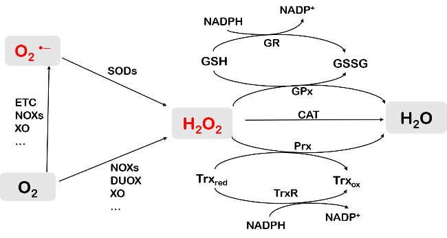
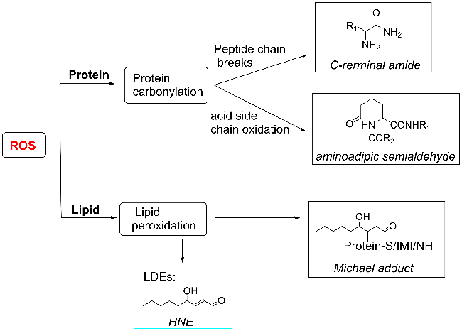
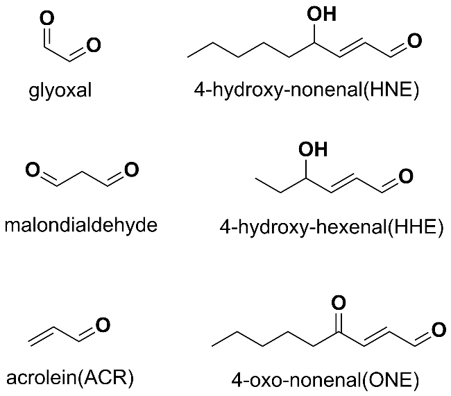
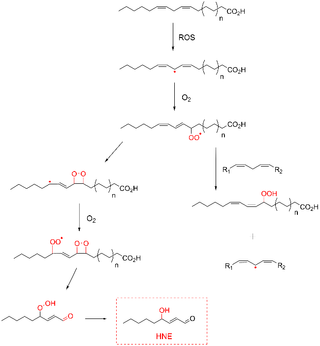
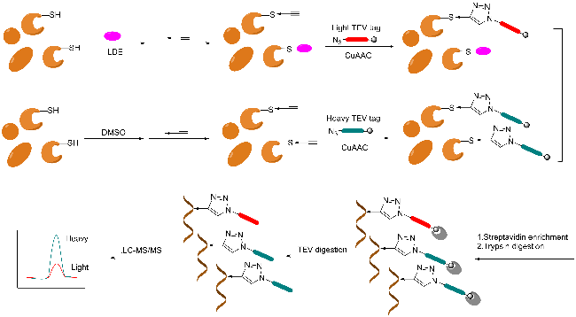
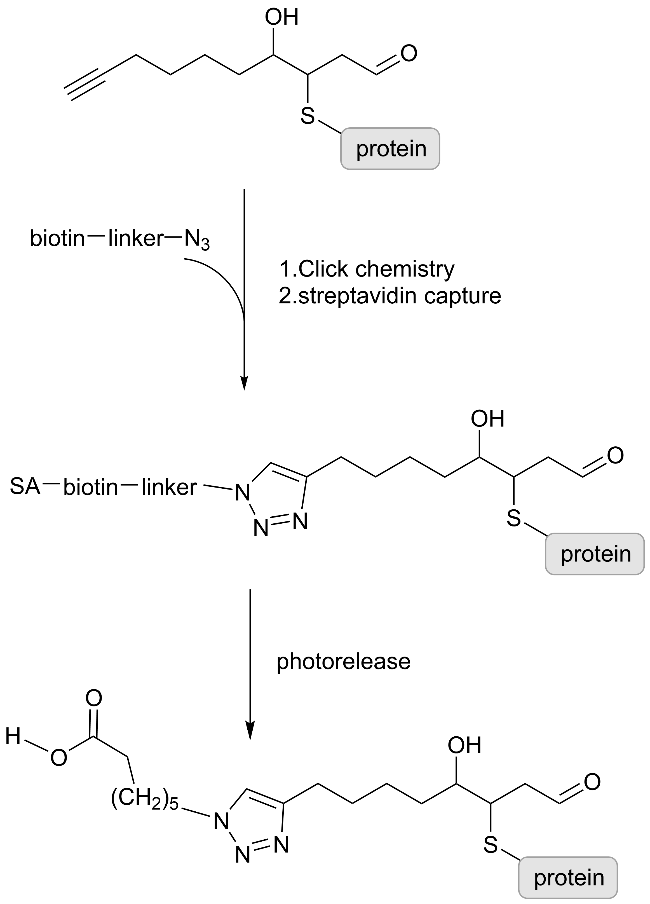
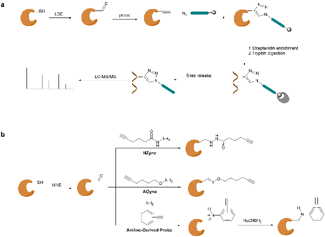
Protein carbonylation modification is an irreversible post-translational modification (PTM) that plays a vital role in modulating protein function. The profiling of intracellular protein carbonylation can provide important information for the investigation of the molecular mechanisms of oxidative stress-related protein signaling networks and pathologies of related diseases. Here, we provide a meticulous description and systematic synthesis of recent research progress in protein carbonylation profiling assays development, especially for mass spectrometry-based chemoproteomic platforms for global profiling of protein lipoxidation. Oxidative stress has been regarded as the result of intracellular reactive oxygen species (ROS) exceeding the buffering capacity of antioxidant defenses, triggering oxidative damage towards lipids, DNA, and proteins. Protein carbonylation (PCO) can be produced either directly by amino acid side chain oxidation, protein backbone cleavage pathways, or indirectly via the formation of adducts between protein nucleophilic side chains and lipid peroxidation products or glycosylation products. We focus on the analysis and detection of protein carbonylation caused by lipid-derived electrophiles (LDEs), and highlight the recent development of protein LDEs profiling assays, especially for mass spectrometry (MS)-based chemoproteomic strategies. Due to the low relative abundance, poor chemical stability, and lack of specific physicochemical properties (e.g. absorption or fluorescence), many carbonylated proteins could not be detected directly, and their detection and quantification rely on the recognition with specific chemical probes. With these probes, mass spectrometry-based chemo-proteomic platforms emerge as powerful tools for comprehensive profiling of protein carbonylation, offering unparalleled sensitivity and specificity, facilitating the identification of protein targets and modification sites critical for elucidating the molecular mechanisms underlying disease progression.

Contents

1 Introduction

2 Sources of oxidative stress and protein carbonylation

2.1 Oxidative stress

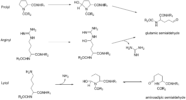
2.2 Sources of protein carbonylation

3 Analytical detection methods for protein carbonylation modifications

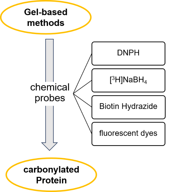
3.1 Gel-based approach

3.2 Gel-free method based on mass spectrometry

4 Conclusion and outlook

Cite this article

Chunyu Wang , Ziming Tang , Chunrong Liu . Protein Carbonylation Modification and Its Analytical Detection Assays[J]. Progress in Chemistry, 2025 , 37(6) : 801 -811 . DOI: 10.7536/PC240809

1 Introduction

Proteins, as fundamental organic components of cells, not only participate in constructing the basic framework of living systems, but also play crucial roles in driving the processes of life activities. mRNA initiates the translation process of proteins by binding to ribosomes in the cytoplasm, while tRNA transports corresponding amino acids that assemble into specific amino acid sequence polypeptide chains within the ribosomes. However, precursor proteins after translation often lack physiological activity and must undergo proper folding and modification to perform their functions. Through the covalent conjugation of functional groups or proteins, the proteolytic dissociation of regulatory subunits, or the degradation of entire proteins, post-translational modification (PTM) can alter the structure[1], function, localization, activity, and interactions[2] of proteins, thereby increasing the functional diversity of the proteome and playing important roles in biological processes such as cell signaling, metabolic regulation, and cell fate determination, ultimately influencing numerous core cellular activities. PTM can occur at any stage of the cell's life cycle, on different amino acid side chains or peptide bonds, and can result in various functions, further adding complexity to the proteome. Meanwhile, abnormal PTMs are often significant indicators of cellular stress and dysfunction.
Oxidative stress (OS) is generally considered to occur when the accumulation of reactive oxygen species (ROS) in cellular systems exceeds the capacity of endogenous antioxidant mechanisms to scavenge them, leading to oxidative damage involving lipids, DNA, and proteins[3]. Protein carbonylation modification is an irreversible post-translational modification of proteins and also one of the important manifestations of oxidative stress. Protein carbonylation can be directly produced by protein peroxidation (through oxidation of amino acid side chains and cleavage of the protein backbone), or indirectly generated by lipid compound peroxidation and sugar oxidation (via adduct formation between protein nucleophilic side chains and lipid peroxidation or glycation products)[4]. Under pathological conditions associated with oxidative stress, the level of protein carbonyls significantly increases[5-6]. For example, compared with young control groups, the level of protein carbonylation in the brains of elderly individuals increases[7]; the carbonylation levels of certain proteins in the brains of Alzheimer's disease patients are significantly higher than those in healthy controls[8]; and the level of protein carbonylation is elevated in heart failure-cachexia rats[9]. Therefore, protein carbonylation is an important potential biomarker closely associated with aging and various diseases. At the same time, protein carbonylation can regulate protein function, thereby influencing related signal transduction networks[10-11], and participates in a series of physiological processes. Thus, carbonylation plays a crucial regulatory role in protein function. Detecting protein carbonylation, especially identifying the carbonylation sites in biological samples, is of significant importance, as it can provide valuable information for studying the molecular mechanisms of oxidative stress-related protein interaction networks and associated diseases[12].
Proteomics research, hailed as "post-genomics," is a scientific discipline that studies the properties of protein sets on a large scale. Its research focuses include protein expression levels, post-translational modifications, and interactions between proteins or between proteins and other biomolecules[13]. With the rapid development of mass spectrometry (MS) instruments and experimental methods, MS-based proteomics has become a reliable and indispensable tool for elucidating biological processes at the protein level.
This review introduces the sources of oxidative stress and protein carbonylation modifications, and focuses on the detection of protein carbonylation modifications caused by lipid-derived electrophilic reagents (LDEs) resulting from the peroxidation of lipid compounds due to ROS attack. It discusses recent advances in methods for detecting and identifying protein carbonyls, particularly the development of mass spectrometry-based proteomics techniques for protein carbonylation. Finally, this article provides an outlook and discussion on the future development of analytical methods for detecting protein carbonylation modifications.

2 Oxidative Stress and the Origin of Protein Carbonylation Modifications

2.1 Oxidative Stress

Oxygen participates in cellular aerobic respiration, and almost all aerobic organisms require oxygen for energy supply through mitochondrial respiration. Reactive oxygen species (ROS) mainly include highly reactive oxygen-containing small molecules such as H2O2, O2•-, and •OH, which are generally considered natural byproducts of cellular aerobic metabolism. The primary source of ROS is the superoxide anion radical (O2•-), mainly generated during mitochondrial respiration due to electron leakage from the mitochondrial electron transport chain (ETC). Additionally, enzymes involved in electron transfer, such as NADPH oxidases (NOXs) and xanthine oxidase (XO)[14], can also catalyze the production of O2•-. H2O2 is primarily formed through the dismutation of superoxide radicals catalyzed by SOD, and can also be directly generated from enzymatic reactions involving NOX4 and xanthine oxidase[15]. Under normal conditions, ROS levels in cells remain low, and certain ROS, such as H2O2, play roles in cell signaling. When cellular ROS levels become excessive, oxidative stress occurs, and the cellular antioxidant network works in coordination; catalase (CAT), peroxiredoxin (Prx), glutathione peroxidase (GPx), and others act together through various ROS scavenging mechanisms to alleviate the effects of oxidative stress[16-17] (Figure 1).
图1 ROS产生及其调节途径

Fig.1 ROS production and its regulatory pathways

Oxidative stress can lead to the peroxidation of important biomacromolecules such as lipids, DNA, and proteins, causing irreversible damage. If this condition persists for a prolonged period, it may result in partial loss of cellular physiological functions and even cell death[18]. Consistently, oxidative stress has been reported to be closely associated with the progression of aging and the development of various diseases[19], including cancer, cardiovascular disease, atherosclerosis, hypertension, diabetes, Alzheimer's disease, Parkinson's disease, and rheumatoid arthritis[20].

2.2 Sources of Protein Carbonylation Modifications

Protein carbonylation (Protein carbonylation, PCO) refers to the process in which reactive carbonyl groups, mainly aldehydes and ketones, are introduced into protein structures under oxidative stress[21], which causes changes in protein structure and function, exacerbates oxidative stress or regulates a series of downstream pathways, and participates in many physiological processes and pathological processes of diseases. As early as 1962, Garrison et al.[22] first demonstrated the formation of carbonylation modifications in proteins through radiation. Since then, scientists have conducted long-term research on it and found that protein carbonylation mainly has four sources: cleavage of the protein backbone, oxidation of amino acid side chains in proteins, modification of proteins by lipid peroxidation products, and oxidative products of glycation[23] (Figure 2).
图2 蛋白质羰基化的来源

Fig.2 Source of protein carbonylation

2.2.1 Breakage of the Protein Backbone

The α-carbon atoms of amino acids in the polypeptide backbone of proteins can react with reactive oxygen species (ROS), generating alkoxyl radicals. Subsequently, these radicals can undergo cleavage in two different ways: one via diamidation reaction, and the other through α-amidation reaction cleavage[24], as shown in Fig. 3a, b. In the first case (Fig. 3a), the C—C bond at the radical initiation site breaks, forming a diamide structure on the side of the polypeptide fragment containing the radical, while the other side of the polypeptide fragment forms an isocyanate group. In the second case (Fig. 3b), the C—N bond at the radical initiation site breaks, converting the polypeptide fragment from the N-terminal of the protein into an amide, while the polypeptide segment from the C-terminal of the protein forms an N-α-ketoamide.
图3 通过(a)二酰胺和(b)α-酰胺化途径裂解肽链主链

Fig.3 The peptide chain backbone is cleaved by (a) diamide and (b) α-amidation pathways

2.2.2 Oxidation of Amino Acid Side Chains in Proteins

In addition, when protein side chains (especially those of proline, arginine, lysine, and threonine) are attacked by reactive oxygen species, one or more carbonyl groups can also be formed. These amino acid side chain oxidation products mainly include 2-pyrrolidone obtained from oxidation of proline residues, glutamic acid semialdehyde obtained from oxidation of arginine and proline residues, aminoadipic acid semialdehyde obtained from oxidation of lysine residues, and 2-amino-3-ketobutyric acid obtained from oxidation of threonine residues. Among these, glutamic-1-semialdehyde, the oxidation product of arginine and proline residues, and aminoadipic acid semialdehyde, the oxidation product of lysine residues, may be the two most abundant specific carbonylation products in cells.
图4 氨基酸侧链氧化产生的羰基衍生物

Fig.4 Carbonyl derivatives produced by oxidation of amino acid side chains

图5 Pro, Arg, Lys氧化生成谷氨酸半醛和氨基己二酸半醛

Fig.5 Pro, Arg, Lys are oxidized to form glutamic acid semialdehyde and aminoadipic acid semialdehyde

2.2.3 Modification of Proteins by Lipid Peroxidation Products

Lipid peroxidation generates a series of reactive carbonyl compounds, including α and β-unsaturated aldehydes, di-aldehydes, and ketoaldehydes[27], which are also known as lipid-derived electrophiles (LDEs), primarily including acrolein (ACR), malondialdehyde (MDA), and 4-hydroxy-2-nonenal (HNE), among others (Figure 6).
图6 常见的几种LDEs结构

Fig.6 Common LDEs structures

Taking the generation of HNE as an example, ROS attack unsaturated fatty acids to form fatty acid radicals. These radicals then react with oxygen to form peroxyl radicals (LOO·). LOO· can further react with adjacent polyunsaturated fatty acids to generate hydroperoxides and alkyl radicals, initiating a chain reaction that damages more fatty acids. LOO· may also react with neighboring double bonds, leading to cleavage of the fatty acid backbone and the formation of 4-hydroxynonenal, which is finally reduced to 4-hydroxy-2-nonenal[28-29] (Fig. 7).
图7 脂质过氧化生成HNE

Fig.7 Lipid peroxidation to generate HNE

After the formation of these reactive carbonyl species, they can be conjugated onto nucleophilic amino acids such as cysteine, histidine, and lysine through Michael addition reactions or via the formation of Schiff bases[29], thereby introducing reactive carbonyl groups into proteins and producing protein carbonylation modifications (Figure 8). Research has shown that compared to healthy individuals, the levels of lipid peroxidation compounds such as 4-hydroxy-2-nonenal and acrolein are elevated in the brains of patients with Alzheimer's disease, and increased levels of lipid peroxidation products are also observed in cerebrospinal fluid and plasma[30-34]. Therefore, studying covalent modifications by lipid-derived electrophiles (LDEs) is not only crucial for understanding the regulatory mechanisms of electrophilic chemical signaling, but also holds significant reference value for the development of drugs targeting related diseases, such as antioxidants and targeted covalent inhibitors[35-36].
图8 HNE与亲核氨基酸的加成

Fig.8 Addition of HNE to nucleophilic amino acids

2.2.4 Glycoxidation Products Modify Proteins

Reactive carbonyl groups can be generated by the addition of small molecular aldehydes such as glyoxal (MG) [37], methylglyoxal (MGO) [38], and 3-deoxyglucosone [39] (3-DG), which are produced from the degradation of glycolytic intermediates, to proteins. Additionally, the non-enzymatic Maillard reaction between amino acids and reducing sugars [40], following a series of chemical rearrangements, can also introduce reactive carbonyl groups into protein side chains.
This article mainly focuses on the analysis and detection of protein carbonylation caused by lipid peroxidation-derived electrophiles (LDEs) resulting from reactive oxygen species (ROS) attacking lipid compounds.

3 Analysis and Detection Methods for Protein Carbonylation Modification

The identification and quantitative analysis of protein carbonylation can be performed at different levels. For instance, carbonylated proteins can be derivatized using 2,4-dinitrophenylhydrazine (DNPH), followed by spectrophotometric measurement or immunodetection using DNPH-specific antibodies in gels or by ELISA, enabling the detection and quantification of global protein carbonylation[41]. However, these methods can only determine overall levels of carbonylation, not the types of modified proteins or the specific sites of modification. Mass spectrometry-based methods allow more detailed analysis of protein modifications. Therefore, mass spectrometry-based proteomics is currently the most suitable approach for studying protein carbonylation sites and their potential molecular functions. At the same time, due to characteristics of carbonylated proteins such as low relative abundance, poor chemical stability, and lack of specific physicochemical properties, they are generally difficult to analyze directly by mass spectrometry. Specific chemical probes are often required to derivatize and enrich carbonylated proteins prior to mass spectrometric analysis, especially for quantitative analysis. Moreover, based on sample preparation methods, current approaches for detecting and identifying protein carbonylation are mainly divided into two categories: gel-based methods and gel-free methods.

3.1 Gel-Based Methods

Electrophoresis is a gel-based technique widely used for analyzing the protein composition of biological samples. Two-dimensional gel electrophoresis (2-DE) separates complex mixtures containing thousands of protein components into individual protein spots by combining two orthogonal physical separation techniques: isoelectric focusing (first dimension) and polyacrylamide gel electrophoresis (second dimension). Combining specific detection methods for protein carbonylation (such as immunoblotting) with two-dimensional gel electrophoresis not only enables the separation and identification of protein carbonylation but also allows quantification of the extent of carbonylation for each protein relative to its total amount. Carbonylated proteins lack specific absorption or fluorescent properties, making direct detection and quantification difficult to achieve. Therefore, specific chemical probes are often required to perform derivatization treatments. Currently, scientists have developed a series of chemical probes for detecting protein carbonyls in polyacrylamide gels, including DNPH, tritiated sodium borohydride ([3H]NaBH4), biotin hydrazide probes, and fluorescent probes (Figure 9).
图9 基于凝胶的方法用于检测羰基化蛋白质

Fig.9 Gel-based method for detecting carbonylation proteins

DNPH (2,4-dinitrophenylhydrazine) can react with ketone and aldehyde functional groups to form 2,4-dinitrophenylhydrazone (DNP-hydrazone), which exhibits a characteristic ultraviolet absorption at approximately 370 nm when measured using a spectrophotometer. The quantification of protein carbonylation after derivatization can be achieved by measuring the absorbance at 370 nm. Levine et al.[42] first published a spectrophotometric method based on this reaction and used immunoassays with protein blotting to detect protein carbonylation, evaluating the relative sensitivity of several plasma proteins to oxidative modification. They derivatized proteins with DNPH, separated them via SDS-gel electrophoresis, and screened using antibodies against DNP[43]. Today, immunoblot detection of carbonylated proteins (Carbonyl Western Blot) is widely applied in academic research. This process primarily consists of four major steps: (1) DNPH derivatization of carbonylated proteins under acidic conditions; (2) gel electrophoresis; (3) electrotransfer to a PVDF membrane; and (4) antibody-based detection. Yang Guofeng et al.[44] applied this method, excising oxidatively modified protein spots for in-gel enzymatic digestion and identifying some oxidatively modified proteins in the human brain through tandem mass spectrometry.
Protein carbonyls can be reduced by sodium borohydride ([3H]NaBH4), during which tritium (3H) is selectively incorporated into the carbonylated proteins. Tritiated proteins are separated by SDS-PAGE, and the target protein bands can be excised, dissolved from the gel, and measured for radioactivity to quantify protein carbonylation. This method utilizes SDS-PAGE and does not require removal of unreacted NaBH4 from the labeling reaction mixture, making it applicable for measuring carbonylation of specific proteins[45].
Hydrazide can specifically react with carbonyl groups through the formation of Schiff bases, and the reaction needs to be stabilized by reduction with sodium cyanoborohydride[46]. The binding affinity between biotin and avidin (or streptavidin) is one of the strongest known non-covalent interactions[47]. Regnier et al.[46] developed a method for detecting carbonylated proteins in two-dimensional electrophoresis using biotinylation and avidin-fluorescein isothiocyanate (FITC) affinity staining. In this method, carbonylated proteins were derivatized with biotin-hydrazide and detected using avidin labeled with fluorescein. Twenty carbonylated proteins were identified in the yeast proteome after hydrogen peroxide stimulation[12].
Since then, scientists have developed a series of fluorescent hydrazide probes such as fluorescein-5-thiosemicarbazide[48-50], Cy5 and Cy3 hydrazides, and BodipyFL hydrazide[51] for the detection of protein carbonylation. Chaudhuri et al.[48] used the fluorescein-5-thiocarbohydrazide (FTC) probe to label proteins in mouse liver and, combined with two-dimensional gel electrophoresis, directly measured changes in the levels of protein carbonylation in specific proteins.
Two-dimensional gel electrophoresis is a highly effective and widely used technique for protein analysis. However, it also has certain limitations, such as poor reproducibility, low sensitivity, and difficulty in detecting low-abundance proteins[52-53]. Two-dimensional difference gel electrophoresis (2D-DIGE) effectively addresses the issue of reproducibility. DIGE labels protein samples with fluorescent dyes (such as Cy2, Cy3, and Cy5), allowing the simultaneous analysis of 2-3 samples on the same gel and simplifying the comparison of several two-dimensional gels by scanning at different wavelengths[54].

3.2 Gel-Free Methods Based on Mass Spectrometry

3.2.1 Indirect Identification Method Based on Competitive Reaction Strategy

As previously mentioned, the relative abundance of protein carbonylation modifications is low and their chemical stability is poor, making them difficult to analyze directly by mass spectrometry. Therefore, chemists often use specific chemical probes to derivatize and enrich carbonylated proteins before conducting mass spectrometry analysis, particularly quantitative analysis. Based on differences in their target sites, strategies for capturing protein reactive carbonyl groups can be divided into two approaches: indirect identification and direct identification. The indirect identification strategy refers to the method of indirectly identifying cysteine carbonylation modifications through a competitive reaction strategy. Specifically, proteins are first treated with LDEs capable of specifically modifying the active sites of protein cysteines; the remaining unmodified cysteine active sites are then labeled using chemical probes specific for cysteine thiol groups. At the same time, a control group without LDEs is established. Combining this with the isotopic tandem orthogonal proteolysis-activity-based protein profiling (isoTOP-ABPP) strategy and quantitative proteomics methods enables the identification of specific carbonylated proteins. Among them, the iodoacetamide alkyne probe (IA-alkyne) is an electrophilic small-molecule probe widely used in ABPP[55]. Wang et al.[56] developed a competitive isoTOP-ABPP strategy to quantify the reactivity of a specific LDE toward cysteines across the human proteome at a global level. The main procedure is as follows: (1) Treat one proteome sample with LDE and set up a control group treated with DMSO (dimethyl sulfoxide), followed by labeling of unmodified reactive cysteine sites using IA probes; (2) Based on click chemistry strategy (copper-catalyzed azide-alkyne cycloaddition, CuAAC), the IA-labeled cysteines in the samples are conjugated with isotopically labeled cleavable biotin-azide tags; (3) The IA-labeled proteins are enriched using streptavidin beads, followed by trypsin digestion and cleavage of the linker to release IA-alkyne-labeled peptides[57]; (4) The samples are analyzed using liquid chromatography-tandem mass spectrometry (LC-MS/MS) (Figure 10). The quantitative ratio (R value, derived from LDE-treated versus untreated samples) for each cysteine-containing peptide accurately reflects the extent of LDE modification at each cysteine under a given LDE concentration. A higher R value indicates higher sensitivity to LDE modification.
图10 基于竞争性反应策略对半胱氨酸羰基化修饰的间接鉴定

Fig.10 Indirect identification of cysteine carbonylation modifications based on competitive reaction strategies

The advantage of the competitive isoTOP-ABPP strategy lies in its ability to directly introduce compound molecules as competitive inhibitors into the proteome, eliminating the cumbersome chemical synthesis steps required to convert them into molecular probes[58]. However, this method is currently limited to the analysis of LDE-cysteine interactions, making it difficult to study modifications of LDEs on histidine and lysine residues. Additionally, the application of this method is restricted due to the high cost of isotopically labeled amino acid reagents and the lengthy, low-yield synthesis processes associated with cleavable light and heavy tag reagents[59].

3.2.2 Direct Identification Method Based on Chemical Probes

Compared with indirect identification methods that rely on competitive reaction strategies, direct identification methods for protein carbonylation, which involve the direct reaction of probes with carbonyl groups at carbonylation sites or carbonylation modifications, offer higher sensitivity and can effectively reduce false positive interference. Based on the action targets of the probes, direct identification methods can also be divided into two categories: metabolic labeling and targeted direct labeling of carbonyl groups.
Metabolic labeling detects specific carbonylation types by using LDE analog probes containing "click handles" (Fig. 11). Marnett et al. introduced azide or alkyne groups into the alkyl terminus of HNE structures and used these HNE analog probes to label proteins in human colon adenocarcinoma cells. Subsequently, biotin-conjugated complementary alkynes or azides were added to perform click chemistry reactions, thereby biotinylating the target proteins. After enrichment and purification using streptavidin beads, LC-MS/MS analysis identified 538 HNE-modified proteins. Additionally, Porter et al. developed a photocleavable azide-biotin reporter tag that specifically releases HNE-alkyne probe-modified proteins or peptides from streptavidin beads after enrichment. Codreanu et al. conducted a series of quantitative modification experiments using alkynyl analogs of the lipid electrophilic reagents 4-hydroxy-2-nonenal and 4-oxo-2-nonenal to investigate the functional impacts of LDE alkylation damage. Building upon this, Liebler et al. employed isotopic labeling combined with photocleavable azide-biotin reagents to selectively capture and quantify cellular targets labeled by HNE-alkyne probes, enabling large-scale, in situ, site-specific identification and quantitative analysis of approximately 400 HNE modification sites.
图11 基于LDEs类似物探针的蛋白质羰基检测方法

Fig.11 Protein carbonyl detection method based on LDEs analogue probe

ONE shares a similar structure with HNE, but their reactivities differ[64]. Generally, HNE preferentially reacts with cysteine residues in the proteome, whereas ONE exhibits higher chemical affinity toward lysine residues. In neuronal cells, ONE demonstrates greater reactivity and cytotoxicity than HNE. Yang et al.[65] utilized an isotopically labeled, photocleavable azido-biotin reagent to selectively capture and quantify protein targets labeled by an ONE alkynyl analog (aONE), revealing the diversity and dynamics of ONE modifications within cells through quantitative analysis.
However, metabolic labeling also has notable limitations. For example, the synthesis of specific LDEs analog probes is cumbersome and costly; it is not suitable for smaller LDE structures, such as ACR, since any chemical modification can affect its reactivity[66].
For another direct identification method, direct labeling targeting reactive carbonyl groups, various carbonyl-reactive reagents have been developed and applied to label LDEs-modified proteins in diverse biological samples. Among these, probes based on hydrazide or hydroxylamine biotinylation are the most widely used.
The hydrazide group of biotin reacts with the active carbonyl groups formed after protein carbonylation modification to form acyl hydrazone bonds for linkage. Utilizing the high affinity of streptavidin beads, biotinylated proteins are specifically captured and enriched. Following trypsin digestion, the proteins are identified and quantified using LC-MS/MS. Soreghan et al.[67] applied a hydrazide biotin probe combined with liquid chromatography-tandem mass spectrometry (LC-MS/MS) analysis to identify more than 100 carbonylated proteins in the brain proteome of aged mice, including some low-abundance proteins previously missed by immunoaffinity-based methods. Codreanu et al.[68] used this method for the first comprehensive analysis of HNE-modified proteins in the human proteome; they treated RKO cells (human colon adenocarcinoma cells) with varying concentrations of HNE, labeled the HNE-modified proteins with a hydrazide-biotin probe, enriched them using streptavidin beads, and performed LC-MS/MS analysis to identify HNE protein targets, revealing numerous important intracellular systems affected by HNE modification, including those involved in protein translation, folding, and degradation. Liu Changfeng et al. used biotin hydrazide labeling, avidin bead enrichment, and tandem mass spectrometry to isolate and identify carbonylated proteins in the striatum of aged rats and integrated bioinformatics to search for mass spectrometry identification information.[69]
Hydroxylamine can react with active carbonyl groups to form oxime bonds. N-Aminooxymethyl carbonyl hydrazide D-biotin, also known as aldehyde-reactive probe (ARP), is a biotinylated hydroxylamine derivative. Chavez et al.[70] used ARP to enrich target proteins and employed tandem mass spectrometry to analyze and identify HNE-labeled protein targets. This approach has been applied to proteomic studies of the human monocytic cell line THP-1[71] and cardiac mitochondria from mouse tissues[72], successfully identifying multiple protein targets and residue sites modified by LDE. Notably, the oxime bonds formed by hydroxylamine and active carbonyl groups exhibit significantly higher stability compared to the hydrazone bonds generated through hydrazide reactions[73]. Furthermore, this transformation does not require an additional reduction step, simplifying the procedure. Consequently, hydroxylamine-based reaction probes (ARP) have become a preferred alternative to hydrazide biotin reagents and are widely utilized in various studies[74-75].
However, the method of identifying carbonylated proteins using biotin probes faces issues of false positive identifications, such as certain proteins or peptides that can bind non-specifically to avidin, and some endogenous proteins that naturally carry biotin[76-77]. To overcome the potential problem of nonspecific background proteins, Meany et al.[78] employed stable isotope labeling of affinity-purified proteins; this strategy combines biotin hydrazide labeling, avidin affinity chromatography, and multiplex iTRAQ reagent-based stable isotope labeling techniques to enrich and identify more than 200 carbonylated proteins in rat muscle mitochondrial extracts.
Hydrazide or hydroxylamine biotinylation probes can efficiently enrich various endogenous carbonylated proteins. However, the introduction of a bulky biotin group during this process may affect the activity of small molecules, thereby influencing the probe's labeling efficiency toward target proteins[79]. At the same time, it also increases the complexity of mass spectrometry (MS) analysis[80].
Griffin et al.[81] proposed a tag-free strategy, in which solid-phase hydrazide (SPH) reagent was innovatively utilized. Following enzymatic digestion of proteins, HNE-modified peptides specifically enrich through the formation of covalent yet reversible hydrazone bonds via reaction between the hydrazide immobilized on glass beads and the HNE-modified peptides. After washing away unreacted peptides, the hydrazone bonds are cleaved under acidic conditions to release the HNE-modified peptides for LC-MS/MS detection. This method avoids the addition of extra tags (e.g., biotin and fluorophores) onto aldehyde groups, facilitating the analysis of HNE modification sites. Using this approach, they identified 15 HNE-modified proteins in HNE-treated yeast. Similarly, Maier et al.[82] combined d0/d4-succinic anhydride labeling with hydrazide-functionalized microbead (Affi-Hz gel)-based enrichment to detect, identify, and relatively quantify site-specific carbonylated proteins/peptides in biological matrices. Prokai et al.[83] first performed differential dimethyl labeling of proteins, followed by enrichment of carbonylated peptides using solid-phase hydrazide chemistry prior to mass spectrometry analysis, enabling quantification and site identification of protein carbonylation modifications. Additionally, Lu et al.[84] developed a method for specifically enriching HNE-modified peptides based on a fluorine derivatization probe and fluorine-based solid-phase microextraction (FSPE). The hydrophobicity of the fluorine tag significantly enhances the signal of HNE-modified peptides, while FSPE enables selective enrichment of fluorine-derivatized HNE-modified peptides directly from salt solutions and complex mixtures without requiring an additional desalting step prior to LC-MS/MS analysis, thereby simplifying the workflow.
Considering the limitations of biotinylated labeling reagents, scientists have developed bioconjugation reactions based on reactive carbonyl groups, enabling rapid and highly specific covalent labeling and forming stable covalent adducts. Based on this reaction, probes containing click chemistry handles such as alkynes were synthesized. Subsequently, biotin containing azide groups and cleavable linkers was introduced via click chemistry reactions. After enrichment, photocleavage or enzymatic cleavage to remove biotin, followed by purification, large-scale quantitative studies of derivatized carbonylated proteins or peptides were achieved.
Wang et al.[66] developed a novel chemical proteomics approach to identify both the types and sites of proteins that can be modified by HNE (Figure 12). In this strategy, hydrazide-derived probe (HZyne) and aminooxy-derived probe (AOyne) containing bioorthogonal alkyne groups were first synthesized and compared, revealing that AOyne exhibited better labeling efficiency in HNE-treated proteomes than HZyne. Subsequently, using the AOyne probe combined with reductive dimethyl labeling technology[86-87] and TOP-ABPP (tandem orthogonal proteolysis-ABPP) technology[88], more than 4,000 HNE-modified proteins and approximately 400 modification sites were identified in the HEK293T cell proteome.
图12 (a)基于化学探针的蛋白质组学工作流程;(b)Wang等开发的酰肼探针、羟胺衍生探针和苯胺衍生探针

Fig.12 (a) Chemical probe-based proteomics workflow; (b) HZyne, AOyne, and Aniline-derived probe developed by Wang et al

Wang et al.[89] also applied aniline-derived probes to detect protein carbonylation modifications in ferroptosis, identifying more than 400 endogenous LDE-modified proteins and over 20 modification sites. They further ranked the sensitivity of these sites using the isoTOP-ABPP strategy and analyzed their roles in biological metabolic pathways through bioinformatics approaches.
Among these labeling reagents, hydrazide probes exhibit poor capture efficiency. The imine products formed by their reaction with active carbonyl groups are structurally unstable and require reduction with sodium cyanoborohydride. However, sodium cyanoborohydride shows poor selectivity during reduction, making the overall experimental procedure more complex. The capture efficiency of hydroxylamine-derived probes and aniline-derived probes is significantly affected by pH, showing notably lower efficiency under neutral or weakly basic conditions compared to acidic environments. Moreover, the products are unstable under basic conditions and tend to decompose during trypsin digestion, leading to the detachment of click handles. Therefore, the development of new labeling reagents with high efficiency, specificity, and excellent product stability remains an urgent need.

4 Conclusion and Prospect

In recent years, with the continuous advancement of research on protein carbonylation modifications, the detection and analysis methods for protein carbonylation in cells have been somewhat expanded, enhancing the understanding of the biological functions of carbonylation modifications. However, current detection of protein carbonylation still faces many challenges. Present studies mostly focus on modifications of protein thiol groups, while modifications of lysine and histidine residues are less explored. This may be mainly due to site-specific preferences in chemical labeling reactions on one hand, and possibly due to the selectivity of enrichment methods on the other. Therefore, designing novel, more efficient and broad-spectrum labeling approaches combined with appropriate enrichment strategies will facilitate the identification of lysine and histidine carbonylation sites. Regarding Schiff modifications formed by carbonyl compounds, such as methylglyoxal, with nitrogen-containing amino acid residues in proteins, relevant studies are limited because the modifications are unstable and difficult to analyze and identify by direct mass spectrometry. Developing new labeling reactions is crucial for advancing detection methodologies. Additionally, due to the relatively low abundance of protein carbonylation modifications, existing labeling and detection methods suffer from low labeling efficiency, poor selectivity, unstable products, and complicated experimental procedures. Developing probes with improved selectivity and labeling efficiency, as well as analytical methods for comprehensive profiling or precise capture of specific protein carbonylation modifications, will enable broader application in various biological samples, facilitating the understanding of pathological and physiological processes involving protein carbonylation and promoting the development of new drug targets.
[1]
Ding Y. Master's Dissertation of Zhengzhou University, 2022.

(丁一. 郑州大学硕士论文, 2022).

[2]
Ulrich H D. Trends Cell Biol., 2005, 15(10): 525.

[3]
Stadtman E R. Science, 1992, 257(5074): 1220.

[4]
Madian A G, Regnier F E. J. Proteome Res., 2010, 9(8): 3766.

[5]
Kong W W, Zhang X. Chin. J. Gerontol., 2021, 41(2): 436

(孔文文, 张昕. 中国老年学杂志, 2021, 41(2): 436).

[6]
Dalle-Donne I, Giustarini D, Colombo R, Rossi R, Milzani A. Trends Mol. Med., 2003, 9(4): 169.

[7]
Smith C D, Carney J M, Starke-Reed P E, Oliver C N, Stadtman E R, Floyd R A, Markesbery W R. Proc. Natl. Acad. Sci. U. S. A., 1991, 88(23): 10540.

[8]
Aksenov M Y, Aksenova M V, Butterfield D A, Geddes J W, Markesbery W R. Neuroscience, 2001, 103(2): 373.

[9]
Kong W W, Zhang X. Tianjin Med. J., 2021, 48(1): 436

(孔文文, 张昕. 天津医药, 2020, 48(1): 30).

[10]
Wong C M, Cheema A K, Zhang L H, Suzuki Y J. Circ. Res., 2008, 102(3): 310.

[11]
Wong C, Marcocci L, Liu L, Suzuki Y J. Antioxid. Redox Signal., 2010, 12(3): 393.

[12]
Zhou J, Qin H, Ye M. J.Instrum. Anal., 2020, 39(1): 82

(周家华, 秦洪强, 叶明亮. 分析测试学报, 2020, 39(1): 82).

[13]
He Q, Fang X, Zhu T, Han S, Zhu H, Li S. Metabolites, 2019, 9(8): 166.

[14]
Kelley E E, Khoo N K H, Hundley N J, Malik U Z, Freeman B A, Tarpey M M. Free. Radic. Biol. Med., 2010, 48(4): 493.

[15]
Schieber M, Chandel N S. Curr. Biol., 2014, 24(10): R453.

[16]
Gill S S, Tuteja N. Plant Physiol. Biochem., 2010, 48(12): 909.

[17]
Lennicke C, Cochemé H M. Mol. Cell, 2021, 81(18): 3691.

[18]
Mullineaux P M, Baker N R. Plant Physiol., 2010, 154(2): 521.

[19]
Baraibar M A, Ladouce R, Friguet B. J. Proteomics, 2013, 92: 63.

[20]
Valko M, Leibfritz D, Moncol J, Cronin M T D, Mazur M, Telser J. Int. J. Biochem. Cell Biol., 2007, 39(1): 44.

[21]
Colombo G, Clerici M, Garavaglia M E, Giustarini D, Rossi R, Milzani A, Dalle-Donne I. J. Chromatogr. B, 2016, 1019: 178.

[22]
Garrison W M, Jayko M E, Bennett W. Radiat. Res., 1962, 16: 483.

[23]
Birben E, Sahiner U M, Sackesen C, Erzurum S, Kalayci O. World Allergy Organ. J., 2012, 5(1): 9.

[24]
Stringfellow H M, Jones M R, Green M C, Wilson A K, Francisco J S. J. Phys. Chem. A, 2014, 118(48): 11399.

[25]
Møller I M, Rogowska-Wrzesinska A, Rao R S P. J. Proteomics, 2011, 74(11): 2228.

[26]
Requena J R, Chao C C, Levine R L, Stadtman E R. Proc. Natl. Acad. Sci. U. S. A., 2001, 98(1): 69.

[27]
Møller I M, Jensen P E, Hansson A. Annu. Rev. Plant Biol., 2007, 58: 459.

[28]
Barrera G, Pizzimenti S, Daga M, Dianzani C, Arcaro A, Cetrangolo G P, Giordano G, Cucci M A, Graf M, Gentile F. Antioxidants, 2018, 7(8): 102.

[29]
Juan C A, Pérez de la Lastra J M, Plou F J, Pérez-Lebeña E. Int. J. Mol. Sci., 2021, 22(9): 4642.

[30]
Markesbery W R. Free. Radic. Biol. Med., 1997, 23(1): 134.

[31]
Galbusera C, Facheris M, Magni F, Galimberti G, Sala G, Tremolada L, Isella V, Guerini F, Appollonio I, Galli-Kienle M, Ferrarese C. Curr. Alzheimer Res., 2004, 1(2): 103.

[32]
Jomova K, Vondrakova D, Lawson M, Valko M. Mol. Cell. Biochem., 2010, 345(1/2): 91.

[33]
Korovesis D, Rubio-Tomás T, Tavernarakis N. Antioxidants, 2023, 12(1): 131.

[34]
Reed T T. Free. Radic. Biol. Med., 2011, 51(7): 1302.

[35]
Gaschler M M, Stockwell B R. Biochem. Biophys. Res. Commun., 2017, 482(3): 419.

[36]
Steinhilber D, Hofmann B. Basic Clin. Pharmacol. Toxicol., 2014, 114(1): 70.

[37]
Wells-Knecht K J, Zyzak D V, Litchfield J E, Thorpe S R, Baynes J W. Biochemistry, 1995, 34(11): 3702.

[38]
Degenhardt T P, Thorpe S R, Baynes J W. Cell. Mol. Biol. (Noisy-le-grand), 1998, 44(7): 1139.

[39]
Ahmad S, Akhter F, Shahab U, Rafi Z, Khan M S, Nabi R, Khan M S, Ahmad K, Ashraf J M, Moinuddin. Semin. Cancer Biol., 2018, 49: 9.

[40]
Mottram D S, Wedzicha B L, Dodson A T. Nature, 2002, 419(6906): 448.

[41]
Fedorova M, Bollineni R C, Hoffmann R. Mass Spectrom. Rev., 2014, 33(2): 79.

[42]
Levine R L, Garland D, Oliver C N, Amici A, Climent I, Lenz A G, Ahn B W, Shaltiel S, Stadtman E R. Methods Enzymol., 1990, 186: 464.

[43]
Shacter E, Williams J A, Lim M, Levine R L. Free. Radic. Biol. Med., 1994, 17(5): 429.

[44]
Yang G, Ji J, Peng L, He S.J. Clin. Rehabil. Tissue Eng. Res., 2011, 15(11): 1990

(杨国锋, 纪建国, 彭立威, 何思志. 中国组织工程研究与临床康复, 2011, 15(11): 1990)..

[45]
Yan L, Sohal R S. Anal. Biochem., 1998, 265(1): 176.

[46]
Yoo B S, Regnier F E. Electrophoresis, 2004, 25(9): 1334.

[47]
Gitlin G, Bayer E A, Wilchek M. Biochem. J., 1987, 242(3): 923.

[48]
Chaudhuri A R, de Waal E M, Pierce A, Van Remmen H, Ward W F, Richardson A. Mech. Ageing Dev., 2006, 127(11): 849.

[49]
Jung Y, Chay K, Song D, Yang S, Lee M W, Ahn B W. Arch. Biochem. Biophys., 1997, 345(2): 311.

[50]
Ahn B, Rhee S G, Stadtman E R. Anal. Biochem., 1987, 161(2): 245.

[51]
Tamarit J, de Hoogh A, Obis E, Alsina D, Cabiscol E, Ros J. J. Proteomics, 2012, 75(12): 3778.

[52]
Zhao Y, Li N, Li L, Yang X, Hao S, Wei Y, Cen J. J. Dalian Ocean Univ., 2016, 31(3): 344

(赵永强, 李娜, 李来好, 杨贤庆, 郝淑贤, 魏涯, 岑剑伟. 大连海洋大学学报, 2016, 31(3): 344).

[53]
Rabilloud T, Chevallet M, Luche S, Lelong C. J. Proteomics, 2010, 73(11): 2064.

[54]
Ünlü M, Morgan M E, Minden J S. Electrophoresis, 1997, 18(11): 2071.

[55]
Maurais A J, Weerapana E. Curr. Opin. Chem. Biol., 2019, 50: 29.

[56]
Wang C, Weerapana E, Blewett M M, Cravatt B F. Nat. Methods, 2014, 11(1): 79.

[57]
Jiang Z, Niu Y, Wang N, Chen Z, Tang B. Univ. Chem., 2023, 38(1): 119

(姜中尧, 牛雅新, 王楠, 陈蓁蓁, 唐波. 大学化学, 2023, 38(1): 119).

[58]
Wang C, Chen N. Acta Chim. Sin., 2015, 73(7): 657.

(王初, 陈南. 化学学报, 2015, 73(7): 657).

[59]
Yang F, Gao J, Che J, Jia G, Wang C. Anal. Chem., 2018, 90(15): 9576.

[60]
Vila A, Tallman K A, Jacobs A T, Liebler D C, Porter N A, Marnett L J. Chem. Res. Toxicol., 2008, 21(2): 432.

[61]
Kim H H, Tallman K A, Liebler D C, Porter N A. Mol. Cell. Proteomics, 2009, 8(9): 2080.

[62]
Codreanu S G, Ullery J C, Zhu J, Tallman K A, Beavers W N, Porter N A, Marnett L J, Zhang B, Liebler D C. Mol. Cell. Proteomics, 2014, 13(3): 849.

[63]
Yang J, Tallman K A, Porter N A, Liebler D C. Anal. Chem., 2015, 87(5): 2535.

[64]
Galligan J J, Rose K L, Beavers W N, Hill S, Tallman K A, Tansey W P, Marnett L J. J. Am. Chem. Soc., 2014, 136(34): 11864.

[65]
Sun R, Fu L, Liu K, Tian C, Yang Y, Tallman K A, Porter N A, Liebler D C, Yang J. Mol. Cell. Proteomics, 2017, 16(10): 1789.

[66]
Chen Y, Cong Y, Quan B, Lan T, Chu X, Ye Z, Hou X, Wang C. Redox Biol., 2017, 12: 712.

[67]
Soreghan B A, Yang F, Thomas S N, Hsu J, Yang A J. Pharm. Res., 2003, 20(11): 1713.

[68]
Codreanu S G, Zhang B, Sobecki S M, Billheimer D D, Liebler D C. Mol. Cell. Proteomics, 2009, 8(4): 670.

[69]
Yuan L, Peng M, Xiang Y, Yang Q, Lei Y, Liu C, Liu X, Tang C, Yin D Z. Chin. J. Biochem. Mol. Biol., 2021, 37(11): 1489

(袁顺灵, 彭美, 向逸, 杨启明, 雷勇, 刘文锋, 刘霞, 汤长发, 印大中. 中国生物化学与分子生物学报, 2021, 37(11): 1489).

[70]
Chavez J, Wu J, Han B, Chung W, Maier C S. Anal. Chem., 2006, 78(19): 6847.

[71]
Chavez J, Chung W G, Miranda C L, Singhal M, Stevens J F, Maier C S. Chem. Res. Toxicol., 2010, 23(1): 37.

[72]
Chavez J D, Wu J, Bisson W, Maier C S. J. Proteomics, 2011, 74(11): 2417.

[73]
Dirksen A, Dawson P E. Bioconjugate Chem., 2008, 19(12): 2543.

[74]
Bollineni R C, Fedorova M, Blüher M, Hoffmann R. J. Proteome Res., 2014, 13(11): 5081.

[75]
Griesser E, Vemula V, Raulien N, Wagner U, Reeg S, Grune T, Fedorova M. Redox Biol., 2017, 11: 438.

[76]
Bayer E A, Wilchek M. J. Chromatogr. A, 1990, 510: 3.

[77]
Labrou N, Clonis Y D. J. Biotechnol., 1994, 36(2): 95.

[78]
Meany D L, Xie H, Thompson L V, Arriaga E A, Griffin T J. Proteomics, 2007, 7(7): 1150.

[79]
Zhou L, Cui T, Hao N, Zhao Y, Zhao B, Liu Y. Biotechnol. Bull., 2023, 39(9): 12

(周璐祺, 崔婷茹, 郝楠, 赵雨薇, 赵斌, 刘颖超. 生物技术通报, 2023, 39(9): 12).

[80]
Spiess P C, Deng B, Hondal R J, Matthews D E, van der Vliet A. J. Proteomics, 2011, 74(11): 2380.

[81]
Roe M R, Xie H, Bandhakavi S, Griffin T J. Anal. Chem., 2007, 79(10): 3747.

[82]
Han B, Hare M, Wickramasekara S, Fang Y, Maier C S. J. Proteomics, 2012, 75(18): 5724.

[83]
Rauniyar N, Prokai L. J. Mass. Spectrom., 2011, 46(10): 976.

[84]
Yuan W, Zhang Y, Xiong Y, Tao T, Wang Y, Yao J, Zhang L, Yan G, Bao H, Lu H. Anal. Chem., 2017, 89(5): 3093.

[85]
Boersema P J, Raijmakers R, Lemeer S, Mohammed S, Heck A J R. Nat. Protoc., 2009, 4(4): 484.

[86]
Hsu J, Chen S. Philos. Trans. A Math. Phys. Eng. Sci., 2016, 374(2079): 20.

[87]
Weerapana E, Speers A E, Cravatt B F. Nat. Protoc., 2007, 2(6): 1414.

[88]
Chen Y, Liu Y, Lan T, Qin W, Zhu Y T, Qin K, Gao J, Wang H, Hou X, Chen N, Friedmann Angeli J P, Conrad M, Wang C. J. Am. Chem. Soc., 2018, 140(13): 4712.

Outlines

/