
Hypochlorous Acid/Hypochlorite (HOCl/ClO-) Specific Fluorescent Probes: Recognition Mechanism and Biological Applications
Received date: 2024-08-19
Revised date: 2024-10-27
Online published: 2025-06-15
Supported by
Key R&D and Transformation Program of Qinghai(2022-QY-210)
Hypochlorous acid/hypochlorite (HOCl/ClO-) are important participants in various physiological and pathological processes in the organisms. Both contribute immune defense throughinflammatory responses, but their overproduction and generation at inappropriate sites will result in oxidative damage of cell membranes, DNA, and proteins. Therefore, in view of the important physiopathological significance of HOCl/ClO-, its specific identification and detection have been an important research topic for researchers. Fluorescence and fluorescent probe methods stand out among many traditional detection methods due to their many advantages. In this paper, some representative research works on HOCl/ClO- specific fluorescent probes for organic small molecules are reviewed from the first case to the present day, categorized according to the recognition mechanisms between fluorescent probes and HOCl/ClO-. The recognition mechanisms and biological applications of HOCl/ClO- specific fluorescent probes are highlighted, and the prospects for the chemical and biological development of HOCl/ClO- specific fluorescent probes are discussed.
1 Introduction
2 Oxidation reaction mechanism
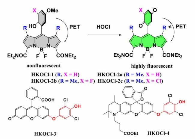
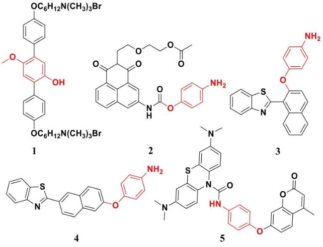
2.1 Oxidation of phenol/aniline analogs
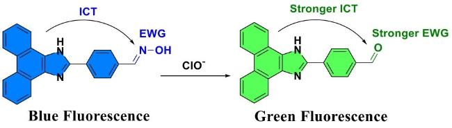
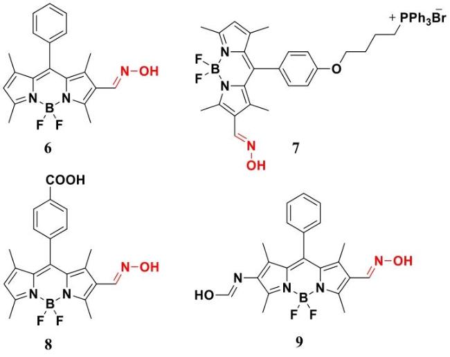
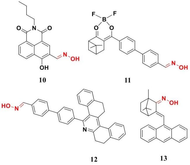
2.2 Oxidation of oximes
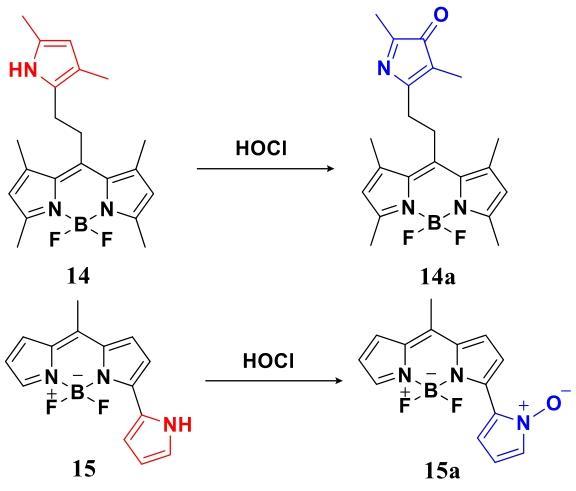
2.3 Oxidation of pyrroles
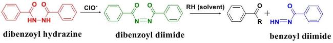
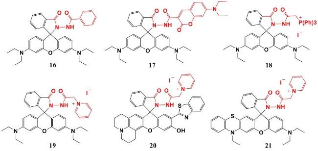
2.4 Oxidation of dibenzoylhydrazines
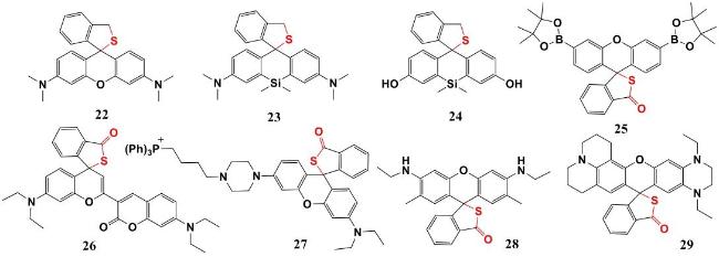
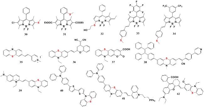
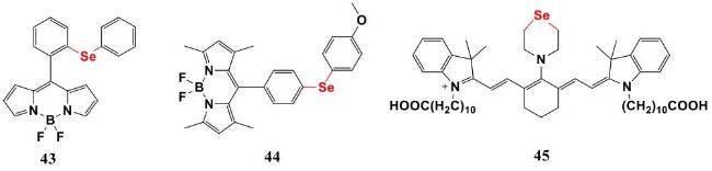
2.5 Sulphur/selenium ether/ester oxidation
3 Electrophilic chlorination reaction mechanism
4 HOCl-mediated cyclization mechanisms
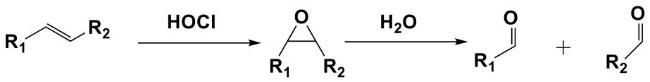
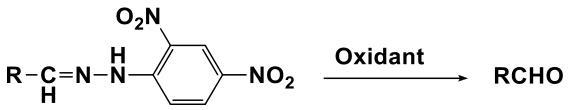
5 Cleavage reaction mechanism based on C=C/C=N bonds
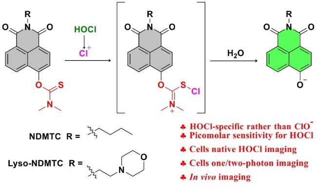
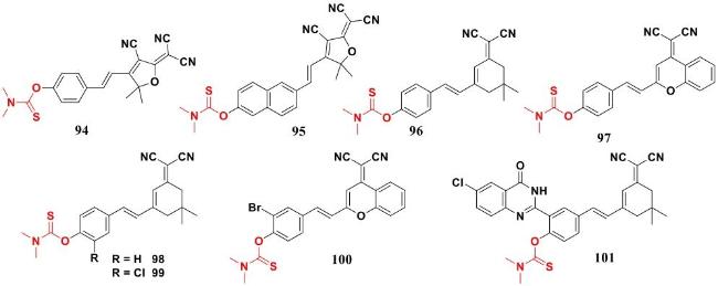
6 Deprotection mechanism based on dimethyl thiocarbamate
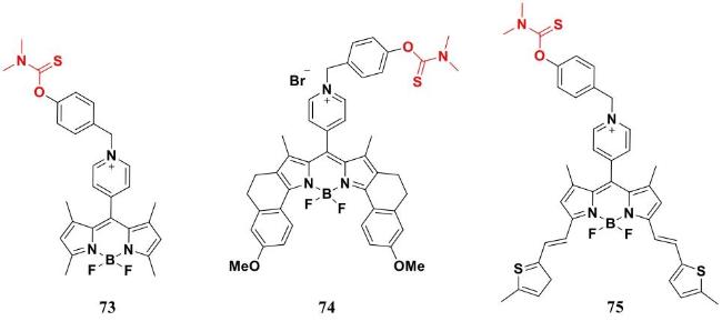
6.1 Based on the BODIPY fluorophore
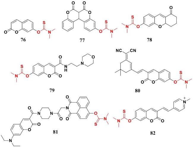
6.2 Based on the coumarin fluorophore
6.3 Based on the naphthalene fluorophore
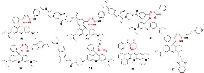
6.4 HBT derivatives as fluorophores
6.5 Based on the resorufin fluorophore
6.6 Based on the cyano fragment fluorophore
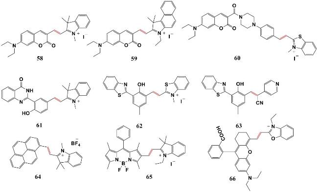
6.7 Based on the hemicyanine xanthene and cyanine fluorophores
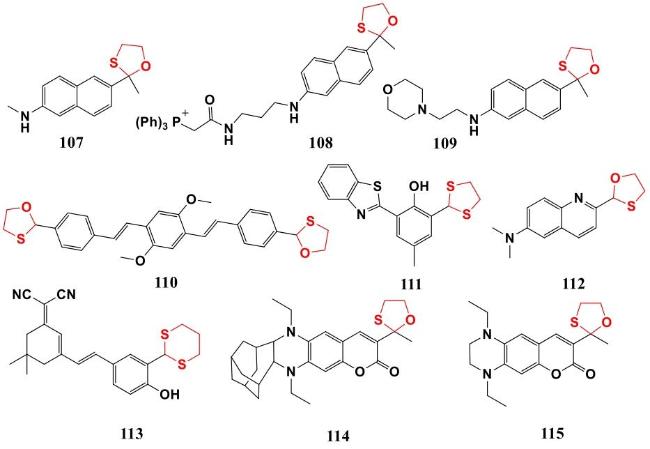
7 Deprotection mechanisms based on oxathiolones/dithiolones
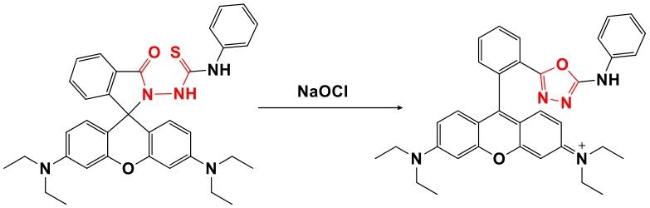
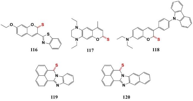
8 Mechanism of desulfurization reactions based on C=S bonds
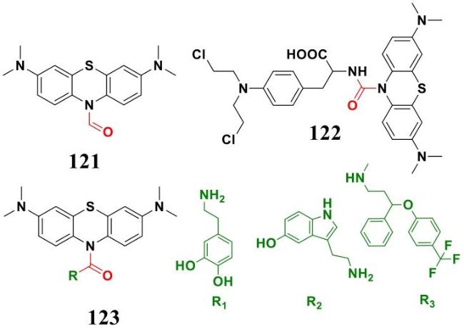
9 Based on other reaction mechanisms
10 Conclusion and outlook
Zhiqiang Zhang , Haichao Li , Ying Long . Hypochlorous Acid/Hypochlorite (HOCl/ClO-) Specific Fluorescent Probes: Recognition Mechanism and Biological Applications[J]. Progress in Chemistry, 2025 , 37(6) : 918 -933 . DOI: 10.7536/PC240803
| [1] |
|
| [2] |
|
| [3] |
|
| [4] |
|
| [5] |
|
| [6] |
|
| [7] |
|
| [8] |
|
| [9] |
|
| [10] |
|
| [11] |
|
| [12] |
|
| [13] |
|
| [14] |
|
| [15] |
|
| [16] |
|
| [17] |
|
| [18] |
|
| [19] |
|
| [20] |
|
| [21] |
|
| [22] |
|
| [23] |
|
| [24] |
|
| [25] |
|
| [26] |
|
| [27] |
|
| [28] |
|
| [29] |
|
| [30] |
|
| [31] |
|
| [32] |
|
| [33] |
|
| [34] |
|
| [35] |
|
| [36] |
|
| [37] |
|
| [38] |
|
| [39] |
|
| [40] |
|
| [41] |
|
| [42] |
|
| [43] |
|
| [44] |
|
| [45] |
|
| [46] |
|
| [47] |
|
| [48] |
|
| [49] |
|
| [50] |
|
| [51] |
|
| [52] |
|
| [53] |
|
| [54] |
|
| [55] |
|
| [56] |
|
| [57] |
|
| [58] |
|
| [59] |
|
| [60] |
|
| [61] |
|
| [62] |
|
| [63] |
|
| [64] |
|
| [65] |
|
| [66] |
|
| [67] |
|
| [68] |
|
| [69] |
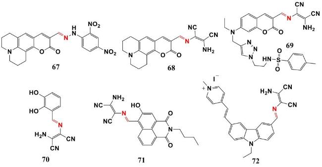
(赵云, 李艳芳, 李蓉晓, 王雅卿, 樊晓霞. 有机化学, 2021, 41(5): 1974).
|
| [70] |
|
| [71] |
(丁炳辉, 韩少辉, 熊海青, 王本花, 左伯军, 宋相志. 有机化学, 20, 43(8): 2878).
|
| [72] |
|
| [73] |
|
| [74] |
|
| [75] |
|
| [76] |
|
| [77] |
|
| [78] |
|
| [79] |
|
| [80] |
|
| [81] |
|
| [82] |
|
| [83] |
|
| [84] |
|
| [85] |
|
| [86] |
|
| [87] |
|
| [88] |
|
| [89] |
|
| [90] |
|
| [91] |
|
| [92] |
|
| [93] |
|
| [94] |
|
| [95] |
|
| [96] |
|
| [97] |
|
| [98] |
|
| [99] |
|
| [100] |
|
| [101] |
|
| [102] |
|
| [103] |
|
| [104] |
|
| [105] |
|
| [106] |
|
| [107] |
|
| [108] |
|
| [109] |
|
| [110] |
|
| [111] |
|
| [112] |
|
| [113] |
|
| [114] |
|
| [115] |
|
| [116] |
|
| [117] |
|
| [118] |
|
| [119] |
|
| [120] |
|
| [121] |
|
| [122] |
|
| [123] |
|
| [124] |
|
| [125] |
|
| [126] |
|
| [127] |
|
| [128] |
|
| [129] |
|
| [130] |
|
| [131] |
|
| [132] |
|
| [133] |
|
| [134] |
|
| [135] |
|
| [136] |
|
| [137] |
|
| [138] |
|
| [139] |
|
| [140] |
|
| [141] |
|
/
| 〈 |
|
〉 |

