Home Journals Progress in Chemistry
Progress in Chemistry

Abbreviation (ISO4): Prog Chem      Editor in chief: Jincai ZHAO

About  /  Aim & scope  /  Editorial board  /  Indexed  /  Contact  / 
Review

A Review of Methods for Evaluating Adverse Skin Reactions

  • Mengyao Bing 1, 2 ,
  • Yao Pei 1, 3 ,
  • Chang'ou Wang 4 ,
  • Gaocai Han 4 ,
  • Qunfang Zhou , 1, 2, 3, * ,
  • Guibin Jiang 1, 2, 3
Expand
  • 1 State Key Laboratory of Environmental Chemistry and Ecotoxicology, Research Center for Eco-Environment Sciences, Chinese Academy of Sciences, Beijing 100085, China
  • 2 School of Environment, Hangzhou Institute for Advanced Study, University of Chinese Academy of Sciences, Hangzhou 310024, China
  • 3 College of Resources and Environment, University of Chinese Academy of Sciences, Beijing 101400, China
  • 4 Beijing Xiaomi Mobile Software Co., Ltd., Beijing 100085, China

Received date: 2024-09-04

  Revised date: 2025-02-13

  Online published: 2025-05-20

Supported by

the Strategy Priority Research Program (Category B) of Chinese Academy of Sciences(XDB0750300)

Abstract

With the rapid development of the economy and society, various types of new chemicals are constantly emerging, and have been widely applied in daily life and work, including medical devices, metal jewelry, beauty and personal care products, and smart wearable products. However, the adverse skin reactions caused by contact with these daily products seriously threaten human health and reduce the quality of life of patients. Therefore, it is of great significance to evaluate the adverse skin reactions of daily necessities and their ingredients. These evaluations aid in identifying potentially hazardous chemicals, and guiding the effective management of the product manufacturing. Traditional methods for evaluating adverse skin reactions have relied heavily on animal experiments. But in light of concerns regarding animal welfare and the need for improving test throughput and prediction efficacy of methods, great efforts have been made to develop various in vivo and in vitro alternative methods. Against this backdrop, the mechanisms of adverse skin reactions, especially for skin irritation/corrosion, atopic dermatitis and allergic contact dermatitis, and their evaluation methods were summarized in this review, based on a large number of studies published in recent years. Finally, the shortcomings and perspectives of research in this field are prospected.

Contents

1 Introduction

2 Adverse skin reactions and evaluating methods

2.1 Non-allergic skin reactions

2.2 Allergic skin reactions

3 Evaluating methods for ACD

3.1 Animal experiments

3.2 In vivo alternative assays

3.3 In vitro alternative assays

4 Conclusion and outlook

Cite this article

Mengyao Bing , Yao Pei , Chang'ou Wang , Gaocai Han , Qunfang Zhou , Guibin Jiang . A Review of Methods for Evaluating Adverse Skin Reactions[J]. Progress in Chemistry, 2025 , 37(7) : 1002 -1010 . DOI: 10.7536/PC240902

1 Introduction

Skin symptoms manifesting as varying degrees of dryness, erythema, pruritus, edema, wheals, papules, vesicles, erosion, and so forth can all be referred to as skin adverse reactions. Skin adverse reactions can be triggered by various factors, such as genetic and environmental influences[1],drug effects[2-3],and exposure to chemicals in living and working environments[4-6].Based on the underlying mechanisms through which these triggers induce symptoms, skin adverse reactions can be classified into two major categories: allergic and non-allergic reactions. Non-allergic skin adverse reactions primarily involve skin irritation and corrosion[7];while allergic skin adverse reactions include atopic dermatitis (AD)[8]and allergic contact dermatitis (ACD)[9],among others.
In daily work and life, there are numerous chemicals and their formulated products that pose risks of irritation or sensitization, such as medical supplies, metal accessories, beauty and personal care products, and smart wearable devices. Skin adverse reactions and diseases caused by these substances pose a widespread and serious threat to the health of workers and consumers in related industries[5,9-10]. Therefore, evaluating the skin adverse reactions of daily-use products and their raw material chemical components is of great significance for avoiding health safety hazards, identifying potential risk substances, promoting product quality improvement, and enhancing industry risk management awareness.
For a long time, animal experiments using albino rabbits, guinea pigs, mice, and other species have been the most commonly used methods for evaluating skin adverse reactions. However, these methods are costly, have long experimental cycles, cause pain and discomfort to animals, and rely primarily on manual observation of skin pathological symptoms as endpoints, which introduces subjectivity in judgment and makes quantitative description difficult. Meanwhile, many countries and regions are increasingly regulating skin risk management for daily-use products and their ingredients, and animal welfare or ethical considerations are leading to restrictions or outright bans on the use of animal experiments in the evaluation process[11-12]. Therefore, developing sensitive, effective, and high-throughput non-animal evaluation methods has become a research hotspot in skin safety assessment in recent years. This article aims to review representative skin adverse reactions and their evaluation methods, with a particular focus on allergic contact dermatitis, in order to provide scientific guidance for the differentiation, identification, and analytical evaluation of various skin adverse reactions.

2 Skin Adverse Reactions and Their Evaluation

2.1 Non-allergic cutaneous adverse reactions

Skin irritation is one of the most common adverse skin reactions in humans[13]. According to the United Nations Globally Harmonized System of Classification and Labelling of Chemicals, skin irritation refers to reversible damage to the skin caused by contact with a substance or mixture, whereas skin corrosion refers to irreversible damage to the skin, characterized by observable necrosis of the epidermis and dermis. In addition, cumulative exposure to mild irritants may lead to the development of irritant contact dermatitis (ICD)[7].
For the safety assessment of skin irritation and corrosivity of chemical substances, the most commonly used classical method is animal testing primarily involving albino rabbits. This method evaluates whether a compound has skin irritancy potential by observing whether visible symptoms such as erythema and edema appear after albino rabbits are exposed to the compound through the skin[14]. This method is direct, effective, and highly sensitive; however, it causes harm to experimental animals, and studies have shown that results obtained from rabbit experiments may differ to some extent from actual human skin reaction outcomes[15]. Therefore, driven by the need to protect experimental animals and improve the predictive capability of the methods, various alternative approaches have gradually emerged. For example, the Rat skin transcutaneous electrical resistance test (TER) method uses isolated rat epidermal tissue to obtain skin slices, and assesses skin irritancy/corrosivity by measuring the transcutaneous electrical resistance of these slices after exposure to the compound[16]. The In vitro membrane barrier test method (MBT), on the other hand, is based on an artificial non-biological polymeric skin model. It evaluates whether the membrane barrier is damaged by detecting changes in the pH of the skin model after exposure to the compound, thereby identifying and classifying the irritancy/corrosivity of the test substance[17]. Additionally, the currently most favored method is the reconstructed human epidermis (RhE) test method. Since irritant or corrosive chemicals can penetrate the stratum corneum through diffusion or erosion, exerting toxic effects on underlying cells, assessing the cellular viability of RhE tissue after exposure to the test substance can reflect the irritancy or corrosivity of the compound[18-19].

2.2 Allergic cutaneous adverse reactions

2.2.1 Atopic Dermatitis

AD is a chronic allergic skin disease prone to recurrence, with main symptoms including dry skin, intense itching, erythema, and poorly defined lesion boundaries. It is often accompanied by allergic rhinitis and/or asthma, and has a higher incidence during childhood[8,20]. The pathogenesis of AD is complex, involving interactions between genetic and environmental factors. Specific factors include impaired skin barrier function, such as loss-of-function mutations in filaggrin; abnormal cell-mediated immune responses, particularly an imbalance in the ratio of cytokines from T helper 1 (Th1) and T helper 2 (Th2) cells; IgE-mediated type I hypersensitivity reactions; and stimulation by environmental factors (such as formaldehyde and detergents)[21]. Due to its complex mechanisms, lack of specific biomarkers, high symptom heterogeneity among patients[22], and susceptibility to being confused with ICD, ACD, and other eczematous diseases[7], clinical differential diagnosis and laboratory evaluation of AD present significant challenges.
Currently, the Hanifin-Rajka criteria are primarily used for the clinical diagnosis of AD, serving as the most widely applied "gold standard." It includes 4 major criteria and 23 minor criteria. Patients must meet at least 3 of the major criteria and 3 of the minor criteria simultaneously to be diagnosed with AD. The 4 major criteria are as follows: ① pruritus; ② typical morphology and distribution (e.g., involvement of flexural skin surfaces); ③ chronic recurrent course; ④ personal/family history of allergies[23]. This standard has high sensitivity and accuracy, but its complex content makes it difficult to remember and requires a high level of expertise from physicians, leading to certain challenges in clinical application[24]. Therefore, based on the Hanifin-Rajka criteria, physicians and researchers worldwide have been working to develop simpler and more effective AD diagnostic standards tailored to the characteristics of diseases in their respective countries or regions. These include the UK Working Party criteria[25], the AAD guidelines from the United States[26], and the Zhang criteria (also known as the Chinese criteria), published in 2016 and more suitable for the Chinese population[27], among others.
In the laboratory, ovalbumin (OVA) or a hapten can be used to sensitize mice, inducing dermatitis-like lesions and establishing an AD disease model. Studies have shown that repeated and prolonged exposure to a hapten can trigger skin inflammation in mice similar to that observed in human AD[28]. Meanwhile, bone marrow-derived macrophages from mice have also been demonstrated to serve as a cellular model for AD research, capable of mimicking inflammatory infiltration during the pathogenesis of AD[29].
In summary, current research on the pathogenesis, triggers, and evaluation models of AD remains incomplete. No specific biomarkers suitable for the diagnosis and evaluation of AD have yet been identified, and further studies are still needed.

2.2.2 Allergic contact dermatitis

ACD is an adaptive inflammatory response generated by the body after skin exposure to allergens, classified as a T-cell-mediated type IV delayed hypersensitivity reaction. Its clinical manifestations include skin itching, erythema, edema, and even blisters[9,30]. The potential allergens causing ACD are small-molecule haptens present in the environment. Due to their inherent electrophilicity, they can bind to nucleophilic centers of skin proteins, forming sensitizing complexes that trigger contact sensitization. It is worth noting that some chemicals, although incapable of binding to proteins themselves, can be converted into haptens under certain conditions. Depending on the conversion conditions, these substances can further be categorized into pre-haptens (chemical conversion) and pro-haptens (biological activation)[31].
Local patch test (Buehler test, BT)[32]and Guinea pig maximization test (GPMT)[33]and other whole-animal experimental methods, as well as later in vivo alternative methods based on animals, such as the Mouse ear swelling test (MEST)[34]and the Local lymph node assay (LLNA)[35],have long been widely used for skin sensitization testing. However, all of the above animal-based experimental methods suffer from lengthy durations, high costs, and ethical concerns regarding animal welfare. Therefore, developing sensitive and effective non-animal methods for assessing skin sensitization potential has become a research hotspot in recent years.
Based on the collection and analysis of extensive relevant studies, the Organisation for Economic Cooperation and Development (OECD) has summarized the biological processes of ACD into an adverse outcome pathway (AOP), which is divided into four key events (Key Event Ⅰ~Ⅳ) and the final adverse outcome[36]. These four key events include the formation of hapten-protein complexes, activation of keratinocytes (KCs), activation of dendritic cells (DCs), and activation and proliferation of T cells (Figure 1). On this basis, in vitro skin sensitization assessment methods for each key event in this AOP have gradually developed and matured. Representative methods include the Direct peptide reactivity assay (DPRA)[37], the KeratinoSensTMassay[38], and the Human cell line activation test (h-CLAT)[39]. The development of these methods has significantly contributed to reducing animal testing, enabling high-throughput screening, and improving detection sensitivity.
图1 过敏性接触性皮炎的不良结局通路

Fig. 1 Adverse outcome pathway (AOP) of allergic contact dermatitis (ACD)

3 Evaluation methods for the skin sensitization effects of ACD

3.1 Whole-animal experiments

The whole-animal test is a classic method for evaluating skin sensitization effects. It determines the sensitizing potential of a test substance by directly observing whether repeated exposure of the substance to animal skin induces allergic skin reactions such as erythema and edema, as well as assessing the severity of these reactions. Guinea pigs are commonly used as experimental animals. Among the whole-animal test methods, the most representative are the BT[32]and GPMT[33]. Both methods include an induction phase and an elicitation phase: during the induction phase, the compound is applied topically or injected intradermally multiple times over 2 to 3 weeks to guinea pigs, simulating the initial exposure to the compound; in the elicitation phase, the test compound is applied to new sites distinct from those treated during the induction phase. After application, the experimental animals are observed for allergic reactions, and the sensitizing potential of the compound is determined based on the number of animals showing positive allergic responses[40]. Overall, the whole-animal test has been developed relatively early and its system is mature; however, it also faces issues such as long testing cycles, subjective observation indicators, limited quantitative evaluation capabilities, high costs, and animal ethics concerns. As a result, it has largely been replaced by in vitro and ex vivo alternative tests.

3.2 In vivo alternative methods

In vivo alternative methods for evaluating skin sensitization effects mainly include MEST[34]and LLNA[35]. The MEST method is based on the principle that when mice develop contact allergy in their ears, the auricles swell and thicken. It evaluates the sensitizing potential of compounds by measuring the degree of ear swelling after compound exposure, involving both an induction and a challenge phase. In the induction phase, the test substance solution is applied topically multiple times to the abdominal or dorsal skin of mice. In the challenge phase, the test substance solution is applied topically to one side of the mouse's ear, while the other side is treated with solvent. After 1 to 2 days, the thickness of both auricles is precisely measured, and the quantitative endpoint is ultimately defined as the percentage increase in auricular thickness on the compound-treated side compared to the solvent control side[34]. The LLNA method, on the other hand, is based on the phenomenon that T cells become activated and proliferate in lymph nodes during the sensitization process. Therefore, quantitatively assessing the proliferation of lymph node cells after compound exposure can determine the sensitizing potential of the test substance. The general procedure involves applying the test substance or control solvent multiple times to both sides of the mouse's ears. Approximately 72 hours after the last treatment, the draining lymph nodes from both sides of the mouse's ears are excised, and single-cell suspensions of lymph node cells are prepared under sterile conditions and cultured. Finally, the proliferation capacity of draining lymph node T cells is determined by measuring the incorporation of [3H]thymidine, thereby reflecting the sensitizing potential of the test substance[35]. Compared to whole-animal experiments, the advantage of in vivo alternative methods lies in their quantitative and objective endpoints for sensitization assessment, as well as the use of relatively inexpensive mice instead of guinea pigs, and a shorter testing cycle. However, in vivo alternative methods still face issues such as high costs, complex testing procedures, and reliance on a large number of experimental animals. Therefore, since the 21st century, in vitro alternative methods have gradually flourished.

3.3 In vitro alternative methods

In vitro alternative methods are primarily developed based on the key events in the aforementioned adverse outcome pathway for skin sensitization. Among these, relatively mature in vitro models for assessing skin sensitization have been established, primarily relying on key events I to III, significantly reducing the reliance on experimental animals in sensitization assessments. Meanwhile, combination evaluation systems based on two or more key events are also undergoing preliminary exploration.

3.3.1 DPRA

DPRA is based on Key Event 1 in the AOP. As mentioned earlier, whether a chemical substance is an ACD allergen hinges on whether it is a hapten—that is, whether it has the ability to bind to nucleophilic centers of skin proteins to form hapten-protein complexes. The DPRA method, developed on this basis, uses high-performance liquid chromatography to detect and quantify the peptide depletion rate (polypeptide reactivity) after incubating a chemical substance with synthetic peptides containing lysine or cysteine. Ultimately, polypeptide reactivity is graded according to the peptide depletion rates of cysteine and lysine, thereby determining the sensitizing potential of the compound[37]. As an in vitro chemical analysis method, DPRA requires simple reagents and conditions at a relatively low cost, but it does have certain requirements for experimental equipment and can only detect haptens, not potential skin sensitizers such as pro-haptens or proto-haptens. Additionally, it is worth noting that this method is also unsuitable for testing metal compounds, as the binding of metals to proteins occurs through mechanisms other than covalent bonding[41].

3.3.2 KeratinoSens™/LuSens assay

The KeratinoSens™ and LuSens methods are based on Key Event II, which is the activation of keratinocytes. The Keap1/Nrf2-ARE signaling pathway, composed of the repressor protein Keap1 (Kelch like ECH associated protein 1), the transcription factor Nrf2 (Nuclear factor erythroid 2-related factor 2), and the antioxidant response element (ARE), is the toxicological pathway through which sensitizers act on the skin. In the KeratinoSens™ method, a luciferase reporter gene under the control of a single-copy ARE element from the human AKR1C2 gene is stably integrated into human immortalized keratinocytes (HaCaT). This allows the Keap1/Nrf2-ARE signaling pathway response to be detected using a luciferase reporter gene system, thereby assessing the activation status of HaCaT cells and determining the sensitizing potential of the test substance[38]. The principle of the LuSens experiment is similar to that of KeratinoSens™; the only difference between the two methods lies in the ARE used, with the former employing the ARE derived from the rat NQO1 gene instead of the human AKR1C2 gene[42]. These methods offer advantages such as high throughput, simplicity of technology, and good predictive performance, allowing for multi-concentration testing to obtain full dose-response curves. However, the key mechanism underlying Keap1/Nrf2-ARE pathway activation appears to depend on the binding of compounds to cysteine residues of the Keap1 protein. Therefore, when the test substance exhibits specificity for lysine residues, false-negative results are likely to occur. In such cases, the results can be combined with those from the DPRA test for comprehensive analysis[43].

3.3.3 h-CLAT

h-CLAT is a skin sensitization assay developed based on the activation of dendritic cells by key events III. During ACD, the expression of adhesion molecule CD54 and costimulatory molecule CD86 on the surface of dendritic cells is upregulated. Therefore, by detecting and quantifying the upregulation of CD54 and CD86 expression on dendritic cells before and after exposure to the test substance, the sensitizing potential of the substance can be assessed. In h-CLAT, THP-1 cells (a human acute monocytic leukemia cell line) are used to simulate the activation process of dendritic cells. After exposure to the test substance, THP-1 cells are stained with fluorescent antibody dyes targeting surface proteins CD54 and CD86. Finally, flow cytometry is used to measure the corresponding fluorescence signal intensity. The ratio of fluorescence intensity between the test substance exposure group and the solvent control group reflects the activation status of THP-1 cells, thereby allowing assessment of the sensitizing potential and potency of the compound[39]. h-CLAT can be used to test various compounds with different physicochemical properties, reaction mechanisms, and sensitizing potencies. It exhibits high sensitivity in detecting strong sensitizers but may produce false-negative results for weak to moderate sensitizers with a certain probability[44]. Additionally, due to the insufficient metabolic capacity of THP-1 cells, this test method may also yield negative sensitization results when testing prohaptenes and hapten precursors[45-46].

3.3.4 U937 cell line activation experiment

Similar to the h-CLAT method based on THP-1 cells, the U937 cell line activation test (U-SENSTM) is also based on Key Event III, but it uses U937 cells (a human histiocytic lymphoma cell line). After exposing compounds to U937 cells, flow cytometry is used to detect the expression of CD86, a typical activation marker on the surface of U937 cells. The sensitizing potential of the test substance is then assessed by comparing the fluorescence intensity ratio between the exposure group and the solvent control group[47]. One major advantage of this method is its ability to effectively detect prohaptenes and haptenes[45], and it exhibits higher sensitivity than h-CLAT in detecting weak sensitizers[44]. However, substances that damage cell membranes, such as surfactants, can cause nonspecific increases in CD86 expression, leading to false-positive results[47]. Therefore, positive results for surfactant-containing substances need to be further verified using other methods.

3.3.5 Comprehensive Test Evaluation Strategy

In summary, existing scoring systems have limited predictive capabilities for bleeding events, and their results are inconsistent[25,30,33].

3.3.6 Cell co-culture method

Similar to the starting point of the IATA strategy, test methods based solely on individual key events cannot fully capture the complete sensitization mechanism. Moreover, predictive models constructed using only a single cell type still have considerable room for improvement in terms of detection sensitivity and accuracy. Therefore, in vitro cell models that co-culture two or more types of cells, such as KCs, DCs, and T cells, to incorporate complex intercellular interactions have gradually emerged. The co-culture system of HaCaT and THP-1 cells (Coculture activation test, COCAT), developed by Hennen et al., simultaneously covers key events II and III in the AOP, effectively simulating the interaction between KCs and DCs during ACD development. This system represents an advancement over h-CLAT and U-SENSTM, which are based solely on DCs. Experimental results demonstrate that COCAT exhibits a significant upregulation of CD86 and CD54 compared to h-CLAT, while also lowering the sensitization threshold for compounds[50-51]. Other studies employing co-cultures of KCs and DCs have utilized different types of KCs; in addition to HaCaT cells, these include primary human keratinocytes[52]and NCTC2544 cells[53]. Furthermore, co-cultures involving KCs and T cells[54], DCs and T cells[55], and KCs, DCs, and T cells[56]have all been shown to upregulate the expression of relevant molecular markers and provide effective evaluation of compound sensitization potential. However, these methods started relatively late and are still in the early stages of development. They have not yet undergone extensive testing with numerous compounds or validation by different research teams, and thus require further in-depth study and refinement.
表1 皮肤不良反应的研究方法

Table 1 Methods for evaluating adverse skin reactions

Adverse skin reaction Method Principle Advantage and disadvantage Ref
Skin irritation/corrosion Animal experiment Experimental animals (mainly albino rabbits) are exposed percutaneously to chemicals and the appearance and severity of symptoms such as erythema and edema are observed to determine the skin irritation potential. Direct and effective test;
Harm to experimental animals;
Subjective endpoints
14
15
TER The test chemical is applied to the epidermal surfaces of skin discs for up to 24 hours. After exposure, reduction in transcutaneous resistance of skin discs can be used to assess the corrosiveness. In vitro alternative methods;
Simple testing procedures;
High testing costs
16
MBT Detect the membrane barrier damage caused by test chemicals after the application of the test chemical to the surface of the synthetic macromolecular membrane barrier. 17
RhE test method The test chemical is applied topically to a three-dimensional RhE model, comprised of non-transformed human-derived epidermal keratinocytes. Irritant/corrosive chemicals are identified by their ability to decrease cell viability in RhE model. 18
19
Allergic contact dermatitis Animal experiment During induction phase, experimental animals (mainly guinea pigs) obtain repeated treatment to simulate the first contact to a chemical. In the challenge phase, the animals are checked if they show allergic reactions to the non-irritant concentration. Well-established testing system;
Harm to experimental animals;
Subjective endpoints;
Long experimental cycle
32
33
40
MEST Evaluate skin sensitization by measuring the increase in mouse ear thickness following topical application of a test chemical. This swelling response reflects the induction of a localized immune response, primarily mediated by T cells, indicating the sensitization potential of the test chemical. In vivo alternative methods;
Quantitative and objective endpoints;
Harm to experimental animals;
Complicated testing procedures;
High testing costs
34
LLNA Evaluate skin sensitization by measuring the proliferation of lymphocytes in the draining lymph nodes after chemical exposure. This proliferation is quantified using radioactive labeling or other markers. 35
DPRA The depletion rate of peptides following co-incubation of chemicals with synthetic peptides containing lysine or cysteine is detected and quantified using high-performance liquid chromatography. The peptide reactivity is classified based on the depletion rates of cysteine and lysine peptides, thereby assessing the sensitization potential of chemicals. Simple reagents and conditions; Low testing costs;
Limited to detecting haptens, unable to identify prohaptens or prehaptens;
Not suitable for testing metals
37
41
KeratinoSensTM/ LuSens Use a luciferase reporter gene system to detect the response of the Keap1/Nrf2-ARE signaling pathway, and to measure the activation of keratinocytes to determine the sensitization potential of the test chemical. High throughput and predictability;
Susceptible to false-negative results;
38
42,43
h-CLAT Up-regulation of CD54 and CD86 expressions in dendritic THP-1 cells after chemical exposure is investigated by flow cytometry to measure the sensitization potential of chemicals. Highly sensitive to strong sensitizers;
Possible false-negative results in prohapten and prehapten testing
39
45,46
U-SENSTM The principle is basically the same as that of the h-CLAT, but only CD86 is determined in U937 cells. Effective for detection of prohaptens and prehaptens, but not for surfactants 44
45,47
Cell coculture Co-culture of two or more of KCs, DCs and T cells to encompass complex cellular interactions. Cover two or more key events;
Lack of extensive experimental validation
50~
56

4 Conclusion and Outlook

Since the 21st century, evaluation methods for various skin adverse reactions have evolved from animal-based assays to in vitro alternative methods, gradually reducing reliance on animals. In addition to the representative methods mentioned in this article, many improved or novel approaches have also been continuously developed[57], such as gene expression analysis (e.g., the genomic allergen rapid detection method GARD™skin[58]), combined testing of cell phenotypes and cytokines (CD86-IL8 method[59]), and the use of skin organoids to simulate environmental exposure and its adverse effects on human skin[60]. These methods have significantly enriched the methodology for evaluating skin adverse reactions, providing diverse options and references for relevant industries and researchers. Meanwhile, the convenience, sensitivity, and accuracy of these alternative methods have steadily improved, leading to their widespread application in compound safety testing, clinical drug screening, and the assessment of potential skin hazards associated with daily-use products and their raw materials.
However, as mentioned earlier, current methods and models for assessing skin adverse reactions still have many shortcomings, such as complex procedures, high requirements for instruments and equipment, excessive costs, and the need for further improvement in sensitivity and accuracy, as well as their limited applicability to specific types of compounds. Therefore, further innovation and development are needed in related methodological research, aiming to strike a balance among detection procedures, costs, throughput, and performance of models or methods. In addition, environmental triggers, pathogenesis, and associated biomarkers of skin adverse reactions, especially allergic reactions such as AD and ACD, remain incompletely understood. Researchers in this field must continue their efforts to deepen our understanding of the biological processes and mechanisms underlying various skin adverse reactions.
In summary, elucidating the pathological mechanisms of various skin adverse reactions, identifying sensitive and effective biomarkers, and developing evaluation models or methods that accurately reflect the real biological processes and mechanisms of skin responses will become important directions for future research on skin adverse reaction assessment. This will enable accurate evaluation of the potential and potency of compounds or actual samples in causing skin adverse reactions.

We would like to thank Xiao Dan and Ding Qian from Beijing Xiaomi Mobile Software Co., Ltd. for their guidance on the content related to skin adverse reactions in this article.

[1]
Boguniewicz M, Leung D Y M. J. Allergy Clin. Immunol., 2006, 117(2): S475.

[2]
Mockenhaupt M. Chem. Immunol Allergy, 2012, 97: 1.

[3]
Karavaizoglu C, Suleyman A, Yavuz Alıc R, Dolu K O, Yucel E, Demirkale Z H, Cimen S S, Ozdemir C, Tamay Z U. Int. Arch. Allergy Immunol., 2024, 185(1): 56.

[4]
Huang L N, Zhong Y P, Liu D, Wang X H, Gong C Y, Wen S, Elias P M, Yang B, Man M Q. Contact Dermat., 2018, 79(6): 365.

[5]
Khatsenko K, Khin Y, Maibach H. Dermatitis, 2020, 31(5): 283.

[6]
Symanzik C, Johansen J D, Weinert P, Babić Ž, Hallmann S, Havmose M S, Kezic S, Macan M, Macan J, Strahwald J, Turk R, van der Molen H F, John S M, Uter W. Contact Dermat., 2022, 86(5): 333.

[7]
Bains S N, Nash P, Fonacier L. Clin. Rev. Allergy Immunol., 2019, 56(1): 99.

[8]
Leung D Y, Bieber T. Lancet, 2003, 361(9352): 151.

[9]
Kaplan D H, Igyártó B Z, Gaspari A A. Nat. Rev. Immunol., 2012, 12(2): 114.

[10]
Smylie A, Gill N, Oosterhuis R, Mathura P, Elliott J, Straube S, Dytoc M. J. Cutan. Med. Surg., 2021, 25(2): 216.

[11]
European Union. Regulation (EC) No 1223/2009 of the European Parliament and of the Council of 30 November 2009 on Cosmetic Products. Office J. Europ Union, 2009, Vol 342.

[12]
European Union. Directive 2010/63/EU of the European Parliament and of the Council of 22 September 2010 on the Protection of Animals Used for Scientific Purposes. Office J. Europ Union, 2010, Vol 276.

[13]
Welss T, Basketter D A, Schröder K R. Toxicol. Vitro, 2004, 18(3): 231.

[14]
OECD. Test No. 404: Acute Dermal Irritation/Corrosion. Paris: Organisation for Economic Cooperation and Development, 2015.

[15]
Jírová D, Basketter D, Liebsch M, Bendová H, Kejlová K, Marriott M, Kandárová H. Contact Dermat., 2010, 62(2): 109.

[16]
OECD. Test No. 430: In Vitro Skin Corrosion: Transcutaneous Electrical Resistance Test Method (TER). Paris: Organisation for Economic Cooperation and Development, 2015.

[17]
OECD. Test No. 435: In Vitro Membrane Barrier Test Method for Skin Corrosion. Paris: Organisation for Economic Cooperation and Development, 2015.

[18]
OECD. Test No. 431: In Vitro Skin Corrosion: Reconstructed Human Epidermis (RHE) Test Method. Paris: Organisation for Economic Cooperation and Development, 2019.

[19]
OECD. Test No. 439: In Vitro Skin Irritation: Reconstructed Human Epidermis Test Method. Paris: Organisation for Economic Cooperation and Development, 2021.

[20]
Bieber T. N. Engl. J. Med., 2008, 358(14): 1483.

[21]
David Boothe W, Tarbox J A, Tarbox M B. Management of Atopic Dermatitis. Cham: Springer International Publishing, 2017, 21.

[22]
Hassan S, Hamideh N, Poulos C, Cheema S, Rangwani S, Lio P A. Dermatitis, 2021, 32(1S): S8.

[23]
Hanifin J M, Rajka G. Acta Derm. Venereol., 1980, 60: 44.

[24]
Reynolds M, Gorelick J, Bruno M. J. Drugs Dermatol., 2020, 19(3): 244.

[25]
Williams H C, Jburney P G, Pembroke A C, Hay R J. Br. J. Dermatol., 1994, 131(3): 406.

[26]
Eichenfield L F, Tom W L, Chamlin S L, Feldman S R, Hanifin J M, Simpson E L, Berger T G, Bergman J N, Cohen D E, Cooper K D, Cordoro K M, Davis D M, Krol A, Margolis D J, Paller A S, Schwarzenberger K, Silverman R A, Williams H C, Elmets C A, Block J, Harrod C G, Smith Begolka W, Sidbury R. J. Am. Acad. Dermatol., 2014, 70(2): 338.

[27]
Liu P, Zhao Y, Mu Z L, Lu Q J, Zhang L, Yao X, Zheng M, Tang Y W, Lu X X, Xia X J, Lin Y K, Li Y Z, Tu C X, Yao Z R, Xu J H, Li W, Lai W, Yang H M, Xie H F, Han X P, Xie Z Q, Nong X, Guo Z P, Deng D Q, Shi T X, Zhang J Z. Chin. Med. J., 2016, 129(7): 757.

[28]
Inagaki N, Nagai H. J. Pharmacol. Sci., 2009, 110(3): 251.

[29]
Yang L, Fu J R, Han X, Zhang C Y, Xia L, Zhu R H, Huang S H, Xiao W F, Yu H M, Gao Y J, Liang Q Y, Li W, Zhou Y F. J. Allergy Clin. Immunol., 2022, 149(6): 2021.

[30]
Karlberg A T, Baron J, Merk H. Advances in Molecular Toxicology. Amsterdam: Elsevier, 2008, 87.

[31]
Karlberg A T, Börje A, Duus Johansen J, Lidén C, Rastogi S, Roberts D, Uter W, White I R. Contact Dermat., 2013, 69(6): 323.

[32]
Buehler E V. Arch. Dermatol., 1965, 91(2): 171.

[33]
Magnusson B, Kligman A M. J. Investig. Dermatol., 1969, 52(3): 268.

[34]
Gad S C, Dunn B J, Dobbs D W, Reilly C, Walsh R D. Toxicol. Appl. Pharmacol., 1986, 84(1): 93.

[35]
Kimber I, Mitchell J A, Griffin A C. Food Chem. Toxicol., 1986, 24(6/7): 585.

[36]
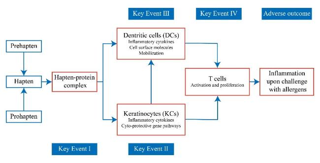
OECD. The Adverse Outcome Pathway for Skin Sensitisation Initiated by Covalent Binding to Proteins. Paris: Organisation for Economic Cooperation and Development, 2014.

[37]
Gerberick G F. Toxicol. Sci., 2004, 81(2): 332.

[38]
Emter R, Ellis G, Natsch A. Toxicol. Appl. Pharmacol., 2010, 245(3): 281.

[39]
Ashikaga T, Yoshida Y, Hirota M, Yoneyama K, Itagaki H, Sakaguchi H, Miyazawa M, Ito Y, Suzuki H, Toyoda H. Toxicol. Vitro, 2006, 20(5): 767.

[40]
Maurer T. Methods, 2007, 41(1): 48.

[41]
OECD. Test No. 442C: In Chemico Skin Sensitisation: Assays Addressing the Adverse Outcome Pathway Key Event on Covalent Binding to Proteins. Paris: Organisation for Economic Cooperation and Development, 2024.

[42]
Ramirez T, Mehling A, Kolle S N, Wruck C J, Teubner W, Eltze T, Aumann A, Urbisch D, van Ravenzwaay B, Landsiedel R. Toxicol. Vitro, 2014, 28(8): 1482.

[43]
OECD. Test No. 442D: In Vitro Skin Sensitisation: Assays Addressing the Adverse Outcome Pathway Key Event on Keratinocyte Activation. Paris: Organisation for Economic Cooperation and Development, 2024.

[44]
OECD. Test No. 442E: In Vitro Skin Sensitisation: In Vitro Skin Sensitisation Assays Addressing the Key Event on Activation of Dendritic Cells on the Adverse Outcome Pathway for Skin Sensitisation. Paris: Organisation for Economic Cooperation and Development, 2024.

[45]
Fabian E, Vogel D, Blatz V, Ramirez T, Kolle S, Eltze T, van Ravenzwaay B, Oesch F, Landsiedel R. Arch. Toxicol., 2013, 87(9): 1683.

[46]
Ashikaga T, Sakaguchi H, Sono S, Kosaka N, Ishikawa M, Nukada Y, Miyazawa M, Ito Y, Nishiyama N, Itagaki H. Altern. Lab. Anim., 2010, 38(4): 275.

[47]
Piroird C, Ovigne J M, Rousset F, Martinozzi-Teissier S, Gomes C, Cotovio J, Alépée N. Toxicol. Vitro, 2015, 29(5): 901.

[48]
OECD. Guidance Document on the Reporting of Defined Approaches and Individual Information Sources to Be Used within Integrated Approaches to Testing and Assessment (IATA) for Skin Sensitisation. Paris: Organisation for Economic Cooperation and Development, 2017.

[49]
Otsubo Y, Nishijo T, Miyazawa M, Saito K, Mizumachi H, Sakaguchi H. Regul. Toxicol. Pharmacol., 2017, 88: 118.

[50]
Hennen J, Aeby P, Goebel C, Schettgen T, Oberli A, Kalmes M, Blömeke B. Toxicol. Sci., 2011, 123(2): 501.

[51]
Hennen J, Bloemeke B. ALTEX-Altern. Anim. Exp., 2017, 34(2): 279.

[52]
Cao Y P, Ma P C, Liu W D, Zhou W Q, Tao Y, Zhang M L, Li L J, Chen Z Y. Immunopharmacol. Immunotoxicol., 2012, 34(2): 196.

[53]
Galbiati V, Maddalon A, Iulini M, Marinovich M, Corsini E. Toxicol. Vitro, 2020, 68: 104929.

[54]
Van Den Bogaard E H, Tjabringa G S, Joosten I, Vonk-Bergers M, Van Rijssen E, Tijssen H J, Erkens M, Schalkwijk J, Koenen H J P M. J. Investig. Dermatol., 2014, 134(3): 719.

[55]
Huppert C, Paris C, Langonné I, Muller S, Mathiot J, Abdessadeq H, Gagnaire F, Battais F, Sponne I. Contact Dermat., 2018, 79(2): 67.

[56]
Sonnenburg A, Stahlmann R, Kreutz R, Peiser M. Arch. Toxicol., 2023, 97(6): 1677.

[57]
De Ávila R I, Lindstedt M, Valadares M C. Regul. Toxicol. Pharmacol., 2019, 108: 104445.

[58]
Johansson H, Albrekt A S, Borrebaeck C A K, Lindstedt M. Toxicol. Vitro, 2013, 27(3): 1163.

[59]
Parise C B, Sá-Rocha V M, Moraes J Z. Toxicol. Vitro, 2015, 30(1): 318.

[60]
Hong Z X, Zhu S T, Li H, Luo J Z, Yang Y, An Y, Wang X, Wang K. Mil. Med. Res., 2023, 10: 40.

Outlines

/