
Reaction Mechanism of Chemodynamic Therapy and Its Applications in Anti-Tumor Treatment
Received date: 2024-11-18
Revised date: 2025-01-10
Online published: 2025-07-30
Supported by
the National Key Research and Development Program of China(2022YFA1503001)
the National Natural Science Foundation of China(22172174)
the Natural Science Foundation of Shaanxi Province of China(2025JC-YBMS-137)
the High-Level Talent Program of Shaanxi Province of China
Chemodynamic therapy (CDT) refers to a method that utilizes metal ion-mediated Fenton/Fenton-like reactions to catalyze the generation of highly cytotoxic hydroxyl radicals from hydrogen peroxide,effectively killing tumor cells. It offers advantages such as tumor specificity,minimal side effects,and a treatment process initiated solely by internal tumor substances like H2O2 and glutathione without the need for external stimuli. However,the high concentration of glutathione in the tumor microenvironment,insufficient endogenous hydrogen peroxide,and hypoxia hinder the therapeutic effect of CDT. To enhance its effectiveness,researchers have explored various metal ion-mediated Fenton/Fenton-like reactions,leading to the proposed combination of CDT with multiple other therapies. This article reviews the reaction mechanisms of CDT and its collaborative applications with various therapies in anti-tumor treatment. It begins by discussing the catalytic reaction mechanisms of CDT mediated by different metal ions,delving into the advantages and disadvantages of various ions in catalyzing Fenton or Fenton-like reactions. Subsequently,it details the latest research progress on the combination of CDT with other therapies,such as photothermal therapy,chemotherapy,and photodynamic therapy,in anti-tumor treatments. Finally,the article proposes future research directions for the development of chemodynamic therapy and highlights key issues that need to be considered to further promote its clinical research applications.
1 Introduction
2 Mechanism for Fenton reaction mediated by various metal ions
2.1 Iron-mediated mechanism for Fenton reaction
2.2 Copper-mediated mechanism for Fenton-like reaction
2.3 Other metal ion-mediated mechanisms for Fenton-like reactions
3 CDT-based combination therapies and their anti-tumor applications
3.1 Combination therapy of PTT and CDT
3.2 Combination therapy of chemotherapy and CDT
3.3 Combination therapy of PDT and CDT
3.4 Combination therapy of other therapies and CDT
4 Conclusion and outlook
Mengyu Han , Rong Chen , Qiao Li , Hong Li , Yi Jia . Reaction Mechanism of Chemodynamic Therapy and Its Applications in Anti-Tumor Treatment[J]. Progress in Chemistry, 2025 , 37(8) : 1091 -1104 . DOI: 10.7536/PC241102
表1 不同金属介导的CDT治疗肿瘤的类型及联合疗法应用情况Table 1 Types of different metal-mediated CDT for the treatment of tumors and the application of combination therapy |
| Metal | Material name | Antitumor type | Combination therapy | Antitumor efficacy | Ref |
|---|---|---|---|---|---|
| Fe | MN@CuO2 NPs | Melanoma | CDT/PTT | Several tumors of the MN@CuO2+NIR group were completely eradicated after treatment | 43 |
| HSA-GOx-TPZ-Fe3+-TA | Breast cancer | CDT/Starvation therapy | HGTFT displayed the highest tumor growth inhibition (TGI) rate of 97.8% | 72 | |
| M-mFeP@O2-G | Mouse osteosarcoma | CDT/PTT/Starvation therapy | M-mFeP@O2-G nanoparticles irradiated with an 808 nm laser have the best tumor inhibition effect in vivo,with a TGI of 90.50% | 81 | |
| GPM@Fe | Breast cancer | CDT/PTT | The tumor suppression rate in the GPM@Fe plus laser group was 94.79% | 44 | |
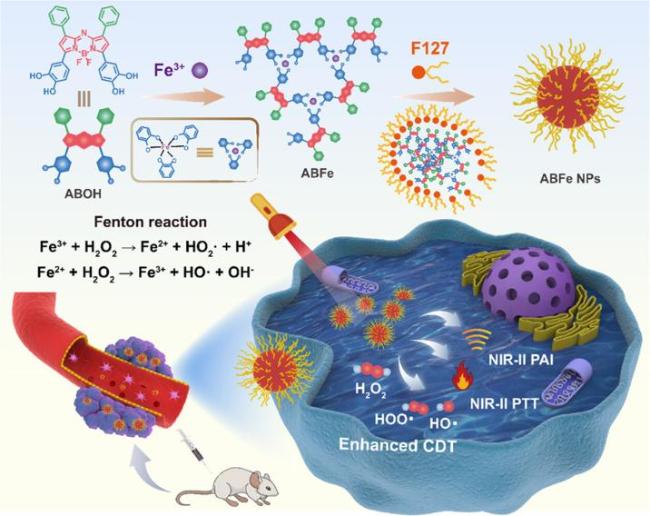
| ABFe NPs | Breast cancer | CDT/PTT | Tumors in the ABFe NPs+laser group were almost eliminated,with a final tumor volume of 300 mm3,which was 15% of the PBS group | 49 | |
| BDP-Fe NPs | Cervical cancer | CDT/PTT | Tumor growth in the NPs+laser group was completely inhibited,the scar was peeling off and the tumor disappeared | 50 | |
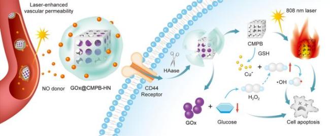
| Cu | GOx@CMPB-HN | Breast cancer | CDT/ Starvation therapy/PTT | The calculated tumor inhibition rates of the GOx@CMPB-HN+laser group was up to 90.29% | 73 |
| CCD NPs | Breast cancer | CDT/chemotherapy | The tumors of mice treated with the CCD NPs were completely eliminated after 18 days of treatment | 58 | |
| Cu9S8 NPs | Breast cancer | CDT/PTT | The tumors of mice in the hollow Cu9S8+laser group almost completely disappeared | 45 | |
| bioCu2-xTe NSs | Breast cancer | CDT/PTT | The remarkable tumor growth inhibition was clearly seen after treatment with bioCu2-xTe NSs plus NIR-II laser irradiation | 48 | |
| CuO2/DDP@SiO2 | liver cancer | CDT/chemotherapy | On day 19,the tumor volume of CuO2/DDP@SiO2 group was 299.1±94.9 mm3,shows no obvious tumor growth after discontinuing medication | 61 | |
| Mo | Mo2C-derived POM | Cervical cancer | CDT/PTT | Tumors in POM+laser group were essentially eliminated in 2 days | 51 |
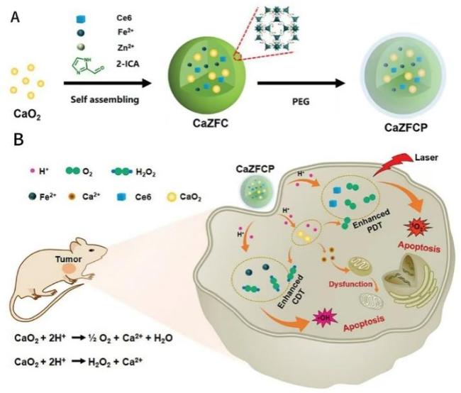
| Ca | CaO2@ZIF-Fe/Ce6@PEG NPs | Breast cancer | CDT/PDT | The group treated with CaZFCP+L (combining CDT with PDT,90.4%) displayed much higher tumor growth inhibition efficacy | 65 |
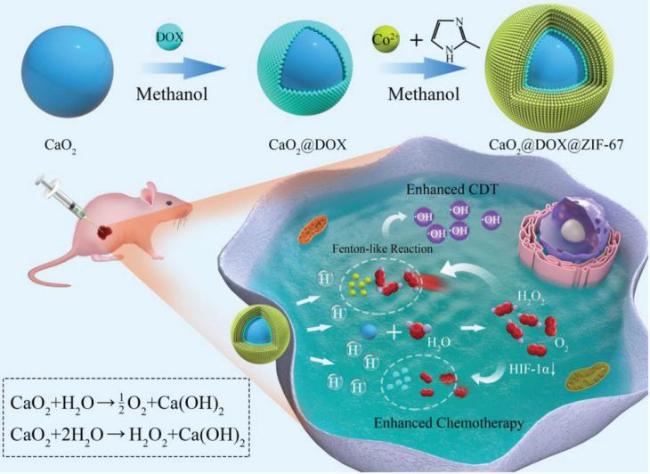
| CaO2@DOX@ZIF-67 NPs | Breast cancer | CDT/chemotherapy | The group treated with CaO2@DOX@ZIF-67 exhibited the strongest tumor suppression effect | 56 | |
| Co | Co-SAs@NC | Breast cancer | CDT/chemotherapy | Co-SAs@NC+DOX combined therapy elicited a 92% inhibition rate | 57 |
| Zn | ZnO2@Au@ZIF-67 NPs | Breast cancer | CDT/Starvation therapy | The tumor size remained almost unchanged in the group treated with ZnO2@Au@ZIF-67 NPs | 74 |
| Pt | A-Pt-IR NP | Breast cancer | CDT/PTT/chemotherapy | A-Pt-IR NP+L exhibited the highest antitumor efficiency and almost completely eradicated the tumors | 82 |
| Mn | PCN@MnO2@DOX@HA | Colon cancer | CDT/PDT | PMDH-induced combination therapy achieved the relatively optimal effect on the tumor-bearing mice | 66 |
| MnO2/Ag3SbS3 | Breast cancer | CDT/PDT | The tumor growth was mostly inhibited or even eliminated when treated with MA+L after 14 days of treatment | 68 | |
| LDNPs@Fe/Mn-ZIF-8 NPs | Cervical cancer | CDT/PDT | The results showed that the volume growth of tumors was evidently inhibited | 69 |
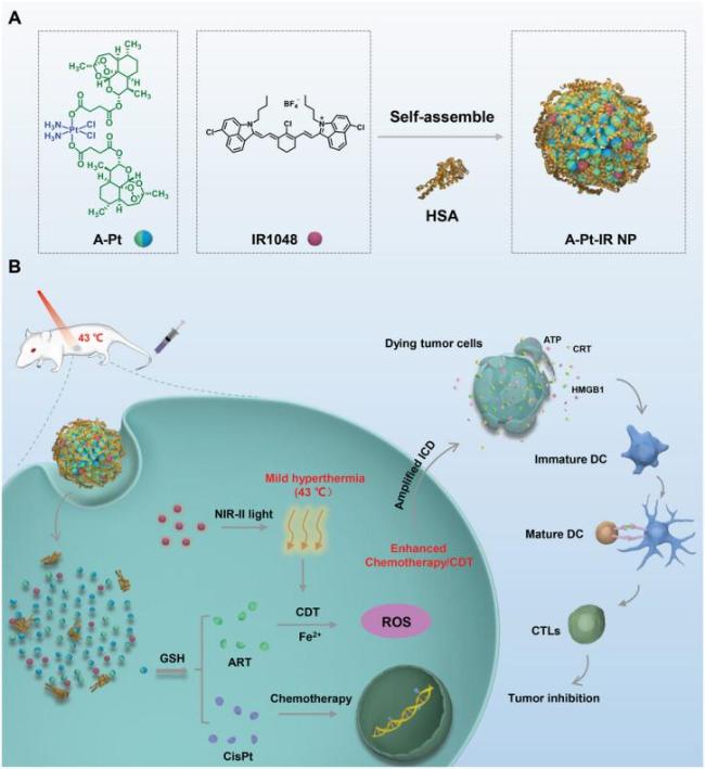
图10 NIR-II光增强化疗/CDT示意图:(A) A-Pt和IR1048的化学结构;(B) 静脉注射后,A-Pt-IR NP可在4T1乳腺癌小鼠模型的肿瘤部位有效蓄积,并通过内吞作用进入细胞发挥作用的示意图[82]Fig.10 Schematic diagram of NIR-II light-enhanced chemotherapy/CDT. (A) Chemical structure of A-Pt and IR1048. (B) Schematic diagram of A-Pt-IR NP that can effectively accumulate at the tumor site in a mouse model of 4T1 breast cancer after intravenous injection and enter cells through endocytosis[82] |
| [1] |
|
| [2] |
|
| [3] |
|
| [4] |
|
| [5] |
|
| [6] |
|
| [7] |
|
| [8] |
|
| [9] |
|
| [10] |
|
| [11] |
|
| [12] |
|
| [13] |
|
| [14] |
|
| [15] |
|
| [16] |
|
| [17] |
|
| [18] |
|
| [19] |
|
| [20] |
|
| [21] |
|
| [22] |
|
| [23] |
|
| [24] |
|
| [25] |
(唐昭敏, 江舒婷, 王郁东, 唐婉兰, 舒娟, 张骥阳, 何浩洋, 陈孔军. 材料导报, 2023, 37(21): 75).
|
| [26] |
|
| [27] |
|
| [28] |
|
| [29] |
|
| [30] |
|
| [31] |
|
| [32] |
|
| [33] |
|
| [34] |
|
| [35] |
|
| [36] |
|
| [37] |
|
| [38] |
|
| [39] |
|
| [40] |
|
| [41] |
|
| [42] |
|
| [43] |
|
| [44] |
|
| [45] |
|
| [46] |
|
| [47] |
|
| [48] |
|
| [49] |
|
| [50] |
|
| [51] |
|
| [52] |
|
| [53] |
|
| [54] |
|
| [55] |
|
| [56] |
|
| [57] |
|
| [58] |
|
| [59] |
|
| [60] |
|
| [61] |
|
| [62] |
|
| [63] |
|
| [64] |
|
| [65] |
|
| [66] |
|
| [67] |
|
| [68] |
|
| [69] |
|
| [70] |
|
| [71] |
|
| [72] |
|
| [73] |
|
| [74] |
|
| [75] |
|
| [76] |
|
| [77] |
|
| [78] |
|
| [79] |
|
| [80] |
|
| [81] |
|
| [82] |
|
| [83] |
|
| [84] |
|
/
| 〈 |
|
〉 |
