Home Journals Progress in Chemistry
Progress in Chemistry

Abbreviation (ISO4): Prog Chem      Editor in chief: Jincai ZHAO

About  /  Aim & scope  /  Editorial board  /  Indexed  /  Contact  / 
Review

Research Progress of Antiviral Coatings

  • Liu Jun ,
  • Ye Daiyong , *
Expand
  • Department of Chemical Engineering, School of Chemistry and Chemical Engineering, South China University of Technology,Guangzhou 510640, China
* Corresponding author e-mail:

Received date: 2022-09-19

  Revised date: 2022-11-24

  Online published: 2023-02-16

Abstract

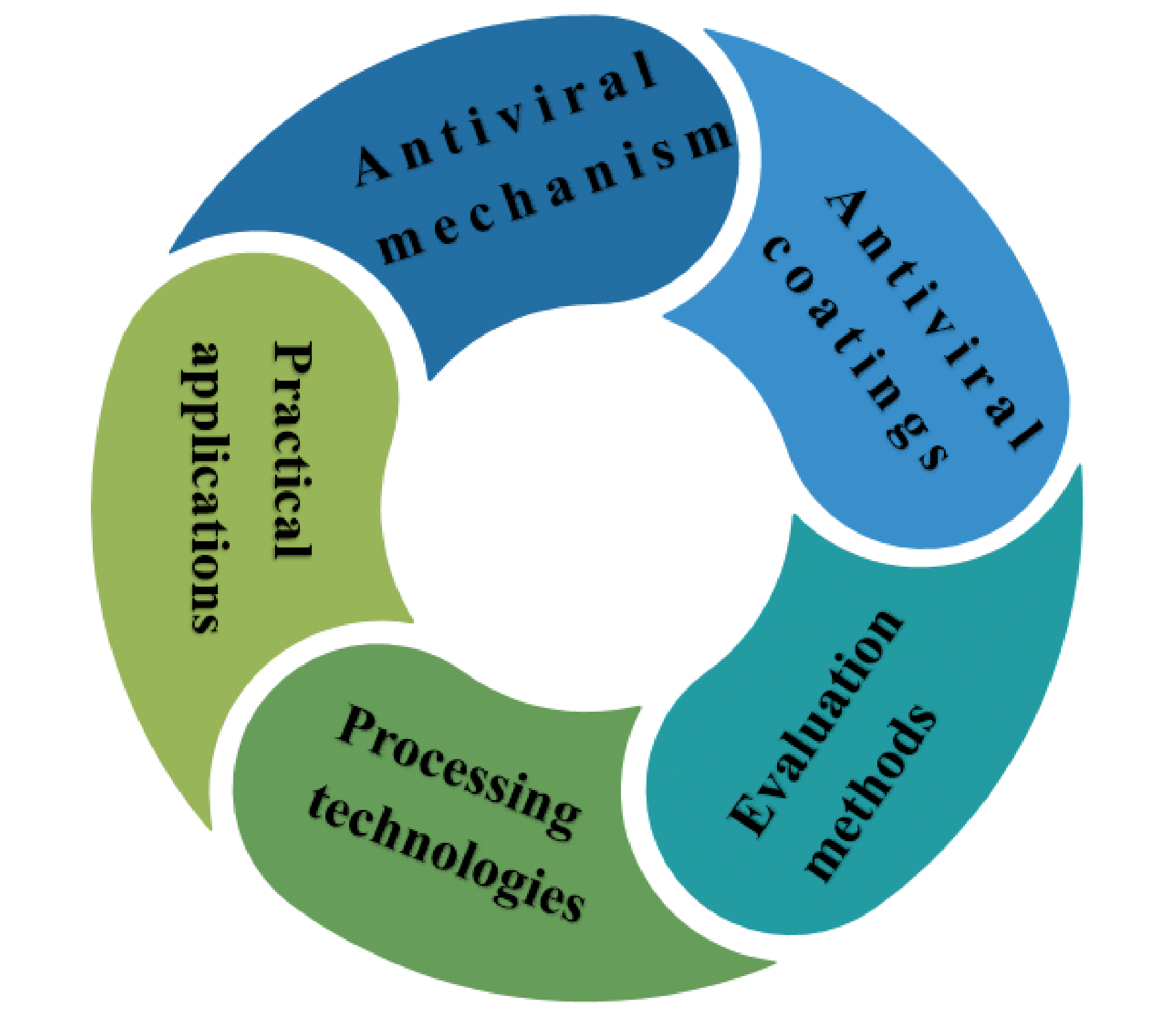
With the large-scale spread of COVID-19 around the world, it has caused serious damage to the health of people around the world. In addition to being transmitted by various droplets, viruses can also be transmitted by human touch of contaminated surfaces. However, as a commonly used surface antiviral method, disinfectants have the disadvantage of discontinuously inactivating viruses, which is bad for inhibiting the spread of various infectious viruses. Therefore, it is urgent to protect the surface of daily objects from virus pollution to eliminate the spread of various respiratory viruses (such as Corona Virus Disease 2019, SARS-CoV-2). From this point of view, it is very important to design and develop effective antiviral coatings. This paper discusses the working mechanisms, performance evaluation methods, processing technologies, practical applications and research progress of nanoparticle antiviral coatings and polymer antiviral coatings for SARS-CoV-2, and also proposes some strategies to design more effective antiviral coatings from the perspective of different types of antiviral coatings. Although some of these antiviral coatings are still in the experimental stage, they still show great potential in the antiviral field.

Cite this article

Liu Jun , Ye Daiyong . Research Progress of Antiviral Coatings[J]. Progress in Chemistry, 2023 , 35(3) : 496 -508 . DOI: 10.7536/PC220917

Contents

1 Introduction
2 Antiviral mechanism
2.1 Direct inactivation of virus
2.2 Inhibiting virus infection of host cells
2.3 Inhibition of virus proliferation
3 Antiviral coatings
3.1 Nanomaterial antiviral coatings
3.2 Antiviral polymer coatings
4 Evaluation methods of antiviral coatings
5 Processing technologies of antiviral coatings
6 Practical applications of antiviral coatings
6.1 Antivirus mask
6.2 Antivirus fabrics
6.3 Surface of other solid objects
7 Conclusion and outlook

1 Introduction

In recent years, infectious diseases caused by viruses have become more and more serious around the world, such as Ebola virus, norovirus, influenza virus and the new coronavirus causing severe acute respiratory syndrome (SARS-CoV-2) (Figure 1), which have seriously affected the health, social stability and economic development of people all over the world[1]. As we all know, the main modes of transmission of viruses are divided into "droplet transmission" and "contact transmission". Droplet transmission is mainly caused by saliva splashing caused by people communicating, sneezing and coughing. Contact transmission is when people come into contact with contaminated surfaces and the virus spreads[2]. Studies have shown that coronaviruses can survive on metal, glass, wood, fabrics, plastics and other surfaces for hours or even days[3]. In our daily life, we inevitably touch the surfaces of various objects, which invisibly increases the risk of virus infection. However, as a commonly used surface disinfection method, disinfectant can only kill the virus before disinfection, but can not achieve the effect of continuous disinfection. Therefore, how to effectively deal with the spread of various infectious viruses and get out of the plight of the epidemic is a top priority for the world. From this point of view, the solution to the problem is to find components (materials and coatings) with antiviral or virucidal activity to design personal protective equipment, sanitary tools and other equipment.It is of great significance to protect the health and safety of the people of the world by protecting the surfaces of daily objects from virus contamination and limiting the spread of viruses[4].
图1 冠状病毒的结构[1]

Fig. 1 Structure of coronavirus[1]. Copyright 2020, American Chemical Society

At present, antiviral coatings are mainly divided into nanomaterial antiviral coatings and polymer antiviral coatings. Based on these two types of antiviral coatings, the mechanism of virus inhibition, different types of antiviral coatings, evaluation and processing methods of antiviral coatings and antiviral products with practical applications are discussed in this paper.At the same time, the advantages and disadvantages of various antiviral materials and the future development prospects were summarized, which provided the direction for future research.

2 Inactivation mechanism of antiviral coating

It is of great significance to study the inactivation mechanism of antiviral materials for inactivating viruses and coping with the spread of infectious diseases. Different antiviral materials have different inactivation mechanisms. At present, the inactivation mechanism of antiviral coating mainly includes direct inactivation of virus, inhibition of virus infection of host cells and inhibition of virus proliferation.

2.1 Direct inactivation of virus

The inactivation mechanism of some nanomaterials with disinfection function is mainly through the release of metal ions or the production of reactive oxygen species (ROS). On the one hand, these substances destroy the capsid protein or genetic material of the virus to inactivate it[5]; On the other hand, metal ions, in addition to causing damage to the structure of the virus, also contribute to the generation of ROS, thereby further improving the antiviral performance[6]. In addition, some nanomaterials can directly inactivate viruses after combining with viruses because of their unique structures. For example, the antiviral ability of graphene oxide (GO) and its derivatives is related to its layered structure and sharp edges[7]. The photothermal effect of nanomaterials also contributes to direct virus inactivation. Under light, as the surface temperature of the material increases, the protein or genetic material of the virus is deformed, thus killing the virus.

2.2 Inhibition of viral infection of host cell

The infectivity of viruses lies in their interaction with host cells after entering host cells, and then replicating themselves. Therefore, it is of great significance to inhibit the infection of host cells by viruses to reduce the infectivity of viruses. Cagno et al. Found that nano-Au coated with mercapto-undecanesulfonic acid could inhibit the binding of influenza virus and vesicular stomatitis virus to host cells, thereby inhibiting viral activity[8]. In addition, some studies have shown that nano-Ag can directly attach to the glycoprotein on the surface of the virus after interacting with the virus, thus inhibiting the virus from entering the host cell and reducing the infectivity of the virus[9].

2.3 Inhibition of viral proliferation

Inhibition of virus proliferation is one of the important ways to effectively inhibit virus transmission. Some metal nanomaterials can bind to viruses and then inhibit the proliferation of viruses by inhibiting the synthesis of RNA. This mechanism is also applicable to non-metallic nanomaterials. Tong et al. Prepared glycyrrhizic acid-carbon dots (Gly-CDs) by hydrothermal synthesis[10]. It has been shown that Gly-CDs can regulate the expression of some host restriction factors by stimulating cells, including DDX53 and NOS3, which are directly related to the proliferation of PRRSV.
Although studies on the inactivation mechanism of antiviral coatings have been extensively explored, further in-depth analysis is needed. Because the mechanism of different antiviral materials is different, and the type of virus also has an impact on the inactivation mechanism, which also brings a lot of difficulties to the study of antiviral mechanism. At present, a considerable part of the antiviral mechanism is deduced from the antibacterial mechanism, while the inactivation mechanism is also lack of detailed and systematic research. Therefore, in addition to the study of antiviral coating, it is also necessary to study its inactivation mechanism and guide the development of antiviral products in the future.

3 Antiviral coating

With the spread of COVID-19 around the world, finding ways to effectively resist the spread of the virus and reduce the possibility of people being infected has become the focus of attention. Compared with previous antiviral methods, the antiviral coating has the characteristics of continuous inhibition and killing of viruses, which helps to prevent the deposition of viruses on the surface of objects. Antiviral coatings include nano-material antiviral coatings (such as copper-based nano-materials, silver-based nano-materials, zinc oxide and titanium dioxide, carbon-based nano-materials and some new nano-materials) and polymer antiviral coatings.

3.1 Nano material anti-virus coating

3.1.1 Metal and metal oxide nanomaterials

3.1.1.1 Cu-based nanomaterials

Copper is relatively less toxic to cells because it is an essential trace element for normal metabolism in the human body, and it also has good antibacterial and antiviral properties, which makes it possible to be used in antiviral coatings[11]. Antiviral mechanisms of copper to date include interference with viral replication during translocation into the nucleus or during gene replication[12]; Nanoparticles destroy viral proteins, enabling the virus to enter the nuclear membrane and prevent cellular enzymes from building new viruses[13]; Free copper ions released from nanoparticles produce ROS, which affect the integrity of the viral envelope or capsid through oxidation, resulting in viral inactivation[14]. Viral proteins are destroyed by cleavage of disulfide bonds, which are essential for protein folding and function[15].
Since the outbreak of COVID-19, the demand for masks has been increasing. Although commonly used masks can effectively prevent the spread of the virus, masks can block the virus while the virus is easy to deposit on their surface, which greatly increases the risk of human infection. In order to better prevent the spread of the virus, Kumar et al. Used a natural biopolymer (shellac) as an adhesive to attach nano-Cu to the mask, thus forming an antiviral coating[16]. The shellac/nano-Cu composite coating can endow the surface with good hydrophobicity to repel respiratory droplets and reduce the deposition of viruses on the surface, thus achieving a self-cleaning effect. In addition, the coating has excellent photocatalytic and photothermal properties. Under sunlight, the temperature of the coating rises rapidly to above 70 ℃, which can produce a large number of free radicals to destroy the virus membrane and inhibit the activity of the virus (Figure 2).
图2 在太阳光照射下,通过光热、光催化和自清洁过程使呼吸飞沫中的病毒失活的示意图[16]

Fig. 2 Schematic drawing of inactivation of virus in respiratory droplets by photothermal effect, photocatalytic effect and self-cleaning under solar irradiation[16]. Copyright 2020, American Chemical Society

Copper oxide (CuO) and cuprous oxide (Cu2O) are the two most commonly used antibacterial and antiviral Cu-based nanomaterials. Castro Mayorga et al. Developed an antiviral biodegradable coating by adding 0.05 wt% nano-CuO to PHBV polymer[17]. The dispersion and uniformity of nano-CuO in polymer can be significantly improved by electrospinning without significantly changing the oxygen permeability, mechanical and optical properties.This allows only a low nanoparticle loading to exhibit good bactericidal and virucidal performance against foodborne pathogens Salmonella enterica, Listeria monocytogenes, and murine norovirus. Mazurkow et al. Have done some research on the difference of antiviral activity between CuO and Cu2O[18]. They performed Zeta potential measurements on these two metal oxides and found that the isoelectric point of Cu2O is 11.0, while that of CuO is 7.4, suggesting that materials containing Cu2O may have a higher positive charge on the surface at pH = 7 (such as Fig. 3). Under electrostatic interaction, the positive surface charge of Cu2O materials can better attract negatively charged viruses (such as bacteriophage MS2), which may contribute to the inactivation of viruses. The electrostatic interaction of Cu2O further expands the antiviral mechanism of Cu-based nanomaterials, which provides different ideas for future researchers.
图3 噬菌体MS2(病毒)、CuO、Cu2O和Cu的zeta电位曲线[18]

Fig. 3 Comparison of zeta potential curves for MS2 bacteriophage (virus), CuO, Cu2O and Cu[18]. Copyright 2019, American Chemical Society

From the above results, it can be seen that there are many kinds of copper-based nanoparticles with antiviral properties, and only a low concentration and a short contact time can produce a significant inhibitory effect on viruses. However, copper is a heavy metal, although heavy metals generally have good bactericidal and antiviral effects, but heavy metals are toxic, when reaching a certain dose in the human body, it will affect health, and even cause poisoning. When copper-based nanomaterials are used as antiviral coatings on the surface of daily objects, it will inevitably increase the contact time between people and them, which is easy to pose a threat to human health. These deficiencies limit the wide application of copper-based nanoparticles in the field of antiviral, therefore, future antiviral research on copper-based nanoparticles should pay more attention to how to reduce cytotoxicity in order to reduce the harm to the human body.

3.1.1.2 Ag-based nanoparticles

As one of the precious metals, silver has been widely used in many fields because of its many excellent properties, especially its unique antibacterial properties, which makes it a good antibacterial material in wound care products, surgical instruments, disinfection filters and so on. At the same time, silver is also an important antiviral material, which inactivates viruses by interacting with viral envelope or viral surface proteins, blocking viral penetration into cells, interacting with viral genomes, and interacting with viral replication factors[19]. At present, the research on the antiviral mechanism of silver-based nanoparticles mainly focuses on the direct chemical reaction between silver-based nanoparticles and different groups (sulfhydryl, amino, carboxyl, phosphate and imidazole groups) on the lipid envelope or surface protein of the virus, resulting in the destruction of the complete structure of the virus or the inhibition of enzyme activity, so as to achieve the effect of virus inactivation[20].
Castro-Mayora et al. Studied the inactivation effect of nanosilver particles and silver nitrate (AgNO3) on feline calicivirus (FCV) and mouse norovirus (MNV)[21]. It was found that the main factor affecting the antiviral activity of nano-Ag and AgNO3 was the concentration. With the increase of concentration, the antiviral activity of nano-Ag and AgNO3 can be significantly improved, and when the concentration is higher than 2. 1 mg/L, the activity of nano-Ag can be maintained for 150 days, but the effective period of AgNO3 is only half of the former, which may be attributed to the instability of Ag+, which is easy to be reduced or agglomerated. In addition, the study further explored the antiviral properties of the coating loaded with nano-Ag. They used electrospinning technology to prepare nano-Ag composite coatings based on PHBV (Figure 4). Because the nano-Ag is firmly fixed in the coating and can better release Ag+, the activity of the norovirus can be inhibited only by adding a very low content of the nano-Ag.
图4 电纺纤维的SEM图像:(A)不含纳米Ag(PHBV18),(B)具有纳米Ag的(PHVB18/纳米Ag),(C)纤维的尺寸分布[21]

Fig. 4 SEM images of electrospun fibers, (A)without AgNP (PHBV18), (B) with AgNP (PHVB18/AgNP), and(C)size distribution of fibers[21]. Copyright 2017, Elsevier

The size and surface modification of nanoparticles are also important factors affecting the antiviral properties of silver-based nanoparticles. He et al. Found that the zeta potential of modified materials was positively correlated with the virucidal effect by studying nano-Ag with different surface modifications[22]. In addition, they also explored the effect of different sizes of nano-Ag on antiviral performance. The results showed that the antiviral effect of nano-Ag depended on the particle size. The smaller the particle size, the higher the percentage of surface atoms and the more unsaturated bonds. This structure provides many adsorption and reaction sites for various types of reactions, and these particles can be easily combined with foreign atoms through chemical bonds. As a result, they are more able to attach to the virus and react with it. In order to further improve the antiviral performance of silver-based nanoparticles, effective synthesis and dispersion methods should be adopted from the size of the nanoparticles themselves, so that the nanoparticles can exist in a stable, dispersed and small size form.
In addition to nano-Ag, other nano-silver compounds also have excellent antiviral properties. Du et al. Investigated the potential mechanism of Ag2S nanoclusters on the inhibition of PEDV[23]. They synthesized Ag2S with an average size of 5.3 nm using glutathione as a capping agent. The nanocluster can reduce the activity of PEDV by 99.9% within 12 H, and the toxicity to cells is almost negligible. Studies have shown that Ag2S nanoclusters may reduce the activity of PEDV by inhibiting viral RNA synthesis and viral budding, and that ISG proteins and proinflammatory cytokines play a key role in this process (Figure 5). PEDV is a coronavirus, which has a similar structure and composition to SARS-CoV-2. Therefore, the potential antiviral mechanism of Ag2S nanoclusters has important reference significance for the inhibition of novel coronavirus.
图5 Ag2S 纳米簇抗病毒活性的潜在机制[23]

Fig. 5 Possible mechanisms of the antiviral activity of Ag2S NCs[23]. Copyright 2018, American Chemical Society

The bactericidal and antiviral properties of silver-based nanoparticles are mostly release mechanism, and the effect is not lasting enough. In addition, similar to copper-based nanoparticles, silver-based nanoparticles also belong to the category of heavy metals. Silver is difficult to metabolize in the human body and will be enriched in the human body, and when it reaches a certain dose in the body, it will have certain neurotoxicity and genotoxicity. In addition, because the surface area of nano-silver is much larger than that of ordinary silver, when it is used as an antiviral coating material, the contact area with the air is very large, and the oxidation reaction is more likely to occur, which makes it difficult to ensure its stability.

3.1.1.3 Zinc oxide (ZnO)

The use of ZnO has a long history, which can be traced back to ancient Egypt 4000 years ago, when it was used to treat some bacterial diseases. In modern science, nano-ZnO is widely used in many fields because of its good photocatalytic, photochemical, bactericidal and antiviral properties. Compared with other heavy metal nanoparticles, nano-ZnO is safe and harmless to human body, and it can be used in daily necessities such as cosmetics or food supplements. In addition, by using melt mixing, electrospinning or solvent casting, nano-ZnO is combined with various polymers to make food packaging, which can effectively improve the shelf life and safety of food. The antiviral mechanism of nano-ZnO is complex and diverse, and its mechanism of action with different kinds of viruses may be different. Fig. 6 shows the antiviral mechanism of nano-ZnO in general: interfering with virus replication by releasing Zn2+, including free virus inactivation, inhibition of virus uncoating, viral genome transcription, and viral protein translation and polyprotein processing[24]. In addition, the photocatalytic activity of nano-ZnO also plays an important role in the inhibition of viruses. Under the irradiation of a specific light source, nano-ZnO can damage the plasma membrane by producing reactive oxygen species, resulting in virus inactivation. Therefore, good antiviral activity and low cytotoxicity make nano-ZnO have broad prospects in the application of antiviral surface coating.
图6 纳米ZnO的抗病毒机制[24]

Fig. 6 Antiviral mechanism of nano ZnO[24]. Copyright 2019, Advances in Nutrition

EI-Megharbel et al. Synthesized a nano-ZnO particle with a certain inhibitory effect on the novel coronavirus[25]. The diameter of the ZnO nanoparticles was determined to be about 40 ~ 60 nm by transmission electron microscopy. Under the scanning electron microscope, the morphology of nano-ZnO is like crushed ice (as shown in Figure 7). The results showed that nano-ZnO at the concentration of 526 ng/mL had strong antiviral activity against SARS-CoV-2, but when the concentration was greater than 292. 2 ng/mL, it had certain toxic side effects on host cells. In order to reduce the damage of nano-ZnO to host cells, they suggested that nano-ZnO should be modified to combine with different compounds, which can not only effectively fix nanoparticles, but also reduce the size of particles, and the small size of nano-ZnO means lower cytotoxicity and better antiviral activity. As for the mechanism of nano-ZnO inhibiting the activity of SARS-CoV-2, researchers believe that nano-ZnO may produce Zn2+ and various reactive oxygen species, which can destroy proteins, lipid membranes and nucleic acids, leading to the inactivation of SARS-CoV-2.
图7 纳米ZnO的SEM图像[25]

Fig. 7 SEM images of nano ZnO[25]. Copyright 2022, Coatings

Considering that pure nano-ZnO may have adverse effects on human health and safety, it is imperative to reduce its cytotoxicity in order to make nano-ZnO used as a surface antiviral protective coating for daily objects. Studies have shown that the size of nano-ZnO has a negative correlation with its antiviral properties and a positive correlation with its cytotoxicity. To this end, Ghaffari et al. Used polyethylene glycol (PEG) to modify the surface of nano-ZnO to endow nanoparticles with higher stability, lower toxicity, and controllable antiviral effect[26].
After surface modification by PEG, nano-ZnO showed good dispersion (Fig. 8). Studies have shown that the highest non-toxic concentration of PEG-ZnO nanoparticles is 200 μg/mL, and the inhibition rate against H1N1 is 94.6%, while the non-toxic concentration of pure nano-ZnO is only 75 μg/mL, and the inhibition rate against H1N1 is 52.2% (as shown in Figure 9 and Figure 10), which indicates that the surface modification of nano-ZnO can not only reduce the cytotoxicity but also improve the antiviral ability. The addition of PEG reduces the size of nano-ZnO, while smaller nanoparticles have better reactivity, larger surface area to volume ratio, which may be the reason for the enhanced antiviral activity.
图8 (a)纳米ZnO和(b)PEG-ZnO纳米颗粒的FE-SEM图,(c)PEG-ZnO纳米颗粒的TEM图[26]

Fig. 8 FE-SEM images of ZnO-NPs (a) and ZnO-PEG-NPs (b); TEM image of ZnO-PEG-NPs (c)[26]. Copyright 2019, Springer Nature

图9 (a)纳米ZnO、(b)PEG-ZnO纳米颗粒、(c)聚乙二醇和(d)奥司他韦对MDCK-SIAT1细胞的细胞毒性[26]

Fig. 9 Cytotoxicity of ZnO-NPs (a), ZnO-PEG-NPs (b), polyethylene glycol (c), and oseltamivir (d) on MDCK-SIAT1 cells[26]. Copyright 2019, Springer Nature

图10 实时PCR测定四种化合物对H1N1流感病毒的抑制率[26]

Fig. 10 The inhibitory rates of the four compounds against H1N1 influenza virus determined by Real-Time PCR assay[26]. Copyright 2019, Springer Nature

Although nano-ZnO has good antiviral activity and low cytotoxicity, it is easy to agglomerate because of its large specific surface area and high specific surface energy. On the other hand, the surface polarity of nano-ZnO is strong, and it is not easy to disperse uniformly in organic media, which greatly limits its performance. Simply adding nano-ZnO to the polymer can endow the coating with antiviral properties, but it may have the disadvantage of toxic side effects. Therefore, it is necessary to modify the surface of ZnO nanoparticles, which can not only improve the dispersion and stability of nanoparticles, but also help to exert their antiviral activity. Compared with copper-based and silver-based nanoparticles, nano-ZnO needs to undergo photochemical reaction under the irradiation of a specific light source to further enhance its antiviral activity in order to give full play to its performance, which also limits the application scope of nano-ZnO.

3.1.1.4 Titanium dioxide (TiO2)

Because of its excellent photocatalytic activity, TiO2 has attracted much attention in the field of antibacterial and antiviral. After absorbing UVA photons, TiO2 can catalyze the following redox reactions of oxygen or water molecules.Reduction reaction :O2+e- O 2 -, O 2 - +H+⇆·O2H, ·O2H+e-+H+→H2O2,The oxidation reaction is :OH-+H+→·OH, producing a variety of reactive oxygen species, such as hydroxyl radicals, neutral hydroxyl ions, and superoxide anions ( O 2 -)[27]. Reactive oxygen species cause TiO2 to exert bactericidal and antiviral functions by damaging viral or bacterial membranes, protein capsids, or RNA. Compared with other nanoparticles, the biggest feature of TiO2 is that it needs to be irradiated by a specific wavelength light source to produce antiviral effect. In addition, the toxicity of TiO2 to human body is almost negligible, which has prompted many researchers to do a lot of research on it.
Compared with the common nano TiO2, the surface functionalized nano TiO2 has better bactericidal and antiviral activity. Some researchers have prepared a plasma Ag0-TiO2 nanocomposite coating[28]. The coating has good photocatalytic effect. Under visible light irradiation (λ ≥ 435 nm), the generated ROS can degrade or oxidize proteins. Therefore, the coating showed good antiviral activity against alphaherpesvirus in both wet and dry environments.
The wavelength and intensity of the excitation light source are strictly controlled by the nanometer TiO2, which greatly limits the utilization efficiency of the nanometer TiO2. To this end, Nakano et al. Developed a CuxO/TiO2 composite with antiviral activity even under dark conditions[29]. The material can effectively inactivate SARS-CoV-2 and its Delta variant. The results showed that when the CuxO/TiO2 was coated on the glass plate, the virus titer decreased to the detection limit within 3 H in the dark. In addition, the antiviral properties of the coating were improved under visible light irradiation, and the virus titer decreased to the detection limit only after 2 H (fig. 11). It has been found that the antiviral activity of CuxO/TiO2 is related to the damage of spike protein and RNA of SARS-CoV-2.
图11 灭活各种变异株:(a)不同变异株在可见光照射下的病毒滴度变化;(b)Delta变异株的病毒滴度的变化[29]

Fig. 11 Inactivation of various types of variants. (a) changes of virus titer of different variants under visible light irradiation and (b) changes of virus titer of Delta variant[29]. Copyright 2022, Springer Nature

The advantages of excellent photocatalytic activity, extremely low cytotoxicity, and simple and cheap industrial scale production make it theoretically feasible for nanosized TiO2 to be an excellent antiviral material. However, similar to nano-ZnO, nano-TiO2 also requires a specific light source to produce antiviral activity, but the difference is that nano-ZnO can inhibit viral activity by releasing Zn2+ even without external light source. However, if the nano TiO2 is not irradiated by a light source, it is difficult to exert bactericidal and antiviral properties. The need for a specific light source greatly limits the applicability of nano-TiO2. Therefore, many researchers try to reduce the power and intensity of the light source as much as possible and increase the excitation wavelength range of nano-TiO2, so that it can be more widely used.

3.1.2 Carbon-based nanomaterials

With the increasing severity of the COVID-19 epidemic, there is an urgent need to develop a variety of effective and low-toxicity antiviral coatings to protect the safety of people around the world. Non-toxic and harmless to human body is one of the basic requirements of antiviral coatings. However, most of the current antiviral materials have more or less toxic side effects, which limits their practical application. Carbon-based nanomaterials, as a kind of materials with good biocompatibility and excellent antiviral activity, have shown broad prospects in the field of antiviral. The antiviral research progress of various carbon-based nanomaterials is summarized in Table 1. So far, carbon-based antiviral nanomaterials mainly include fullerenes, carbon nanotubes, graphene, carbon dots and so on. Because the antiviral properties of carbon-based nanomaterials are still in the initial stage, their antiviral mechanisms are still being studied and improved. The main mechanisms recorded in the literature are: direct interaction with viruses, light-induced mechanism, and the generation of reactive oxygen species[30,31].
表1 碳基抗病毒纳米材料研究进展

Table 1 Progress of carbon-based antiviral nanomaterials

Material Virus Antiviral effect ref
Graphene oxide/
Polydimethylsiloxane
HAdV5, HSV-1, CoV The coating reduces titers of HAdV5 by 1.8 log, HSV-1 by 2.2 log, and CoV by 2.4 log 32
Polyethylenimine-carbon dots VSV Activated by visible light to effectively and efficiently inactivate VSVs 33
Fullerene derivatives HIV, Influenza viruses Promising antiviral activity against HIV and influenza viruses. 34
Carbon nanotube SARS-CoV-2 Exhibit excellent barrier and antiviral effects against SARS-CoV-2 35
Carbon-based nanomaterials play an important role in the field of anti-virus because of their excellent physical and chemical properties. However, at present, the preparation of carbon-based nanomaterials is expensive, the steps are complicated, and there may be defects that affect the performance. In addition, there is a lack of systematic and comprehensive research on the theoretical knowledge of the antiviral mechanism of carbon-based nanomaterials, which hinders the further development of this material. These ubiquitous shortcomings pose a challenge to the practical application of carbon-based nanomaterials in antiviral applications. Therefore, it is necessary to improve the preparation technology to synthesize carbon-based nanomaterials with complete structure in a simple and efficient way. At the same time, we should improve the mechanism of virus inactivation, let the theory guide the research, and then promote the development of antiviral field.

3.1.3 Other antiviral nanomaterials

Most of the antiviral nanomaterials mentioned above have been well studied and developed. In addition, there are also some relatively new nanomaterials to be developed in the field of anti-virus.
Cerium (Ce) is a metal nanoparticle with unique properties. Compared with other metal nanomaterials, the most significant advantages of nano-Ce are its low cytotoxicity and high safety. Nano-Ce has attracted much attention in the biomedical field because of its antiviral, anti-inflammatory, antibacterial and anticancer properties[36]. There are many theoretical explanations for the antiviral mechanism of nano-Ce. On the one hand, studies have shown that CeO2 can stimulate dendritic cells to produce interleukins, which are of great significance in regulating cytokines[37]. On the other hand, CeO2 can also reduce the damage to cell structure by inhibiting ROS, which is beneficial to maintain the normal function of cells during inflammation[38]. At present, many studies have shown that nano-Ce and its oxides have excellent antiviral properties. Some researchers have obtained highly stable antiviral agents with Ag-modified nano-CeO2, and have specific antiviral activity against OC43 coronavirus and RV14 virus pathogens[39]. In addition, there was a synergistic effect between Ce and molybdate ions, and the combination of them showed high antiviral activity against SARS-CoV[40]. Although nano-Ce is an excellent material in the antiviral field, the current research reports on nano-Ce mainly focus on the antiviral activity, and there are few studies on its antiviral application. In addition, in order to obtain Ce nanoparticles with excellent properties and good biocompatibility, it is necessary to control the synthesis of Ce nanoparticles.
Among the nanomaterials, 2D MXene has many advantages, such as abundant surface functional groups, excellent hydrophilicity, excellent photocatalytic and photothermal properties, excellent surface structure and conductivity, which play an important role in the fight against viruses. These superior properties allow 2D MXene to be used in the preparation of masks, face shields, and biomedical instruments, among others[41]. When 2D MXene with good hydrophilicity and abundant functional groups is used as a coating, it can capture viruses and inactivate or kill them by interacting with external spike proteins[41]. The efficient photothermal performance of 2D MXene can inactivate viruses through simple photothermal conversion, which can endow protective equipment with self-disinfection function[42]. As the COVID-19 epidemic becomes more and more serious, the antiviral research on 2D MXene is constantly improving (Table 2). Although 2D MXene materials were discovered a decade ago, research on their antiviral aspects is in its infancy. In addition, some characteristics of 2D Mxene are easily affected by external environmental conditions, which is not conducive to its performance. More importantly, the adhesion of MXene on metal or polymer is poor, which makes it easy to peel off as a coating. All these shortcomings have affected its wide application and commercialization. Therefore, more in-depth research on the antiviral effect of 2D MXene is needed in the future.
表2 2D MXene在抗病毒方面的研究进展

Table 2 Research progress of 2D MXene in anti-virus

Material Virus Antiviral effect ref
Ti3C2Tx SARS-CoV-2/clade GR 99% reduction of the viral copy numbers 43
Ti3C2-Au-MPS PRRSV、SARS-CoV-2 The infection of PRRSV and SARS-CoV-2 can be blocked 44
In 2022, Chinese researchers developed a two-dimensional CuInP2S6(CIPS) nanosheet with broad-spectrum resistance to COVID-19[45]. CIPS has a high selective binding ability, which binds to the receptor of spike protein on SARS-CoV-2 and its variant strains (Delta and Omicron), and then macrophages can quickly engulf and eliminate the virus, thus achieving the effect of capturing and promoting host cells to eliminate the virus. In addition, CIPS is a safe, biocompatible and biodegradable 2D nanomaterial. These excellent characteristics make CIPS promising as a good surface coating or detergent.

3.2 Antiviral polymer coating

Polymers are widely used in daily life, and most of the protective coatings on the surface of objects are composed of polymers. As a kind of water-soluble polymer with special functions, polyelectrolyte contains ionizable groups in its structural unit, which can be divided into polycation and polyanion according to the ionized charge. The polycationic polymer can ionize positive charges, and the negatively charged virus is adsorbed to the surface of the coating under electrostatic action, which subsequently destroys the structure of the virus, causing RNA to leak out of the virus and eventually inactivating the virus (Figure 12)[46]. This kind of coating not only has bactericidal and antiviral properties, but also has good durability, and can still maintain relatively stable performance after multiple washings. Table 3 shows the research progress of the main antiviral polymer coatings, among which the polymer coatings with quaternary ammonium salt groups have attracted much attention because of their excellent bactericidal and antiviral functions. In addition to developing new and effective antiviral polymer coatings, modifying polymer coatings to endow them with super-hydrophobicity can make it difficult for water droplets carrying viruses or bacteria to adhere to the surface of the coating, which hinders the survival of viruses, and is also one of the important strategies to further enhance antiviral activity in the future.
图12 聚阳离子涂层灭活包膜病毒的机理示意图[46]

Fig. 12 Mechanism of enveloped virus inactivation by polycation coating[46]. Copyright 2011, Proceedings of the National Academy of Sciences

表3 聚合物抗病毒涂层

Table 3 Polymer antiviral coatings

Material Virus Antiviral effect ref
N,N-
dodecyl,
methyl PEI
Poliovirus Rotavirus Approximately 100% virucidal activity 47
Silane-functionalized polyionenes SARS-CoV-2 Exhibiting potent bactericidal (>99.999%) and virucidal (7-log PFU reduction) activities 48
Surface-Grafted quaternary ammonium polymer MHV-A59, SuHV-1 A 4.3-log reduction in infectious MHV-A59 virus and a 3.3-log reduction in infectious SuHV-1 virus 49
SurfaceWise2 HCoV-229E, SARS-CoV-2 The inhibition rate of the two viruses is above 99.9% 50

4 Evaluation method of anti-virus coating

At present, the evaluation methods of antiviral coatings are relatively unified, and the usual evaluation indicators mainly include antiviral activity, cytotoxicity, optimal antiviral dose and antiviral materials of antiviral coatings.
Inhibition, etc. Antiviral activity is one of the most important indicators to evaluate the performance of antiviral coatings. Plaque assay and TCID50 are commonly used methods to evaluate the antiviral activity of coatings[51][26]. The advantages of these two methods are low cost and simple operation, but there are some problems such as time-consuming, high detection limit and poor repeatability, which are not conducive to the scientificity and rigor of the study. Compared with the above two methods, real-time fluorescent quantitative PCR has the characteristics of high sensitivity, accuracy and repeatability[52].
It has gradually become the main means to evaluate the antiviral activity of materials. The cytotoxicity of antiviral coatings is evaluated to ensure that the material itself does not cause too much damage to the human body, and the usual method is MTT colorimetric method[26]. It is characterized by high sensitivity and economy. In addition, the optimal dosage of antiviral materials can be preliminarily obtained by combining the experimental results of antiviral activity and cytotoxicity. Indirect immunofluorescence assay (IFA) and Western blotting play an important role in evaluating the inhibitory effect of antiviral materials[53]. However, these methods suffer from the disadvantages of being time and labor consuming and not suitable for handling a large number of samples simultaneously. Although remarkable achievements have been made in the research and development of antiviral coatings, the development of detection methods for antiviral coatings is relatively slow, which is not conducive to the overall development of this field. A rapid and highly sensitive method has been developed to evaluate the virucidal activity of antiviral coatings (fig. 13)[54]. The method is to evaluate the antiviral activity of the surface coating by measuring the luciferase activity of SeV virus. In the future, it is necessary to further improve the evaluation methods and means of antiviral coatings, improve the applicability, accuracy and sensitivity of the methods, so as to better promote the development of antiviral coatings.
图13 表面涂层抗病毒实验的示意图[54]

Fig. 13 Schematic representation of an antiviral assay for surface coatings[54]. Copyright 2022, ACS Applied Bio Materials

5 Processing technology of anti-virus coating

In order to convert antiviral materials and coatings into antiviral products and apply them in real life, some preparation and processing technologies are needed to protect the human body from viral infection. Electrospinning technology is often used to prepare products with antiviral function. Using this technology, nanomaterials can be immobilized in polymers and processed into corresponding antiviral products. On the one hand, this technology can effectively fix nanomaterials, so that they are not easy to fall off, and better play an antiviral role[21]; On the other hand, this technique also helps to improve the dispersion and uniformity of nanoparticles[17]. Because of its unique advantages, electrospinning technology plays an important role in the processing and production of antiviral protective fabrics and clothing. Dip coating technology is another way to prepare and process antiviral products. The method is characterized by simplicity, high production efficiency and easy coating. Some researchers have immersed the mask in a solution of nanomaterials, so that the nanomaterials are evenly attached to the fibers of the mask to resist viral infection[55]. In addition, solvent casting is also a good processing technology. Moreno et al. Added natural antiviral agents to agar or alginate, and then used solvent casting to form antiviral films and prepare antiviral coatings for food packaging[56]. The preparation and processing technology of antiviral coating is an important way to transform scientific research achievements into practical application. Through the development of more simple and efficient processing technology, more antiviral products can be prepared to ensure the physical and mental health of the human body.

6 Practical application of antiviral coating

As the COVID-19 epidemic becomes more and more serious worldwide, there is an urgent need for effective means to deal with the spread of the virus. Because the virus can exist on the surface of objects for hours or even days, disinfection of the surface is the top priority in epidemic prevention and control. Disinfectant is a commonly used surface disinfection method, but in order to effectively kill viruses, it is usually necessary to use disinfectant regularly and frequently, which is very troublesome and inefficient. Therefore, by coating an antiviral coating on the surface of an object, the activity of the virus can be continuously and efficiently inhibited, thereby protecting the physical and mental health of the human body. At present, the practical application of antiviral coatings is mainly focused on masks, fabrics and other solid surfaces.

6.1 Antiviral mask

During epidemics of infectious diseases, masks are important to prevent the spread of viruses through droplets. Usually, medical protective masks are composed of three layers of cloth, which play a role in filtering harmful particles. However, the filtered pathogens easily remain on the surface of the mask, which poses a potential risk. In order to effectively kill the virus and improve the protective effect of the mask, it is an economical and simple way to coat the mask with antiviral coating. Lin et al. Prepared a graphene-embedded carbon film mask by ultrasonic compaction (Fig. 14)[57]. The mask has the characteristics of good superhydrophobicity, light disinfection and reusability. Under sunlight, the temperature of the mask can quickly reach 110 ℃. The mask is expected to help address the risk of COVID-19 because the spike protein of the coronavirus is sensitive to temperature. In addition to using the photothermal effect to disinfect, researchers have modified masks to inhibit viral activity. Kumar et al. Used a simple dip-coating technique to uniformly coat Cu @ ZIF-8 nanowires on the mask fibers (Fig. 15)[55]. Due to the continuous release of Cu2+ and Zn2+, only a low concentration of nanowires is needed to make the mask antiviral.
图14 石墨烯嵌入碳膜口罩的制备流程:(a) GNEC薄膜的沉积过程,(b) GNEC口罩的制造工艺[57]

Fig. 14 Preparation process of graphene nanosheet-embedded carbon (GNEC) film mask. (a) Deposition process of GNEC film. (b) Fabrication process of the GNEC mask[57]. Copyright 2020, Nano Research

图15 浸涂在(a) 0.1 mg/mL, (b) 0.25 mg/mL, (c) 0.5 mg/mL, (d) 1 mg/mL Cu@ZIF-8 NWs分散体中的面具过滤器的SEM图像[55]

Fig. 15 SEM images of face mask filter coated by dipping in dispersions of a) 0.1 mg/mL, b) 0.25 mg/mL, c) 0.5 mg/mL, and d) 1 mg/mL Cu@ZIF-8 NWs[55]. Copyright 2021, Advanced Functional Materials

6.2 Antiviral fabric

In the face of the risks and challenges brought by the epidemic, in addition to the development of antiviral protective masks, the development of fabrics with self-disinfection function is also essential. Disinfection wipes are commonly used in surface disinfection in daily life. Some researchers have used nano-Ag to treat cellulose wipes, so that they can have the functions of sterilization and disinfection at the same time (Figure 16)[58]. The results showed that the wet tissue had an inhibition rate of 48. 3% against MERS-CoV and showed good antibacterial activity against a variety of bacteria. In addition, they used cotton yarn treated with nano-Ag to make winter sweaters (Figure 17) to reduce the risk of infection. Similarly, Karagoz et al. Made nanofibers from a solution containing polymethyl methacrylate, nano-ZnO and nano-Ag by electrospinning and integrated them into protective clothing to form a multifunctional fabric[59]. The protective clothing can sterilize and resist viruses, and can achieve the effect of self-cleaning under the action of photocatalysis.
图16 负载有纳米Ag的单独包装湿巾[58]

Fig. 16 Individual packages of the wet wipes loaded with the silver nanoparticles[58]. Copyright 2021, International Journal of Biological Macromolecules

图17 用纳米Ag处理的棉纱制成的抗菌和抗病毒冬季毛衣[58]

Fig. 17 Antimicrobial and antiviral winter sweater made of cotton yarns treated with AgNPs[58]. Copyright 2021, International Journal of Biological Macromolecules

6.3 Other solid surface

In daily life, whether in offices, public areas, schools or hospitals, people inevitably come into contact with the surfaces of various objects (such as glass, metal, plastic, etc.), which will increase the possibility of contact with viruses. Therefore, some common solid surfaces need to be treated to protect people from virus infection. In order to develop a universal antiviral coating, Tiwary et al. Used polyvinyl alcohol (PVA) as the base coating and added copper-graphene (Cu-Gr) nanocomposites to form a transparent antiviral surface coating[60]. With the synergistic effect of copper and graphene, the coating can interfere with the replication process of the virus and inactivate the virus within 30 min. Therefore, the transparent coating can be applied to solid surfaces such as masks, mobile phone screens, door handles, medical devices, etc., to make the surface sterile without affecting the appearance of the object.
Although good progress has been made in the development of antiviral coatings for personal protective equipment and surface protection, there are still many problems to be improved, such as lack of fluidity and permeability, difficult temperature control and low efficiency of pathogen inactivation. Therefore, it is very important to modify the antiviral coating and develop more effective antiviral products.

7 Conclusion and prospect

While the techniques described above are effective in inactivating viruses on a variety of surfaces, they still have some drawbacks that limit their practical use in everyday life. For example, the general applicability of some antiviral coatings is poor, and their effects on different types of viruses vary greatly. In the process of large-scale production, it is difficult to ensure that the size of nanomaterials is completely consistent or there are defects, which may lead to agglomeration in the coating, thus affecting the antiviral performance. The toxicity of antiviral materials is still a big problem, and cytotoxicity and antiviral activity are difficult to be compatible. In order to further improve the practicability of antiviral coatings and develop more effective antiviral coatings, the following aspects can be further studied: (1) For nanomaterials, the synthesis route should be further optimized to reduce the production cost and ensure the integrity and consistency of the structure, so as to prepare new functional coatings with high efficiency and low cost, and promote the commercialization of antiviral coatings; (2) carrying out functional modification on the nanoparticles with antiviral or virucidal properties to improve the dispersibility, stability and compatibility of the material in the polymer and maximize the antiviral capability of the coating; (3) Nanoparticles should be better fixed on the surface of the coating to prevent them from separating from the coating, so as to improve the durability and durability of the antiviral coating; (4) Attention should be paid to balancing the antiviral activity and cytotoxicity of metal nanoparticles, so as to improve the antiviral performance as much as possible under the condition of ensuring non-toxic and harmless to human body.
[1]
Eldin A Osman E, Toogood P L, Neamati N. ACS Infect. Dis., 2020, 6(7): 1548.

[2]
Mehraeen E, Salehhi M A, Behnezhad F, Moghaddam H R, SeyedAhmad S. Infectious Disorders-Drug Targets., 2021, 21(6): 27.

[3]
Ghosh S K. Nova Surface-Care Centre Pvt: Maharashtra, India, 2020.

[4]
Huang H, Fan C, Li M, Nie H L, Wang F B, Wang H, Wang R, Xia J, Zheng X, Zuo X, Huang J. ACS Nano, 2020, 14(4): 3747.

[5]
Reina G, Peng S Y, Jacquemin L, Andrade A F, Bianco A.. ACS Nano, 2020, 14(8): 9364.

[6]
Moschini E, Colombo G, Chirico G, Capitani G, Dalle-Donne I, Mantecca P. Scientific Reports, 2023, 13(1): 2326.

[7]
Innocenzi P, Stagi L. Chemical Science., 2020, 11(26): 6606.

[8]
Cagno V, Gasbarri M, Medaglia C, Gomes D, Clement S, Stellacci F, Tapparel C. Antimicrobial Agents and Chemotherapy, 2020, 64.

[9]
Morris D, Ansar M, Speshock J, Ivanciuc T, Qu Y, Casola A. Viruses, 2019, 11(8): 732.

[10]
Tong T, Hu H G, Zhou J W, Deng S F, Zhang X T, Tang W T, Fang L R, Xiao S B, Liang J G. Small, 2020, 16(13): 1906206.

[11]
Turnlund J R. Am. J. Clin. Nutr., 1998, 67(5): 960S.

[12]
Tavakoli A, Hashemzadeh M S. Journal of Virological Methods, 2020, 275: 113688.

[13]
Maruzuru Y, Shindo K, Liu Z M, Oyama M, Kozuka-Hata H, Arii J, Kato A, Kawaguchi Y. Journal of Virology, 2014, 88(13): 7445.

[14]
Chatterjee A K, Chakraborty R, Basu T. Nanotechnology, 2014, 25(13): 135101.

[15]
Minoshima M, Lu Y, Kimura T, Nakano R, Ishiguro H, Kubota Y, Hashimoto K, Sunada K. Journal of Hazardous Materials., 2016, 312: 1.

[16]
Kumar S, Karmacharya M, Joshi S R, Gulenko O, Park J, Kim G H, Cho Y K. Nano Lett., 2021, 21(1): 337.

[17]
Castro Mayorga J L, Fabra Rovira M J, Cabedo Mas L, Sánchez Moragas G, LagarÓn Cabello J M. J. Appl. Polym. Sci., 2018, 135(2): 45673.

[18]
Mazurkow J M, Yüzbasi N S, Domagala K W, Pfeiffer S, Kata D, Graule T. Environ. Sci. Technol., 2020, 54(2): 1214.

[19]
Rai M, Deshmukh S D, Ingle A P, Gupta I R, Galdiero M, Galdiero S. Critical reviews in microbiology., 2016, 42(1): 46.

[20]
Chen Y N, Hsueh Y H, Hsieh C T, Tzou D Y, Chang P L. International Journal of Environmental Research and Public Health, 2016, 13(4): 430.

[21]
Castro-Mayorga J L, Randazzo W, Fabra M J, Lagaron J M, Aznar R, Sánchez G. LWT-Food Science and Technology, 2017, 79: 503.

[22]
He Q, Lu J, Liu N, Lu W Q, Li Y, Shang C, Li X, Hu L G, Jiang G B. Nanomaterials, 2022, 12(6): 990.

[23]
Du T, Liang J G, Dong N, Lu J, Fu Y Y, Fang L R, Xiao S B, Han H Y. ACS Appl. Mater. Interfaces, 2018, 10(5): 4369.

[24]
Read S A, Obeid S, Ahlenstiel C, Ahlenstiel G. Advances in nutrition., 2019, 10(4): 696.

[25]
El-Megharbel S M, Alsawat M, Al-Salmi F A, Hamza R Z. Coatings, 2021, 11(4): 388.

[26]
Ghaffari H, Tavakoli A, Moradi A, Tabarraei A, Bokharaei-Salim F, Zahmatkeshan M, Farahmand M, Javanmard D, Kiani S J, Esghaei M, Pirhajati-Mahabadi V, Monavari S H, Ataei-Pirkooh A. Journal of Biomedical Science, 2019, 26(1): 70.

[27]
Nosaka Y, Nosaka A Y. Chem. Rev., 2017, 117(17): 11302.

[28]
Boldogk? i Z, Csabai Z, Tombácz D, Janovák L, Balassa L, Deák Á, TÓth P S, Janáky C, Duda E, DÉkány I. Front. Bioeng. Biotechnol., 2021, 9: 709462.

[29]
Nakano R, Yamaguchi A, Sunada K, Nagai T, Nakano A, Suzuki Y, Yano H, Ishiguro H, Miyauchi M. Scientific Reports., 2022, 12(1): 1.

[30]
Innocenzi P, Stagi L. Chem. Sci., 2020, 11(26): 6606.

[31]
Ayub M, Othman M H D, Khan I U, Yusop M Z M, Kurniawan T A. Surfaces and Interfaces., 2021, 27: 101460.

[32]
Galante A J, Yates K A, Romanowski E G, Shanks R M Q, Leu P W. ACS Applied Nano Materials, 2022, 5(1): 718.

[33]
Adcock A F, Wang P, Ferguson I S, Obu S C, Sun Y P, Yang L J. ACS Applied Bio Materials, 2022, 5(7): 3158.

[34]
Kraevaya O A, Bolshakova V S, Peregudov A S, Chernyak A V, Slesarenko N A, Markov V Y, Lukonina N S, Martynenko V M, Sinegubova E O, Shestakov A F, Zarubaev V V, Schols D, Troshin P A. Organic Letters, 2021, 23(18): 7226.

[35]
Lee S, Nam J S, Han J, Zhang Q, Kauppinen E I, Jeon I. ACS Applied Nano Materials, 2021, 4(8): 8135.

[36]
Allawadhi P, Khurana A, Allwadhi S, Joshi K, Packirisamy G, Bharani K K. Nano Today, 2020, 35: 100982.

[37]
Zandi M, Hosseini F, Adli A H, Salmanzadeh S, Behboudi E, Halvaei P, Khosravi A, Abbasi S. Biomedicine © Pharmacotherapy, 2022, 156: 113868.

[38]
Cui W, Wang Y, Luo C, Xu J, Wang K, Han H, Yao K. Materials Today Nano, 2022, 18: 100218.

[39]
Neal C J, Fox C R, Sakthivel T S, Kumar U, Fu Y, Drake C, Parks G D, Seal S. ACS Nano, 2021, 15(9): 14544.

[40]
Ito T, Sunada K, Nagai T, Ishiguro H, Nakano R, Suzuki Y, Nakano A, Yano H, Isobe T, Matsushita S, Nakajima A. Materials Letters, 2021, 290: 129510.

[41]
Huang Y, Yang C, Xu X, Xu W, Liu S. Acta Pharmacologica Sinica, 2020, 41(9): 1141.

[42]
Huang Z M, Cui X, Li S L, Wei J C, Li P, Wang Y T, Lee C S. Nanophotonics, 2020, 9(8): 2233.

[43]
Unal M A, Bayrakdar F, Fusco L, Besbinar O, Shuck C E, Yalcin S, Erken M T, Ozkul A, Gurcan C, Panatli O, Summak G Y, Gokce C, Orecchioni M, Gazzi A, Vitale F, Somers J, Demir E, Yildiz S S, Nazir H, Grivel J C, Yilmazer A. Nano Today, 2021, 38: 101136.

[44]
Tong T, Tang W, Xiao S, Liang J. Advanced Nobiomed Research, 2022, 2(10): 2200067.

[45]
Zhang G F, Cong Y L, Liu F L, Sun J F, Zhang J T, Cao G L, Zhou L Q, Yang W J, Song Q L, Wang F J, Liu K, Qu J, Wang J, He M, Feng S, Baimanov D, Xu W, Luo R H, Long X Y, Liao S M, Fan Y P, Li Y F, Li B, Shao X M, Wang G C, Fang L J, Wang H Y, Yu X F, Chang Y Z, Zhao Y L, Li L, Yu P, Zheng Y T, Boraschi D, Li H C, Chen C Y, Wang L M, Li Y. Nat. Nanotechnol., 2022, 17(9): 993.

[46]
Hsu B B, Wong S Y, Hammond P T, Klibanov A M. Proceedings of the National Academy of Sciences, 2011, 108(1): 61.

[47]
Larson A M, Hsu B B, Rautaray D, Haldar J, Chen J Z, Klibanov A M. Biotechnol. Bioeng., 2011, 108(3): 720.

[48]
Qiu Q H, Yang C, Wang Y M, Alexander C A, Yi G S, Zhang Y G, Qin X H, Yang Y Y. Biomaterials, 2022, 284: 121470.

[49]
Sorci M, Fink T D, Sharma V, Singh S, Chen R, Arduini B L, Dovidenko K, Heldt C L, Palermo E F, Zha R H. ACS Applied Materials © Interfaces, 2022, 14(22): 25135.

[50]
Ikner L A, Torrey J R, Gundy P M, Gerba C P. American Journal of Infection Control., 2021, 49(12): 1569.

[51]
Ghosh S, Mukherjee R, Basak D, Haldar J. ACS Applied Materials © Interfaces, 2020, 12(25): 27853.

[52]
Li R, Hou Y, Huang J, Pan W, Ma Q, Shi Y, Li C, Zhao J, Jia Z, Jiang H, Zheng K, Huang S, Dai J, Li X, Hou X, Wang L, Zhong N, Yang Z. Pharmacological Research, 2020, 156: 104761.

[53]
Ou X Y, Liu Y, Lei X B, Li P, Mi D, Ren L L, Guo L, Guo R X, Chen T, Hu J X, Xiang Z C, Mu Z X, Chen X, Chen J Y, Hu K P, Jin Q, Wang J W, Qian Z H. Nature Communications, 2021, 12(1): 2144.

[54]
Sano M, Morisita K, Onizawa Y, Takagi T, Sumaru K. ACS Applied Bio Materials, 2022, 5(11): 5174.

[55]
Kumar A, Sharma A, Chen Y, Jones M M, Vanyo S T, Li C N, Visser M B, Mahjan S D, Sharma R K, Swihart MT. Adv. Funct. Mater., 2021, 31: 2008054.

[56]
Moreno M A, Bojorges H, Falco I, Sanchez G, Lopez-Carballor G, Lopez-Rubio A, Zampini I C, Isla M a I, Fabra M J. Food Hydrocolloids, 2020, 102: 105595.

[57]
Lin Z Z, Wang Z, Zhang X, Diao D F. Nano Research, 2021, 14(4): 1110.

[58]
Hamouda T, Ibrahim H M, Kafafy H H, Mashaly H M, Mohamed N H, Aly N M. International Journal of Biological Macromolecules, 2021, 181: 990.

[59]
Karagoz S, Kiremitler N B, Sarp G, Pekdemir S, Salem S, Goksu A G, Onses M S, Sozdutmaz I, Sahmetlioglu E, Ozkara e s, Ceylan A, Yilmaz E. ACS Appl. Mater. Interfaces, 2021, 13: 5678.

[60]
Das Jana I, Kumbhakar P, Banerjee S, Gowda C C, Kedia N, Kuila S K, Banerjee S, Das N C, Das A K, Mnana I, Tiwary C S, Mondal A. ACS Appl. Nano Mater., 2021, 4: 352.

Outlines

/