Home Journals Progress in Chemistry
Progress in Chemistry

Abbreviation (ISO4): Prog Chem      Editor in chief: Jincai ZHAO

About  /  Aim & scope  /  Editorial board  /  Indexed  /  Contact  / 
Review

The Nucleic Acid Detection and CRISPR-Based Microfluidic Point-of-Care Biosensing: Research and Applications

  • Zihao Zhao ,
  • Liang Zhao , * ,
  • Xiayan Wang
Expand
  • State Key Laboratory of Materials Low-Carbon Recycling, Center of Excellence for Environmental Safety and Biological Effects, College of Chemistry and Life Science, Department of Chemistry, Beijing University of Technology, Beijing 100124, China

Received date: 2025-03-04

  Revised date: 2025-05-16

  Online published: 2025-09-10

Supported by

National Natural Science Foundation of China(22174007)

Outstanding Students Project for undergraduates BJUT, and the Spark Fund for undergraduates, Beijing University of Technology

Abstract

Nucleic acid testing is the gold standard and technological cornerstone for the modern diagnosis of pathogenic infections. As a deployable public health surveillance technology, Point-of-Care Testing (POCT) has demonstrated significant value in infectious disease prevention and control, personalized precision medicine, and medical scenarios with limited resources. POCT technology can rapidly provide diagnostic information, significantly improve patient outcomes, and optimize the allocation of medical resources. As an emerging technology, microfluidic chips have become a key component in POCT due to their low reagent consumption, high integration, and automation. By integrating laboratory functions onto a single chip, microfluidic devices have achieved full-process automation of sample processing, signal amplification, and detection, greatly enhancing the efficiency and accuracy of testing. Moreover, when combined with isothermal amplification techniques (such as LAMP) and CRISPR-Cas technology, microfluidic chips can rapidly and sensitively detect pathogens, making them suitable for on-site screening of various infectious diseases. Currently, POCT devices based on microfluidic chips have been successfully applied in the detection of pathogens such as SARS-CoV-2, demonstrating the advantages of speed, portability, and high sensitivity. This review aims to summarize the development of nucleic acid detection and the research progress on the combination of CRISPR-Cas technology and microfluidic chips to explore their current applications and future prospects for POCT.

Contents

1 Introduction

2 Significance of point-of-care nucleic acid testing for pathogens

3 Conventional nucleic acid testing

3.1 PCR-Based nucleic acid testing

3.2 Isothermal-amplification-based pathogen nucleic acid testing

3.3 Other methods

4 CRISPR-Cas biosensor-based nucleic acid testing

4.1 Cas12a-Based nucleic acid detection

4.2 Cas13a-Based nucleic acid detection

4.3 Other CRISPR systems

5 CRISPR-Cas nucleic acid detection on microfluidic chips

5.1 Multiplexed detection on microfluidic chips

5.2 Amplification-free detection on microfluidic chips

5.3 Equipment-free microfluidic POCT for rapid detection

6 Conclusion and prospects

Cite this article

Zihao Zhao , Liang Zhao , Xiayan Wang . The Nucleic Acid Detection and CRISPR-Based Microfluidic Point-of-Care Biosensing: Research and Applications[J]. Progress in Chemistry, 2025 , 37(10) : 1397 -1409 . DOI: 10.7536/PC20250303

1 Introduction

Since the 21st century, the increasing frequency of global public health events has made rapid and precise pathogen detection technology a core technical indicator in disease prevention and control systems. In the field of pathogen detection and diagnosis, nucleic acid detection technology is widely recognized as the “gold standard.” Currently, point-of-care testing (POCT) technology for pathogen nucleic acids, which combines microfluidic chips with the CRISPR-Cas system, has emerged as a cutting-edge research direction in molecular diagnostics. This technology boasts advantages such as sub-femtomolar detection limits, single-base resolution specificity, and the ability to complete tests within a short time[1]. Microfluidic technology achieves the integration of nucleic acid extraction, isothermal amplification, and optical detection modules through microscale fluid manipulation, thereby constructing a fully enclosed “sample-in, result-out” (Sample-to-answer) detection system[2]. CRISPR biosensors leverage the nucleic acid recognition and trans-cleavage activities of Cas effector proteins to establish a cascade amplification detection mechanism based on dual-mode fluorescence/lateral flow signaling. By integrating with microfluidic chips, this approach further overcomes the traditional reliance of molecular diagnostics on specialized equipment and technical personnel[3-4]. The integration and organic combination of these two technologies have opened up an entirely new technological pathway for pathogen nucleic acid detection. Their fusion provides a novel diagnostic paradigm for resource-poor regions that aligns with the ASSURED principles (Affordable, Sensitive, Specific, User-friendly, Rapid, Equipment-free, Deliverable), making it of significant value in emergency responses to sudden infectious disease outbreaks[1,5].
This article briefly reviews the fundamental development trajectory of nucleic acid detection (Figure 1),and summarizes the latest research advances in on-site, point-of-care nucleic acid detection technologies for pathogens based on microfluidic devices and CRISPR biosensors. It focuses on their applications in pathogen nucleic acid detection, analyzes the current challenges facing these technologies, and outlines future development directions. By delving into the principles, advantages, and application examples of these technologies, the article aims to provide a comprehensive reference framework for researchers in related fields.
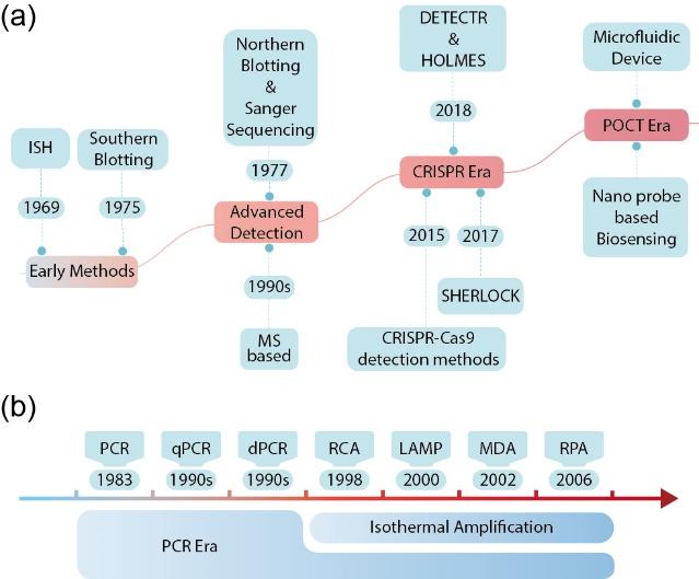
图1 核酸检测技术发展历程概览:(a) 核酸检测技术的发展历程;(b) 核酸扩增技术的发展历程。ISH,原位杂交;MS,质谱;DETECTR,DNA内切酶靶向CRISPR转录报告系统;HOLMES,1 h低成本多功能高效系统;SHERLOCK,特异性高灵敏度酶促报告解锁技术; PCR,聚合酶链式反应;RT-PCR,逆转录聚合酶链式反应;qPCR,定量聚合酶链式反应;dPCR,数字聚合酶链式反应;RCA,滚环扩增;LAMP,环介导等温扩增;MDA,多重置换扩增;RPA,重组酶聚合酶扩增

Fig.1 Overview of the evolution of nucleic acid detection technologies. (a) The development history of nucleic acid detection technologies. (b) The development history of nucleic acid amplification technologies. Abbreviations: ISH, in situ hybridization; MS, mass spectrometry; DETECTR, DNA endonuclease-targeted CRISPR trans reporter; HOLMES, one-hour low-cost multipurpose highly efficient system; SHERLOCK, specific high-sensitivity enzymatic reporter UnLOCKing; PCR, polymerase chain reaction; RT-PCR, reverse transcription-polymerase chain reaction; qPCR, quantitative polymerase chain reaction; dPCR, digital polymerase chain reaction; RCA, rolling circle amplification; LAMP, loop-mediated isothermal amplification; MDA, multiple displacement amplification; RPA, recombinase polymerase amplification

2 The Significance of On-Site, Real-Time Pathogen Nucleic Acid Testing

During the outbreak of the novel coronavirus pandemic, nucleic acid testing has served as the “gold standard” for determining viral infection[6-7],and its timeliness has not only secured valuable treatment time for patients but also played a crucial role in breaking the chain of epidemic transmission[8-9].Traditional pathogen nucleic acid detection methods primarily rely on PCR (polymerase chain reaction) or real-time qPCR (quantitative fluorescence PCR) technology[10].Although these methods exhibit high sensitivity and specificity in pathogen identification, they also have several significant limitations. On the one hand, conventional PCR and qPCR assays are time-consuming, typically requiring more than 40 minutes to obtain test results[11],making it difficult to meet the clinical demand for rapid diagnosis. On the other hand, both detection methods require operation by trained technicians, involve relatively complex procedures, and place high demands on laboratory equipment, generally necessitating a professional laboratory environment[12].These factors limit their application in primary healthcare settings or on-site environments, preventing rapid point-of-care testing and making it challenging to meet the urgent needs for rapid clinical diagnosis and public health emergency response deployment[13].
In contrast, the emergence of on-site, real-time pathogen nucleic acid detection technologies has brought about a breakthrough in pathogen detection[14]. POCT, also known as near-patient testing, bedside testing, or extra-laboratory testing, is a rapid testing method performed near or at the patient’s location. Its core concept is to generate test results quickly so that appropriate treatment can be initiated in a timely manner, thereby improving clinical or economic outcomes[15]. Compared with traditional PCR and qPCR methods, POCT technology offers significant advantages: it can complete testing in a short time (typically within minutes to tens of minutes), greatly reducing diagnostic turnaround time. In addition, POCT devices are typically portable, easy to operate, can be used without specialized training, and can function in non-laboratory settings[16]. These features enable POCT technology to overcome the limitations of conventional nucleic acid detection methods, providing strong support for clinical diagnosis, disease monitoring, and public health emergency response[17-18].

3 Traditional nucleic acid testing

Traditional nucleic acid detection technologies have laid the foundation for research in molecular biology and genetics, with the most representative techniques being blot hybridization and Sanger sequencing. Blot hybridization was first invented by Southern[19]in 1975, based on agarose gel electrophoresis technology. This technique, known as the Southern Blot, is used to determine whether specific DNA fragments are present in a sample. In this process, denatured DNA fragments that have been cut by restriction enzymes are separated by gel electrophoresis, and then transferred to membranes such as nylon or nitrocellulose via capillary action. Finally, a labeled probe is used to hybridize with the target DNA sequence for identification. The Southern Blot technique can be used for genomic structural analysis of DNA[20],detection of gene mutations[21],and analysis of restriction fragment length polymorphisms (RFLP)[22],among other applications. Moreover, this technique remains indispensable for analyzing long DNA fragments (>1 kb) and for genomic research on unsequenced species. Building on the Southern Blot, Alwine’s team[23]made innovative improvements to the technique and successfully developed the Northern Blot, a method specifically designed for RNA detection. In addition to blotting techniques such as the Southern Blot and Northern Blot, the most widely used technique in nucleic acid analysis is Sanger sequencing. This pioneering method has provided a powerful tool for the precise analysis of DNA sequences. Developed in 1977 by Sanger and his team[24],Sanger sequencing is based on the principle that DNA polymerase synthesizes a new DNA strand on a DNA template, and that the addition of special dideoxynucleotide triphosphates (ddNTPs) terminates DNA chain elongation, thereby generating a series of DNA fragments of different lengths. The length of each fragment corresponds to the type of base in the DNA sequence. By separating these fragments through electrophoresis and reading the sequence information using fluorescent labeling, the DNA sequence can ultimately be determined. The invention of Sanger sequencing laid the foundation for genomics research and served as a cornerstone technology for the Human Genome Project.

3.1 PCR-based nucleic acid testing

Currently, the mainstream technology for nucleic acid detection involves using polymerases to specifically synthesize target DNA before performing detection. Polymerase chain reaction (PCR) technology was proposed by Mullis[25]in 1985. Its core principle is to use an in vitro enzymatic synthesis reaction catalyzed by DNA polymerase, combined with temperature cycling controlled by a thermocycler, to achieve exponential amplification of the target DNA sequence. This molecular-level thermodynamic control mechanism enables target DNA fragments to be amplified by 106to 109-fold within 2 hours. With its high sensitivity, high specificity, and high efficiency, PCR technology has been widely applied in fields such as basic life science research, molecular medical diagnostics, agriculture, forensic science, and environmental monitoring, greatly advancing technological progress in these areas[26]. Building on PCR technology, researchers have subsequently developed a variety of derivative techniques. For example, by combining reverse transcriptase (RT) with PCR technology, reverse transcription PCR (RT-PCR) was created, thereby expanding the application scope of PCR in RNA virus detection, biomedical research, and clinical diagnostics. In addition, in the 1990s, Vogelstein and Kinzler[27]proposed the concept of digital PCR (dPCR). dPCR achieves absolute quantification of nucleic acids by partitioning the reaction system into micro-units and combining this with fluorescence signal detection. Compared with traditional PCR, dPCR offers higher sensitivity, specificity, and precision, and it demonstrates significant advantages, particularly in the detection of low-abundance targets and in copy number variation analysis[28-29].

3.2 Pathogen Nucleic Acid Detection Based on Isothermal Amplification

The implementation of PCR technology relies on Taq DNA polymerase, which is heat-resistant and enables efficient DNA amplification under high-temperature conditions. In contrast, isothermal amplification technologies have gradually gained attention due to their simplicity, speed, and efficiency. This method does not require complex temperature-cycling equipment and can achieve rapid nucleic acid amplification at a constant temperature, making it well-suited for use in resource-constrained settings. Nucleic acid sequence-based amplification (NASBA) primarily relies on the synergistic action of AMV reverse transcriptase, T7 RNA polymerase, and ribonuclease H (RNase H). A pair of specific primers is used to amplify RNA under constant 42°C conditions[30]. Rolling circle amplification (RCA) is an isothermal DNA amplification technique that uses short primers and a DNA polymerase with strand-displacement activity (such as φ29 DNA polymerase) to generate long single-stranded DNA molecules from a circular DNA template at a constant temperature[31]. RCA is characterized by its high efficiency and isothermal nature, enabling rapid amplification of target DNA[32]; however, its efficiency is lower when amplifying larger circular DNA templates, and it depends on single-primer binding and extension. Multiple-primer rolling circle amplification (MP-RCA) achieves efficient amplification through the use of random primers and φ29 DNA polymerase, enhancing both amplification efficiency and selectivity[33]. Building on the principles of MP-RCA, the field of single-cell sequencing has further developed multiple displacement amplification (MDA) technology, which uses random primers and φ29 DNA polymerase under mild conditions to perform whole-genome amplification of single-cell genomes, thereby further improving amplification uniformity and coverage[34]. This technology has driven the advancement of single-cell sequencing and provides strong technical support for research in areas such as cellular heterogeneity, developmental biology, and disease mechanisms[35]. In addition, loop-mediated isothermal amplification (LAMP)[36-39]and recombinase polymerase amplification (RPA) are widely used in pathogen detection, genotyping, and other fields, and are particularly well suited for POCT applications[40-43].

3.3 Other methods

In addition, various other innovative detection methods have achieved precise analysis of pathogen nucleic acids through unique technologies. Nucleic acid mass spectrometry is an advanced nucleic acid detection method that combines multiplex PCR, high-throughput chips, and time-of-flight mass spectrometry (MALDI-TOF-MS). By precisely measuring mass differences among nucleic acid molecules, this technique can rapidly and efficiently detect genetic variants, polymorphisms, methylation patterns, and other characteristics[44-45]. The technology offers advantages such as high throughput, high sensitivity, ease of operation, and cost-effectiveness[46-47]. In situ hybridization (ISH) or fluorescence in situ hybridization (FISH) is a technique used to detect specific nucleic acid sequences in cell or tissue sections. It achieves localization and detection of target genes or transcripts by enabling specific hybridization between labeled nucleic acid probes and the target nucleic acid sequences[48-49]. These technological advances provide multidimensional complementarity with isothermal amplification systems: isothermal amplification focuses on rapid nucleic acid enrichment, mass spectrometry excels in fine variant analysis, and in situ hybridization enables spatially resolved detection.

4 Nucleic Acid Detection Technology Based on CRISPR-Cas Biosensing

In the technological evolution of building multimodal detection platforms, the field of molecular diagnostics is undergoing a paradigm shift driven by gene-editing technologies. The CRISPR (Clustered regularly interspaced short palindromic repeats) structure was discovered in bacteria and archaea, and its function is associated with bacterial immune defense mechanisms[50-51]. Charpentier, Doudna, and others[52]revealed that the CRISPR-Cas9 system can serve as a programmable gene-editing tool, significantly advancing the development of gene-editing technology. With its high specificity and sensitivity, CRISPR-Cas technology has greatly accelerated pathogen nucleic acid detection, enabling precise testing within a short time and providing strong support for rapid diagnosis[53-54]. The core principle of CRISPR-Cas nucleic acid detection technology is based on the dual functionality of Cas proteins (such as Cas12/13/14): on the one hand, specific recognition is achieved through precise complementary pairing between crRNA and the target nucleic acid sequence; on the other hand, activation of non-specific nuclease activity (Trans-cleavage activity) cleaves reporter molecules (such as fluorescent probes), thereby converting target-binding events into detectable optical, electrochemical, or other signals (Figure 2). By coupling this technology with isothermal amplification techniques (such as RPA or LAMP), it can achieve single-molecule-level detection sensitivity within 30–60 minutes[43,55]. Based on the CRISPR-Cas system, biosensing technologies innovatively integrate gene recognition with signal transduction mechanisms, opening up an entirely new dimension for pathogen nucleic acid detection.
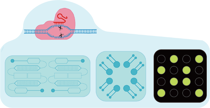
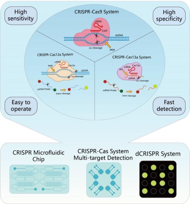
图2 CRISPR-Cas系统在核酸检测中的应用,该系统以其高特异性、高灵敏度、检测快速和操作简便的特点,正在推动核酸检测技术的革新

Fig.2 The application of the CRISPR-Cas system in nucleic acid detection is driving innovation in the field. This system is characterized by its high specificity, high sensitivity, rapid detection capabilities, and ease of operation

4.1 Nucleic acid detection using Cas12a

In recent years, real-time nucleic acid detection technologies based on the CRISPR-Cas12 system have garnered widespread attention. CRISPR-Cas12a (formerly known as Cpf1), as a unique member of the Type II CRISPR-Cas system, has demonstrated significant advantages in the field of nucleic acid detection[56](Fig. 2). Compared with other Cas proteins, Cas12a not only accurately recognizes and cleaves double-stranded DNA target sequences under the guidance of crRNA, but also activates its non-specific single-stranded DNA cleavage activity upon target binding. This unique trans-cleavage property provides an ideal molecular tool for developing highly sensitive nucleic acid detection methods[57].
DETECTR (DNA endonuclease-targeted CRISPR trans reporter) technology was developed in 2018 by the Doudna team[58]. The underlying principle is that, after the Cas12a protein, guided by gRNA, recognizes and cleaves the target double-stranded DNA, it activates its non-specific single-stranded DNA cleavage activity, thereby cleaving a fluorescently labeled single-stranded DNA reporter molecule and generating a detectable signal[59]. This method offers rapid detection with high sensitivity and specificity, and the results are visually intuitive, but it is limited to detecting DNA samples only and requires the design of specific guide sequences. HOLMES (One-hour low-cost multipurpose highly efficient system) technology was proposed in 2018 by Li et al.[60]. It similarly exploits the dual cleavage activity of Cas12a under gRNA guidance, featuring rapid detection with high sensitivity and specificity, but is also limited to the detection of DNA samples[61]. HOLMESv2 represents a further upgrade, employing a heat-stable Cas12b protein that can specifically distinguish single nucleotide polymorphisms (SNPs) and accurately detect viral RNA, human cell mRNA, and circular RNA. By combining LAMP amplification technology, HOLMESv2 enables one-step quantitative nucleic acid detection under isothermal conditions, effectively preventing cross-contamination between samples. In addition, it can be combined with bisulfite treatment to precisely quantify the degree of DNA methylation, demonstrating extremely broad application prospects in molecular diagnostics and epigenetic research[62]. The sPAMC (Suboptimal PAM of Cas12a-based test with enhanced flexibility, speed, sensitivity and reproducibility) technology developed by Lu et al.[63] utilizes a suboptimal PAM sequence and combines isothermal amplification (RPA) with Cas12a-mediated detection to achieve rapid, highly sensitive, and specific detection of SARS-CoV-2 RNA. Furthermore, Shi et al.[64] developed a DNA diagnostic technology called CONAN (CRISPR-Cas-only amplification network). This method integrates Cas12a’s stringent target recognition, helicase activity, and trans-cleavage activity into a positive feedback loop to achieve isothermal amplification-based detection of genomic DNA. The method boasts advantages such as single-enzyme operation, one-step procedure, real-time detection, high sensitivity, and single-base specificity. In optimizing traditional Cas12a-based detection systems, Yue et al.[65], by optimizing reaction conditions and employing droplet-based Cas12a detection with dual crRNA targeting, significantly improved detection sensitivity, quantitative analysis capability, and specificity compared to the single-crRNA approach, providing a more precise tool for molecular diagnostics. Meanwhile, Ding et al.[66] developed an all-integrated dual CRISPR-Cas12a (AIOD-CRISPR) detection technology that uses a dual crRNA design to overcome the limitations imposed by PAM sequences in traditional Cas12a-based detection. Coupled with a "one-pot" strategy, this technology significantly enhances detection sensitivity, enabling the detection of extremely low copy numbers of SARS-CoV-2 nucleic acid.
Although the Cas12a enzyme has traditionally been used for DNA detection, the SAHARA (Split activator for highly accessible RNA analysis) technology ingeniously leverages Cas12a to enable RNA detection. The core principle is that when a short DNA strand binds near the PAM sequence, the distal end of the PAM sequence can bind to RNA, thereby activating Cas12a’s trans-cleavage activity and enabling efficient RNA detection. This technology not only allows the use of the Cas12a enzyme for detecting RNA substrates but also enhances sensitivity to mutations at specific sites[67]..

4.2 Nucleic acid detection using Cas13a

Following the Cas12a technology, CRISPR-Cas13a, as a representative member of the Type VI CRISPR-Cas system, has demonstrated great potential in the field of point-of-care nucleic acid testing due to its precise RNA recognition and cleavage capabilities[68-70]. Guided by crRNA, Cas13a can specifically recognize target RNA and, upon activation, exhibits non-specific cleavage of surrounding single-stranded RNA, providing an ideal mechanism for signal amplification. In 2017, the Zhang lab[71]first proposed the SHERLOCK technology, which leverages this "collateral cleavage" property. By designing fluorescently labeled RNA reporter molecules, SHERLOCK enables highly sensitive detection of target RNA, thereby significantly advancing the development of POCT applications. Subsequently, Liu et al.[72]developed the FIND-IT technology, which uses Cas13a and the Csm6 nuclease in tandem. Under amplification-free conditions and in combination with chemically stable activators, this technology enables rapid detection of SARS-CoV-2viral RNA, with a sensitivity of approximately 30 molecules per microliter and a detection time of within 20 minutes. In addition, Fozouni et al.[4]proposed an amplification-free CRISPR-Cas13a detection technology that, when combined with a mobile phone microscope, enables high-sensitivity, low-cost on-site detection.

4.3 Other CRISPR Systems

In addition to detection technologies based on Cas12a and Cas13a, other CRISPR systems also demonstrate unique advantages in nucleic acid detection. The CRISDA (CRISPR-Cas9-triggered nicking endonuclease-mediated strand displacement amplification) technology was invented in 2018 by Zhou et al.[73]It leverages the precise recognition and cleavage capabilities of the Cas9 nickase (Cas9n) to generate a single-strand nick on the non-target strand of the target DNA, and through DNA polymerase–mediated strand displacement amplification, achieves highly sensitive and specific detection of the target DNA. The LEOPARD (Leveraging engineered tracrRNAs and on-target DNAs for parallel RNA detection) technology was proposed in 2021 by Jiao et al.[74]This technology reprograms tracrRNA (Rptr) to specifically hybridize with the target RNA, forming an atypical crRNA that guides the Cas9 nuclease to recognize and cleave the complementary DNA sequence. Another technique that uses Rptr’s specific binding to the target RNA to activate the Cas9 protein is AGATHA (Atypical gRNA-activated transcription halting alarm). After activation, Cas9 binds to a specific AGATHA DNA, blocking transcription by disrupting the secondary structure at its 3’ end, while continuously generating Broccoli RNA aptamers that bind to fluorescent substrates to produce a signal. By designing multiple Rptr variants, the AGATHA technology achieves highly specific, strongly amplified multiplex RNA detection, demonstrating rapid and precise detection capabilities[75].

5 CRISPR-Cas Nucleic Acid Detection on Microfluidic Chips

In nucleic acid detection technology, achieving high sensitivity, high specificity, and rapid, convenient detection has long been a research hotspot. Due to their inherent characteristics—such as small reaction space, short diffusion distances within the chamber, fast reaction kinetics, and flexible, versatile design—microfluidic chips combined with CRISPR-Cas systems have demonstrated tremendous application potential.
Yu et al.[76]developed the PAMDA SlipChip technology, which employs a dual-layer microfluidic chip design and uses a "pearl chain" structure to achieve automated droplet partitioning and precise distribution, thereby effectively supporting digital LAMP-CRISPR detection of SARS-CoV-2;Park et al.[77]developed the deCOViD technology, which miniaturizes a CRISPR/Cas12a–assisted RT-RPA detection system and integrates it into a chip, enabling qualitative and quantitative detection to be completed in 15 and 30 minutes, respectively, thus providing a reliable solution for on-site testing in POCT scenarios.
In addition, a series of integrated approaches have further advanced the development of end-to-end, automated detection technologies on microfluidic platforms. The DISCoVER technology developed by Chandrasekaran et al.[78]integrates pre-processing of saliva samples, LAMP amplification, and CRISPR-Cas13-based detection on a single chip, enabling on-site viral RNA detection within 60 minutes. In 2023, Zhou et al.[79]developed the M3-CRISPR platform, which enables multiplexed, low-cost, and highly specific nucleic acid detection. The system incorporates a heating film to precisely control the reaction temperature on the chip, ensuring efficient RPA and CRISPR reactions (Fig. 3a). The Shen team[80]has developed the LOC-CRISPR technology, which integrates viral nucleic acid extraction, recombinase polymerase amplification, and CRISPR/Cas12a–based cleavage reaction detection on a microfluidic chip, enabling end-to-end contamination-free detection. He et al.[81]have developed a high-throughput, all-liquid-phase African swine fever virus (ASFV) detection technology based on CRISPR-Cas12a, which, combined with fluorescence signal readout and a portable detection system, enables rapid and accurate ASFV detection. Meanwhile, Qin et al. have developed a fully microfluidic nucleic acid detection technology using CRISPR-Cas13a for rapid detection of Ebola virus RNA (Fig. 3b). Samples are automatically mixed and hybridized on the microfluidic chip, after which the non-specific cleavage products of Cas13a are measured in real time using a custom-integrated fluorescence reader. This isothermal, all-liquid-phase diagnostic method does not require amplification, is simple to operate, and offers high sensitivity, providing an efficient, low-cost solution for rapid on-site diagnosis, particularly suitable for infectious disease detection in more remote areas[82](Fig. 3b).
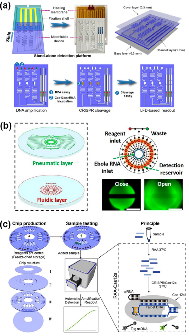
图3 将微流控技术与CRISPR-Cas系统整合用于POCT:(a) M3-CRISPR平台的构成(加热膜、固定壳和滑动组件)[79];(b) CRISPR-Cas13a全微流控核酸检测芯片由气动层和流体层组成[82];(c) CASMEAN核酸检测技术的芯片结构和样本测试流程[83]

Fig.3 Integration of microfluidic technology and the CRISPR-Cas system for point-of-care testing (POCT). (a) The components of the M3-CRISPR platform (heating film, fixation shell, and sliding component). Reprinted from ref 79; (b) The CRISPR-Cas13a fully microfluidic nucleic acid detection chip consists of a pneumatic layer and a fluidic layer. Reprinted from ref 82; (c) The chip structure and sample testing procedure of the CASMEAN nucleic acid detection technology. Reprinted from ref 83

Chen et al.[83]developed an automated POCT technology called “CASMEAN” (Cas12a-assisted microfluidic nucleic acid analysis device) (Figure 3c). By integrating recombinase-aided amplification (RAA) with the CRISPR-Cas12a system and utilizing a centrifugal microfluidic chip, this technology enables highly sensitive and specific nucleic acid detection. The CASMEAN platform preloads RAA-Cas12a reagents, thereby avoiding aerosol contamination and quantification challenges associated with traditional separate amplification and detection steps, while significantly enhancing detection efficiency and sensitivity. Subsequently, Chen et al.[84]built upon this work to develop a dual CRISPR/Cas12a–assisted RT-RAA detection technology. By combining this technology with a centrifugal microfluidic platform, the detection time was dramatically reduced to 30 minutes. Ramachandran et al.[85]developed a CRISPR-Cas12 nucleic acid detection method based on electric-field-driven microfluidic technology. By using an electric field gradient on a microfluidic chip to control and accelerate the CRISPR reaction, this method enables rapid, highly sensitive detection of SARS-CoV-2viral RNA. This approach not only increases detection speed but also significantly reduces reagent consumption, making it particularly suitable for resource-limited settings. In addition, researchers have explored strategies that combine CRISPR nucleic acid detection with electrochemical methods. Najjar et al.[86]designed a 3D-printed microfluidic chip that integrates nucleic acid extraction, concentration, amplification, and electrochemical sensor-based detection. This chip can detect RNA by cleaving biotinylated ssDNA probes to reduce the current signal, and it can simultaneously measure antibody levels in saliva. The entire detection process takes only about 2 hours, demonstrating significant potential for rapid COVID-19 diagnosis and immune assessment.

5.1 Multiplexed Detection on Microfluidic Chips

Among the numerous detection technologies described above, most are limited to detecting a single target, making it difficult to meet the demand for simultaneous multi-target analysis in complex biological systems. Multi-target detection technologies aim to overcome the limitation of traditional methods, which can only target a single analyte, thereby addressing the need for concurrent analysis of multiple indicators in complex biological systems. Microfluidic chip-based technologies enable the combination of multiple targets and stepwise reactions by precisely controlling ultra-small reaction volumes, providing an ideal platform for high-throughput, multiplexed detection.
CARMEN (Combinatorial arrayed reactions for multiplexed evaluation of nucleic acids) technology was developed by the Blainey team in 2020. It achieves self-organized pairing of nanoliter-volume reaction systems in the form of droplets within a micro-well array, and uses fluorescence microscopy to read the color codes of the droplets, thereby identifying sample–assay pairs (Fig. 4a). By merging droplets under an electric field to initiate the reaction, CARMEN can simultaneously detect thousands of sample–crRNA target pairs, ensuring both high sensitivity and high specificity while effectively reducing detection costs[87]. Building on CARMEN technology, mCARMEN represents a further upgrade: by implementing multiplexed nucleic acid testing for multiple samples and pathogens on commercial microfluidic chips, it significantly streamlines clinical workflows and enhances its potential for broader applications in the POCT field[88](Fig. 4b). Xu et al.[89]developed a multiplex nucleic acid detection technology called MiCaR (Microfluidic space coding for multiplexed nucleic acid detection via CRISPR-Cas12a and recombinase polymerase amplification) (Fig. 4c). This technology integrates microfluidic spatial coding and, using a single fluorescent probe, can simultaneously detect 30 nucleic acid targets, completing highly sensitive and specific rapid detection within 40 minutes. In addition, MiCaR technology demonstrates broad application prospects in rapid pathogen diagnostics. With its simple operation, low cost, and suitability for POCT, it provides an efficient solution for multiplex nucleic acid testing in resource-limited settings. Another technology, the Microfluidic dual-droplet device (M-D3), combines CRISPR-Cas12a with RPA technology, using pressure/vacuum-driven generation of microdroplets to separately encapsulate Cas12a/crRNA, fluorescent reporter molecules, and samples, thereby enabling rapid and sensitive simultaneous detection of HPV16 and HPV18 (Fig. 4d). With its high sensitivity, rapid detection capability, and extremely low sample consumption, this technology exhibits great application potential in the field of multiplex nucleic acid POCT, particularly suitable for clinical management and large-scale population screening[90].
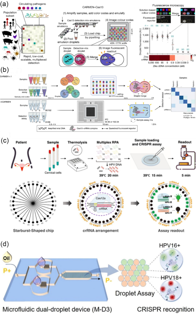
图4 微流控芯片上的CRISPR体系多靶标检测:(a) CARMEN-Cas13技术用于检测人类和动物中的多种病原体。图中黑点代表液滴,蓝色数字表示重复次数,红线为中位数,用于生成热图[87];(b) 上部分为CARMEN v.1工作原理图,下部分为mCARMEN工作原理图[88];(c) MiCaR技术的检测流程和原理。该技术使用30通道星形芯片,每个通道加载特定HPV亚型的Cas12a/crRNA复合体[89];(d) 微流控双滴平台技术示意图。图中为微流控双滴平台的芯片示意图及其检测结果示意图[90]

Fig.4 Multiplexed detection of CRISPR systems on microfluidic chips. (a) The CARMEN-Cas13 technology for detecting multiple pathogens in humans and animals. The black dots represent droplets, the blue numbers indicate the number of replicates, and the red lines denote the medians used to generate the heatmap. Reprinted from ref 87. (b) The upper part illustrates the working principle of CARMEN v.1, while the lower part shows the working principle of mCARMEN. Reprinted from ref 88. (c) The detection process and principle of the MiCaR technology. This technology employs a 30-channel star-shaped chip, with each channel loaded with a Cas12a/crRNA complex specific to a particular HPV subtype. Reprinted from ref 89. (d) Schematic of the microfluidic dual-droplet platform technology. The figure shows the chip diagram of the microfluidic dual-droplet platform and the schematic of its detection results. Reprinted from ref 90

5.2 Amplification-free detection on microfluidic chips

In clinical sample testing, pathogen concentrations are often extremely low, and traditional nucleic acid detection methods typically require amplification prior to detection. However, amplification-free nucleic acid detection technology based on microfluidic chips, through innovative design, enables direct detection of low-concentration samples, offering a new solution for clinical diagnosis.
Tian et al.[91]developed the "ultra-local Cas13a assay" technology, which confines the RNA-triggered Cas13a catalytic system within cell-sized reactors. By leveraging microdroplet technology to simultaneously enhance the local concentrations of both the target RNA and the reporter molecule, this approach enables absolute digital single-molecule quantification of multiple RNA molecules. With its high sensitivity, specificity, and the advantage of not requiring nucleic acid amplification, this technology shows great promise for broad applications in clinical diagnostics, pathogen detection, and liquid biopsies. In particular, its simplicity and low cost make it an ideal tool for viral load detection. Similarly, Yue et al.[65]developed the "microdroplet Cas12a assay" technology, which optimizes Cas12a reaction parameters and confines the reaction within microdroplets, thereby successfully achieving absolute single-molecule-level quantification of DNA while avoiding the risk of cross-contamination associated with traditional amplification methods (Figure 5a).
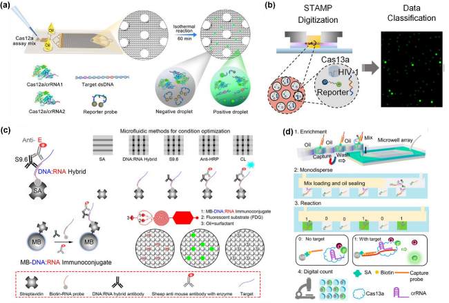
图5 微流控芯片上的无扩增检测:(a) 微滴Cas12a检测技术:通过芯片生成液滴,包含Cas12a、crRNA、报告探针和靶DNA,进行等温反应后,荧光成像计数液滴以定量DNA[65];(b) STAMP数字CRISPR-Cas13a检测技术图解。芯片大致结构示意图和其最终结果图[92];(c) 数字微流控杂交检测技术通过链霉亲和素(SA)和DNA:RNA杂交抗体捕获DNA:RNA杂交体。该技术使用连续流通道芯片生成、孵育和成像液滴,以实现核酸检测[93];(d)无扩增数字 CRISPR/Cas13a检测:生物素化探针捕获目标RNA,磁珠固定Cas13a/crRNA复合物,油密封微孔中检测荧光信号[94]

Fig.5 Amplification-free detection on microfluidic chips. (a) Droplet-based Cas12a detection technology: Droplets containing Cas12a, crRNA, reporter probes, and target DNA are generated on the chip. After isothermal reaction, droplets are imaged and counted by fluorescence to quantify DNA. Reprinted from ref 65. (b) Schematic of the STAMP digital CRISPR-Cas13a detection technology. The diagram shows the general structure of the chip and its final results. Reprinted from ref 92. (c) Digital microfluidic hybridization detection technology captures DNA:RNA hybrids using streptavidin (SA) and DNA:RNA hybridization antibodies. This technology generates, incubates, and images droplets on a continuous-flow channel chip to achieve nucleic acid detection. Reprinted from ref 93. (d) Amplification-free digital CRISPR/Cas13a detection: Biotinylated probes capture target RNA, and magnetic beads immobilize the Cas13a/crRNA complex. Fluorescent signals are detected in oil-sealed microwells. Reprinted from ref 94

The STAMP (Self-digitization through automated membrane-based partitioning) technology employs a different strategy[92].This technology uses automated membrane partitioning to separate the reaction system into tiny, independent reaction units by mixing the sample with the detection reagent and then loading the mixture onto a polycarbonate membrane, thereby enabling amplification-free, highly sensitive, and specific absolute quantification of HIV-1 viral RNA (Figure 5b). Meanwhile, the “digital microfluidic hybridization detection technology” developed by Mou et al.[93]integrates picoliter-scale droplet technology with magnetic microbeads to achieve highly sensitive HPV nucleic acid detection without amplification. This technology is easy to operate, low in cost, and fast in detection, making it suitable for POCT and providing an efficient solution for viral load testing in resource-limited settings (Figure 5c). Notably, the amplification-free digital CRISPR/Cas13a detection technology developed by Wang et al.[94]innovatively combines magnetic bead capture and enrichment with micro-well dispersion detection, ensuring that each micro-well contains only one magnetic bead, thereby significantly enhancing detection sensitivity. This method can complete detection in just 50 minutes, avoiding the complex procedures and potential biases associated with traditional amplification methods, and provides an efficient and reliable solution for detecting low-concentration samples (Figure 5d). These amplification-free detection technologies not only simplify the operational process and reduce costs but also open up new possibilities for POCT.

5.3 Microfluidic POCT rapid detection without the need for specialized testing equipment

Among the detection methods mentioned above, most rely on external equipment to complete the testing process. In the field of microfluidic POCT, reducing dependence on specialized equipment is a crucial direction for enhancing the portability and practicality of testing.
The MAV chip technology innovatively designs a magnetic bead-single-stranded DNA-platinum nanoparticle (BDNP) complex as a reporting molecule. This system leverages the strong binding affinity between biotin and streptavidin, as well as the stable coordination bond between thiol and platinum nanoparticles, to achieve visual quantitative detection without the need for fluorescence (Figure 6). Upon activation of the Cas12a enzyme, its trans-cleavage activity releases platinum nanoparticles, which then catalyze the decomposition of hydrogen peroxide to produce oxygen, driving the movement of red ink in the chip’s channels. The test result can be intuitively determined by observing the distance the ink has moved[95]. This technology not only enables visual quantitative detection of target nucleic acids but also eliminates the reliance on fluorescent labeling in traditional Cas12a assays, significantly enhancing the convenience and practicality of the detection process.
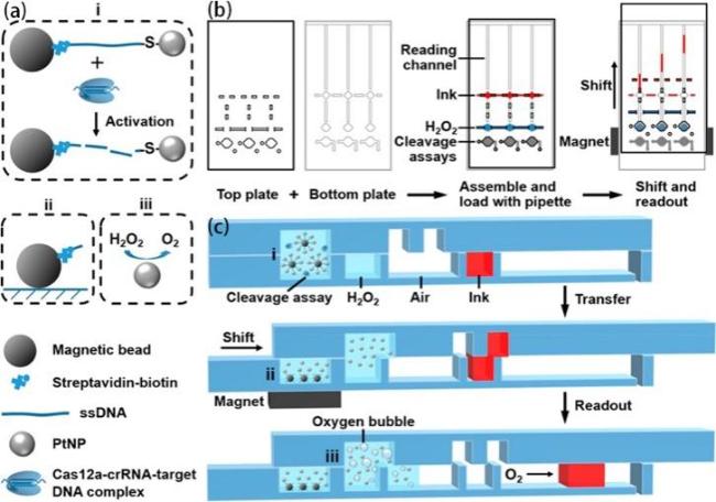
图6 基于铂纳米信号探针的CRISPR-Cas12a检测的磁辅助V芯片(MAV-chip)的工作原理:(a) (i~iii)展示了磁珠操作、目标识别以及通过氧气气泡形成实现信号生成的过程;(b) MAV芯片的上层和下层展示图、结合图和滑动后展示图;(c) MAV 芯片在分析/试剂加载、偏移和读数状态下的横截面示意图[95]

Fig. 6 Working principle of the platinum nanoreporter-based CRISPR-Cas12a detection system on the MAV-chip.(a) (i-iii) Illustrate the processes of magnetic bead manipulation, target recognition, and signal generation via oxygen bubble formation. (b) Illustrations of the upper and lower layers of the MAV-chip, the combined layers, and the chip after sliding. (c) Cross-sectional schematics of the MAV-chip in the analytical/reagent loading, offset, and readout states. Reprinted from ref 95

6 Summary and Outlook

In summary, the combination of microfluidic chips and CRISPR-Cas technology has brought about a revolutionary breakthrough in the POCT field, demonstrating unique advantages particularly in the rapid detection of pathogen nucleic acids. Currently, several companies have advanced the commercialization of such technologies: for example, Mammoth Biosciences has launched a CRISPR detection card that can complete diagnosis within 15 minutes; Sherlock Biosciences has developed multiplex pathogen detection kits based on the SHERLOCKv2 platform; and Wezhen Bio in China has introduced NuRapid-Dx®, the world's first self-testing CRISPR product for tuberculosis.These commercialization examples validate the clinical feasibility and market potential of this technology (Table 1).
表1 商业化的CRISPR生物传感检测技术的比较

Table 1 Comparison of commercialized CRISPR biosensing detection technologies

Company Name Core Technology
Platform
Main Cas Proteins/Systems Primary Targets/Application Scenarios
Mammoth Biosciences DETECTR™、DETECTR BOOST® Cas12a、Cas13a dsDNA/ssDNA/RNA (e.g., SARS-CoV-2, HPV typing, detection of multiple pathogens)
Sherlock Biosciences SHERLOCK™ Cas12a、Cas13a ssRNA (e.g., Zika virus, SARS-CoV-2), tumor mutations, antibiotic resistance genes
MicroDiag Biomedicine NuRapid-Dx® Cas12a dsDNA (e.g., Mycobacterium tuberculosis, other pathogens)
Detect™ (Lucira Health) Detect™ Cas13 ssRNA (e.g., SARS-CoV-2)
ToloBio HOLMES™ Cas12 DNA/RNA(e.g.,Pathogens, tumors, genetic diseases, SNP sites, and methylation sites)
In future technology translation, the key challenges that need to be addressed include: First, standardizing the industrial production of microfluidic chips and enhancing the integration and automation of chip operations to simplify complex nucleic acid detection processes, enabling rapid and accurate testing in resource-constrained settings. This capability is particularly crucial in responding to sudden public health emergencies, such as the COVID-19 pandemic, providing strong support for disease prevention and control. Second, highly specialized customization of key reagents and enzymatic reactions within the CRISPR-Cas system—such as pre-embedding lyophilized reagents targeting specific pathogen genes on the chip—ensures both portability and ease of operation while improving the reliability of test results and the feasibility of field applications. Third, further developing multi-parameter detection technologies: by combining microfluidic chips with CRISPR-Cas technology, the advantage of simultaneous detection of multiple targets can be leveraged to screen for various pathogens without compromising detection accuracy, thereby significantly enhancing testing efficiency and clinical diagnostic value.
In recent years, POCT technology has been undergoing a paradigm shift from “miniaturization” to “intelligence.” Traditional microfluidic-chip-based detection methods rely on laboratory personnel to subjectively interpret the results. This result representation method, which depends on subjective visual interpretation, inevitably introduces operator-dependent errors, thereby affecting the accuracy and reproducibility of the detection results. The introduction of AI technology enables objective, quantitative analysis of experimental results through deep learning algorithms, effectively eliminating the subjective errors inherent in traditional manual interpretation and significantly enhancing the accuracy and reproducibility of detection results[96-97]. Similarly, in CRISPR-based biosensing systems, artificial intelligence technology also demonstrates powerful optimization capabilities. The CRISPR off-target effect refers to the phenomenon whereby, during gene editing, the CRISPR/Cas system, due to insufficient specificity in the binding between the guide RNA (gRNA) and the target DNA sequence, causes the Cas enzyme to cleave non-target sites—a major challenge in the application of CRISPR technology. In gene editing, this effect may trigger genomic mutations such as insertions, deletions, or translocations, thereby affecting gene function or causing genomic instability. In nucleic acid detection, the CRISPR off-target effect may lead to false-positive or false-negative detection results[98-99]. DeepCRISPR, developed by Liu et al.[100], is the first computational platform to apply AI to the prediction of sgRNA targeting efficiency and off-target effects. The application of this technology has significantly enhanced the detection performance and result reliability of CRISPR systems while substantially reducing off-target effects arising from non-specific binding. Looking ahead, the deep integration of AI technology with microfluidic nucleic acid detection technology based on CRISPR biosensors will bring about revolutionary advances. Through continuous learning from massive amounts of detection data using deep learning algorithms, the system can continuously optimize reaction condition parameters and dynamically adjust signal thresholds, significantly improving the detection rate of weakly positive samples and reducing the risk of false positives. On the other hand, it may be possible to use AI technology to design novel nucleases that rival natural Cas proteins, thereby significantly reducing or even completely eliminating the off-target effects of CRISPR technology[101]. Such intelligent nucleic acid detection platforms are not only suitable for clinical diagnostics but also hold broad application prospects in areas such as environmental monitoring, food safety, and biosafety, providing strong technological support for precision medicine and public health prevention and control.
[1]
Li P T, Xiong H W, Yang B, Jiang X Y, Kong J L, Fang X E. Trac Trends Anal. Chem., 2022, 157: 116812.

[2]
Whitesides G M. Nature, 2006, 442(7101): 368.

[3]
Avaro A S, Santiago J G. Lab Chip, 2023, 23(5): 938.

[4]
Fozouni P, Son S, Díaz de León Derby M, Knott G J, Gray C N, D’Ambrosio M V, Zhao C Y, Switz N A, Kumar G R, Stephens S I, Boehm D, Tsou C L, Shu J, Bhuiya A, Armstrong M, Harris A R, Chen P Y, Osterloh J M, Meyer-Franke A, Joehnk B, Walcott K, Sil A, Langelier C, Pollard K S, Crawford E D, Puschnik A S, Phelps M, Kistler A, DeRisi J L, Doudna J A, Fletcher D A, Ott M. Cell, 2021, 184(2): 323.

[5]
Land K J, Boeras D I, Chen X S, Ramsay A R, Peeling R W. Nat. Microbiol., 2019, 4(1): 46.

[6]
Zhang S Y, Su X S, Wang J, Chen M Y, Li C Y, Li T D, Ge S X, Xia N S. Crit. Rev. Anal. Chem., 2022, 52(2): 413.

[7]
Wang T, Liu Y, Sun H H, Yin B C, Ye B C. Angew. Chem. Int. Ed., 2019, 131(16): 5436.

[8]
He P, Song L G, Xie H, Liang J Y, Yuan D Y, Wu Z D, Lv Z Y. Infect. Dis. Poverty, 2016, 5: 25.

[9]
Wu J G, Liu J S, Li S J, Peng Z Y, Xiao Z, Wang X F, Yan R C, Luo J F. Travel. Med. Infect. Dis., 2020, 37: 101673.

[10]
Huggett J F, O’Sullivan D M, Cowen S, Cleveland M H, Davies K, Harris K, Moran-Gilad J, Winter A, Braybrook J, Messenger M. Mol. Aspects Med., 2024, 97: 101275.

[11]
Sheridan C. Nat. Biotechnol., 2020, 38(5): 515.

[12]
Afzal A. J. Adv. Res., 2020, 26: 149.

[13]
Huang Y, Liu G D, Zhang X J. Prog. Chem., 2020, 32(9): 1241.

(黄炎, 刘国东, 张学记. 化学进展, 2020, 32(9): 1241).

[14]
Zhao L, Zhao Z H, Li N, Wang X Y. Talanta, 2025, 286: 127457.

[15]
Price C P. Bmj, 2001, 322(7297): 1285.

[16]
Kang T J, Lu J M, Yu T, Long Y, Liu G Z. Biosens. Bioelectron., 2022, 206: 114109.

[17]
Niemz A, Ferguson T M, Boyle D S. Trends Biotechnol., 2011, 29(5): 240.

[18]
Shang M Y, Guo J C, Guo J H. Sens. Diagn., 2023, 2(5): 1123.

[19]
Southern E M. J. Mol. Biol., 1975, 98(3): 503.

[20]
Gebbie L. Cereal Genomics:Methods and Protocols. NY: Humana Press, 2014, 1099: 159.

[21]
Mellars G, Gomez K. PCR Mutation Detection Protocols. NJ: Humana Press, 2010, 281.

[22]
Brown T A. DNA, 2001, 1(2): 3.

[23]
Alwine J C, Kemp D J, Stark G R. Proc. Natl. Acad. Sci. U. S. A., 1977, 74(12): 5350.

[24]
Sanger F, Nicklen S, Coulson A R. Proc. Natl. Acad. Sci. U. S. A., 1977, 74(12): 5463.

[25]
Mullis K, Faloona F, Scharf S, Horn G, Erlich H. Cold Spring Harbor symposia on quantitative biology. NY: Cold Spring Harbor Laboratory Press, 1986. 51: 263.

[26]
Post J C. Jama, 2000, 283(12): 1544.

[27]
Vogelstein B, Kinzler K W. Proc. Natl. Acad. Sci. U. S. A., 1999, 96(16): 9236.

[28]
Wils G, Hamerlinck L, Trypsteen W, Van Den Eeckhaut C, Weiss J, Abdel Nour A, Vergult S, Vandesompele J. Methods Mol. Biol., 2025, 2880: 17.

[29]
Bai H, Hu J, Liu T, Wan L, Dong C, Luo D S, Li F, Yuan Z X, Tang Y M, Chen T L, Wang S, Gou H N, Zhou Y Z, Ying B W, Huang J, Hu W W. Lab Chip, 2025, 25(3): 413.

[30]
Compton J. Nature, 1991, 350(6313): 91.

[31]
Lizardi P M, Huang X H, Zhu Z R, Bray-Ward P, Thomas D C, Ward D C. Nat. Genet., 1998, 19(3): 225.

[32]
Nallur G, Luo C, Fang L, Cooley S, Dave V, Lambert J, Kukanskis K, Kingsmore S, Lasken R, Schweitzer B. Nucleic Acids Res., 2001, 29(23): e118.

[33]
Dean F B, Nelson J R, Giesler T L, Lasken R S. Genome Res., 2001, 11(6): 1095.

[34]
Dean F B, Hosono S, Fang L H, Wu X H, Faruqi A F, Bray-Ward P, Sun Z Y, Zong Q L, Du Y F, Du J, Driscoll M, Song W M, Kingsmore S F, Egholm M, Lasken R S. Proc. Natl. Acad. Sci. U. S. A., 2002, 99(8): 5261.

[35]
Wang J B, Fan H C, Behr B, Quake S R. Cell, 2012, 150(2): 402.

[36]
Notomi T, Okayama H, Masubuchi H, Yonekawa T, Watanabe K, Amino N, Hase T. Nucleic Acids Res., 2000, 28(12): e63.

[37]
Garg N, Sahu U, Kar S, Ahmad F J. Sci. Rep., 2021, 11: 9859.

[38]
Zhu Y S, Shao N, Chen J W, Qi W B, Li Y, Liu P, Chen Y J, Bian S Y, Zhang Y, Tao S C. Anal. Chim. Acta, 2020, 1140: 30.

[39]
Yao Y H, Zhao N, Jing W W, Liu Q, Lu H J, Zhao W, Zhao W, Yuan Z M, Xia H, Sui G D. Sens. Actuat. B Chem., 2021, 333: 129521.

[40]
Piepenburg O, Williams C H, Stemple D L, Armes N A. PLoS Biol., 2006, 4(7): e204.

[41]
Hong L, Huang Q, Zhou Y H, Zheng Q, Wang S P, Chen F F, Chang X Y, Jiang G S, Zha L S. Transbound. Emerg. Dis., 2024, 2024(1): 3680778.

[42]
Wang P, Zhang X, Shen H, Lu Q W, Li B Z, Liu X, Gao S. Sens. Actuat. B Chem., 2023, 374: 132853.

[43]
Liu Y, Chao Z J, Ding W, Fang T F, Gu X X, Xue M, Wang W, Han R, Sun W P. Cell. Mol. Biol. Lett., 2024, 29: 49.

[44]
Zhao X, Xiao D. Chin. J. Prev. Med., 2024, 58(1): 98

(赵欣, 肖迪. 中华预防医学杂志, 2024, 58(1): 98).

[45]
Do T, Guran R, Adam V, Zitka O. Analyst, 2022, 147(14): 3131.

[46]
Singhal N, Kumar M, Kanaujia P K, Virdi J S. Front. Microbiol., 2015, 6: 791.

[47]
Lasch P, Beyer W, Bosch A, Borriss R, Drevinek M, Dupke S, Ehling-Schulz M, Gao X W, Grunow R, Jacob D, Klee S R, Paauw A, Rau J, Schneider A, Scholz H C, Stämmler M, Thanh Tam L T, Tomaso H, Werner G, Doellinger J. Sci. Data, 2025, 12: 187.

[48]
Pardue M L, Gall J G. Proc. Natl. Acad. Sci. U. S. A., 1969, 64(2): 600.

[49]
Jin L, Lloyd R V. J. Clin. Lab. Anal., 1997, 11(1): 2.

[50]
Wiedenheft B, Sternberg S H, Doudna J A. Nature, 2012, 482(7385): 331.

[51]
Horvath P, Barrangou R. Science, 2010, 327(5962): 167.

[52]
Jinek M, Chylinski K, Fonfara I, Hauer M, Doudna J A, Charpentier E. Science, 2012, 337(6096): 816.

[53]
Pardee K, Green A A, Takahashi M K, Braff D, Lambert G, Lee J W, Ferrante T, Ma D, Donghia N, Fan M, Daringer N M, Bosch I, Dudley D M, O’Connor D H, Gehrke L, Collins J J. Cell, 2016, 165(5): 1255.

[54]
Santiago-Frangos A, Hall L N, Nemudraia A, Nemudryi A, Krishna P, Wiegand T, Wilkinson R A, Snyder D T, Hedges J F, Cicha C, Lee H H, Graham A, Jutila M A, Taylor M P, Wiedenheft B. Cell Rep. Med., 2021, 2(6): 100319.

[55]
Joung J, Ladha A, Saito M, Segel M, Bruneau R, Huang MW, Kim N G, Yu X, Li J, Walker B D, Greninger A L, Jerome K R, Gootenberg J S, Abudayyeh O O, Zhang F. MedRxiv, 2020, https://www.medrxiv.org/content/10.1101/2020.05.04.20091231v1

[56]
Zetsche B, Gootenberg J S, Abudayyeh O O, Slaymaker I M, Makarova K S, Essletzbichler P, Volz S E, Joung J, van der Oost J, Regev A, Koonin E V, Zhang F. Cell, 2015, 163(3): 759.

[57]
Li S Y, Cheng Q X, Liu J K, Nie X Q, Zhao G P, Wang J. Cell Res., 2018, 28(4): 491.

[58]
Chen J S, Ma E B, Harrington L B, Da Costa M, Tian X R, Palefsky J M, Doudna J A. Science, 2018, 360(6387): 436.

[59]
Fang L J, Yang L S, Han M Y, Xu H M, Ding W S, Dong X J. Front. Bioeng. Biotechnol., 2023, 11: 1158672.

[60]
Li S Y, Cheng Q X, Wang J M, Li X Y, Zhang Z L, Gao S, Cao R B, Zhao G P, Wang J. Cell Discov., 2018, 4: 20.

[61]
Gao R X, Liu X X, Xiong Z Q, Wang G Q, Ai L Z. Food Res. Int., 2024, 193: 114767.

[62]
Li L X, Li S Y, Wu N, Wu J C, Wang G, Zhao G P, Wang J. ACS Synth. Biol., 2019, 8(10): 2228.

[63]
Lu S H, Tong X H, Han Y, Zhang K, Zhang Y Z, Chen Q B, Duan J Y, Lei X L, Huang M H, Qiu Y, Zhang D Y, Zhou X, Zhang Y, Yin H. Nat. Biomed. Eng, 2022, 6(3): 286.

[64]
Shi K, Xie S Y, Tian R Y, Wang S, Lu Q, Gao D H, Lei C Y, Zhu H Z, Nie Z. Sci. Adv., 2021, 7(5): eabc7802.

[65]
Yue H H, Shu B W, Tian T, Xiong E H, Huang M Q, Zhu D B, Sun J, Liu Q, Wang S C, Li Y R, Zhou X M. Nano Lett., 2021, 21(11): 4643.

[66]
Ding X, Yin K, Li Z Y, Lalla R V, Ballesteros E, Sfeir M M, Liu C C. Nat. Commun., 2020, 11: 4711.

[67]
Rananaware S R, Vesco E K, Shoemaker G M, Anekar S S, Sandoval L S W, Meister K S, Macaluso N C, Nguyen L T, Jain P K. Nat. Commun., 2023, 14: 5409.

[68]
Abudayyeh O O, Gootenberg J S, Essletzbichler P, Han S, Joung J, Belanto J J, Verdine V, Cox D B T, Kellner M J, Regev A, Lander E S, Voytas D F, Ting A Y, Zhang F. Nature, 2017, 550(7675): 280.

[69]
East-Seletsky A, O’Connell M R, Knight S C, Burstein D, Cate J H D, Tjian R, Doudna J A. Nature, 2016, 538(7624): 270.

[70]
Abudayyeh O O, Gootenberg J S, Konermann S, Joung J, Slaymaker I M, Cox D B T, Shmakov S, Makarova K S, Semenova E, Minakhin L, Severinov K, Regev A, Lander E S, Koonin E V, Zhang F. Science, 2016, 353(6299): aaf5573.

[71]
Gootenberg J S, Abudayyeh O O, Lee J W, Essletzbichler P, Dy A J, Joung J, Verdine V, Donghia N, Daringer N M, Freije C A, Myhrvold C, Bhattacharyya R P, Livny J, Regev A, Koonin E V, Hung D T, Sabeti P C, Collins J J, Zhang F. Science, 2017, 356(6336): 438.

[72]
Liu T Y, Knott G J, Smock D C J, Desmarais J J, Son S, Bhuiya A, Jakhanwal S, Prywes N, Agrawal S, Díaz de León Derby M, Switz N A, Armstrong M, Harris A R, Charles E J, Thornton B W, Fozouni P, Shu J, Stephens S I, Kumar G R, Zhao C Y, Mok A, Iavarone A T, Escajeda A M, McIntosh R, Kim S, Dugan E J, Consortium I T, Pollard K S, Tan M X, Ott M, Fletcher D A, Lareau L F, Hsu P D, Savage D F, Doudna J A. Nat. Chem. Biol., 2021, 7(9): 982.

[73]
Zhou W H, Hu L, Ying L M, Zhao Z, Chu P K, Yu X F. Nat. Commun., 2018, 9: 5012.

[74]
Jiao C L, Sharma S, Dugar G, Peeck N L, Bischler T, Wimmer F, Yu Y Y, Barquist L, Schoen C, Kurzai O, Sharma C M, Beisel C L. Science, 2021, 372(6545): 941.

[75]
Liu Y, Pinto F, Wan X Y, Yang Z G, Peng S G, Li M X, Cooper J M, Xie Z, French C E, Wang B J. Nat. Commun., 2022, 13: 1937.

[76]
Yu Z Q, Xu L, Lyu W Y, Shen F. Lab Chip, 2022, 22(16): 2954.

[77]
Park J S, Hsieh K, Chen L B, Kaushik A, Trick A Y, Wang T H. Adv. Sci., 2021, 8(5): 2003564.

[78]
Chandrasekaran S S, Agrawal S, Fanton A, Jangid A R, Charrez B, Escajeda A M, Son S, McIntosh R, Tran H, Bhuiya A, de León Derby M D, Switz N A, Armstrong M, Harris A R, Prywes N, Lukarska M, Biering S B, Smock D C J, Mok A, Knott G J, Dang Q, Van Dis E, Dugan E L, Kim S, Liu T Y, Consortium I T, Hamilton J R, Lin-Shiao E, Stahl E C, Tsuchida C A, Giannikopoulos P, McElroy M, McDevitt S, Zur A, Sylvain I, Ciling A, Zhu M, Williams C, Baldwin A, Moehle E A, Kogut K, Eskenazi B, Harris E, Stanley S A, Lareau L F, Tan M X, Fletcher D A, Doudna J A, Savage D F, Hsu P D. Nat. Biomed. Eng, 2022, 6(8): 944.

[79]
Zhou H, Xu Z C, He L, Wang Z J, Zhang T, Hu T, Huang F W, Chen D J, Li Y, Yang Y H, Huang X Y. Anal. Chem., 2023, 95(6): 3379.

[80]
Shen J N, Chen Z, Xie R B, Li J F, Liu C Y, He Y Q, Ma X P, Yang H, Xie Z J. Biosens. Bioelectron., 2023, 237: 115523.

[81]
He Q, Yu D M, Bao M D, Korensky G, Chen J H, Shin M, Kim J, Park M, Qin P W, Du K. Biosens. Bioelectron., 2020, 154: 112068.

[82]
Qin P W, Park M, Alfson K J, Tamhankar M, Carrion R, Patterson J L, Griffiths A, He Q, Yildiz A, Mathies R, Du K. ACS Sens., 2019, 4(4): 1048.

[83]
Chen Y, Mei Y X, Zhao X H, Jiang X Y. Anal. Chem., 2020, 92(21): 14846.

[84]
Chen Y, Zong N, Ye F D, Mei Y X, Qu J X, Jiang X Y. Anal. Chem., 2022, 94(27): 9603.

[85]
Ramachandran A, Huyke D A, Sharma E, Sahoo M K, Huang C H, Banaei N, Pinsky B A, Santiago J G. Proc. Natl. Acad. Sci. U. S. A., 2020, 117(47): 29518.

[86]
Najjar D, Rainbow J, Sharma Timilsina S, Jolly P, de Puig H, Yafia M, Durr N, Sallum H, Alter G, Li J Z, Yu X G, Walt D R, Paradiso J A, Estrela P, Collins J J, Ingber D E. Nat. Biomed. Eng., 2022, 6(8): 968.

[87]
Ackerman C M, Myhrvold C, Thakku S G, Freije C A, Metsky H C, Yang D K, Ye S H, Boehm C K, Kosoko-Thoroddsen T F, Kehe J, Nguyen T G, Carter A, Kulesa A, Barnes J R, Dugan V G, Hung D T, Blainey P C, Sabeti P C. Nature, 2020, 582(7811): 277.

[88]
Welch N L, Zhu M L, Hua C, Weller J, Mirhashemi M E, Nguyen T G, Mantena S, Bauer M R, Shaw B M, Ackerman C M, Thakku S G, Tse M W, Kehe J, Uwera M M, Eversley J S, Bielwaski D A, McGrath G, Braidt J, Johnson J, Cerrato F, Moreno G K, Krasilnikova L A, Petros B A, Gionet G L, King E, Huard R C, Jalbert S K, Cleary M L, Fitzgerald N A, Gabriel S B, Gallagher G R, Smole S C, Madoff L C, Brown C M, Keller M W, Wilson M M, Kirby M K, Barnes J R, Park D J, Siddle K J, Happi C T, Hung D T, Springer M, MacInnis B L, Lemieux J E, Rosenberg E, Branda J A, Blainey P C, Sabeti P C, Myhrvold C. Nat. Med., 2022, 28(5): 1083.

[89]
Xu Z C, Chen D J, Li T, Yan J Y, Zhu J, He T, Hu R, Li Y, Yang Y H, Liu M L. Nat. Commun., 2022, 13: 6480.

[90]
Zhao Y, Chen D J, Xu Z C, Li T, Zhu J, Hu R, Xu G Y, Li Y, Yang Y H, Liu M L. Anal. Chem., 2023, 95(6): 3476.

[91]
Tian T, Shu B W, Jiang Y Z, Ye M M, Liu L, Guo Z H, Han Z P, Wang Z, Zhou X M. ACS Nano, 2021, 15(1): 1167.

[92]
Nouri R, Jiang Y Q, Politza A J, Liu T Y, Greene W H, Zhu Y S, Nunez J J, Lian X J, Guan W H. ACS Nano, 2023, 17(11): 10701.

[93]
Mou L, Hong H H, Xu X J, Xia Y, Jiang X Y. ACS Nano, 2021, 15(8): 13077.

[94]
Wang D, Wang X D, Ye F D, Zou J, Qu J X, Jiang X Y. ACS Nano, 2023, 17(8): 7250.

[95]
Shao N, Han X, Song Y N, Zhang P C, Qin L D. Anal. Chem., 2019, 91(19): 12384.

[96]
Pouyanfar N, Harofte S Z, Soltani M, Siavashy S, Asadian E, Ghorbani-Bidkorbeh F, Keçili R, Hussain C M. Trends Environ. Anal. Chem., 2022, 34: e00160.

[97]
Zhang Y H, Li J X, Zhou Y, Zhang X, Liu X H. Sensors, 2024, 24(13): 4350.

[98]
Guo C T, Ma X T, Gao F, Guo Y X. Front. Bioeng. Biotechnol., 2023, 11: 1143157.

[99]
Slaymaker I M, Gao L Y, Zetsche B, Scott D A, Yan W X, Zhang F. Science, 2016, 351(6268): 84.

[100]
Chuai G H, Ma H H, Yan J F, Chen M, Hong N F, Xue D Y, Zhou C, Zhu C Y, Chen K, Duan B, Gu F, Qu S, Huang D S, Wei J, Liu Q. Genome Biol., 2018, 19: 80.

[101]
Ruffolo J A, Nayfach S, Gallagher J, Bhatnagar A, Beazer J, Hussain R, Russ J, Yip J, Hill E, Pacesa M, Meeske A J, Cameron P, Madani A. BioRxiv, 2024, https://doi.org/10.1101/2024.04.22.590591.

Outlines

/