
Quinoline-Based Fluorescent Probes in the Detection of Ions and Small Moleculars
Received date: 2025-02-17
Revised date: 2025-07-13
Online published: 2025-10-15
Supported by
Key Research and Development Program of Lianyungang(SF2341)
Fluorescent probes have gained significant attention in the fields of chemical sensor and bioimaging due to their excellent optical properties and broad application potential. Quinoline and its derivatives, as an important class of fluorophores, exhibit remarkable advantages in the detection of ions and molecules owing to their unique structures and tunable photophysical properties. This review summarizes the development of quinoline-based fluorescent probes for environmental monitoring, bioanalysis, and medical diagnostics, with a focus on their fluorescence response mechanisms, coordination chemistry characteristics, and practical applications. Previous work demonstrates that the structural modification and functional design of quinoline derivatives enable the preparation of highly selective and sensitive fluorescent probes, which serve as powerful tools for detecting target analytes in complex systems. In conclusion, this review not only outlines prospective research directions for quinoline-based fluorescent probes but also provides valuable insights and guidance for advancing related research fields.
1 Introduction
2 Common mechanisms of probes
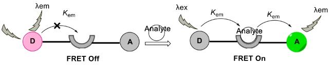
2.1 Fluorescence resonance energy transfer
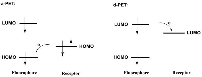
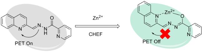
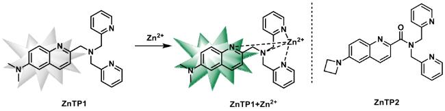
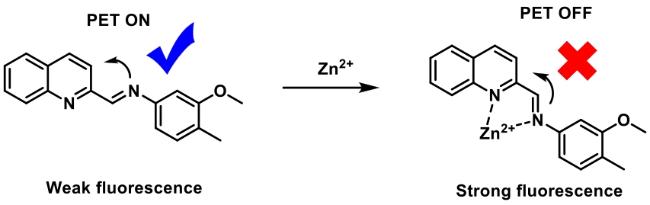
2.2 Photoinduced electron transfer
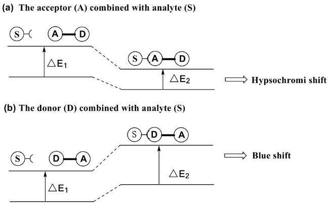
2.3 Intramolecular charge transfer
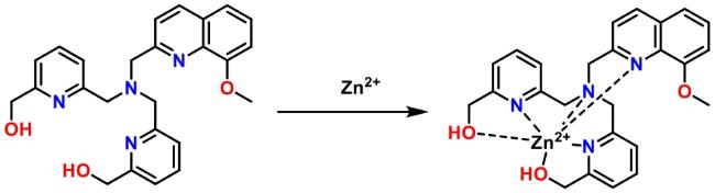
2.4 Chelation enhanced fluorescence
3 Progress of fluorescent probes based on quinoline derivatives in ion detection
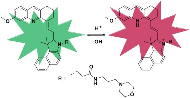
3.1 Fluorescent probes for H+ detection
3.2 Fluorescent probes for Zn2+ detection
3.3 Fluorescent probes for Cd2+ detection
3.4 Fluorescent probes for Cu+/Cu2+ detection
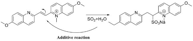
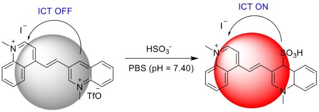
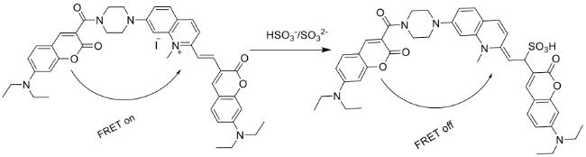
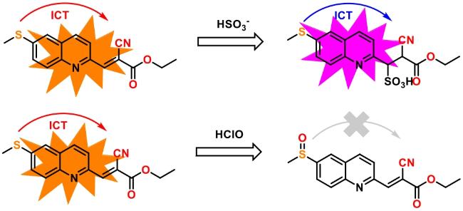
3.5 Fluorescent probes for the detection of SO2, HSO3-, SO32-
4 Advances in fluorescent probes based on quinoline derivatives for small molecule detection
4.1 Fluorescent probes for the detection of small molecules of reactive oxygen species
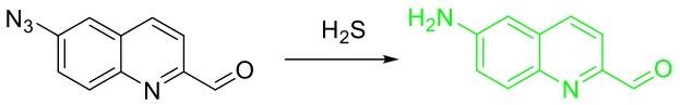
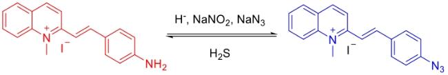
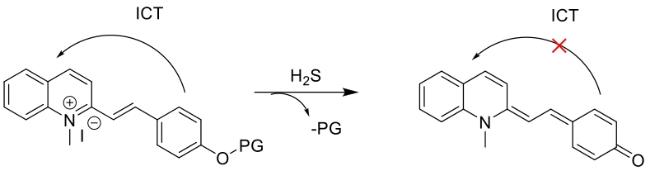
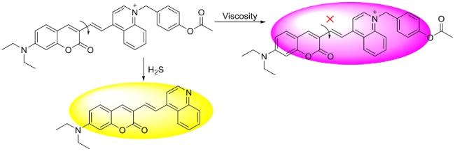
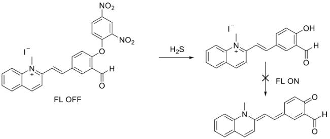
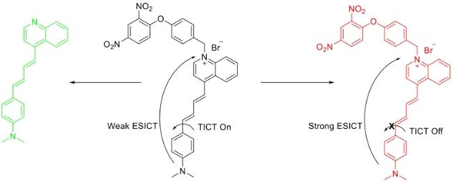
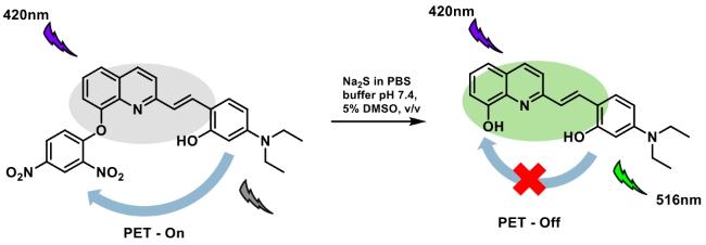
4.2 Fluorescent probes for the detection of H2S
5 Conclusion and outlook
Xu Tang , Liang Jiang , Shuguang Zhang , Xiaoyun Chen . Quinoline-Based Fluorescent Probes in the Detection of Ions and Small Moleculars[J]. Progress in Chemistry, 2025 , 37(10) : 1438 -1455 . DOI: 10.7536/PC20250204
表1 离子荧光探针相关参数Table 1 Related parameters of ions fluorescent probes |
| Probe | Analyte | λex/λem (nm) | Type of sensing | Response mechanism | Response time | Detection limit |
|---|---|---|---|---|---|---|
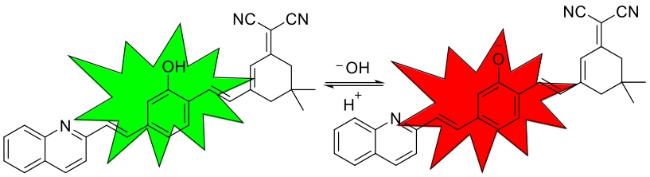
| Q1 | H+、OH- | 580/670、480/610 | Ratiometric | Conjugated effect | Extremely fast | - |
| Q2 | H+、OH- | 430/626、514/734 | Ratiometric | ICT | - | - |
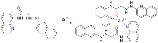
| Q3 | Zn2+ | 400/570 | Turn-on | CHEF | - | 0.66 µmol/L |
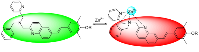
| Q4 | Zn2+ | 750/515 | Ratiometric | ICT | <2 s | 25 nmol/L |
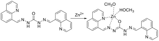
| Q6 | Zn2+ | 340/505 | Turn-on | CHEF | - | 0.07 µmol/L |
| Q7 | Zn2+ | 375/484 | Off-on | CHEF、PET | <3 s | 72 nmol/L |
| Q8 | Zn2+ | 317/445 | Turn-on | CHEF、PET | - | 154 nmol/L |
| Q9 | Zn2+ | 370/571 | Turn-on | CHEF、PET | - | 0.104 µmol/L |
| Q10 | Zn2+ | 405/550 | Turn-on | CHEF | 5 min | - |
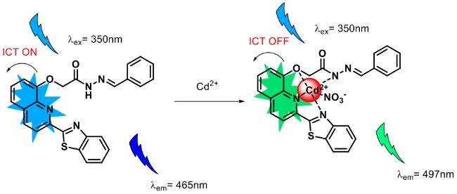
| Q11 | Cd2+ | 360/507 | Turn-on | ICT、CHEF | - | 3.52 nmol/L |
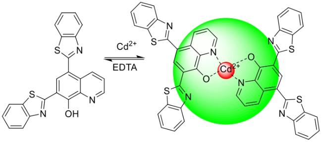
| Q12 | Cd2+ | 313/525 | Turn-on | CHEF | - | 0.1 µmol/L |
| Q13 | Zn2+、Cd2+ | 300/520、300/480 | Turn-on | CHEF、PET | - | 69 nmol/L |
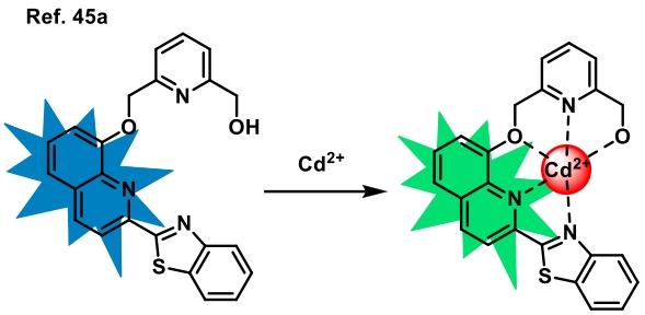
| Q14 | Cd2+ | 350/497 | Turn-on | ICT、CHEF | 10 s | 0.14, 0.29 µmol/L |
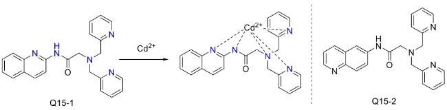
| Q15 | Zn2+、Cd2+ | -/350、-/380 | Turn-on | PET、tautomerism | - | 0.15, 0.13 µmol/L |
| Q16 | Cu+ | 410/530 | Turn-on | ICT | - | 1.03 µmol/L |
| Q17 | Cu2+ | 449/505 | Off-on | PET | - | 0.06 µmol/L |
| Q18 | Cu2+、S2- | 450/521 | Off-on | PET | <1 min | 15.2 nmol/L、15 µmol/L |
| Q19 | Cu2+ | 340/467 | Turn-off | AIE | - | 28.1 nmol/L |
| Q20 | Cu2+ | 365/430、516 | Ratiometric | ICT | <1 min | 87 nmol/L |
| Q21 | Cu2+、S2- | 450/515 | Off-on | PET | - | 0.289, 0.01 µmol/L |
| Q22 | Cu2+、S2- | 367/545 | Off-on | PET | - | - |
| Q23 | SO2(HSO3-) | 330/570、330/445 | Ratiometric | Addition、 Conjugated effect | 90 s | 0.29 µmol/L |
| Q24 | HSO3-、SO32- | 550/620 | Turn-on | Addition、 Conjugated effect | <15 s | 0.11 µmol/L |
| Q25 | HSO3-、SO32- | 395/655、395/490 | Ratiometric | FRET、Addition | 50 min | 0.1 µmol/L |
| Q26 | HSO3-、SO32- | 493/710 | Turn-on | ICT、Addition | 50 s | 72 nmol/L |
| Q27 | HSO3-、HClO | 365/565、365/420 | Ratiometric | ICT、Addition | 2.5 min | 12.6, 56.8 nmol/L |
表2 小分子荧光探针相关参数Table 2 Related parameters of small molecule fluorescent probes |
| Probe | Analyte | λex/λem (nm) | Type of sensing | Response mechanism | Response time | Detection limit |
|---|---|---|---|---|---|---|
| Q28 | H2O2 | 440/700 | Turn-on | ICT | 40 min | 3 nmol/L |
| Q29 | H2O2 | 550/700 | Turn-on | ICT | 30 min | 0.85 µmol/L |
| Q30 | H2O2 | 405/580、405/464 | Ratiometric | ICT | - | - |
| Q31 | H2O2 | 725/772 | Turn-on | ICT | 5 min | 0.17 µmol/L |
| Q32 | H2O2 | 430/617 | Turn-on | ICT | 18 min | 35.5 nmol/L |
| Q33 | H2O2 | 394/550 | Turn-off | ICT | 40 min | 1.447 µmol/L |
| Q34 | H2O2 | 360/420、360/505 | Ratiometric | ICT | 60 min | - |
| Q35 | H2O2 | 440/570 | Turn-on | ICT | 40 min | 13 nmol/L |
| Q36 | H2O2 | 300/450、300/366 | Ratiometric | ICT | 30 min | - |
| Q37 | Cu2+、ClO- | 401/490、401/460 | Off-on | LMCT、Oxidation | 2 s | 0.57 µmol/L |
| Q38 | HClO | 450/550 | Turn-off | ICT | 25 s | 6.5 nmol/L |
| Q39 | HClO | 360/470 | Turn-on | AIE | 30 min | 89.25 nmol/L |
| Q40 | H2S | 335/533 | Turn-on | ICT | 3.4 min | 0.08 µmol/L |
| Q41 | H2S | 385/525、521/605 | Ratiometric | ICT | 60 min | 0.1 µmol/L |
| Q42 | H2S | 380/490 | Turn-off | ICT | 15 min | 54 nmol/L |
| Q43 | H2S | 450/570 | Turn-on | ICT | 9 min | 5.7 µmol/L |
| Q44 | H2S | 400/544 | Turn-on | ICT | 150 s | 15 nmol/L |
| Q45 | H2S | 400/634 | Turn-on | ICT | - | 0.295 pmol/L |
| Q46 | H2S | 420/516 | Turn-on | PET | 30 min | 269 nmol/L |
| [1] |
|
| [2] |
|
| [3] |
|
| [4] |
|
| [5] |
|
| [6] |
|
| [7] |
|
| [8] |
|
| [9] |
|
| [10] |
|
| [11] |
|
| [12] |
|
| [13] |
|
| [14] |
|
| [15] |
|
| [16] |
|
| [17] |
|
| [18] |
|
| [19] |
|
| [20] |
|
| [21] |
|
| [22] |
|
| [23] |
|
| [24] |
|
| [25] |
|
| [26] |
|
| [27] |
|
| [28] |
|
| [29] |
|
| [30] |
|
| [31] |
|
| [32] |
a)
b)
|
| [33] |
|
| [34] |
|
| [35] |
|
| [36] |
|
| [37] |
|
| [38] |
|
| [39] |
(王磊. 东华大学硕士论文, 2021).
|
| [40] |
|
| [41] |
|
| [42] |
|
| [43] |
|
| [44] |
|
| [45] |
|
| [46] |
|
| [47] |
|
| [48] |
|
| [49] |
|
| [50] |
|
| [51] |
|
| [52] |
|
| [53] |
|
| [54] |
|
| [55] |
|
| [56] |
|
| [57] |
|
| [58] |
|
| [59] |
|
| [60] |
|
| [61] |
|
| [62] |
|
| [63] |
|
| [64] |
|
| [65] |
|
| [66] |
|
| [67] |
|
| [68] |
|
| [69] |
|
| [70] |
|
| [71] |
|
| [72] |
|
| [73] |
|
| [74] |
|
| [75] |
|
| [76] |
|
| [77] |
|
| [78] |
|
| [79] |
|
| [80] |
|
| [81] |
|
| [82] |
|
| [83] |
|
| [84] |
|
| [85] |
|
| [86] |
|
| [87] |
|
| [88] |
|
| [89] |
|
| [90] |
|
| [91] |
|
| [92] |
|
| [93] |
|
| [94] |
|
| [95] |
|
| [96] |
|
| [97] |
|
| [98] |
|
| [99] |
|
| [100] |
|
| [101] |
(孙晨阳. 江苏科技大学硕士论文, 2022).
|
| [102] |
|
| [103] |
|
| [104] |
|
| [105] |
|
| [106] |
|
| [107] |
|
| [108] |
|
| [109] |
|
| [110] |
|
| [111] |
|
| [112] |
|
| [113] |
|
| [114] |
|
| [115] |
|
| [116] |
|
| [117] |
|
| [118] |
|
| [119] |
|
| [120] |
|
| [121] |
|
| [122] |
|
| [123] |
|
| [124] |
|
| [125] |
|
| [126] |
|
| [127] |
|
| [128] |
|
| [129] |
|
| [130] |
|
| [131] |
|
| [132] |
|
| [133] |
|
| [134] |
|
| [135] |
|
| [136] |
|
| [137] |
|
| [138] |
|
| [139] |
|
| [140] |
|
| [141] |
|
| [142] |
|
/
| 〈 |
|
〉 |
