Home Journals Progress in Chemistry
Progress in Chemistry

Abbreviation (ISO4): Prog Chem      Editor in chief: Jincai ZHAO

About  /  Aim & scope  /  Editorial board  /  Indexed  /  Contact  / 
Review

Saccharide Sensors Based on Phenylboronic Acid Derivatives

  • Tan Shi ,
  • Donghui Kou ,
  • Yanan Xue ,
  • Shufen Zhang ,
  • Wei Ma , *
Expand
  • State Key Laboratory of Fine Chemicals, Frontier Science Center for Smart Materials, Dalian University of Technology, Dalian 116024, China
* e-mail:

Received date: 2023-05-19

  Revised date: 2023-08-26

  Online published: 2023-12-10

Supported by

National Natural Science Foundation of China(22278064)

National Natural Science Foundation of China(21878040)

National Natural Science Foundation of China(22238002)

Fundamental Research Funds for the Central Universities(DUT22LAB610)

Research and Innovation Team Project of Dalian University of Technology(DUT2022TB10)

Abstract

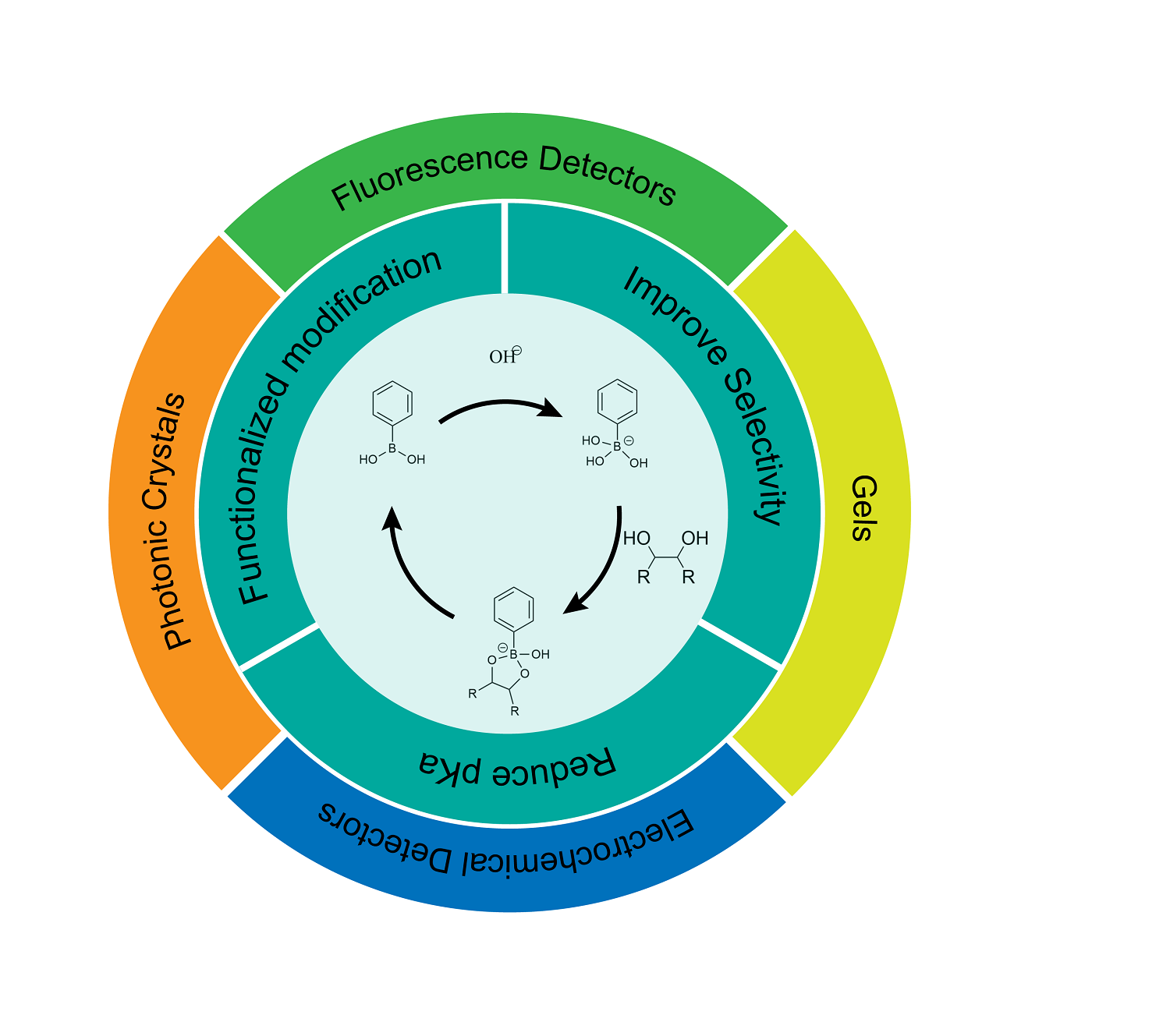
Phenylboronic acid, a kind of synthetic molecule that can covalently bind with saccharide, has attracted wide attention in the field of saccharide detection. It has the characteristics of good stability, strong recognition ability and easy coupling with various detection systems. In this paper, the mechanism of phenylboronic acid binding to saccharide and its specific applications in detection was first introduced. What’s more, the strategies for structural modification, in the manner of introducing electron-withdrawing group or electron-donating group into ortho, meta and para position of the boric acid group on the benzene ring, were mainly discussed, and the progress made in reducing pKa and improving the selectivity according to these strategies were summarized. At the same time, the saccharide sensors based on these new phenylboronic acid derivatives in recent years were also summarized, including electrochemical sensors, fluorescence sensors, gels/microgels and photonic crystals, and their detection principles were discussed. The main analytes are monosaccharides with similar structures, such as glucose and fructose. Finally, the research of these sensors based on phenylboronic acid derivatives was compared, and their advantages and disadvantages were analyzed. Meanwhile, the applications of saccharide sensors based on phenylboronic acid derivatives in the future are prospected from two aspects including the integration of diagnosis and treatment and the identification of saccharide in complex chemical environment.

Contents

1 Introduction

2 Phenylboronic acid and its derivatives

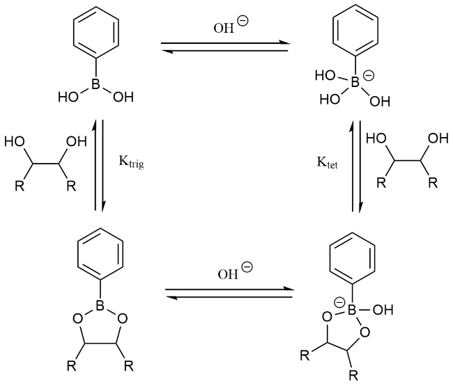
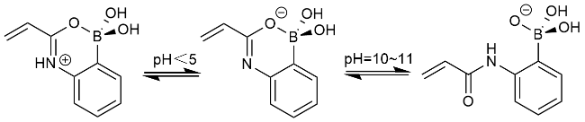
2.1 Reaction principle of phenylboronic acid and saccharides

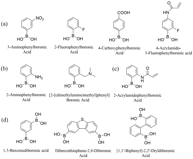
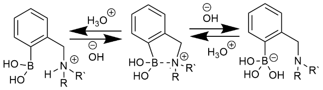
2.2 Structural modification strategy of phenylboronic acid

2.3 Detection principle of saccharides in phenylboronic acid

3 Saccharide sensors based on phenylboronic acid derivatives

3.1 Electrochemical sensors for saccharide detections

3.2 Fluorescent sensors for saccharide detections

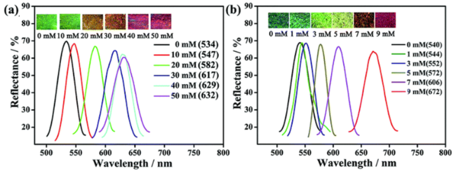
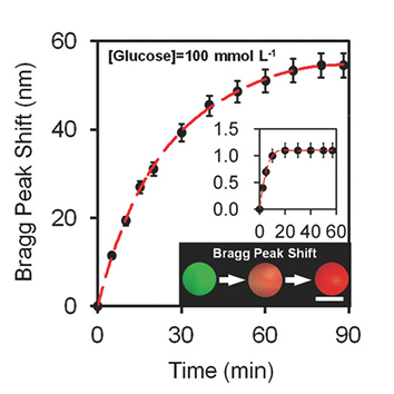
3.3 Photonic crystals for saccharide detections

3.4 Gels for saccharide detections

4 Conclusion and outlook

Cite this article

Tan Shi , Donghui Kou , Yanan Xue , Shufen Zhang , Wei Ma . Saccharide Sensors Based on Phenylboronic Acid Derivatives[J]. Progress in Chemistry, 2024 , 36(1) : 106 -119 . DOI: 10.7536/PC230519

1 Introduction

Carbohydrate is one of the most abundant organic compounds in organisms, and it is also an important condiment and energy supply substance for human beings, which has important physiological significance. For example, glucose is the main metabolic fuel produced by plants to harvest energy from sunlight during photosynthesis[1]; Fructose is the sweetest of all naturally occurring carbohydrates and is widely used commercially in soft drinks, fruit juices, and baked goods[2]. However, studies have shown that the accumulation of sugar in body fluids and excessive sugar intake can cause a variety of chronic diseases, among which diabetes, characterized by chronic hyperglycemia, has become a major killer threatening human health. About 537 million people worldwide have diabetes in 2021, and the number is still increasing, and is expected to increase to 783 million by 2045[3]. The World Health Organization recommends that adults consume no more than 50 G of added sugar per day, preferably less than 25 G, so it is necessary to pay attention to the sugar content in body fluids and food at all times.
Because of the threat of diabetes, most of the research is based on the background of carbohydrate detection in body fluids. Glucose is the most abundant sugar in blood, usually 3.5 ~ 5.5 mM, followed by fructose, which is less than 0.1 mM in normal blood, while the concentration of glucose in tears is one order of magnitude lower than that in blood[4][5]; Fasting blood glucose above 7.0 mM or postprandial blood glucose above 11.1 mM is considered at risk for diabetes. Therefore, glucose is the most important analyte in carbohydrate detection studies, and the concentration range of 0 to 20 mM is considered to be the physiologically relevant concentration range, and the pH range of 7.35 to 7.45 is considered to be the physiologically relevant pH range. In addition, with the development of medical level, there is a large amount of evidence that excessive intake of fructose has adverse effects on visceral metabolism, so fructose is also one of the widely reported analytes[6,7].
At present, the reported carbohydrate sensors mainly include two parts, namely a recognition element and a conversion element. The recognition element refers to the element that interacts with carbohydrate molecules and has a recognition reaction, mainly including enzymes, lectins and phenylboronic acid (PBA) derivatives; The conversion element is to convert the binding of the recognition element and the analyte into a measurable signal, such as fluorescent molecules, building blocks of photonic crystals, etc. For a long time, enzymes, as efficient biocatalysts, have been widely used in the recognition part of carbohydrate detectors, such as glucose oxidase, glucose dehydrogenase and fructose dehydrogenase, which have been used to manufacture electrochemical carbohydrate sensors, and some of them have even been commercialized[8,9][10,11][12,13]. In addition, lectins also play an important role in carbohydrate detection, in which concanavalin A (Con A) combines four Con A molecules to form a tetrameric assembly in the presence of divalent cations.The assembly can be combined with a plurality of glucose molecules, and a plurality of electrochemical carbohydrate sensors, fluorescent carbohydrate sensors, microgel carbohydrate sensors and the like are developed based on the property[14~16]. However, as natural macromolecular compounds, enzymes and lectins can not be recycled in some systems, which makes them have certain limitations, and the characteristics that they are difficult to chemically combine with the conversion element will bring the risk of leakage. In addition, the problems of decreased glucose sensitivity and high price of lectin are not conducive to its application. In 1954, Soboczenski et al. Monitored and proved the formation of esters between aromatic boronic acids and a series of diols. PBA and its derivatives can combine with compounds with 1,2- or 1,3-cis-diol structure, such as glucose and fructose, through covalent bonds, and form relatively stable cyclic esters[17]. This covalent binding has made phenylboronic acid and its derivatives widely used in the recognition of polyhydroxy compounds, and has been coupled with a variety of conversion elements to prepare carbohydrate sensors[22][18~20].
As a new type of carbohydrate recognition compounds, PBA and its derivatives can be combined with various conversion elements through chemical bonds to obtain carbohydrate sensors, and their high stability and durability in physiological environment make them promising for application. In this paper, the recognition principle of PBA, the structural modification strategy of PBA and the application of modified PBA derivatives in carbohydrate detection are summarized. A variety of methods and strategies have been reported to reduce the PBA pKa and improve the detection specificity for the target carbohydrate, and the novel compounds proposed based on these strategies have good carbohydrate binding properties. In this paper, the sensing principle and sensing performance of these derivatives combined with four sensors, including fluorescence, electrochemistry, gel and photonic crystal, are discussed, which provides a platform for the detection of saccharides with high selectivity and low detection limit.

2 Phenylboronic acid and its derivatives

2.1 Principle of reaction between phenylboronic acid and saccharide

The boron atom in the phenylboronic acid structure exists in a sp2 hybrid state, containing an empty orbital, and thus can act as a Lewis acid to accept an extraneous electron[21]. In aqueous solution, the boronic acid group has two morphologies, a neutral planar triangular form and a negatively charged tetrahedral borate form[22,23]. As shown in Fig. 1, there is an equilibrium between the two States of boronic acid, and PBA in the planar trigonal form can accept a pair of electrons from a donor (such as OH)) to form a covalent bond and transform into a sp3 hybridized tetragonal borate anion. At this time, the tetragonal borate anion can first undergo a substitution reaction with the hydroxyl group of the diol compound, and then undergo a ring closure reaction, thereby forming a new B — O bond with the diol structure to form a stable and hydrophilic phenylborate ester.Sugars are a class of organic compounds whose chemical nature is polyhydroxy aldehydes or polyhydroxy ketones and their derivatives, and their binding force is significantly higher than that of other diol structures, so this reaction is often used to detect carbohydrates[24]. Most PBA derivatives have the same carbohydrate response mechanism as PBA, and a few with special response properties will be described in the next section.
图 1 不同形式的苯硼酸在溶液环境中的平衡以及与糖类化合物响应平衡

Fig. 1 Equilibrium of different forms of phenylboronic acid in solution environment and their response equilibria with saccharides

Although both forms of PBA can reversibly react with saccharides to form neutral or charged borate esters, respectively, due to the high ring strain on the boron atom,Neutral borate esters are less stable than anionic ones, and in general the equilibrium constant (Ktet) of anionic tetrahedral borate esters is 2 – 4 orders of magnitude higher than the equilibrium constant (Ktrig) of neutral trigonal ones[25]. In general, neutral borate esters are rapidly converted to neutral boric acid or charged borate esters. The stability of tetrahedral borate esters is mainly affected by the pKa of PBA, the acidity of the diol ligand and the pH of the environment[26,27]. The more stable the anionic tetrahedral borate ester is, the greater the binding force with the diol ligand is, which is more conducive to recognition and detection. The pKa of boronic acid is defined as the pH at which 50% of the boronic acid groups in solution are present as hydroxyborate anions, with optimal binding generally occurring at a pH between the pKa of the phenylboronic acid derivative and the diol ligand[28]. In addition, the binding constant is also affected by the type of solvent, steric hindrance, dihedral angle of the diol and other factors.

2.2 Structural Modification Strategy of Phenylboronic Acid

The pKa of phenylboronic acid is 8. 83, which requires alkaline environment to play its role, so it can not be directly detected in physiological conditions or continuously detected in vivo by embedding and other technical means, which is not conducive to the application of body fluid carbohydrate detection[29][30]. In addition, PBA can bind to a variety of monosaccharides, and the sugar affinity and selectivity of PBA-based sensors are poor, which is not conducive to sugar detection in complex chemical environments. In this regard, a variety of modification strategies to reduce the pKa and improve the selectivity have been reported, and some new PBA derivatives have been obtained. The molecular structures of some common PBA derivatives are shown in Figure 2[31-32].
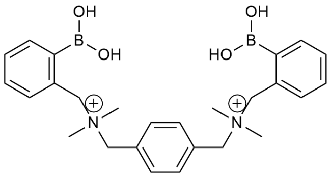
图2 (a)常见吸电子基改性苯硼酸衍生物的分子结构;(b)邻位给电子基改性苯硼酸衍生物的分子结构;(c)2-丙烯酰胺基苯硼酸的分子结构;(d)常见二元苯硼酸衍生物的分子结构

Fig. 2 (a) Molecular structure of common electron- withdrawing modified phenylboronic acid derivatives;(b) molecular structure of ortho-electron-donating modified boronic acid derivatives; (c) molecular structure of 2-acrylamidophenylboronic acid; (d) molecular structure of common dibasic phenylboronic acid derivatives

There are two main ideas to reduce the pKa of PBA: one is to introduce electron-withdrawing groups on the benzene ring, such as nitro, carbonyl and fluorine atoms. The molecular structure of phenylboronic acid derivatives modified by common electron-withdrawing groups is shown in Figure 2a[33][34][35,36]. The geometry of the boronic acid group is closely related to the electron cloud density on the boron atom. When there is an electron-withdrawing group on the benzene ring, the electron cloud density decreases due to the induction effect, the Lewis acidity of the boronic acid group is stronger, the pKa value decreases, and the boric acid group tends to transform into a tetrahedral form. The other is to introduce some electron-donating groups, such as amino, dimethylamino, etc., into the ortho position of the boronic acid group on the benzene ring, and the molecular structure is shown in Figure 2b. The coordination between nitrogen and boron promotes the formation of the tetrahedral boronic acid group, thereby reducing the apparent pKa value[37,38]. B Böhnke et al. Found that due to the formation of the B-N coordination bond, the boron center moves to the sp3 hybridization and can form more stable borate esters with saccharides[39]. At neutral pH, the deprotonated nitrogen coordinates to the boron center to form a tetrahedral boronic acid that can complex with the saccharide diol. At lower pH values, the amine is protonated and the boron atom moves toward the trigonal planar sp2 hybridization. At higher pH values, boric acid and hydroxide ions form boric acid centers and release nitrogen, as shown in Figure 3. This boric acid with intramolecular four-coordinate B — N or B — O bonds is called Wulff boronic acid. Similarly, due to the interaction between boron and oxygen, the introduction of a carbonyl group at the ortho position of boron gives a modified Wulff boronic acid, that is, a boronic acid with an intramolecular three-coordinate B — O bond, which favors the formation of boronic esters over almost the entire pH range[40,41][42].
图3 硼酸基团邻位给电子基官能化的苯硼酸在溶液环境中的电离平衡[39]

Fig. 3 Ionization equilibria of electron-donating group functionalized phenylboronic acid in solution[39]

Also based on coordination interactions, the apparent pKa of PBA can also be reduced by introducing an amide group at the ortho position of the boronic acid group on the benzene ring, as shown in Figure 1C[33]. Lowe et al. Proved that various forms of 2-acrylamidophenylboronic acid (2-AAPBA) intermediates were produced in solution due to the proximity effect of the amide group to the boronic acid using 1H-NMR, B-NMR, C-NMR, FT-IR and MS.As shown in Fig. 4, both negatively charged tetrahedral boronic acid intermediates can be condensed with saccharides to form a new B-O bond with the diol structure, resulting in stable and hydrophilic phenylboronic acid esters, which means that they can be detected even under weak acidic conditions (pH = 5 – 7)[40]. At physiological pH, 2-AAPBA exists predominantly as a zwitterionic tetrahedron. FTIR data show that the tetrahedral boron geometry of 2-AAPBA remains unchanged relative to that of 3-acrylamidophenylboronic acid (3-AAPBA) even in a non-solution environment.
图4 2-丙烯酰胺基PBA在溶液环境中的电离平衡

Fig. 4 Ionization equilibrium of 2-acrylamidophenylboronic acid in solution

The orientation and relative position of the hydroxyl group in the diol compound are the key factors affecting the bonding strength between the diol compound and boric acid. The formation of borate esters requires the combination of diols with homo/near-planar configuration with boric acid, so the binding constants of PBA with saccharides are usually higher than those of other diols, while the content of "envelope" configuration with homo-planar cis-dihydroxyl in D-fructose is higher than that of other saccharides.Therefore, the binding force of d-fructose to most aryl monoboronic acids is usually stronger, the binding constant of PBA to fructose is about 4370 M−1, while the binding constant to glucose is 110 M−1, and the binding force of phenylboronic acid to monosaccharides generally follows the order of fructose > galactose > mannose > glucose[43]. In order to achieve selective recognition of other saccharides, substituents can be introduced to the benzene ring. Hoeg-Jensen et al. Found that ortho-methyl-substituted phenylboronic acid preferred to selectively bind to D-glucose rather than D-fructose in physiological buffer[44][45].
In order to solve the problems of poor selectivity of monoboronic acid and poor binding force with saccharides, some researchers have designed binary phenylboronic acid derivatives, that is, there are two boronic acid sites that can bind to diols at the same time in one molecule. The molecular structure of common binary phenylboronic acid derivatives is shown in Figure 2D. By changing the position of the boronic acid group or the length of the spacer group of these binary PBA derivatives to regulate their binding force to different sugars, and based on the multivalent effect, the binding constants of these diboronic acids to sugars are generally higher[46]. Glucose in the form of α-furanose is a special saccharide compound that can bind two boronic acid moieties through two pairs of binding sites at its 1,2- and 3,5,6-positions (1:2 binding), and this binding is driven by different thermodynamic energies from the 1:1 binding (binding of one boronic acid moiety through only one pair of binding sites)[25][47]. Selective recognition of glucose is thus achieved when two boronic acid groups are designed to bind simultaneously to two pairs of cis-diols (1,2- and 5,6-cis-diols of glucose) in a synergistic manner; For example, Bazan et al. Developed a new dicationic diboronic acid structure, as shown in Figure 5, which has good glucose affinity and selectivity[48]. At physiological pH, 0 ~ 30 mM glucose can be recognized by conductivity monitoring, and interference from other sugars (such as maltose, fructose, sucrose, lactose, and galactose) is negligible. Based on this idea, polymers containing multiple phenylboronic acid groups can also replace single molecules with diboronic acid to achieve similar effects. For example, Zhang et al. Prepared a glucose-selective poly (N-isopropylacrylamide-2-acrylamidophenylboronic acid) (P (NIPAM-2-AAPBA)) microgel.It can form glucose-binary borate ester complexes, and the gel particle size decreases with increasing glucose concentration due to further crosslinking within the polymer network[49].
图5 双阳离子二硼酸分子(DBA2+)的分子结构[48]

Fig. 5 Molecular structure of diboronic acid (DBA2+)[48]

In addition to the above modifications based on detection performance, there are some functionalized modifications so that PBA derivatives can be better combined with various detection systems. The first step is to introduce functional groups on the benzene ring for chemical bonding, such as carbon-carbon double bonds, amino and carboxyl groups, so that phenylboronic acid can be introduced into the polymer system. In addition, there are some modification strategies such as fluorophore modification, redox modification, diol complexation (such as dopamine, catechol diol) and so on.

3 Carbohydrate Sensor Based on Phenylboronic Acid Derivatives

Phenylboronic acid derivatives can be bonded to the sensing device through the functional group on the benzene ring, thus realizing the coupling with most sensing methods. Some studies have prepared electrochemical sensors based on PBA derivatives by redox modification of phenylboronic acid or direct bonding on the electrode surface[50,51]; There are also different types of fluorescent sensors obtained by combining PBA with a fluorescent group[52]. In addition, researchers have integrated PBA derivatives with carbon-carbon double bonds and other functional monomers into the building blocks of gels/microgels or photonic crystals to achieve carbohydrate sensing[53,54][55].

3.1 Lectrochemical sensor for saccharide detection

The electrochemical reaction and the change of electrochemical properties can be caused by the electron transfer and the increase of charged substances when phenylboronic acid is combined with diols. Based on this, the electrochemical parameters such as current and potential are used as the detection basis to provide a theoretical basis for the electrochemical detection of sugars[56]. PBA derivatives have been used to construct various types of electrochemical sensors, mainly based on redox-active electrochemical probes and various PBA modified electrodes[57,58][59,60].
PBA is an electrochemically inert compound, which needs to be modified before it can be used as a redox-active probe for electrochemical sensing. Zheng et al. Used 2-fluorophenylboronic acid and dopamine (DA) as probe sets to study the electrochemical properties of DA and its binding with 2-fluorophenylboronic acid in phosphate buffers of different pH values by cyclic voltammetry[61]. Fructose displaces DA in combination with 2-fluorophenylboronic acid after the addition of fructose to the solution, resulting in the release of DA. The regenerative oxidation current of DA increased with increasing fructose concentration. The peak current was linear with fructose concentration in the range of 0.3 – 5.0 mM, and the detection limit was 0.1 mM. Hayashita et al. Detected fructose using a supramolecular complex formed between β-cyclodextrin (β-CDs) and PBA chemically modified ferrocene (4-Fc-PBA)[62]. 4-Fc-PB is the only molecule with axial orientation in the CDs cavity, as shown in fig. 6; Referring to the calibration curve associated with the oxidation peak, the detection limit was (0.43 ± 0.05) mM, while the detection limit associated with the reduction peak was (1.14 ± 0.14) mM. Sakata et al. Combined poly (3,4-ethylenedioxythiophene) with poly (styrenesulfonate) (PEDOT: PSS) with a second polyacrylamide network containing PBA to obtain a channel of organic electrochemical transistors (OECTs) with high conductivity[63]. The OECT channel can directly detect and amplify the electrochemical signal reflecting the change of glucose concentration, and has a sensitive response to 2-15 mM glucose.
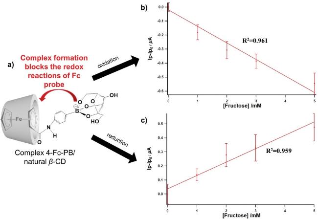
图6 氧化还原改性后的4-二茂铁-苯硼酸(4-Fc-PBA)/天然β-环糊精(β-CDs)复合物(a)检测果糖的机理;(b)基于氧化峰和(c) 基于还原峰的校准线(果糖浓度高达5 mM)[62]

Fig. 6 (a) Mechanism of fructose detection by 4-Ferrocene- phenylboronicacid (4-Fc-PBA)/natural β-cyclodextrins (β-CDs) calibration lines based on (b) oxidation peaks and on (c) reduction peaks (fructose concentration up to 5 mM)[62]

In addition, PBA-modified electrodes have also been used to construct carbohydrate sensors. On the one hand, the change of electrode surface potential can be used as an output signal to detect carbohydrates by coating PBA-containing polymers on the electrode. Hosoyamada et al. Developed an electrochemical glucose sensor by constructing 150 nm-thick phenylboronic acid-modified mesoporous silica (PBA-MPSi) on a Ta2O5 electrode[64]. The binding of glucose to PBA will change the surface charge density of MPSi, thereby changing the Ta2O5 electrode potential, which is measured by a detector. Examined in human serum solutions from 0 to 20 mM glucose, a surface potential shift of 16 mV was observed at a low glucose level of 2.8 mM and 48 mV at a glucose level of 22.8 mM, as shown in Figure 7. On the other hand, PBA can also be used to modify the electrode surface to prepare voltammetric carbohydrate sensors, which are expected to achieve low detection limit and high signal intensity of carbohydrate detection. Liu et al. Synthesized 3,4-ethylenedioxythiophene-fluorophenylboronic acid (EDOT-FPBA) monomer, which was Electropolymerized on a glassy carbon electrode (GCE) at a constant voltage to obtain poly (EDOT-FPBA)/GCE[65]. Glucose can be detected in the range of 0.05 ~ 25 mM by electrochemical impedance spectroscopy, and the detection limit is as low as 0.05 mM at pH = 7.0. Fan et al. Developed a poly (3-aminophenylboronic acid) -reduced graphene oxide (PAPBA-RGO) modified electrode to detect fructose and xylitol[66]. When the electrode was immersed in fructose or xylitol solution, the boric acid group of PAPBA was complexed with fructose or xylitol, and the steric effect was produced on the electrode, so that the ferricyanide in the solution could not enter the electrode. With the increase of the concentration of fructose or xylitol, the peak current of ferricyanide decreased. The response range of fructose and xylitol was 1×10⁻¹²~1×10⁻²M, and the detection limits of fructose and xylitol were 1×10⁻¹²M and 6×10⁻¹³M, respectively.
图7 涂有PBA-MPSi的Ta2O5电极在不同浓度的葡萄糖-人血清溶液中的表面电位(ΔVout)偏移图(根据Langmuir吸附等温线拟合近似曲线)[64]

Fig. 7 Plot of shift in the surface potential (ΔVout) at different concentrations of glucose dissolved in human serum (Approximate curve was fitted by Langmuir adsorption isotherm) [64]

3.2 Carbohydrate detection fluorescent sensor

At present, a variety of fluorescent probes based on different luminescent substances and sensing principles have been used for carbohydrate detection. On the one hand, the competitive reaction of saccharide and fluorescent substance containing dihydric alcohol to PBA probe can be utilized, PBA derivative is used as quencher or de-quencher, and the concentration of saccharide is related to the fluorescence intensity to obtain competitive reaction type fluorescent sensors, including quenching-type competitive reaction type fluorescent sensors and recovery-type competitive reaction type fluorescence sensors[67,68]; On the other hand, the PBA can be directly modified by a fluorophore to obtain a novel carbohydrate-sensitive fluorescent probe, and the change of fluorescence intensity generated when the probe is combined with the carbohydrate is used as a signal reflecting the concentration of the carbohydrate to realize the detection of the carbohydrate[69].
Hydroxyanthraquinone fluorescent compounds are the research focus of quenching competitive reaction fluorescent sensors, among which alizarin red S (ARS) is one of the fluorescent substances used to couple with PBA derivatives for glucose detection, which has been widely used in early research and continues to this day. Wang et al. Found that the active proton of hydroxyanthraquinone would cause its fluorescence quenching, while the binding of phenylboronic acid to the catechol diol of ARS would remove the active proton and eliminate the fluorescence quenching, and when the carbohydrate competitively bound to the phenylboronic acid group, the proton of ARS would quench the fluorescence again[70]. The fluorescence intensity decreased gradually in the fructose concentration range of 0 – 100 mM, and decreased 8-fold at 100 mM, with a detection limit of 0.1 mM. Qi et al. Developed a fluorescent probe for the determination of serum glucose[71]. The probe consists of a multifunctional azlactone polymer as a linker, aminophenylboronic acid, and alizarin red as a signal moiety. The excitation/emission peaks of the polymeric fluorescent probe were located at 468/567 nm. The fluorescence intensity decreased linearly in the glucose concentration range of 0.1 – 14 mM, indicating good glucose discrimination. Li et al. Fabricated a rainbow structural color chip with alizarin red S-diphenylboronic acid-2-aminoethyl ester (ARS-DPBA) as the recognition part based on the enhancement effect of the photonic band gap of photonic crystal (PC) on the fluorescence at the same wavelength[72]. The variable photon stop band of the stretch chromophoric PC in the chip enhances the fluorescence intensity in different degrees in a wide spectral region, amplifies the binding force difference between the phenylboronic acid group and different saccharides, and can distinguish 14 similar structural saccharides such as D-glucose, D-fructose, D-mannose, D-ribose and the like, and the minimum detection concentration is 10−4mM.
In addition, some recovery-type competitive reaction fluorescent sensors have been reported, which reduce the difficulty of synthesis and link the fluorescent moiety to the PBA moiety by a reversible borate bond. Wang et al. Synthesized a novel composite fluorescent probe by assembling polyhydroxy carbon quantum dots (CDs) on PBA-modified magnetic nanoparticles through reversible dynamic covalent bonds[73]. The light-absorbing black magnetic nanoparticles mask the fluorescence of polyhydroxy CD, which is released and the fluorescence is restored in the presence of glucose in solution. As shown in fig. 8, it has good responsiveness over a large concentration range (0 ~ 100 mM), and has a linear range of 0.2 ~ 20.0 mM and a detection limit of 0.15 μM. Shuai et al. Used aminated graphene oxide (GON) modified by PBA and 1,1 '-ferrocenedicarboxylic acid (FCA) as a fluorescence quencher (FCA-GON-BPA), and tetraphenylethylene derivative (TPBD) modified by cis-diol with aggregation-induced emission (AIE) characteristics as a fluorophore. After adding glucose, the fluorophore was replaced by glucose to form a new cyclic borate ester, and the fluorescence of TPBD was restored. The detection range was 5 ~ 100 mg[74].
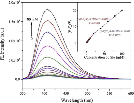
图8 在pH值为8的PBS溶液中,恢复型竞争反应类荧光复合探针(5 mg·mL−1)对不同浓度葡萄糖响应的荧光光谱(插图: 复合探针的(F−F0)/F0对葡萄糖浓度的半对数图)[73]

Fig. 8 Fluorescence response of composite probe (5 mg·mL−1) upon addition of various concentrations of glucose in a pH 8 PBS solution. Inset: semilogarithmic plot of (F−F0)/F0 of composite probe vs the concentration of glucose [73]

Some phenylboronic acid-modified fluorescent probes have also been synthesized, which are usually based on the photoinduced electron transfer (PET) mechanism and the internal charge transfer (ICT) mechanism, but the detection mechanism of some probes is not clear[75][76]. Hibi et al. Synthesized a novel fluorescent probe for fructose detection by combining the phenylboronic acid group with the lipophilic fluorescent dye boronyldipyrromethane (BODIPY)[77]. The probe showed a linear fluorescence response to d-fructose concentration in the range of 100 – 1000 μm with a detection limit of 32 μm. Lee et al. Developed an amphiphilic peptide-based monoboronic acid probe with pyrene fluorophore, which can detect glucose (Glu) in aqueous solution with high selectivity under physiological pH conditions, with a detection limit as low as 3.1 µM and a response time shorter than 10 min[78].
Although this kind of fluorescent probe is difficult to synthesize, it can distinguish carbohydrates through molecular design, which is difficult for other types of sensors to achieve. Some researchers have achieved selective recognition of carbohydrates by compounding PBA derivative molecules containing chromophores in the cavity of cyclodextrin and adjusting the spatial structure between the derivative molecules and the cavity or between the derivative molecules[79]. Hayashita et al. Synthesized an anthracene-type fluorine-containing phenylboronic acid fluorescent carbohydrate probe (4- (anthracene-2-carbamoyl) -3-fluorophenylboronic acid), which can recognize carbohydrates at physiological pH[80]. When the hydrophobic moieties of two probe molecules were immobilized simultaneously within the cavity of γ-cyclodextrin (γ-CyD), the dimer fluorescence increased with the addition of glucose. Based on the splitting mode of the Cotton effect, the complex of two boronic acids and a specific sugar molecule (glucose and galactose) forms a fixed distorted structure, so the complex has a much higher response to glucose and galactose than other saccharides. The research group also tried to prepare a glucose-specific fluorescent sensor by immobilizing a molecule of the probe in the cavity of a supramolecular complex formed by fluorophenylboronic acid and β-cyclodextrin (FPB-β-CyD)[81]. The probe molecule is precisely positioned to react with the fluorophenylboronic acid on the cyclodextrin and two pairs of cis-diols of D-glucose at the same time, and the probe molecule shows strong fluorescence enhancement to D-glucose at 431 nm.There was almost no response to other saccharides (d-fructose, d-galactose, d-mannose, d-ribose, and d-xylose), and the fluorescence intensity increased linearly in the range of d-glucose concentrations from 0 to 5 mM and peaked at about 20 mM.
Suzuki group also reported a series of fluorescent chemosensors based on inclusion complexes of γ-cyclodextrin (γ-CyD) with fluorescent probes, which can specifically recognize D-psicose and D-glucose, respectively[82]. The structures and synthesis processes of two benzoxazole-based probe molecules 1 and 2 are shown in Figure 9a. Substituted carboxyl groups at different positions on the benzene ring of phenylboronic acid are condensed with the amino group of the fluorescent molecule, named 1/γCyD and 2/γCyD, respectively. 1/γCyD is selective for D-psicose, with an equilibrium constant more than 10-fold higher than the rest of D-fructose, D-glucose, and D-galactose. The detection limit of 1/γCyD for d-psicose was 6.9 mM at pH = 8.0. The research group also synthesized probes with heterocyclic boronic acid and fluorophenylboronic acid structures, and the structures of the probes are shown in Figure 9b. By complexing the two molecular probes into the γ-cyclodextrin cavity, a series of selective fluorescent sensors 1F/γ-CyD, 2N/γ-CyD, and 3Ph/γ-CyD were developed, which have higher binding force compared with other saccharides due to the bipolar recognition of the pseudo-diboronic acid moiety on D-glucose[83]. As shown in fig. 10, 1F/γ-CyD responded to D-glucose, D-fructose, D-galactose and L-glucose, and the response to D-glucose was the largest; 2 N/γ-CyD showed a significant fluorescence response almost exclusively to D-glucose. The detection limits of 1F/γ-CyD and 2N/γ-CyD for d-glucose in aqueous solution at pH = 7.4 were 1.1 and 1.8 mM, respectively.
图9 (a)苯并噁唑基复合探针分子1和2的结构和合成示意图[82];(b) 探针分子1F、2N、3Ph的分子结构[83]

Fig. 9 (a) Structures and synthetic procedure of fluorescence probe 1 and 2[82]; (b) structures of fluorescence probe 1F, 2N and 3Ph[83]

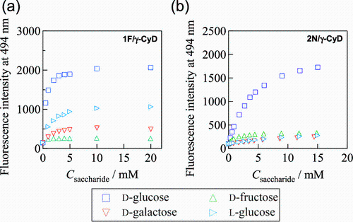
图 10 苯硼酸改性复合荧光探针(a) 1F/γ-CyD和(b) 2N/γ-CyD在不同糖浓度(D-葡萄糖、D-果糖、D-半乳糖和L-葡萄糖)的二甲基亚砜(DMSO)/水中时在494 nm处的荧光强度(1F/γ-CyD和2N/γ-CyD的激发波长分别为328和378 nm)[83]

Fig. 10 Fluorescence intensities at 494 nm of 1F/γ-CyD (a) and 2N/γ-CyD (b) at various concentrations of saccharides (D-glucose, D-fructose, D-galactose, and L-glucose) in DMSO/water (2/98 in v/v) (The excitation wavelengths were set as 328 nm for 1F/γ-CyD and 374 nm for 2N/γ-CyD)[83]

3.3 Carbohydrate detection gel

For a long time, gel/microgel has been widely combined with phenylboronic acid derivatives to achieve carbohydrate sensing. As early as 1994, Sakurai et al reported the preparation of carbohydrate response polymers based on PBA groups. After the phenylboronic acid groups were incorporated into the three-dimensional polymer network,When it combines with saccharide in a ratio of 1:1, it will promote the increase of charged substances and hydrophilicity, and the resulting Donnan potential and enhanced free energy of mixing are the key factors for detection, which will drive the swelling of polymers, and the concentration of saccharide can be judged according to the degree of swelling[84][33][85][86]; When the phenylboronic acid group is combined with glucose at a ratio of 2:1, the degree of crosslinking increases and the polymer segment shrinks; When it is cross-linked with other diol structures (such as dopamine, catechol, etc.), the introduction of saccharides will compete with these diol compounds for boric acid binding sites, and the degree of cross-linking will change with the concentration of saccharides. At present, a variety of gels/microgels with different response characteristics have been developed, including gels/microgels directly doped with PBA monomers into polymer chains, gels constructed by the competitive reaction of carbohydrates and other diols with PBA, and multi-response microgels obtained by copolymerizing a variety of stimulus-responsive functional monomers.The saccharide concentration can be converted into a variety of signal outputs by correlating the saccharide concentration with the parameters of particle size, swelling degree, crosslinking degree, storage modulus and critical solution temperature, respectively.
The gel directly doped with PBA monomer in the polymer chain segment is the most widely used, and the construction is relatively simple. Yun et al. Developed a poly (acrylamide-polyethylene glycol diacrylate-3-acrylamidophenylboronic acid) glucose-responsive hydrogel optical fiber[87]. The glucose response is shown in Fig. 12. The saturation response time of the sensor is about 20 min. When the glucose concentration is gradually increased to 12.0 mM, the hydrogel fiber swells by 6%; With the decrease of glucose concentration, the hydrogel fibers returned to their original diameter size, and the de-complexation time was about 30 min. In order to solve the problem of long response time, researchers have focused their research on nanosized microgels. Wu et al. Obtained a gel named SPBA by crosslinking polymerization of N, N N ´ -bis (propenyl) perylene-3,4,9,10-tetracarboxydiimide and 4-vinylphenylboronic acid[88]. Based on the formation of non-covalent hydrophobic/CH-π interaction between perylene diimide and the axial CH group of glucose and the covalent binding between the PBA group and the cis-diol of glucose, the gel has a good recognition effect on glucose. At physiological pH = 7.4, there is selective swelling to glucose in the concentration range of 0.0 ~ 30.0 mM, as shown in Fig. 11. The initial particle size of the gel is 300 nm, and the particle size is doubled in 30 mM glucose solution. The association constant between gel and glucose is about 212 M−1, which is significantly larger than 78, 42 and 52 M−1 of fructose, galactose and mannose. Reducing the size of microgel can further improve the response speed. Luo et al. Prepared poly (N, N-dimethylacrylamide-acrylamide-acrylamidophenylboronic acid) microgel by emulsion polymerization.The particle size was 59. 28 nm, and then the chitosan (CS) crosslinked by glutaraldehyde (GA) was used to prepare the composite hydrogel containing CS by physically embedding the microgel, which had a rapid swelling response to glucose[89]. In addition, some studies have combined gel with other detection methods to improve the detection sensitivity and reduce the detection limit. Zhu et al. Polymerized sodium alginate (SA, 99%), N, N N ´, N N ´ -tetramethylethylenediamine (TEMED), acrylamide (AAm), N, N-methylenebisacrylamide (MBA) and PBA derivatives to obtain glucose-responsive gel and coated it on the electrode of quartz crystal microbalance (QCM) to convert the mass change of the gel into electrical signal output, with a detection limit of 0.15 mg·L−1 and a linear range of 0.5~120 mg·L−1 for glucose[90].
图11 SPBA微凝胶的(a)SEM图和(b)葡萄糖响应行为(5.0 mM PBS,25.0 ℃)[88]

Fig. 11 (a) SEM images and (b) glucose response behavior of microgels (5.0 mM PBS, 25.0 ℃)[88]

图 12 24 ℃水凝胶纤维在葡萄糖传感(pH=7.4,1.0~12.0 mM, 24 ℃)中的可重复使用性[87]

Fig. 12 Reusability of the hydrogel fibers in sensing glucose(pH=7.4, 1.0~12.0 mM, 24 ℃)[87]

It is also one of the effective methods to use the competitive reaction of carbohydrates and other diols with PBA to construct dynamic covalent bonds for sensing, which is expected to achieve the application of drug controlled release in vivo in the integration of diagnosis and treatment. For example, Wu et al. Prepared injectable polyethylene glycol (PEG) hydrogel by mixing dopamine-functionalized four-arm polyethylene glycol (4-arm-PEG-DA) and phenylboronic acid-modified four-arm polyethylene glycol (4-arm-PEG-PBA) at pH = 9.0 using borate-dopamine complexation[91]. The hydrogel has a ternary response of pH, glucose and dopamine, and the glucose and the dopamine compete for complexation in a glucose solution, so that the crosslinking degree of the hydrogel is reduced, and swelling occurs. Webber et al. Complexed rigid diboronic acid-modified four-arm polyethylene glycol (4aPEG) with polyhydroxylated four-arm polyethylene glycol to obtain dynamic and self-repairing hydrogels[92]. The elastic modulus of the hydrogel decreased significantly with the increase of glucose concentration, and the hydrogel had higher affinity for glucose and lower affinity for other carbohydrates at physiologically relevant concentrations (0 ~ 22 mM). Yeh et al. Developed a novel alginate hydrogel by crosslinking alginate dialdehyde (ADA) with phenylboronic acid-functionalized polyethyleneimine (PBA-PEI) through imine bond and borate ester bond, and introduced two orthogonal dynamic covalent crosslinking networks[93]. The highly sensitive borate ester bonds in the network enable the PBA-PEI/ADA hydrogel to change the degree of crosslinking with binding to glucose, fructose, and in addition to respond to hydrogen peroxide, as shown in Fig. 13.
图13 竞争反应型水凝胶对葡萄糖(0.1 M)、果糖(0.1 M)和H2O2(3%)的响应[93]

Fig. 13 Stimuli-responsiveness of hydrogels against glucose (0.1 M), fructose (0.1 M), and H2O2 (3%)[93]

There are also studies on the regulation and selection of functional monomers to make the polymer have temperature-dependent carbohydrate response performance, and based on this, carbohydrate detection gels/microgels with temperature-sensitive characteristics are obtained. Wu et al. Copolymerized 3-acrylamidophenylboronic acid with crosslinking agents polyethylene glycol dimethacrylate (PEGDMA), 2- (2-methoxyethoxy) ethyl methacrylate, oligo (ethylene glycol) methyl ether methacrylate, and fluorescent moieties (DAEAM) to obtain a microgel core, and then moderately grew a crosslinked polyacrylamide gel layer on the surface[94]. PEGDMA contains a short polyethylene glycol (PEG) side chain, which endows the microgel with the characteristics of cation localization and temperature response; On the one hand, the microgel can position cations near the glucose-diborate to form an ion pair, and stabilize the glucose-diborate anion complex by minimizing the electrostatic repulsion between the anions and the borate; On the other hand, the microgel can change its volume with temperature, thus realizing the absorption and elimination of internal cations. In this design, the gel shows a switchable behavior with increasing glucose concentration, with more cations being squeezed out above the critical solution temperature, the gel tends to swell by forming glucose-monoborate anion complexes, and shrink by forming glucose-diborate anion complexes below the critical solution temperature. The change of particle size is shown in Fig. 14. When the temperature is lower than 29.0 ℃, the particle size shrinks to 1/2 of the initial particle size in 20 mM glucose solution at 25.0 ℃; When the temperature is between 29.0 and 33.0 ℃, there is no reaction; When the temperature is above 33.0 ℃, the particle size in 20 mM glucose solution at 37 ℃ is doubled. Zhang et al. Synthesized a saccharide (including glucose, mannose, galactose, and fructose) -sensitive poly (N-isopropylacrylamide-co-2-acrylamidophenylboronic acid) (P (NIPAM-co-2-AAPBA)), which responded by lowering the lower critical solution temperature (LCST) of the polymer rather than by changing the degree of ionization[95]. Taking the microgel with 15 mol% AAPBA as an example, at lower glucose concentrations (0 ~ 100 mM), glucose binds to the PBA groups on the polymer chain, the molecular weight of the microgel increases, and the LCST decreases from 33 ℃ to 29 ℃; At higher concentrations (100 ~ 1000 mM), glucose acts as an additive to inhibit the LCST of the polymer, reducing the LCST from 29 ℃ to 20 ℃.
图14 微凝胶在不同浓度葡萄糖的pH = 7.4 PBS溶液中的<Dh>[Glu]/<Dh>0.0 mM与温度的关系[94]

Fig. 14 Glucose-dependent <Dh>[Glu]/<Dh>0.0 mM of microgels as a function of the solution temperature, all measurements were made in PBS of pH = 7.4[94]

3.4 Carbohydrate detection photonic crystal

Photonic crystals (PCs) are periodic micro-nano structures composed of high and low refractive index materials with Photonic band gaps, which can prevent the propagation of light with specific wavelengths. When the photonic band gap falls within the visible range, it will produce visible structural colors. By integrating PBA into the building unit of the photonic crystal, the photonic crystal is endowed with the ability to change the lattice constant and refractive index with the change of glucose concentration, so the glucose sensing PCs can change color in a wide visible spectral range, thus realizing the naked eye recognition and detection of glucose concentration[96,97][98]. At present, the main photonic crystals studied include three-dimensional photonic crystals based on opal and inverse opal, novel 2DPC gratings, one-dimensional Bragg stacks, PC probes, and lyotropic liquid crystal PCs.
The earliest photonic crystal used for saccharide detection is three-dimensional photonic crystal based on opal structure, which usually has high color saturation. For example, Asher et al. Embedded polystyrene (PS) microsphere array into the polymer network of phenylboronic acid hydrogel to obtain a colloidal crystal array (PCC)[99]. In the glucose concentration range of 0 ~ 20 mM, the distance between PS microspheres increased with the increase of hydrogel swelling, the photonic band gap shifted to red, the structural color changed from blue to red, and the detection limit was as low as 10 μmol/L. Zhu et al. Designed and prepared a novel sensor for detecting glucose concentration in tears by depositing colloidal crystal arrays composed of three-dimensional polystyrene (PS) microspheres and 4-formylphenylboronic acid modified polyvinyl alcohol (PVA) hydrogel matrix on polymethylmethacrylate contact lenses.The initial state is green. At low concentrations of 0 ~ 3 mM, one glucose molecule binds to two phenylboronic acid sites simultaneously, and the structural color shifts to blue. For glucose concentrations of 3 ~ 50 mM, the reflection wavelength of the sensor shifts from 468 nm to 567 nm[100]. Gao et al. Obtained dispersed poly (methyl methacrylate-N-isopropylacrylamide-acrylamidophenylboronic acid) (P (MMA-NIPAM-AAPBA)) spheres by polymerization, and fabricated three-dimensional photonic crystals by vertical convection self-assembly[101]. When phenylboronic acid is combined with glucose, the hydrophilicity of the polymer is enhanced, the polymer is swollen, and the photonic band gap is red-shifted due to the expansion of the polymer sphere. The PC sensor can be used to detect glucose from 3 to 20 mM with a response time of several minutes. The diffraction wavelength of PC shifts from 497 nm to 572 nm, and the color changes from bright blue to emerald green. The digital photo is shown in Figure 15. Gu et al. Obtained glucose-responsive photonic crystal (GCC) by constructing SiO2 colloidal crystal arrays in fluorophenylboronic acid (FPBA) matrix. When the glucose concentration increased from 100 mg·dL-1 to 400 mg·dL-1, the reflection peak of GCC red-shifted from 443 nm to 570 nm, the color changed from purple to green, and the response time was less than 5 min[102]. Subsequently, the photonic crystal was assembled on a hard microneedle array, so that it could be embedded under the skin for continuous monitoring.
图15 基于蛋白石结构的P(MMA-NIPAM-AAPBA)三维光子晶体传感器在(a) 0 mM和(b) 20 mM葡萄糖浓度下的数码照片[101]

Fig. 15 Digital photos of P(MMA-NIPAM-AAPBA) three-dimensional photonic crystal sensor based on opal structure at (a) 0 mM and (b) 20 mM glucose concentration[101]

However, these photonic crystals based on opal and colloidal crystal arrays have no pores or small pores, which affect the mass transfer rate. Therefore, based on the improvement of response speed, some glucose-responsive photonic crystals with three-dimensional large pore structure have also been developed. Zhao et al. Used 3-acrylamidophenylboronic acid (AAPBA) as a functional monomer and ethylene glycol dimethacrylate (EGDMA) as a crosslinking agent to prepare an inverse opal hydrogel sensor with a pore size of about 120 nm by a sacrificial template method[103]. The optimal pH of the sensor is 9, the response time is less than 5 min, and there is no obvious signal drift in 10 cycles of response experiments, and the concentration range of phenylboronic acid can be matched by adjusting the amount of phenylboronic acid. As shown in fig. 16, when 0.26 mol of AAPBA was added during polymerization, the reflection wavelengths of 526, 582, 617, and 639 nm corresponded to glucose concentrations of 0, 10, 20, 30, and 40 mM, respectively; When the amount of AAPBA increased to 0. 52 mol, the reflection wavelength of 540 ~ 672 nm corresponded to the glucose concentration of 0 ~ 9 mM, respectively. Zhang Pingping et al. Developed a wearable and portable microneedle photonic crystal sensor[104]. Polyacrylamide gel was first prepared by the sacrificial template method, and then hydrolyzed with NaOH and functionalized with phenylboronic acid to obtain glucose-reactive hydrogel film. In 0 ~ 20 mM glucose solution, the reflection wavelength increased from 533 nm to 582 nm. The microneedle array was constructed on the membrane using PDMS. The microneedles were attached to the skin of pigs treated with glucose for measurement, and the color changed significantly after about 35 minutes, and the wavelength corresponding to the reading color was consistent with the results of commercial blood glucose meter, which is expected to achieve in situ blood glucose detection on the body surface.
图16 加入(a) 0.26 mol (b) 0.52 mol AAPBA制备的反蛋白石传感器随葡萄糖浓度增大而红移的反射光谱和照片[103]

Fig. 16 Reflectance spectrum and photos of inverse opal sensor prepared by adding (a) 0.26 mol (b) 0.52 mol of AAPBA with the increase of glucose concentration[103]

In addition, some new 2DPC gratings, one-dimensional Bragg stacks, PC probes and lyotropic liquid crystal PCs have been developed as carbohydrate sensors, which provide the possibility for carbohydrate detection in different application scenarios. Wu et al. Embedded PS microspheres in a modified acrylamide hydrogel cross-linked with phenylboronic acid and dopamine groups to obtain glucose detection 2DPC[105]. The hydrogel was illuminated vertically with monochromatic light, and a halo (Debye diffraction ring) appeared below the hydrogel. The high bonding ability between sugar diol and phenylboronic acid enables sugar to replace dopamine to form borate ester, which leads to the expansion of hydrogel sensor and the contraction of Debye diffraction ring, thus realizing the response of glucose and related sugars. Yetisen et al. Engraved phenylboronic acid-functionalized silver bromide nanocrystals with a multilayer spacing of about 200 nm into a poly (acrylamide-polyethylene glycol diacrylate) hydrogel mold to obtain glucose-sensitive PC[106]. As shown in fig. 17, in the 100.0 mM glucose solution, the color gradually changed from green to red, and the average sensitivity was calculated to be 0.2 mM based on the reflection peak. Guan Jianguo et al. Constructed a 10-nm-thick poly (3-acrylamidophenylborate-N-2-hydroxyethylacrylamide) hydrogel shell on the surface of magnetic nanochains (PNC) by selective concentration polymerization of monomers in dimethyl sulfoxide-water microheterogeneous binary solvent, and obtained glucose-responsive photonic crystals by placing the PNC dispersion in a magnetic field[107]. With the increase of glucose concentration from 0 to 20 mM, the dispersion changes from green to red, the reflection peak shifts by more than 80 nm, the color shows a clear transition at about 7 mM, and the response time is within a few seconds. Park et al. Used small molecular lyotropic liquid crystal and polyacrylic acid to construct an interwoven network (IPN) photonic film, and then modified it with phenylboronic acid to obtain a glucose-responsive array sensor. According to the reflection peak, the detection limit was calculated to be 0.35 mM, the linear range was 1 ~ 12 mM, and the sensor had good interference ability in serum[108]. Teramoto et al. Formed cholesteric lyotropic liquid crystals (CLC) by evaporating an aqueous solution of phenylboronic acid-modified hydroxypropyl cellulose (PBA-HPC), and the resulting film had a structural color based on circularly polarized light[109]. The increase of glucose concentration in solution causes the change of cholesterol helical spacing and structural color. The color of the structural color film obtained from the evaporation of 70 wt% PBA-HPC solution changed from blue to red in the concentration range of 0 – 3 mg/mL glucose.
图17 在100 mM葡萄糖溶液中布拉格堆叠的反射峰随时间的变化(插图显示Bragg堆叠的比色读数以及没有3-APBA的对照实验,比例尺: 2.0 mm)[106]

Fig. 17 Peak shift of the Bragg stacks as a function of time in 100 mM glucose solution. Insets show colorimetric readouts of the Bragg stacks, and the control experiment without 3-APBA. Scale bar: 2.0 mm [106]

4 Conclusion and prospect

In this paper, the reaction principle and detection mechanism of phenylboronic acid with saccharides were summarized, and some new phenylboronic acid derivatives and their research progress in the field of saccharide detection were reviewed. The results show that phenylboronic acid derivatives have good recognition of saccharides, are easy to be integrated into various detection systems as recognition molecules/probes, and have better stability and lower risk of inactivation or leakage compared with other commonly used saccharide recognition molecules. Some carbohydrate detectors based on new PBA derivatives can accurately identify the isomers of different carbohydrates and some monosaccharides, and have broad application prospects.
For the reported carbohydrate sensors based on phenylboronic acid derivatives, fluorescent sensors and electrochemical sensors can achieve lower detection limits and higher sensitivity, even at submicromolar concentrations.It is easier to integrate the phenylboronic acid derivatives modified by complex structures into the sensor, so as to achieve better detection effect, and even to distinguish a variety of different sugars; However, it is difficult to combine with subcutaneous embedding for continuous blood glucose monitoring, and it may cause signal drift in complex solution environment (such as blood, urine, etc.) And it is difficult to play a role. In addition, electrochemical sensors need additional power input, and fluorescence sensors also need additional detection means (such as spectrometers), which are difficult to judge only by naked eyes. However, gel/microgel and photonic crystal have relatively strong anti-interference, and gel/microgel has been widely studied as a carrier of insulin and glucagon, which has the potential to achieve automatic blood glucose management in vivo; The detection of photonic crystals is more intuitive and easy to operate, but it is difficult to achieve high sensitivity and rapid detection at the same time in the current research, and the molecular structure and morphology size of the building unit need to be designed and optimized.
Although important research results have been achieved in the development of new phenylboronic acid derivatives and carbohydrate sensors, the integration of diagnosis and treatment, that is, the coupling of detection and treatment, still needs to be promoted. Clinically, frequent insulin injections make diabetic patients miserable. Patients not only need real-time monitoring of sugars in body fluids, but also desire automatic blood sugar management. Therefore, a non-invasive intelligent electronic skin system that integrates detection and treatment may be an important development direction in the future. In addition, the application of sugar detection in chemical systems with complex components, such as various foods and drinks, needs to be expanded. In order to enhance the taste, businesses usually add a variety of sugar components at the same time. Therefore, sugar detectors with stronger anti-interference effect and the ability to identify different sugars need to be developed. Based on this, it is necessary to synthesize phenylboronic acid derivatives with stronger recognition ability, and the molecular structure of PBA derivatives with high selectivity and high binding force is usually complex.Especially when combined in the polymer chain segment, the steric effect will affect the binding force between saccharides and phenylboronic acid sites, so how to strengthen the function while maintaining the volume of smaller molecules is also a problem to be solved.
[1]
Bunn H F, Higgins P J. Science, 1981, 213(4504): 222.

[2]
Taskinen M, Packard C J, Borén J. Nutrients, 2019, 11(9): 1987.

[3]
Liu Y, Zeng S, Ji W, Yao H, Lin L, Cui H Y, Hélder A, Santos, Pan G. Adv. Sci., 2022, 9(3): 2102466.

[4]
Güemes M, Rahman S A, Hussain K. Arch. Dis. Child., 2016, 101(6): 569.

[5]
Kawasaki T, Akanuma H, Yamanouchi T. Diabetes Care, 2002, 25(2): 353.

[6]
Lim S, Taskinen M R, Borén J. Obes. Rev., 2019, 20(4): 599.

[7]
Muriel P, López-Sánchez P, Ramos-Tovar E. Int. J. Mol. Sci., 2021, 22(13): 6969.

[8]
Soto J, Hughes T, Li Y S. ACS Omega, 2019, 4(19): 18312.

[9]
Bian Z, Qi Y, Xue L, Hu A, Yang H, Chu F. Optik, 2022, 255: 168655.

[10]
Jeon W Y, Kim H H, Choi Y B. Membranes, 2021, 11(6): 384.

[11]
Cohen R, Cohen Y, Mukha D, Yehezkeli O. Electrochim. Acta, 2021, 367: 137477.

[12]
Trivedi U B, Lakshminarayana D, Kothari I L, Patel P B, Panchal C J. Sens. Actuator B-Chem., 2009, 136(1): 45.

[13]
Voitechovič E, Vektarienė A, Vektaris G, Jančienė R, Razumienė J, Gurevičienė V. Electroanalysis, 2020, 32(5): 1005.

[14]
Jafarzadeh S, Bargahi N, Shamloo H B, Soleymani J. RSC Adv., 2022, 12(14): 8492.

[15]
Wei M, Li X, Serpe M J. ACS Appl. Polym. Mater., 2019, 1(3): 519.

[16]
Zhang H, Lu Q, Zuo F, Yuan R, Chen S. Sens. Actuator B-Chem., 2017, 241: 887.

[17]
Kuivila H G, Keough A H, Soboczenski E J. J. Org. Chem., 1954, 19(5): 780.

[18]
Wen X, Liu Y, Liu Q, Chen Z, Hu X, Xu C, Chen H, Xing M, Qu H, Zhang M. Opt. Express, 2022, 30(26): 47541.

[19]
Li M T, Zhang H, Liu M, Dong B. J. Mater. Chem. C, 2017, 5(18): 4400.

[20]
Zhu J, Liu W, Zhang B, Zhou D, Fan X, Wang X, Liu X. Nanomaterials, 2022, 12(17): 3065.

[21]
Wang Z, Li S. University Chemistry, 2020, 35(07): 95.

(王卓, 李朔. 大学化学, 2020, 35(7): 95.).

[22]
Wang C, Lin B, Zhu H, Bi F, Xiao S, Wang L, Gai G, Zhao L. Molecules, 2019, 24(6): 1089.

[23]
Kazunori K, Hiroaki M, Masayuki B, Teruo O, Yasuhisa S. J. Am. Chem. Soc., 1998, 120(48): 12694.

[24]
Duin V M, Peters J A, Kieboom A P G, Bekkum V H. Tetrahedron, 1984, 40(15): 2901.

[25]
Wu X, Li Z, Chen X, John S F, Tony D, Jiang Y. Chem. Soc. Rev., 2013, 42(20): 8032.

[26]
Martínez-Aguirre M A, Medrano F, Ramírez-Rave S, Yatsimirsky A K. J. Phys. Org. Chem., 2022, 35(12): e4425.

[27]
Springsteen G, Wang B H. Tetrahedron, 2002, 58(26): 5291.

[28]
Yan J, Springsteen G, Deeter S, Wang B H. Tetrahedron, 2004, 60(49): 11205.

[29]
Valenzuela S A, Howard J R, Park H M, Darbha S, Anslyn E V. J. Org. Chem., 2022, 87(22): 15071.

[30]
Hoare T, Pelton R. Biomacromolecules, 2008, 9(2): 733.

[31]
Li S, Davis E N, Anderson J, Lin Q, Wang Q. Biomacromolecules, 2009, 10(1): 113.

[32]
Matsumoto A, Ikeda S, Harada A, Kataoka K. Biomacromolecules, 2003, 4(5): 1410.

[33]
Zhang C, Losego M D, Braun P V. Chem. Mater., 2013, 25(15): 3239.

[34]
Zhang Y, Wu M, Dai W, Li Y, Wang X, Tan D, Yang Z, Liu S, Xue L, Lei Y. Nanoscale, 2019, 11(13): 6471.

[35]
GhavamiNejad A, Li J, Lu B, Zhou L, Lam L, Giacca A, Wu X Y. Adv. Mater., 2019, 31(30): 1901051.

[36]
Fu Y, Sun Y, Chen M, Xing W, Xu Y, Qian X, Zhu W. Biomacromolecules, 2022, 23(3): 1251.

[37]
Wang Z, Wang J, Li H, Gu Z. Proc. Natl. Acad. Sci. U. S. A., 2020, 117(47):29512.

[38]
Kim K T, Cornelissen J J, Nolte R J, van Hest J C. J. Am. Chem. Soc., 2009, 131(39): 13908.

[39]
Wulff G, Lauer M, Böhnke H. Angew. Chem.-Int. Edit., 1984, 23(9): 741.

[40]
Yang X, Lee M C, Sartain F, Pan X, Lowe C R. Chem.-Eur. J., 2006, 12(33): 8491.

[41]
Dowlut M, Hall D G. J. Am. Chem. Soc., 2006, 128(13): 4226.

[42]
Brooks W L, Deng C, Sumerlin B S. ACS Omega, 2018, 3(12): 17863.

[43]
Lorand J P, Edwards J O. J. Org. Chem., 1959, 24(6): 769.

[44]
Zhang C, Cano G, Braun P V. Adv. Mater., 2014, 26(32): 5678

[45]
Hansen J S, Christensen J B, Solling T I, Jakobsen P, Hoeg-Jensen T. Tetrahedron, 2011, 67(6): 1334.

[46]
Carlo F, Christoph A. S, Marcus W, Oliver S, Stefan H, Beate K, Jens D, Christina G, Ernst-Walter K, Rainer H. Angew. Chem.-Int. Edit., 2012, 51:10472.

[47]
Maseda M, Miyazaki Y, Takamuku T. J. Mol. Liq., 2021, 341: 117343.

[48]
Wang B, Chou K, Queenan B N, Pennathur S, Bazan G C. Angew. Chem.-Int. Edit., 2019, 58(31): 10612.

[49]
Tang Z, Guan Y, Zhang Y. Polym. Chem., 2014, 5(5): 1782.

[50]
Sęk J P, Kaczmarczyk S, Guńka K, Kowalczyk A, Borys K M, Kasprzak A, Nowicka A M. Dalton Trans., 2021, 50(3): 880.

[51]
Granot E, Tel-Vered R, Lioubashevski O, Willner I. Adv. Funct. Mater., 2008, 18(3): 478.

[52]
Chen L Y, Hwang E, Zhang J. Sensors, 2018, 18(5): 1440.

[53]
Cai B, Luo Y, Guo Q, Zhang X, Wu Z. Carbohydr. Res., 2017, 445: 32.

[54]
Wu J, Bremner D H, Li H, Sun X, Zhu L. Mater. Sci. Eng. C-Mater. Biol. Appl., 2016, 69: 1026.

[55]
Robinson S, Dhanlaksmi N. Photonic Sens., 2017, 7(1): 11.

[56]
Anzai J. Mater Sci Eng C Mater Biol Appl., 2016, 67: 737.

[57]
Murakami H, Akiyoshi H, Wakamatsu T, Sagara T, Nakashima N. Chem. Lett., 2000, 29(8): 940.

[58]
Komkova M A, Valeev R G, Kolyagin Y G, Andreev E A, Beltukov A N, Nikitina V N, Yatsimirsky A K, Karyakin A A, Eliseev A A. Mater. Today Chem., 2022, 26: 101070.

[59]
Takahashi S, Kurosawa S, Anzai J I. Electroanalysis, 2008, 20(7): 816.

[60]
Takahashi S, Anzai J. Bunseki Kagaku, 2007, 56(11): 951.

[61]
Li J, Sun Y, Wei Y, Zheng J. Chin. Chem. Lett., 2013, 24(4): 291.

[62]
Casulli M A, Taurino I, Hashimoto T, Carrara S, Hayashita T. Small, 2020, 16(44): 2003359.

[63]
Tseng A C, Sakata T. ACS Appl. Mater. Interfaces, 2022, 14(21): 24729.

[64]
Kajisa T, Hosoyamada S. Langmuir, 2021, 37(46): 13559.

[65]
Bao W, Hai W, Bao L, Yang F, Liu Y, Goda T, Liu J. Mater. Chem. Front., 2021, 5(20): 7675.

[66]
Wu S, Guo H, Wang L, Xin Y, Cheng Y S, Fan W S. Sens. Actuators B-Chem., 2017, 245: 117.

[67]
Wang K, Zhang R, Yue X, Zhou Z, Bai L, Tong Y, Wang B, Gu D, Wang S, Qiao Y, Liu Q, Xue X, Yin Y, Xi R, Meng M. ACS Sens., 2021, 6(4): 1543.

[68]
Deng M, Song G, Zhong K, Wang Z, Xia X, Tian Y. Sens. Actuators B-Chem., 2022, 352: 131067.

[69]
Yang W, He H, Drueckhammer D G. Angew. Chem. Int. Ed., 2001, 40(9): 1714.

[70]
Springsteen G, Wang B. Chem. Commun., 2001(17): 1608.

[71]
Qiao J, Liu Q, Wu H, Cai H, Qi L. Microchim. Acta, 2019, 186(6): 366.

[72]
Xie X, Zhang Z, Jiang Q, Zheng S, Yun Y, Wu H, Li C, Tian F, Su M, Li F. ACS Nano, 2022, 16(12): 20094.

[73]
Li J, Li X, Weng R, Qiang T, Wang X. Sens. Actuator B-Chem., 2020, 304: 127349.

[74]
Ouyang F, Zhang X, Wang T, Shuai Q. Mater. Today Commun., 2022, 33: 104681.

[75]
Cooper C R, James T D. Chem. Lett., 1998, 27(9): 883.

[76]
Cao H, Diaz D I, Dicesare N, Lakowicz J R, Heagy M D. Org. Lett., 2002, 4(9): 1503.

[77]
Kashiwazaki G, Watanabe R, Nishikawa A, Kawamura K, Kitayama T, Hibi T. RSC Adv., 2022, 12(24): 15083.

[78]
Mehta P K, Neupane L N, Lee K H. Sens. Actuators B-Chem., 2023, 375: 132913.

[79]
Rahali S, Belhocine Y, Allal H, Bouhadiba A, Assaba I M, Seydou M. Struct. Chem., 2022, 33: 195.

[80]
Sugita K, Tsuchido Y, Kasahara C, Casulli M A, Fujiwara S, Hashimoto T, Hayashita T. Front. Chem., 2019, 7: 806.

[81]
Sugita K, Suzuki Y, Tsuchido Y, Fujiwara S, Hashimoto T, Hayashita T. RSC Adv., 2022, 12(31): 20259.

[82]
Suzuki Y, Hashimoto T, Hayashita T. RSC Adv., 2022, 12(19): 12145.

[83]
Suzuki Y, Mizuta Y, Mikagi A, Misawa-Suzuki T, Tsuchido Y, Sugaya T, Hashimoto T, Ema K, Hayashita T. ACS Sens., 2023, 8(1): 218.

[84]
Shiino D, Murata Y, Kataoka K, Koyama Y, Yokoyama M, Okano T, Sakurai Y. Biomaterials, 1994, 15(2): 121.

[85]
Brooks W L, Sumerlin B S. Chem. Rev., 2016, 116(3): 1375.

[86]
Zhang Y, Yang J, Zhang J, Li S W, Zheng L S, Zhang Y L, Meng H P, Zhang X G, Wu Z M. J. Mat. Chem. B, 2020, 8(21): 4627.

[87]
Yetisen A K, Jiang N, Fallahi A, Montelongo Y, Ruiz-Esparza G U, Tamayol A, Zhang Y S, Mahmood I, Yang S A, Kim K S, Butt H, Khademhosseini A, Yun S H. Adv. Mater., 2017, 29(15): 1606380.

[88]
Wu Q, Du X, Chang A, Jiang X, Yan X, Cao X, Farooqic Z H, Wu W. Polym. Chem., 2016, 7(42): 6500.

[89]
Wang J, Yang Z, Zhou C, Qiao C, Yuan F, Liu Q, Luo X X. J. Macromol. Sci. Part B-Phys., 2022, 61(4/5): 557.

[90]
Liu N, Xiang X, Sun M, Li P, Qin H, Liu H, Zhou Y, Wang L, Wu L, Zhu J. Biosens. Bioelectron. X, 2022, 10: 100110.

[91]
Shan M, Gong C, Li B, Wu J. Polym. Chem., 2017, 8(19): 2997.

[92]
Xiang Y, Xian S, Ollier R C, Yu S, Su B, Pramudya I, Webber M J. J. Control. Release, 2022, 348: 601.

[93]
Shen K, Yeh Y Y, Chiu T H, Wang R, Yeh Y C. ACS Biomater. Sci. Eng., 2022, 8(10): 4249.

[94]
Zhou M, Lu F, Jiang X, Wu Q, Changa A, Wu W. Polym. Chem., 2015, 6(48): 8306.

[95]
Wang Q, Fu M, Guan Y, James T D, Zhang Y. Sci. China Chem., 2020, 63(3): 377.

[96]
Chen C, Dong Z G, Shen J H, Chen H W, Zhu Y H, Zhu Z G. ACS Omega, 2018, 3(3): 3211.

[97]
Morandi V, Marabelli F, Amendola V, Meneghetti M, Comoretto D. Adv. Funct. Mater., 2007, 17(15): 2779.

[98]
Elshaarani T, Yu H J, Wang L, Abdin Z U, Ullah R S, Haroon M, Khan R U, Fahad S, Khan A, Nazir A, Usman M, Naveed K U. J. Mat. Chem. B, 2018, 6(23): 3831.

[99]
Alexeev V L, Das S, Finegold D N, Asher S A. Clin. Chem., 2004, 50(12): 2353.

[100]
Ruan J, Chen C, Shen J, Zhao X, Qian S, Zhu Z. Polymers, 2017, 9(4): 125.

[101]
Hong X, Peng Y, Bai J, Ning B, Liu Y, Zhou Z, Gao Z. Small, 2014, 10(7): 1308.

[102]
Zeng Y, Wang J, Wang Z, Chen G, Yu J, Li S, Li Q, Li H, Wen D, Gu Z. Nano Today, 2020, 35: 100984.

[103]
Feng X Q, Xu J, Liu Y X, Zhao W P. J. Mat. Chem. B, 2019, 7(22): 3576.

[104]
Zhang P P, Zhu J C, Zhao B J, Xu S H, Wang L, Luo X L. Chin. J. Anal. Chem., 2022, 50(4): 100054.

[105]
Chen Q, Wei Z, Wang S, Zhou J, Wu Z. Microchimica. Acta, 2021, 188(6): 210.

[106]
Jiang N, Butt H, Montelongo Y, Liu F, Afewerki S, Ying G L, Dai Q, Yun S H, Yetisen A K. Adv. Funct. Mater., 2018, 28(24): 1702715.

[107]
Cai J, Luo W, Pan J, Li G, Pu Y, Si L, Shi G, Shao Y, Ma H, Guan J. Adv. Sci., 2022, 9(9): 2105239.

[108]
Munir S, Hussain S, Park S Y. ACS Appl. Mater. Interfaces, 2019, 11(41): 37434.

[109]
Miyagi K, Takano T, Teramoto Y. J. Appl. Polym. Sci., 2022, 139(41): e52984.

Outlines

/