Home Journals Progress in Chemistry
Progress in Chemistry

Abbreviation (ISO4): Prog Chem      Editor in chief: Jincai ZHAO

About  /  Aim & scope  /  Editorial board  /  Indexed  /  Contact  / 
Review

Crystal Material Construction Based on DNA Nanotechnology

  • Peixin Li ,
  • Mushi Peng ,
  • Xuehui Yan ,
  • Yifan Yu ,
  • Ye Tian , *
Expand
  • College of Engineering and Applied Sciences, State Key Laboratory of Analytical Chemistry for Life Science, Chemistry and Biomedicine Innovation Center, Nanjing University, Nanjing 210023, China
* e-mail:

Received date: 2024-01-17

  Revised date: 2024-02-29

  Online published: 2024-07-01

Supported by

National Natural Science Foundation of Jiangsu Province, China(BK20220124)

National Natural Science Foundation of China(22372077)

National Natural Science Foundation of China(92356304)

State Key Laboratory of Analytical Chemistry for Life Science,Nanjing University(5431ZZXM2301)

Abstract

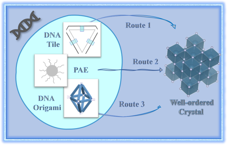
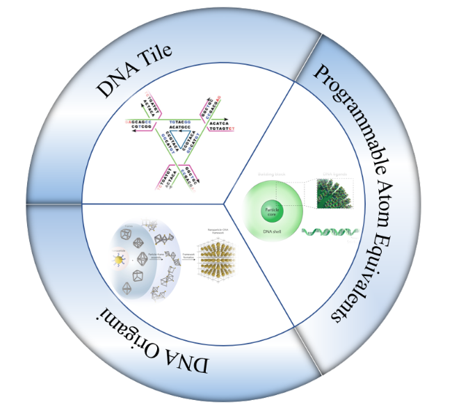
nanocrystal materials have exhibited unique and superior properties in the fields of electronics,optics,and magnetism.the fabrication of nanocrystal materials holds significant importance in the material science for achieving breakthroughs and further developments in their performance.DNA,known for its complementary base pairing characteristics,offers the potential to construct diverse nanostructures and crystals of which the structures and composition can be precisely controlled,thereby enabling the tailored material properties.Currently,the nanocrystal materials fabricated through DNA nanotechnology has been applied in various fields,including catalysts,optical devices,and semiconductor materials.These advancements signify a progressive realization of the fundamental objective for constructing three-dimensional crystals as versatile,periodic molecular frameworks.in this review,we provide a systematic elucidation of the development and recent advancements in the research pertaining to three pivotal techniques employed in the construction of DNA nanocrystals.These techniques include DNA tiles,programmable atomic equivalents,and DNA origami.Moreover,we briefly deliberate on the future direction of utilizing DNA nanotechnology in the construction of nanocrystal materials。

Contents

1 Introduction

2 Construction of nanoparticle crystals based on DNA tiles

3 Direct DNA-mediated nanoparticle crystallization

4 DNA origami-based assembly technique for nanoparticle crystal

5 Conclusion and outlook

Cite this article

Peixin Li , Mushi Peng , Xuehui Yan , Yifan Yu , Ye Tian . Crystal Material Construction Based on DNA Nanotechnology[J]. Progress in Chemistry, 2024 , 36(7) : 949 -960 . DOI: 10.7536/PC240116

1 Introduction

Since human beings began to explore the natural world,crystals have played an important role in all aspects of human production and life.Even in the era of shallow understanding of crystals,people have begun to make rough use of the original properties of crystals in color,hardness and other aspects.It was not until Hauy proposed the periodicity of crystals in 1784 that people realized that crystals should have their unique charm in more physical properties,so they began to study crystals,proposed the structure model of crystals,successfully verified the periodicity of crystals and analyzed its structure.with the development of technology,artificial crystals gradually play an important role in human production and life,and how to construct new crystal materials to achieve functional breakthroughs has become a key issue,among which the crystallization of nanoparticles has attracted special attention from academia.nanoparticles show special properties in electricity,magnetism,optics and so on because of their scale.When nanoparticles are orderly assembled into crystals,they can not only integrate their microscopic properties and show them macroscopically,but also emerge ideal properties beyond their microscopic properties to prepare new materials With various superior properties.the designed nanoparticle crystals can also help us understand the crystallization process and guide the control of material properties.However,although crystals of many substances have been successfully produced,there is still no general and effective method for nano-scale particles,especially some biological macromolecules,to assemble them into desired crystal structures。
DNA nanotechnology is considered to be one of the effective methods to solve the problem of macromolecular crystallization.In 1982,Seeman of New York University first proposed that DNA chains could be self-assembled into three-dimensional frameworks from bottom to top,and pointed out that such DNA frameworks could be used as a general structural template to guide the organization of biological and inorganic nanocomponents into three-dimensional arrays,which was the origin of DNA nanotechnology[1]。 the proposal of DNA nanotechnology is inseparable from the understanding and exploration of DNA structure,which is also closely related to the study of crystals.In the 1950s,Watson and Crick collected microcrystalline DNA fiber diffraction patterns based on Franklin.the discovery of DNA double helix structure opened up a new field of"molecular biology",but the study of nucleic acid crystal structure did not develop rapidly until Caruthers developed a low-cost chemical DNA synthesis method,which greatly reduced the cost of crystallization,and DNA nanotechnology appeared[2]
It has been decades since Seeman proposed DNA nanotechnology,and in that time,several techniques for making DNA crystals have been developed,effectively crystallizing a variety of nanoparticles as well as protein molecules.From the proposal of DNA tile structure in 1983 to the emergence of Programmable Atom Equivalents(PAE)in 1996,a variety of two-dimensional crystals have been gradually constructed.more three-dimensional lattices could be fabricated,and with the invention of DNA origami technology in 2006,the difficulty of constructing More complex structures and crystals with different symmetries was gradually reduced,and DNA nanotechnology continued to develop in the construction of nanocrystalline materials.in this article,we will systematically summarize the development history and research progress of three DNA crystal construction methods,namely DNA tile,PAE and DNA origami(Figure 1),and discuss the potential development direction of three-dimensional DNA crystals in the era of nanotechnology。
图1 DNA瓦片、可编程原子等价物和DNA折纸三种构建晶体材料的重要DNA纳米技术

Fig. 1 Three important DNA nanotechnology techniques for constructing crystal materials: DNA tiles, programmable atomic equivalents, and DNA origami

2 DNA tile-based method for constructing nanoparticle crystal

"tile"structure is the first proposed DNA assembly structure,which uses the"tile"formed by single-stranded DNA as the basic unit,and uses the unpaired bases of single-stranded DNA as the"sticky end"to connect with each other,thus assembling complex and diverse structures.Seeman prepared the first tile structure in 1983,and then DNA tiles gradually developed into more and more complex structures,from which various one-dimensional and two-dimensional lattices were constructed[3][4~7]; It was not until Zheng et al.Constructed a tensegrity triangular structure that a three-dimensional DNA single crystal was produced[8]。 After the possibility of using DNA tiles to construct three-dimensional lattices was proved,a series of three-dimensional lattices were successfully constructed,and the crystals assembled by DNA tiles were applied in various aspects[9~12][13~15]
In 1982,Seeman proposed that branched DNA units could be used to assemble three-dimensional periodic structures through interconnection[1]。 The following year,Seeman et al.Prepared a stable tetramer structure in solution by pairing and linking 16-base-long single-stranded DNA[3]。 Later,Seeman et al.Further synthesized three-arm,five-arm,six-arm,eight-arm and twelve-arm DNA structures,which expanded the richness of DNA branch structures[16~18]。 with the proposal of a variety of DNA structures,Yan et al.Further used 3-arm and 4-arm branched DNA to assemble a polygonal mosaic pattern With Archimedean tiling(Figure 2a),which showed the possibility of DNA assembling two-dimensional quasicrystal structures[19]。 However,as the degree of branching increases,the long arms of more nucleotide base pairs are required to maintain stability,and these branched arm structures are generally not rigid enough to assemble larger periodic structures。
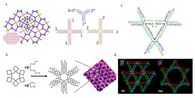
图2 (a)阿基米德图形及构成其的DNA瓦片[19];(b)三种链构成的“六点星”DNA瓦片及其组装的六边形晶格示意图[6];(c)张拉整体三角形示意图[25];(d)使用DNA拉张整体三角形构建的菱方(R3)和六方晶格(P63)[10]

Fig. 2 (a) Archimedean shapes and the DNA tiles composing them[19], Copyright © 2013, American Chemical Society; (b) A schematic diagram of a hexagonal lattice composed of “six-pointed star” DNA tiles consisting of three strands[6], Copyright © 2006, American Chemical Society; (c) A schematic diagram of DNA tensegrity triangles[25]; (d) Diamond (R3) and hexagonal (P63) lattices constructed using DNA tensegrity triangles[10], Copyright © 2021, American Chemical Society.

in 1993,Seeman proposed another structure of"DNA double-crossover molecule"(DX).by introducing crossover with single-stranded DNA In two side-By-side double helices,the new DNA structure is more rigid than before,which makes it possible to further assemble larger structures[20]。 Later,inspired by Wang Hao's tile in the theory of planar close-packing,Seeman et al.Self-assembled three two-dimensional lattices with two different topologies through the specific pairing of sticky ends of different DX structures,demonstrating the potential of the structure in the construction of periodic arrays[4]。 In addition,they assembled a pseudo-hexagonal lattice with triangular DNA tiles each with an edge of DX,and showed that the two sticky ends that the triangular edges have are essential for the formation of the lattice[21]。 On this basis,Mao et al.Designed a three-point star DNA tile and self-assembled a two-dimensional porous hexagonal lattice structure with a size of 1 mm and a hexagonal pore size of 18 nm[22]。 Later,Mao et al.designed a series of DNA tiles such as four-point star,five-point star and six-point star(Fig.2b)to obtain a variety of two-dimensional DNA lattices,and prepared dodecagonal quasicrystal DNA lattices by self-assembly of five-point star and six-point star[5~7][23]。 However,these assemblies are confined to a two-dimensional plane,and the researchers hope to develop a DNA structural unit that can be extended in three-dimensional space。
In 2004,Liu et al.Introduced the tensegrity structure into DNA tiles,constructed tensegrity equilateral triangular DNA tiles,and self-assembled a series of arrays[24]; Although The arrangement in these arrays is still two-dimensional,the DNA tile is not a planar structure,with multiple possible extension directions that are not coplanar,which provides an idea for the construction of three-dimensional lattice structures.the revolutionary progress of dimensional spanning appeared in 2009.Zheng et al.Successfully self-assembled a three-dimensional diamond-shaped DNA single crystal with a size of 250μm and a resolution of 44µm by constructing a stable tensegrity triangular structure with cubic symmetry,which proved that DNA tiles could be precisely controlled to form a three-dimensional periodic structure(Fig.2C)[8]。 After that,a series of diversified structures were synthesized.Wang et al.Used two different tensegrity triangular DNA tiles for co-self-assembly to realize the asymmetric unit assembly DNA crystal structure,and by controlling the combination of different dyes with the two DNA tiles,the crystal showed different colors such as pink,blue-green and purple(the first two colors are superimposed)[9]。 By using non-canonical 5′-AG and 5′-TC cohesive ends and changing the motif of the strand,Lu et al.Constructed hexagonal lattices of the p63 space group and cubic and rhombohedral DNA lattices,respectively,with tensegrity triangular DNA tiles(Figure 2D)[10][11]。 Woloszyn et al.Constructed 18 different DNA lattice structures by expanding the symmetry and asymmetry of the region between triangles,using the covalent connection or cohesive end coordination connection of adjacent tensegrity triangles,and realized the precise regulation of the morphology,symmetry and internal space of DNA crystals[12]。 These works have greatly expanded the material library of early DNA lattice structures。
One of the major obstacles to the practical application of DNA crystals is stability and environmental tolerance.Due to the polyanionic character of the DNA strand,the structure needs to be maintained in a high concentration of cationic solution,and the thermal stability is poor.In order to enhance the stability of DNA crystal structure,Zhao et al.Modified the sequence of tensegrity triangular DNA tiles and introduced a trimeric oligonucleotide molecule(TFOs),which enabled the formation of triplet structure between DNA tiles under acidic conditions(pH=0.5),and reduced the concentration of(NH4)2SO4solution required to maintain stability from 1.2 mol/L to 0.02 mol/L[26]。 Abdallah et al.Used the strategy of photocrosslinking,using TFOs to guide the photocrosslinking agent 4,5',8-trimethylpsoralen to crosslink between the two strands in the DNA tile,which improved the thermal stability[27]。 In addition,Zhang et al.Used bis(2-chloroethyl)amine hydrochloride as a chemical crosslinking agent to crosslink the strands of DNA tiles,which was proved by X-ray diffraction that it did not change the crystal structure,while improving the thermal stability,enzymatic degradation resistance and solution cation dependence of DNA crystals[28]。 Li et al.Used DNA ligase to form covalent bonds at the junction of DNA tiles in the self-assembled DNA crystal,so that the DNA crystal could be maintained at 65℃for 16 H and in water for 42 d[29]。 Improved thermal stability and environmental tolerance have pushed DNA crystals an important step toward practical applications。
Thanks to the development of self-assembly of DNA tiles to form lattices,more and more applications have been developed.Zheng et al.Used rigid triangular DNA tiles to connect gold nanoparticles with DNA tiles through DNA chains,and then self-assembled to obtain two-dimensional gold nanoparticle arrays[30]。 Wang et al.Constructed a three-dimensional organic semiconductor superlattice array by introducing octamer polyaniline molecules into dimeric tensegrity triangular DNA and then self-assembling.the reversible conversion of the redox state of octamer polyaniline molecules is retained in the crystal,and the change of the oxidation state is reflected in the color change of the crystal[13]; the proton-doped jadeite salt was obtained at pH=5,which corresponds to the conductive morphology of polyaniline,providing an idea for the construction of electronic devices using DNA lattices.Hao et al.Prepared a three-state DNA nanodevice by the interaction of dye-containing DNA strands in solution with the chain transfer reaction of DNA crystals to achieve the change of crystal color[14]。 Recently,Zheng et al.Reported a DNA crystal actuator powered by the concerted dissociation or cohesion of thousands of DNA cohesive ends at the designed crystal contacts.It can have reversible and directional expansion and contraction under various stimuli such as temperature,ionic strength,pH and redox potential,with an amplitude of more than 50μm,and can change the accommodation performance of nanoparticles,demonstrating the possibility of DNA crystals for actuation[15]
As the earliest proposed DNA assembly structure,DNA tile has important guiding significance in the formation of one-dimensional and two-dimensional lattices.its assembly method is simple,and it is easy to form large-scale structures.However,limited by the structure of the module itself,the DNA tile is difficult to accommodate larger nanoparticles due to Its small cavity,and the complexity of the assembly structure is also very limited,so it is difficult to form three-dimensional crystals。

3 Nanoparticle Crystallization Using DNA as a Connector

PAE is a unit consisting of a nanoparticle as a core,densely functionalized with a radially oriented DNA shell(Figure 3A).the concept of nanoparticles as"atoms"and DNA as programmable"bonds"has many similarities with traditional chemical bonding,so it is called atomic equivalent.Usually its DNA shell consists of an"anchor"strand attached to The nanoparticle surface and a complementary"linker"strand terminating in a"sticky end"short single-stranded region[31]。 In the mid-1990s,Mirkin et al.and Alivisatos et al.Demonstrated that DNA molecules could be used to synthesize programming materials And obtain amorphous aggregates[32][33]。 Later,in 2008,Mirkin et al.And Gang et al.Successfully obtained three-dimensional ordered crystals of gold nanoparticles with different DNA ligation designs using different methods[34,35]。 They achieved a disorder-to-order breakthrough through fine control of DNA hybridization temperature.in this way,nanoparticles are assembled into various complex structures,such as crystals with new crystal symmetries that do not exist In nature,dynamically changing versus reversibly changing structures,chiral structures,etc[36][37~41][42]
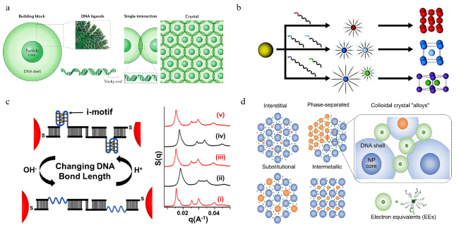
图3 (a)PAE组成及构成晶体示意图[31];(b)通过改变DNA连接体的序列将金纳米粒子-DNA偶联物编程组装成不同的晶体排列[60];(c)pH变化引发的晶格收缩和膨胀[40];(d)EEs与PAE组成的四种胶体晶体合金等价物[61]

Fig. 3 (a) Schematic diagram of PAE composition and crystal structure[31], Copyright © 2022 Wiley‐VCH GmbH; (b) Programmed assembly of gold nanoparticles DNA conjugates into different crystal arrangements by changing the sequence of DNA linkers[60], Copyright © 2013 WILEY‐VCH Verlag GmbH & Co. KGaA, Weinheim; (c) Lattice contraction and expansion caused by pH changes[40], Copyright © 2018, American Chemical Society; (d) Four colloidal crystal alloy equivalents composed of EEs and PAE[61], Copyright © 2019, American Chemical Society

DNA-mediated methods are versatile for nanoparticles with different properties,including inorganic nanoparticles,inorganic-organic nanoparticles(such as metal-organic frameworks),and some biomolecules that can modify DNA strands(such as proteins,enzymes)[43]。 in terms of size,most colloidal nanoparticles can be assembled into ordered Crystals mediated by DNA.crystals composed of gold nanospheres of 5–80 nm have been reported In the literature[44]。 Even larger particles still have the possibility of crystallization,such as triangular bipyramidal gold nanoparticles with a long side of 250 nm and a short side of 177 nm,which can self-assemble into inclusion colloidal crystals mediated by DNA chains[45]。 Previous studies have focused on isotropic nanoparticles,and with the maturity of the theory,anisotropic and complex particles can also form well-defined structures under the guidance of DNA[46~49]。 the shape of the anisotropic particle makes the DNA bond directional,and the regioselective functionalization of DNA can also make it anisotropic to guide the local distribution of nanoparticles In the crystal.in addition,no matter how the chemical composition of the particle changes,only a certain density of single-stranded DNA loaded on the particle surface can successfully crystallize PAE.Mirkin et al.and Gang et al.Independently proposed a general DNA modification method for nanoparticles with different chemical compositions from different perspectives,and synthesized a variety of single-component and multi-component crystals and crystals with different symmetries,which laid the foundation for the proposal of PAE assembly into ordered lattice guiding rules[50,51][52,53]
In order to reasonably predict and design the interaction between PAEs,Mirkin et al.Proposed a complementary contact model(CCM)to guide the synthesis of target crystals[36]。 the model States that the most stable structure is the one that maximizes hybridization between the sticky ends of the DNA connector.In addition,Mirkin et al.Studied the DNA-mediated nanoparticle crystallization process and found that it can be divided into three stages:the initial"random binding"stage leads to disordered PAE aggregates,then the disordered PAE aggregates undergo local reorganization crystallization,and finally the growth of crystalline domain size[54]。 In the second stage,DNA needs to undergo dehybridization and rehybridization to adjust the position of the particle to form an ordered crystal.In this stage,many variables affect the whole crystallization process by affecting the behavior of DNA.For DNA linkers,many parameters of their design will affect the type and quality of crystals,such as the amount of DNA modified on the particle surface,DNA sequence,DNA length,flexibility of DNA linkers,etc.When the density of modified DNA is too low or too high,the interaction between particles will not be suitable to form an ordered structure or hinder the readjustment of particle position to achieve a large ordered structure[51,54]。 Different DNA sequences can affect the interaction between particles,which can affect the formation of crystal lattice,and is one of the important bases For people to control crystal growth.for example,gold nanoparticles modified with self-complementary DNA sequences tend to form close-packed structures showing face-centered cubic crystal symmetry,while gold nanoparticles modified with pairwise complementary DNA linkers tend to form non-close-packed structures showing body-centered cubic symmetry(Fig.3B)[34]; Gang et al.Also used computer to simulate the interaction between PAEs linked by self-complementary DNA,which enriched people's understanding of the role of combinatorial entropy in particle binding at low temperature[55]; Kaitlin et al.Used PAE with different DNA sequences to achieve heterogeneous growth and eliminate the secondary nucleation pathway to improve the uniformity of the crystal[56]。 the lattice constant and interparticle spacing are important properties of crystals.In the PAE crystallization system,the distance between particles can be adjusted by adjusting the length of the DNA connector,and the linear relationship between these two variables can be calculated and predicted by formulas,which also help people understand the behavior of PAE crystallization[57,58]。 It can be considered that the flexibility of DNA connector mainly comes from its single-stranded part,and the longer the single-stranded part is,the more flexible the DNA connector is.the surface curvature of the particle itself makes more free space volume outside the DNA connector,and the local density of the DNA connector is relatively reduced,thus affecting the interaction between particles[59]。 By controlling the parameters of DNA linkers,Mirkin et al.Crystallized nanoparticles of different diameters into crystals of various symmetries,and established a set of mature guidelines for crystal design[36,44]
the preparation of dynamic,reconfigurable and stimulus-responsive nanocrystals provides an effective way to dynamically control the properties and functions of materials,which can greatly enrich the application scenarios of these materials,but the interaction between the units that make up the crystal often destroys the integrity of the crystal once it changes.DNA is an ideal molecule for making such dynamic structures.the flexible DNA shell provides the possibility of dynamic changes in the superlattice due to its structural plasticity and reversibility of interactions.Various DNA tertiary structures,such as hairpin and i-motif,can be obtained by designing DNA sequences.Some small molecules or specific groups can be inserted or modified on DNA molecules to realize the transformation of DNA conformation through special mechanisms[37,40]。 in addition,the valence and concentration of cations themselves can also affect the conformation of DNA In solution[62]。 Mirkin et al.Used hairpin and i-motif(Figure 3C)structures,using DNA chains and pH as external stimuli,respectively,to achieve reversible contraction,expansion and crystal symmetry conversion of superlattices.In addition,they also made shape memory crystals to achieve irregular contraction and rapid recovery of crystals under dehydration and rehydration[38,40,63][41]。 the optical response of The superlattice can be achieved by optical isomerization of azobenzene molecules[64,65]。 Gang et al.Modified the photosensitive group in the DNA linker to improve the stability of the superlattice under DNA denaturation conditions,and could achieve reversible changes between assembly and disassembly by changing the light conditions[65]。 Interestingly,while it is generally believed that the inorganic nanoparticles of PAE provide rigidity to the superlattice,there is also the possibility of using the core of PAE for physical control to change its lattice,and this work enriches people's understanding of the controllable factors in the process of assembling the lattice by PAE[66]
It is worth noting that Girard et al.Recently found that PAEs have a new diffusion behavior beyond the traditional view:they found that when the size and DNA linker density decrease,small PAEs can behave as electron equivalents.EEs),these EE can diffuse and stabilize through the lattice formed by PAE,similar to the diffusion of electrons in metals,and the degree of delocalization and diffusion of EE is affected by temperature and the number of DNA strands bound on the surface[67]。 the new concept of"valence electron"established by PAE and EE is similar to The theory of valence shell electron pair repulsion(VSEPR)in chemical bonds.Using this new mechanism,various colloidal crystal alloys can be designed and synthesized.These include colloidal crystal alloys with atomic analogs(fig.3D)and triple double-top low-symmetry structures without natural equivalents,as well as more low-symmetry and complex colloidal crystal structures[61][68]。 These works show that PAE can construct systems similar to metals,demonstrate more similarities with traditional chemistry,and greatly expand people's understanding of DNA nanotechnology。
After more than 20 years of development,PAE crystallization has become a relatively mature method for superlattice preparation.many factors affecting the growth process have been specifically explored,and the guiding rules have been clearly proposed.However,due to the Many constraints of crystallization,the crystallization behavior is limited by the inherent properties of nanoparticles,such as shape,composition and size,the complexity of the structure formed is limited,and the density of DNA on the surface of particles required to form an ordered structure is large,so the potential of crystallizing nanoparticles into various shapes is still limited。

4 DNA Origami Based Nanoparticle Crystal Assembly

DNA tiles and PAE are powerful methods for building nanoparticle superlattices in DNA nanotechnology and also tailoring DNA structures in the desired way,but these methods still fall short when it comes to using DNA to achieve assembly of nanoparticles into designed crystalline materials.to achieve the fundamental goal of constructing three-dimensional crystals as templates for nanoparticle molecular crystallization scaffolds,such scaffolds need to have several properties:large enough cavities to accommodate nanoparticles,high enough designability to form complex structures and lattices,and decoupling from nanoparticle properties to facilitate their ready incorporation into the structure.the insufficient cavity of the DNA tile structure,the particle core of the PAE being essentially part of the resulting structure,creates a contradiction with the requirements of the required scaffold.the emergence of DNA origami demonstrates the great potential of DNA nanotechnology,which makes people see the ideal technology to form three-dimensional periodic molecular scaffolds。
In 2006,Rothemund of the California Institute of Technology proposed a new method for constructing DNA structures,known as"DNA origami"[69]。 Hundreds of short-stranded DNA(staple strands)are cross-linked to fold a long single-stranded circular DNA(scaffold strand),the scaffold strand is mixed with a correspondingly designed staple strand,maintained in a single-stranded state at a high temperature of 95 deg C,and then gradually cooled in a buffer solution;Driven by base pairing,the staple strand can fold the scaffold strand into the desired monolayer planar pattern(Figure 4A).Using this technology,Rothemund et al.Successfully assembled pre-designed two-dimensional structures such as rectangles,triangles,pentagons and smiling faces.DNA origami has attracted Many non-chemists to study it,because its use in many cases does not require the purification of staple strands,and through simple purification,high yield of DNA origami structures can be obtained,and the assembly method is simple and efficient.many teams have used this method to assemble various complex structures,such as maps of China and dolphins[70][71]。 Since then,Shih et al.Extended the DNA origami method to construct three-dimensional structures,designed a honeycomb lattice(6HB)formed by stacking DNA double helices,which is more rigid than monolayer DNA origami(Fig.4B),and successfully prepared a variety of complex structures using this unit[72]
图4 (a)DNA折纸术的发明[95];(b)多层DNA折纸组成蜂窝状晶格[96];(c)不同DNA折纸框架自组装形成晶格[86];(d)利用DNA折纸八面体制备Wulff形状的单晶[94],比例尺:1 μm

Fig. 4 (a) The invention of DNA origami[95]; (b) Multiple layers of DNA origami form a honeycomb lattice[96],Copyright © 2020, Springer Nature Switzerland AG; (c) Different DNA origami frameworks self assemble to form a lattice[86], Copyright © 2016, Springer Nature Limited; (d) Preparation of Wulff shaped single crystals using DNA origami octahedra[94],scale bar:1 μm

in the development of designing DNA origami structures,the realization of curvature is one of the important advances in expanding the complexity of DNA origami structures.after designing 6HB,Shih et al.Proposed a strategy to expand the shape design of DNA origami in terms of curvature.A normal B-form DNA helix rotates its helical path by 240°every seven base pairs,and in the DNA origami structure of the honeycomb array,because of the connection between adjacent DNA strands,one DNA helix will exert a left-handed or right-handed torque and pull on the adjacent DNA strands After the deletion or insertion of base pairs.Using this principle,Shih et al.First realized DNA origami structures with curvature,including twist,bend and helix,and Yan et al.Also designed curvature rings without scaffolds[73][74]。 in addition,Shih et al.And Yan et al.Have proposed new strategies to achieve curvature without the stress caused by base pair deletion or insertion In DNA structure.Shih et al.Rely on the mechanical tension generated by single-stranded DNA to bend a certain rigid DNA helix to produce curvature[75]; Yan et al.Arranged the DNA scaffold strands into a pre-designed shape with curvature according to certain rules,and fixed them with corresponding DNA nails to offset the ability of the DNA double helix to resist bending due to its rigidity,thus constructing a complex three-dimensional vase DNA origami structure[76]。 These strategies for constructing DNA origami structures with curvature make it possible for scientists to design structures with more complexity,and also expand the possibility of DNA origami applications in nanorobots[77][78,79]
The basic goal of DNA nanotechnology has been to use DNA molecules to construct three-dimensional crystals as periodic molecular scaffolds to precisely arrange nanoparticles that are difficult to crystallize alone.The resulting three-dimensional nanoparticle arrays are particularly influential because such structures provide access to large-scale spatially organized nanomaterials with integrated properties by integrating a large number of nanocomponents[80]。 Although DNA origami can be finely tailored to the size and shape of the structure,and can prepare a variety of unique structures,the interaction between the structures is very complex,and the influence factors of entropy and enthalpy in the assembly process are not clear,so it is very challenging to assemble DNA origami structures into ordered superlattices or three-dimensional DNA crystals[81~83]。 Tian et al.Used 6HB to construct a rigid DNA origami octahedron with top coding,which can realize the regular arrangement of nanoparticles in one or two dimensions by connecting specific coding with nanoparticles,and three-dimensional nanoparticle clusters can also be effectively created based on this method[84]。 Later,Gang et al.Also used 6HB to construct a DNA origami tetrahedron,which defined a tetravalent binding topology that could be assembled with the external connection of nanoparticles to form an FCC superlattice,and produced a diamond-structured crystal by encapsulating an additional nanoparticle within the tetrahedron,which was a major breakthrough in the construction of three-dimensional lattices in the field of DNA origami[85]。 Although DNA origami tetrahedra are not sufficient to assemble into ordered diamond-structured crystals due to possible defects caused by the rotation of bonds,according to previous rough understanding,through the calculation of isotropic nanoparticle binding"footprint",Gang et al.Proved that the assembly of nanoparticles externally connected by DNA origami into FCC structure has thermodynamic advantages,and the electrostatic and steric repulsion between origami tetrahedra is also conducive to the formation of FCC structure.Later,Tian et al.Designed a variety of DNA origami polyhedral frameworks(Fig.4C),which were assembled into three-dimensional crystals by connecting DNA functional chains at the vertices with nanoparticles,further developing this method of constructing three-dimensional lattices[86]
In addition to serving as a linker between nanoparticles,DNA origami itself can interact and assemble into ordered structures,and nanoparticles can be functionalized by DNA to connect to the interior of the DNA framework to carry material functions[87]。 Gang et al.Designed a flat origami module with different"colors",which can create a variety of one-dimensional or two-dimensional structures through different connections between"colors"[88]。 the assembly of DNA nanochambers explores The possibility of using more"color"bonding in different directions and quantities to form ordered arrays in various dimensions[89]。 Wang et al.Constructed a combination module of nanoparticles and DNA origami,and used the"color"and the geometric shape of the module to obtain linear,square and other planar assemblies[90]。 Later,Tian et al.Extended this method to three dimensions.They demonstrated a general method for organizing proteins into three-dimensional lattices.DNA-functionalized proteins are encapsulated into DNA origami polyhedral frameworks,which can be assembled into three-dimensional lattices by complementary hybridization of cohesive ends of DNA single strands between vertices[91]。 Liedl et al.Also used DNA origami to construct a tensegrity triangular structure,which was assembled into a three-dimensional rhombic crystal with a larger cavity of 20 nm[92]。 the ordered structure produced by these structural assemblies is not related to the nanoparticles encapsulated in the DNA origami polyhedral framework,but is completely determined by the topological shape of the DNA origami framework,which makes it possible for more nanoparticles that are difficult to crystallize to form an ordered lattice.They successfully decoupled the inherent properties of nanoparticles from the assembly structure,assembled a variety of nanoparticles(including gold nanoparticles,proteins,enzymes,etc.)into an ordered three-dimensional lattice,and even further prepared a Wulff-shaped DNA origami single crystal(Fig.4D)[91~93][94]
Whether DNA origami is used as a topological connector between nanoparticles or DNA origami frameworks are directly connected to each other to assemble a three-dimensional lattice,the binding valence defined by the structure and the connection mode caused by the geometry of the structure have an important impact on the resulting macrostructure.This allows one to take advantage of DNA origami frameworks with different topologies to control structures of different dimensions and even assemble to get different lattices using the same nanoparticle[97]。 in addition,the design of connecting partial sticky ends can also achieve the purpose of influencing the type of assembled lattice,and even make the crystal produce phase transition under external stimulation.Tian et al.Designed the SE of two kinds of origami octahedra so that they could be connected In different ways,and selectively put nanoparticles into the monomer of origami octahedra to obtain different lattices of nanoparticles[98]; Extending this approach further,multiple units of DNA origami are used as basic assembly modules,which greatly expands the library of superlattice types that can be synthesized by DNA origami[99]。 In addition,because the assembly of DNA origami octahedra into crystals is significantly affected by ionic strength,Dai et al.Successfully assembled DNA origami octahedra into different lattices such as simple cubic(SC)and face-centered cubic(FCC)by simply adjusting the concentration of Mg2+,and realized the phase transition of crystals in different environments[100]。 the phase transition of the crystal can also switch the lattice type between simple cubic(SC)and simple tetragonal(ST)by introducing a pH-sensitive i-motif at the SE,which changes the length of the DNA origami framework connecting strands upon pH change[101]。 Yan et al.Also introduced hairpin sequences,which were opened or closed by the combination of key sequences or lock sequences,thus changing the spacing of the DNA origami frame,and the lattice constant of the crystal changed accordingly,resulting in phase transition,thus constructing a dynamic reconfigurable DNA origami crystal with multiple phase transition capabilities[102]。 From these studies,in order to construct DNA origami crystals with phase transition ability,the key is to introduce changeable molecules that can bind to DNA,stimulate the corresponding molecules to change through external changes,so that a single DNA origami module changes and transmits to the whole DNA origami crystal,so as to achieve the purpose of obtaining different three-dimensional lattices。
DNA origami usually needs a solution with high cationic strength to maintain its structure,and it has strict requirements on the solution environment,so it is easy to dissociate when subjected to strong external stimuli.Improving the stability of DNA origami is conducive to promoting its application in various fields.Ma et al.Used a rigid DNA rod as the connecting part of the DNA origami framework to improve the rigidity and compression resistance of the co-crystal,so that the structure can be maintained in a harsh solution environment[103]。 Another effective way to improve the structural stability of DNA origami is to use silica shell as a protective coating,so that DNA origami can maintain structural integrity in harmful environments such as high temperature or the presence of DNase[105,104]。 three-dimensional DNA origami crystals can also be mineralized with silica,thus preserving them out of solution and allowing detailed analysis of their Three-dimensional structure[104]。 For example,when DNA origami single crystals are directly dried and observed by transmission electron microscopy(TEM),the orderly arrangement of soft DNA crystals can not be carefully observed due to the collapse of soft DNA crystals during drying.After the DNA origami is mineralized by silica,the surface structure of the microcrystal can be observed by scanning electron microscopy(SEM),and the three-dimensional reconstruction of the silicon-coated DNA origami single crystal can be carried out by high-resolution electron microscopy[94]。 In addition,silica-mineralized DNA origami structures can be further coated with superconducting niobium(Nb)or converted to SiC to produce structures that can be applied to various superconducting or optoelectronic materials,greatly exploiting the potential of DNA origami nanotechnology[106][107]
At present,DNA origami is one of the most designable methods of DNA structure making.the connection between DNA origami allows the development of more complex scaffolds,resulting in lattices of various symmetries,and the incorporation of nanoparticles.in this process,the particles do not play a structural role in the assembled architecture,which significantly reduces the requirement to functionalize them with DNA,as long as the nanoparticles can be coupled to the DNA origami unit and can be crystallized in an ordered manner according to the crystal symmetry and unit cell parameters prescribed by the template.Therefore,DNA origami is one of the more advanced methods to design DNA structures for specific tasks,and it is also one of the ideal technologies to form crystals loaded with nanoparticles。

5 Conclusion and prospect

in the past,the assembly of nanoparticles into crystals mostly relied on their physical interparticle interactions,and the lack of control over the bonding strength and directionality made it a great challenge for nanoparticles to construct structures with nanoscale accuracy.in the past few decades,the assembly of nanoparticles into crystals Using the explicit bonding properties of DNA has been realized In DNA tile,PAE and DNA origami technologies.DNA nanotechnology has developed into a powerful method for the design and synthesis of high-precision nanostructures and organizations,and its ability to integrate the properties of nanoparticles has attracted much attention.the chemical programmability of DNA gives DNA nanotechnology a significant advantage over other methods of bottom-up assembly of structures.using DNA nanotechnology,nanoscale structural assembly modules can be precisely constructed,nanoparticles can be assembled into highly ordered macroscopic structures,and three-dimensional crystals can be manufactured,while nanoparticles can be loaded into almost any periodic position In the structure,which can not be achieved by other technologies。
Nowadays,DNA crystals have shown their potential in many scenarios,such as catalysts,optical devices,semiconductor materials and so on[91,108 ~110][111][13,112]。 in the future,it is of great significance to study how to use the self-assembly ability of DNA molecules to construct three-dimensional crystals and explore the function of three-dimensional DNA crystals for the breakthrough of DNA Nanotechnology in biomedicine,chemistry and materials science.the research on how to integrate DNA nanodevices with different functions into a system to realize multifunctional nanodevices will promote the further development of DNA nanotechnology in biosensing,biocomputing and other fields.In addition,how to manufacture DNA crystals on a large scale,develop efficient and controllable preparation methods of DNA nanostructures,and improve the process to reduce the preparation cost of DNA nanotechnology should become an important research direction of DNA nanotechnology,so as to realize the commercial application of DNA nanotechnology and meet its needs in the fields of biomedicine,optical devices,semiconductor materials and so on。
[1]
Seeman N C. J. Theor. Biol., 1982, 99(2): 237.

[2]
Caruthers M H. Science, 1985, 230(4723): 281.

[3]
Kallenbach N R, Ma R I, Seeman N C. Nature, 1983, 305(5937): 829.

[4]
Winfree E, Liu F R, Wenzler L A, Seeman N C. Nature, 1998, 394(6693): 539.

[5]
Zhang C, Su M, He Y, Zhao X, Fang P A, Ribbe A E, Jiang W, Mao C D. Proc. Natl. Acad. Sci. U. S. A., 2008, 105(31): 10665.

[6]
He Y, Tian Y, Ribbe A E, Mao C D. J. Am. Chem. Soc., 2006, 128(50): 15978.

[7]
He Y, Tian Y, Chen Y, Deng Z X, Ribbe A E, Mao C D. Angew. Chem. Int. Ed., 2005, 44(41): 6694.

[8]
Zheng J P, Birktoft J J, Chen Y, Wang T, Sha R J, Constantinou P E, Ginell S L, Mao C D, Seeman N C. Nature, 2009, 461(7260): 74.

[9]
Wang T, Sha R J, Birktoft J, Zheng J P, Mao C D, Seeman N C. J. Am. Chem. Soc., 2010, 132(44): 15471.

[10]
Lu B, Vecchioni S, Ohayon Y P, Sha R J, Woloszyn K, Yang B A, Mao C D, Seeman N C. ACS Nano, 2021, 15(10): 16788.

[11]
Lu B, Vecchioni S, Ohayon Y P, Woloszyn K, Markus T, Mao C D, Seeman N C, Canary J W, Sha R J. Small, 2023, 19(3): 2205830.

[12]
Woloszyn K, Vecchioni S, Ohayon Y P, Lu B, Ma Y L, Huang Q Y, Zhu E, Chernovolenko D, Markus T, Jonoska N, Mao C D, Seeman N C, Sha R J. Adv. Mater., 2022, 34(49): 2206876.

[13]
Wang X, Sha R J, Kristiansen M, Hernandez C, Hao Y D, Mao C D, Canary J W, Seeman N C. Angew. Chem. Int. Ed., 2017, 56(23): 6445.

[14]
Hao Y D, Kristiansen M, Sha R J, Birktoft J J, Hernandez C, Mao C D, Seeman N C. Nat. Chem., 2017, 9(8): 824.

[15]
Zheng M X, Li Z, Zhang C Z, Seeman N C, Mao C D. Adv. Mater., 2022, 34(26): 2200441.

[16]
Wang X, Seeman N C. J. Am. Chem. Soc., 2007, 129(26): 8169.

[17]
Wang Y L, Mueller J E, Kemper B, Seeman N C. Biochemistry, 1991, 30(23): 5667.

[18]
Ma R I, Kallenbach N R, Sheardy R D, Petrillo M L, Seeman N C. Nucleic Acids Res., 1986, 14(24): 9745.

[19]
Zhang F, Liu Y, Yan H. J. Am. Chem. Soc., 2013, 135(20): 7458.

[20]
Fu T J, Seeman N C. Biochemistry, 1993, 32(13): 3211.

[21]
Ding B Q, Sha R J, Seeman N C. J. Am. Chem. Soc., 2004, 126(33): 10230.

[22]
He Y, Chen Y, Liu H P, Ribbe A E, Mao C D. J. Am. Chem. Soc., 2005, 127(35): 12202.

[23]
Liu L F, Li Z, Li Y L, Mao C D. J. Am. Chem. Soc., 2019, 141(10): 4248.

[24]
Liu D G, Wang M S, Deng Z X, Walulu R, Mao C D. J. Am. Chem. Soc., 2004, 126(8): 2324.

[25]
Paukstelis P, Seeman N. Crystals, 2016, 6(8): 97.

[26]
Zhao J M, Chandrasekaran A R, Li Q, Li X, Sha R J, Seeman N C, Mao C D. Angew. Chem. Int. Ed., 2015, 54(34): 9936.

[27]
Abdallah H O, Ohayon Y P, Chandrasekaran A R, Sha R J, Fox K R, Brown T, Rusling D A, Mao C D, Seeman N C. Chem. Commun., 2016, 52(51): 8014.

[28]
Zhang D A, Paukstelis P J. ChemBioChem, 2016, 17(12): 1163.

[29]
Li Z, Liu L F, Zheng M X, Zhao J M, Seeman N C, Mao C D. J. Am. Chem. Soc., 2019, 141(40): 15850.

[30]
Zheng J W, Constantinou P E, Micheel C, Alivisatos A P, Kiehl R A, Seeman N C. Nano Lett., 2006, 6(7): 1502.

[31]
Samanta D, Zhou W J, Ebrahimi S B, Petrosko S H, Mirkin C A. Adv. Mater., 2022, 34(12): 2107875.

[32]
Mirkin C A, Letsinger R L, Mucic R C, Storhoff J J. Nature, 1996, 382(6592): 607.

[33]
Alivisatos A P, Johnsson K P, Peng X G, Wilson T E, Loweth C J, Bruchez M P, Schultz P G. Nature, 1996, 382(6592): 609.

[34]
Park S Y, Lytton-Jean A K R, Lee B, Weigand S, Schatz G C, Mirkin C A. Nature, 2008, 451(7178): 553.

[35]
Nykypanchuk D, Maye M M, van der Lelie D, Gang O. Nature, 2008, 451(7178): 549.

[36]
MacFarlane R J, Lee B, Jones M R, Harris N, Schatz G C, Mirkin C A. Science, 2011, 334(6053): 204.

[37]
Maye M M, Kumara M T, Nykypanchuk D, Sherman W B, Gang O. Nat. Nanotechnol., 2010, 5(2): 116.

[38]
Kim Y, MacFarlane R J, Mirkin C A. J. Am. Chem. Soc., 2013, 135(28): 10342.

[39]
Mason J A, Laramy C R, Lai C T, O’Brien M N, Lin Q Y, Dravid V P, Schatz G C, Mirkin C A. J. Am. Chem. Soc., 2016, 138(28): 8722.

[40]
Zhu J H, Kim Y, Lin H X, Wang S Z, Mirkin C A. J. Am. Chem. Soc., 2018, 140(15): 5061.

[41]
Lee S, Calcaterra H A, Lee S M, Hadibrata W, Lee B, Oh E, Aydin K, Glotzer S C, Mirkin C A. Nature, 2022, 610(7933): 674.

[42]
Ben Zion M Y, He X J, Maass C C, Sha R J, Seeman N C, Chaikin P M. Science, 2017, 358(6363): 633.

[43]
He L C, Mu J, Gang O, Chen X Y. Adv. Sci., 2021, 8(8): 2003775.

[44]
MacFarlane R, Jones M, Senesi A, Young K, Lee B, Wu J S, Mirkin C. Angew. Chem. Int. Ed., 2010, 49(27): 4589.

[45]
Lin H X, Lee S M, Sun L, Spellings M, Engel M, Glotzer S C, Mirkin C A. Science, 2017, 355(6328): 931.

[46]
Young K L, Jones M R, Zhang J, MacFarlane R J, Esquivel-Sirvent R, Nap R J, Wu J S, Schatz G C, Lee B, Mirkin C A. Proc. Natl. Acad. Sci. U. S. A., 2012, 109(7): 2240.

[47]
O’Brien M N, Jones M R, Lee B, Mirkin C A. Nat. Mater., 2015, 14(8): 833.

[48]
Jones M R, MacFarlane R J, Lee B, Zhang J, Young K L, Senesi A J, Mirkin C A. Nat. Mater., 2010, 9(11): 913.

[49]
Lu F, Yager K G, Zhang Y G, Xin H L, Gang O. Nat. Commun., 2015, 6: 6912.

[50]
Zhang C, MacFarlane R J, Young K L, Choi C H J, Hao L L, Auyeung E, Liu G L, Zhou X Z, Mirkin C A. Nat. Mater., 2013, 12(8): 741.

[51]
Zhang Y G, Lu F, Yager K G, van der Lelie D, Gang O. Nat. Nanotechnol., 2013, 8(11): 865.

[52]
Landy K M, Gibson K J, Urbach Z J, Park S S, Roth E W, Weigand S, Mirkin C A. Nano Lett., 2022, 22(1): 280.

[53]
Distler M E, Landy K M, Gibson K J, Lee B, Weigand S, Mirkin C A. J. Am. Chem. Soc., 2023, 145(2): 841.

[54]
MacFarlane R J, Thaner R V, Brown K A, Zhang J, Lee B, Nguyen S T, Mirkin C A. Proc. Natl. Acad. Sci. U. S. A., 2014, 111(42): 14995.

[55]
Sciortino F, Zhang Y G, Gang O, Kumar S K. ACS Nano, 2020, 14(5): 5628.

[56]
Landy K M, Gibson K J, Chan R R, Pietryga J, Weigand S, Mirkin C A. ACS Nano, 2023, 17(7): 6480.

[57]
Hill H D, MacFarlane R J, Senesi A J, Lee B, Park S Y, Mirkin C A. Nano Lett., 2008, 8(8): 2341.

[58]
MacFarlane R J, Lee B, Hill H D, Senesi A J, Seifert S, Mirkin C A. Proc. Natl. Acad. Sci. U. S. A., 2009, 106(26): 10493.

[59]
Senesi A J, Eichelsdoerfer D J, Brown K A, Lee B, Auyeung E, Choi C H J, MacFarlane R J, Young K L, Mirkin C A. Adv. Mater., 2014, 26(42): 7235.

[60]
MacFarlane R J, O’Brien M N, Petrosko S H, Mirkin C A. Angew. Chem. Int. Ed., 2013, 52(22): 5688.

[61]
Wang S Z, Du J S, Diercks N J, Zhou W J, Roth E W, Dravid V P, Mirkin C A. J. Am. Chem. Soc., 2019, 141(51): 20443.

[62]
Samanta D, Iscen A, Laramy C R, Ebrahimi S B, Bujold K E, Schatz G C, Mirkin C A. J. Am. Chem. Soc., 2019, 141(51): 19973.

[63]
Kim Y, MacFarlane R J, Jones M R, Mirkin C A. Science, 2016, 351(6273): 579.

[64]
Zhu J H, Lin H X, Kim Y, Yang M W, Skakuj K, Du J S, Lee B, Schatz G C, Van Duyne R P, Mirkin C A. Adv. Mater., 2020, 32(8): 1906600.

[65]
De Fazio A F, El-Sagheer A H, Kahn J S, Nandhakumar I, Burton M R, Brown T, Muskens O L, Gang O, Kanaras A G. ACS Nano, 2019, 13(5): 5771.

[66]
Urbach Z J, Park S S, Weigand S L, Rix J E, Lee B, Mirkin C A. Angew. Chem. Int. Ed., 2021, 60(8): 4065.

[67]
Girard M, Wang S Z, Du J S, Das A, Huang Z Y, Dravid V P, Lee B, Mirkin C A, Olvera de la Cruz M. Science, 2019, 364(6446): 1174.

[68]
Wang S Z, Lee S M, Du J S, Partridge B E, Cheng H F, Zhou W J, Dravid V P, Lee B, Glotzer S C, Mirkin C A. Nat. Mater., 2022, 21(5): 580.

[69]
Rothemund P W K. Nature, 2006, 440(7082): 297.

[70]
Qian L L, Wang Y, Zhang Z, Zhao J, Pan D, Zhang Y, Liu Q, Fan C H, Hu J, He L. Chin. Sci. Bull., 2006, 51(24): 2973.

[71]
Andersen E S, Dong M D, Nielsen M M, Jahn K, Lind-Thomsen A, Mamdouh W, Gothelf K V, Besenbacher F, Kjems J. ACS Nano, 2008, 2(6): 1213.

[72]
Douglas S M, Dietz H, Liedl T, Hogberg B, Graf F, Shih W. Nature, 2009, 459: 414.

[73]
Dietz H, Douglas S M, Shih W M. Science, 2009, 325(5941): 725.

[74]
Yang Y, Zhao Z, Zhang F, Nangreave J, Liu Y, Yan H. Nano Lett., 2013, 13(4): 1862.

[75]
Liedl T, Högberg B, Tytell J, Ingber D E, Shih W M. Nat. Nanotechnol., 2010, 5(7): 520.

[76]
Han D R, Pal S, Nangreave J, Deng Z T, Liu Y, Yan H. Science, 2011, 332(6027): 342.

[77]
Wang D F, Yu L, Ji B, Chang S, Song J, Ke Y G. Nano Lett., 2020, 20(11): 8236.

[78]
Wang D F, Yu L, Huang C M, Arya G, Chang S, Ke Y G. J. Am. Chem. Soc., 2021, 143(5): 2256.

[79]
Peil A, Xin L, Both S, Shen L Y, Ke Y G, Weiss T, Zhan P F, Liu N. ACS Nano, 2022, 16(4): 5284.

[80]
Kahn J S, Gang O. Angew. Chem. Int. Ed., 2022, 61(3): 2105678.

[81]
Benson E, Mohammed A, Gardell J, Masich S, Czeizler E, Orponen P, Högberg B. Nature, 2015, 523(7561): 441.

[82]
Veneziano R, Ratanalert S, Zhang K M, Zhang F, Yan H, Chiu W, Bathe M. Science, 2016, 352(6293): 1534.

[83]
Kim M, Lee C, Jeon K, Lee J Y, Kim Y J, Lee J G, Kim H, Cho M, Kim D N. Nature, 2023, 619(7968): 78.

[84]
Tian Y, Wang T, Liu W Y, Xin H L, Li H L, Ke Y G, Shih W M, Gang O. Nat. Nanotechnol., 2015, 10(7): 637.

[85]
Liu W Y, Tagawa M, Xin H L, Wang T, Emamy H, Li H L, Yager K G, Starr F W, Tkachenko A V, Gang O. Science, 2016, 351(6273): 582.

[86]
Tian Y, Zhang Y G, Wang T, Xin H L, Li H L, Gang O. Nat. Mater., 2016, 15(6): 654.

[87]
Zhang C, Li X, Tian C, Yu G M, Li Y L, Jiang W, Mao C D. ACS Nano, 2014, 8(2): 1130.

[88]
Liu W Y, Halverson J, Tian Y, Tkachenko A V, Gang O. Nat. Chem., 2016, 8(9): 867.

[89]
Lin Z W, Emamy H, Minevich B, Xiong Y, Xiang S T, Kumar S, Ke Y G, Gang O. J. Am. Chem. Soc., 2020, 142(41): 17531.

[90]
Wang M Y, Dai L Z, Duan J L, Ding Z Y, Wang P, Li Z, Xing H, Tian Y. Angew. Chem. Int. Ed., 2020, 59(16): 6389.

[91]
Tian Y, Lhermitte J R, Bai L, Vo T, Xin H L, Li H L, Li R P, Fukuto M, Yager K G, Kahn J S, Xiong Y, Minevich B, Kumar S K, Gang O. Nat. Mater., 2020, 19(7): 789.

[92]
Zhang T, Hartl C, Frank K, Heuer-Jungemann A, Fischer S, Nickels P C, Nickel B, Liedl T. Adv. Mater., 2018, 30(28): 1870203.

[93]
Dong Y X, Liu J L, Lu X Z, Duan J L, Zhou L Q, Dai L Z, Ji M, Ma N N, Wang Y, Wang P, Zhu J J, Min Q H, Gang O, Tian Y. Nano Lett., 2022, 22(9): 3809.

[94]
Wang Y, Dai L Z, Ding Z Y, Ji M, Liu J L, Xing H, Liu X G, Ke Y G, Fan C H, Wang P, Tian Y. Nat. Commun., 2021, 12: 3011.

[95]
Yan X H, Huang S J, Wang Y, Tang Y Y, Tian Y. Nanomaterials, 2020, 10(10): 2047.

[96]
Ma N N, Minevich B, Liu J L, Ji M, Tian Y, Gang O. Directional assembly of nanoparticles by DNA shapes: towards designed architectures and functionality. DNA Nanotechnology. Cham: Springer, 2020. 157.

[97]
Gao D, Ma N N, Yan X H, Ji M, Zhu J J, Min Q H, Tian Y. Chem. Sci., 2022, 13(1): 283.

[98]
Ji M, Liu J L, Dai L Z, Wang L, Tian Y. J. Am. Chem. Soc., 2020, 142(51): 21336.

[99]
Ji M, Zhou Z Y, Cao W H, Ma N N, Xu W G, Tian Y. Sci. Adv., 2022, 8(47): eadc9755.

[100]
Dai L Z, Hu X X, Ji M, Ma N N, Xing H, Zhu J J, Min Q H, Tian Y. Proc. Natl. Acad. Sci. U. S. A., 2023, 120(28): e2302142120.

[101]
Wang Y, Yan X H, Zhou Z Y, Ma N N, Tian Y. Angew. Chem. Int. Ed., 2022, 61(40): 2208290.

[102]
Yan X H, Wang Y, Ma N N, Yu Y F, Dai L Z, Tian Y. J. Am. Chem. Soc., 2023, 145(7): 3978.

[103]
Ma N N, Dai L Z, Chen Z, Ji M, Wang Y, Tian Y. Nano Lett., 2021, 21(8): 3581.

[104]
Nguyen L, Döblinger M, Liedl T, Heuer-Jungemann A. Angew. Chem. Int. Ed., 2019, 58(3): 912.

[105]
Liu X G, Zhang F, Jing X X, Pan M C, Liu P, Li W, Zhu B W, Li J, Chen H, Wang L H, Lin J P, Liu Y, Zhao D Y, Yan H, Fan C H. Nature, 2018, 559(7715): 593.

[106]
Shani L, Michelson A N, Minevich B, Fleger Y, Stern M, Shaulov A, Yeshurun Y, Gang O. Nat. Commun., 2020, 11: 5697.

[107]
Michelson A, Zhang H H, Xiang S T, Gang O. Nano Lett., 2021, 21(4): 1863.

[108]
Geng C, Paukstelis P J. J. Am. Chem. Soc., 2014, 136(22): 7817.

[109]
Kahn J S, Xiong Y, Huang J, Gang O. JACS Au, 2022, 2(2): 357.

[110]
Wang S T, Minevich B, Liu J F, Zhang H H, Nykypanchuk D, Byrnes J, Liu W, Bershadsky L, Liu Q, Wang T, Ren G, Gang O. Nat. Commun., 2021, 12: 3702.

[111]
Kim S, Zheng C Y, Schatz G C, Aydin K, Kim K H, Mirkin C A. Nano Lett., 2020, 20(11): 8096.

[112]
Wang X, Deshmukh R, Sha R J, Birktoft J J, Menon V, Seeman N C, Canary J W. Angew. Chem. Int. Ed., 2022, 61(5): e202115155.

Outlines

/