Home Journals Progress in Chemistry
Progress in Chemistry

Abbreviation (ISO4): Prog Chem      Editor in chief: Jincai ZHAO

About  /  Aim & scope  /  Editorial board  /  Indexed  /  Contact  / 
综述

Stimulus-Responsive Multifunctional Nucleic Acid Hydrogels Based on Cell Capture and Release

  • Danyu Wang 1 ,
  • Mengke Guo 1 ,
  • Zihan Guo 1 ,
  • Mengyu Huang 1 ,
  • Hua Yi 1 ,
  • Kaixiang Zhang , 1, 2, *
Expand
  • 1 School of Pharmacy, Zhengzhou University, Zhengzhou 450001, China
  • 2 Henan Provincial Key Laboratory of Targeted Therapy and Diagnosis of Major Tumor Diseases, Zhengzhou 450001, China

Received date: 2024-02-22

  Revised date: 2024-07-21

  Online published: 2024-09-15

Supported by

National Natural Science Foundation of China(22122409)

National Natural Science Foundation of China(22377110)

Henan Province Advantageous Discipline Cultivation Fund Project(222301420019)

Abstract

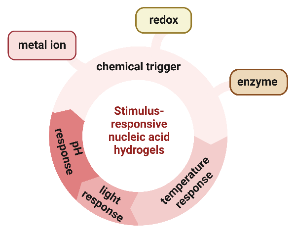
Nucleic acid hydrogels have good hydrophilicity, adjustability and biocompatibility, which have attracted considerable attention in the past few years, especially in the field of biomedicine and smart materials. Nucleic acid hydrogel is stimulus-responsive, meaning that external stimuli such as pH changes, light, temperature variations, and chemical triggers (including metal ion response, redox response, and enzyme response) can induce physical and chemical changes within them. Consequently, they are capable of perceiving their environment and undergoing responsive deformation, enabling precise cell therapy that can be controlled both temporally and spatially. Cell capture and release using stimulus-responsive nucleic acid hydrogels can control and modulate cellular behavior, and can also play an important role in biomedical research and applications, such as targeted drug therapies using the capture and release of specific cell types. Based on this, this paper summarizes the preparation methods of pure nucleic acid hydrogels and polymer-nucleic acid hybrid hydrogels, further discusses the application strategies of different stimuli-responsive nucleic acid hydrogels, and focuses on the research progress of cell capture and release in cell imaging, cell therapy and synergistic drug delivery. Finally, we discuss the urgent problems that need to be addressed in the research of nucleic acid hydrogels, and provide a prospect for their future development.

Contents

1 Introduction

2 Preparation of nucleic acid hydrogels

2.1 Pure nucleic acid hydrogel

2.2 Polymer-nucleic acid hybrid hydrogel

3 Stimulus-responsive nucleic acid hydrogels

3.1 pH response

3.2 Light response

3.3 Temperature response

3.4 Chemical trigger

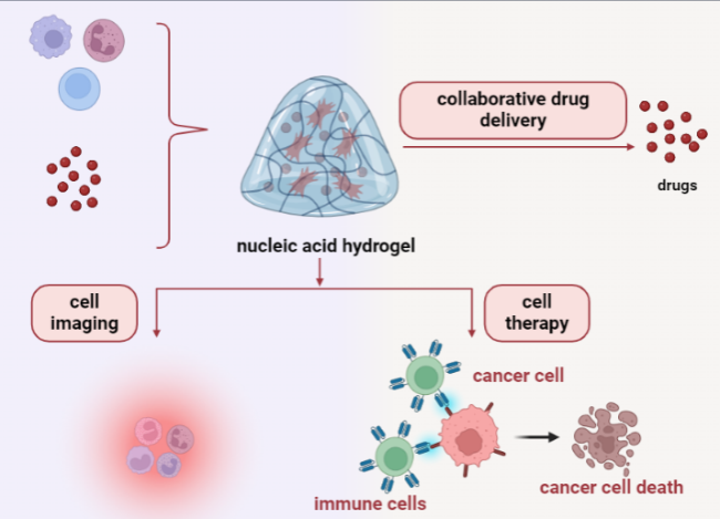
4 Stimulus-responsive nucleic acid hydrogels used for cell capture and release

4.1 Cell imaging

4.2 Cell therapy

4.3 Collaborative drug delivery

5 Conclusion and outlook

Cite this article

Danyu Wang , Mengke Guo , Zihan Guo , Mengyu Huang , Hua Yi , Kaixiang Zhang . Stimulus-Responsive Multifunctional Nucleic Acid Hydrogels Based on Cell Capture and Release[J]. Progress in Chemistry, 2024 , 36(10) : 1567 -1580 . DOI: 10.7536/PC240216

1 Introduction

Based on the programmability, functional diversity, and biocompatibility of nucleic acids, the synthesis of nucleic acid hydrogels and their potential applications in biomedicine have attracted the attention of many researchers. Nucleic acid hydrogels are three-dimensional network polymeric materials formed with the participation of nucleic acids. Because they retain the original biological properties of nucleic acids and the mechanical properties of their own backbone, they have become one of the most focused emerging functional polymer materials in recent years[1]. Nucleic acid hydrogels can be divided into two categories: DNA hydrogels and RNA hydrogels[2]. Among them, RNA hydrogels have poorer stability and a more complex preparation process, which has hindered their widespread application at present. As an important carrier of genetic information in organisms, DNA not only possesses structural diversity and biocompatibility but also relatively high stability. In recent years, with the rapid development of nanotechnology and biotechnology, DNA is considered one of the most promising materials for constructing artificial structures from the nanoscale to the bulk scale. According to the different compositions of nucleic acid hydrogels, they can be classified into two types: pure nucleic acid hydrogels and polymer-nucleic acid hybrid hydrogels[3,4]. Additionally, nucleic acid hydrogels exhibit good hydrophilicity and reversible swelling or deswelling ability in water, meaning they can transition between gel and solution states. On the other hand, nucleic acid hydrogels can achieve controllable stimulus response, undergoing changes in volume and shape under external physical or chemical stimuli[5].
Based on the high biocompatibility and stimulus-responsive deformation characteristics of nucleic acid hydrogels, more and more research is applying them to the capture and release of cells[6]. On one hand, the preparation methods for nucleic acid hydrogels are relatively mild, causing no significant damage to cells, which helps in protecting the physiological functions and structural integrity of the cells. On the other hand, by adjusting the components, structure, and cell loading methods of the hydrogel, different application requirements can be met. More importantly, nucleic acid hydrogels possess highly specific and sensitive recognition capabilities, enabling precise target recognition and localization in applications, capable of converting signals from biomolecules or cell surfaces into detectable signals (such as fluorescence). Additionally, nucleic acid hydrogels can also serve as drug carriers, targeting specific tissues or cells in the form of complexes, thereby achieving precise delivery and release of drugs. Nucleic acid hydrogels can also capture certain immune cells or stem cells, achieving therapeutic purposes at the cellular level. These advantages make nucleic acid hydrogels widely used in biosensing and imaging, drug delivery, and cell therapy.
Based on this, this paper reviews the research progress of stimuli-responsive nucleic acid hydrogels in cell capture and release applications. Firstly, it summarizes the preparation methods of pure nucleic acid hydrogels and polymer-nucleic acid hybrid hydrogels, and reviews the morphological changes that nucleic acid hydrogels undergo under different external stimuli (such as pH, light, temperature, chemical triggers, etc.), providing more theoretical basis for the design of functional nucleic acid hydrogels. Then, the paper focuses on summarizing the applications of stimuli-responsive nucleic acid hydrogels in cell capture and release, including biosensing and cell imaging, drug delivery, and cell therapy, offering ideas for the future development of nucleic acid hydrogels in the field of biomedicine and other areas.

2 Preparation of Nucleic Acid Hydrogels

As research continues to advance, researchers utilize the inherent properties of nucleic acid molecules, introducing other functional molecular elements within them or combining them with other functional biomaterials. To this end, various cross-linking methods have been designed to prepare pure nucleic acid hydrogels or polymer-nucleic acid hybrid hydrogels. The following introduces the preparation methods for these two types of stimuli-responsive nucleic acid hydrogels.

2.1 Pure Nucleic Acid Hydrogel

Pure DNA hydrogels are a type of hydrogel entirely composed of deoxyribonucleotide chains, which are typically formed through hydrogen bonds, physical entanglements, or enzymatic reactions between the chains[3]. In 2006, a research team first reported the successful synthesis of pure DNA hydrogels, designing three types of branched DNA monomers in X, Y, and T shapes[7] (as shown in Figure 1A). Due to these DNA molecules all having complementary sticky ends, they can hybridize with each other and ultimately be linked into a DNA hydrogel with the help of T4 DNA ligase. This study was the first to synthesize pure DNA hydrogels and further demonstrated through experiments that such hydrogels possess tunability and programmability. Importantly, based on these characteristics, different types of DNA hydrogels can be designed to meet various application needs. However, ordinary pure DNA hydrogels have lower sensitivity and take a relatively long time to produce effects. Therefore, developing DNA hydrogels that can rapidly respond to external stimuli is of great significance. For example, Pei Renjun et al.[8] constructed a pure DNA hydrogel using two Y-branched nucleic acid subunits and an aptamer domain for Ochratoxin A. In the presence of Ochratoxin A, this hydrogel underwent a switchable gel-sol transition, and the target molecule could stimulate the rapid degradation of the DNA hydrogel, releasing the encapsulated HRP, thus triggering the H2O2 and ABTS reaction, achieving dual signal amplification in colorimetric detection.
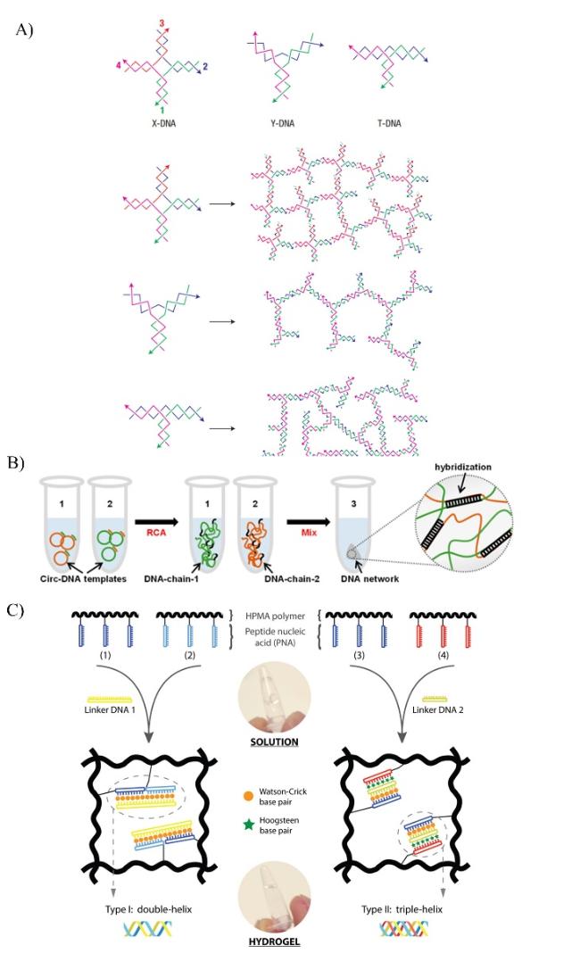
图1 刺激响应型核酸水凝胶的制备。A)X-DNA、Y-DNA和T-DNA构成的核酸水凝胶[7]。 B)通过RCA合成DNA长单链以获得3D DNA网络的过程[11]。C)通过PNA/DNA络合形成的杂化水凝胶[4]

Fig. 1 Preparation of stimulus-responsive nucleic acid hydrogels. A) Nucleic acid hydrogel composed of X-DNA, Y-DNA, and T-DNA[7]. Copyright 2006, Springer Nature B) The process of synthesizing long single strands of DNA by RCA to obtain a 3D DNA network[11]. Copyright 2020, American Chemical Society C) Heterogeneous hydrogels formed by PNA/DNA complexation[4]. Copyright 2015, Elsevier

However, the preparation of pure DNA hydrogels also faces some issues, such as high production costs, complexity, and insufficient stability. Currently, the use of cost-effective, efficient, and accurate nucleic acid amplification methods, especially rolling circle amplification (RCA), for the preparation of pure DNA hydrogels has attracted increasing attention from researchers[9]. RCA is an isothermal nucleic acid amplification technique that can utilize DNA polymerase to cyclically replicate a DNA template under mild conditions, thereby forming long single-stranded DNA (ssDNA) with periodic sequences[10]. Daying Yang et al.[11] reported a method where two ultra-long ssDNAs formed by dual RCA self-assembled into a DNA hydrogel (as shown in Figure 1B). The long ssDNA exhibits high affinity with ALPL protein on the membrane of bone marrow mesenchymal stem cells, enhancing the specificity of cell anchoring, and combined with the moderate storage modulus of the cross-linked network of the DNA hydrogel, it reduces mechanical damage to cells. Similarly, to demonstrate the important role of hydrogen bonds in DNA hydrogels, in 2023, Juan Yan et al.[12] used ssDNA produced by dual RCA as a precursor to prepare pure DNA hydrogels, and by adjusting the degree of hydrogen bonding between ssDNAs, they changed the encapsulation efficiency of AuNPs. This provides new ideas for designing different types of nucleic acid hydrogels and lays the foundation for their applications. However, with the increasing demand for multi-field applications of DNA hydrogels, introducing more isothermal amplification methods and further improving the efficiency, reproducibility, and stability of amplification reactions have become key factors affecting the improvement of DNA hydrogel preparation methods[13].
In addition, using intermolecular G-quadruplexes as the core component of DNA hydrogels can provide sufficient binding force for the construction of nucleic acid hydrogels.[14]With single-stranded and ingeniously designed Y-shaped structures as auxiliary, under normal circumstances, the pH required for the formation and dissociation of the triplet structure based on thymine-adenine-thymine (thymine-adenine-thymine T-A .T) is higher than that of the protonated cytosine-guanine-cytosine (cytosine-guanine- cytosine, C-G .C+) triplet structure. Therefore, such a pure DNA hydrogel can undergo reversible self-assembly of DNA structures controlled by pH, and it can also be converted between gel and liquid states. To further explore the possibilities of pH-stimulus-responsive pure DNA hydrogels, Xiu-Rong Yang et al.[15]introduced both T-A.T and C-G.C+ triplets to construct a pH-activated pure DNA hydrogel, where the triplet structures are the key elements for controlling the formation and dissociation of the pure DNA hydrogel by changing the pH value. At the same time, they incorporated fluorophores and quenchers into the hydrogel, proposing a new strategy for fluorescence detection techniques.

2.2 Polymer-Nucleic Acid Hybrid Hydrogels

DNA hydrogels possess high stimulus responsiveness, encapsulation efficiency, and excellent hydrophilicity, softness, and strong molecular recognition capabilities. It is these advantages that have enabled DNA hydrogels to be applied in many areas over the past nearly 20 years, but there are still some drawbacks that cannot be ignored, such as the limited stability of DNA molecules, negative net charge, and high production costs. Based on this, many researchers have attempted to incorporate nanomaterials like hydrophilic polymers into DNA hydrogels to obtain polymer-nucleic acid hybrid hydrogels with higher biocompatibility and stability and lower costs. For responsive DNA hydrogels, stimulus-responsive DNA sequences or other linkers can be grafted onto polymers, which then self-assemble into a three-dimensional hydrogel through DNA hybridization or other interactions, for example, grafting DNA onto polyacrylamide chains.
In 2015, Kopeček et al.[4] proposed another design concept for hybrid hydrogels. They incorporated multiple peptide nucleic acids (PNA) grafted synthetic water-soluble N- (2-hydroxypropyl) methacrylamide polymers into linker DNA and then crosslinked them (as shown in Figure 1C). The PNA-DNA complex can mediate the self-assembly of polymer chains, leading to the formation of a hydrophilic polymer network. However, when subjected to external stimuli, the interaction between the linkers and the polymers is likely to be disrupted, causing the dissociation of the DNA hybrid hydrogel. In the same year, Willner et al. designed and prepared a polyacrylamide-DNA hybrid hydrogel based on a stimulus-responsive G-quadruplex structure triggered by K+[16]. This hydrogel utilized two crosslinking parts, including self-complementary sticky-ended DNA double strands and guanine-rich DNA sequences that form G-quadruplex structures responsive to stimuli. Moreover, due to the presence of two crosslinks, the hydrogel could form guanine-K+-guanine complexes under conditions where K+ was present, significantly enhancing its stability. This reversible, stimulus-responsive hybrid DNA hydrogel could induce a cyclic transformation among solution, hydrogel, and solid states triggered jointly by G-quadruplex and thermal stimuli. Furthermore, adding 18-crown-6 ether to the hydrogel could eliminate K+ and decompose the G-quadruplex structure, resulting in the formation of an amorphous hydrogel. Among these, the self-complementary dual crosslinks acted as memory codes, allowing the polyacrylamide-DNA hybrid hydrogel to recover its original shape in the presence of K+ and reversibly exhibit K+-responsive phase transitions between gel and quasi-liquid states. These design methods have promoted the development of preparing stimulus-responsive hydrogels.

3 Stimulus-Responsive Nucleic Acid Hydrogels

Nucleic acid hydrogels can be classified into various types based on different stimulus factors, including pH-responsive, light-responsive, temperature-responsive, and chemically triggered types (as shown in Figure 2). These different types of nucleic acid hydrogels can be designed and optimized according to specific application requirements to achieve particular stimulus-responsive behaviors.
图2 不同刺激响应型核酸水凝胶的设计原理

Fig. 2 Design principles of different stimulus-responsive nucleic acid hydrogels. Created with BioRender.com

3.1 pH-Responsive

Based on the tumor-specific acidic microenvironment, pH-responsive DNA hydrogels can specifically control drug release in the tumor microenvironment. The main DNA structures responsive to H+ are G-quadruplexes and i-motifs. The hydrogel backbone is often formed by two ssDNA strands that form G-quadruplex or i-motif structures under acidic conditions, thereby forming a dense DNA hydrogel. In 2009, Dongsheng Liu et al.[17] first reported a pH-responsive hydrogel based on DNA self-assembly. In this study, each end of the Y-shaped DNA contained half of an i-motif sequence, which could form a complete secondary functional structure under acidic conditions, leading to the formation of a pure DNA hydrogel. When the solution pH was high, the i-motif secondary structure dissociated, and the DNA hydrogel would convert into a solution. In 2017, Leilei Tian et al.[18] used RCA technology to introduce pH-sensitive crosslinking sites into the DNA template strand, preparing a pH-responsive hydrogel (as shown in Figure 3A). Because the DNA template for RCA can be freely programmed, it makes the properties of the hydrogel more easily adjustable. These pH-responsive hydrogels, which are simple to prepare, can be used for further research on pH-stimulated drug release and also open up new avenues for the design and development of other intelligent DNA hydrogels.
图3 pH、光和温度响应型核酸水凝胶的应用。A)用于制备pH响应型DNA水凝胶的RCA方法的示意图[18]。B)光控DNA交联水凝胶的机理与设计[21]。C)光热响应的MXene-DNA水凝胶的构建示意图及其应用[27]

Fig. 3 Applications of pH, light, and temperature-responsive nucleic acid hydrogels. A) Schematic diagram of the RCA method used to prepare pH-responsive DNA hydrogels[18]. Copyright 2017, John Wiley and Sons B) Mechanism and design of photocontrolled DNA cross-linked hydrogels[21]. Copyright 2011, American Chemical Society C) Schematic diagram of the construction of photothermally responsive MXene-DNA hydrogels and their application[27]. Copyright 2022, John Wiley and Sons

In addition, specific DNA sequences are closely related to the development of diseases, but how to achieve rapid, low-cost, and highly sensitive target DNA detection remains a significant challenge. Hong et al.[19] combined catalytic hairpin assembly (CHA) technology with capillary action to design a pH-responsive DNA hydrogel for naked-eye detection of target short ssDNA. This system fully amplifies Y-shaped DNA nanostructures through the mutual hybridization of three hairpin DNAs with the target DNA until completely consumed by the CHA cycle. The arms of the final amplified Y-shaped DNA are composed of sticky ends formed by i-motif structures. Notably, under acidic conditions at pH 5, these sequences can self-assemble, forming a target ssDNA and pH dual-responsive DNA hydrogel driven by i-motif cross-linking. This method has high sensitivity for detecting target genes in serum, is effective, and allows for the naked-eye detection of target DNA, thereby overcoming, to some extent, the limitations of expensive analytical equipment and complex operations.

3.2 Light Response

Light is used as a physical stimulus to construct and decompose nucleic acid structures, offering more precise spatiotemporal response characteristics compared to pH. Since the type, power, and wavelength of light can all be precisely regulated by humans, and the stimulation by light is reversible, there has been considerable research work utilizing light-responsive structures to synthesize DNA hydrogels[20]. For example, azobenzene molecules have been employed to design photo-triggered DNA hydrogels. Azobenzene possesses unique photo-controlled properties; under natural light, its conformation changes from cis to trans, while under ultraviolet (UV) conditions, this transformation is reversed. By taking advantage of this characteristic, researchers use azobenzene-functionalized DNA as a crosslinker to build reversible, light-responsive DNA hydrogels. In 2010, Tan Weihong et al.[21] incorporated photosensitive azobenzene as a crosslinker into DNA chains, which were then placed in a DNA-polymer solution, allowing for the formation of DNA hybrid hydrogels that respond differently to various wavelengths of light through the inter-hybridization between the crosslinkers and side-chain DNAs (as shown in Figure 3B). Exposure to external UV light converts azobenzene to benzene, causing the DNA polymer to transition from a gel state to a solution. This study found that light-responsive DNA hydrogels could reversibly control the phase change of the hydrogel through alterations in natural and UV light conditions, thereby enabling precise regulation of cargo loading and release. Additionally, PC-linker, a multifunctional crosslinking agent, can also be used to link DNA strands together via photochemical reactions, making it suitable for constructing light-responsive nucleic acid hydrogels.
In another study, Liu Dongsheng et al.[22]prepared a 1-(4,5-dimethoxy-2-nitrobenzyl) ethoxy photosensitive group-protected thymine deoxyribonucleoside phosphoramidite monomer, which was then introduced into DNA sequences through solid-phase synthesis, thereby achieving optical control of DNA strand complementarity. Subsequently, using this monomer, they successfully prepared a fast photo-responsive DNA supramolecular hydrogel, broadening the diversity of DNA monomers. In addition, photo-responsive molecules can also be used to design responsive DNA hydrogels. Murata et al.[23]fabricated a photo-responsive DNA hydrogel by hybridizing X-shaped DNA with an artificial basecnvK, which could exhibit phase changes at different wavelengths under UV irradiation. When the UV light wavelength is 366 nm, this base can interact with thymine. Notably, the photo-responsive DNA hydrogel undergoes disintegration at 340 nm, allowing for reversible hybridization of the sticky ends and ultimately leading to a gel-sol transition. Furthermore, photo-responsive PVA-DNA hydrogel films can be constructed by crosslinking DNA and polyvinyl alcohol[24]. Upon exposure to UV light and immersion in pure water, the hydrogel film expands. Conversely, if the expanded film is placed in a sodium chloride solution or a cetyltrimethylammonium bromide solution, it will contract. This expansion-contraction process of the DNA film can be repeated multiple times.

3.3 Temperature Response

The hydrogen bonds in the DNA double helix are prone to breaking under high-temperature conditions, leading to DNA denaturation. When the temperature decreases, the double-stranded DNA can re-form into a double helix structure. Utilizing this temperature-sensitive characteristic of DNA, temperature-responsive DNA hydrogels have been widely designed and applied. In 2010, Dongsheng Liu et al.[25] synthesized a DNA hydrogel that is responsive to both heat and enzymes. This hydrogel was assembled through Y-shaped DNA and linkers via sticky-end hybridization. Moreover, when the temperature rises to 50 ℃, the complementary sticky ends of the hydrogel are removed, causing it to transform from a gel to a solution. Additionally, thermoresponsive DNA hydrogels with precise temperature control can be prepared by combining photo-thermal nanomaterials with nucleic acids, making them more suitable for in vivo applications. For instance, Park et al.[26] used gold nanoparticles (AuNPs) with photo-thermal properties to synthesize a self-assembled, temperature-responsive DNA hydrogel for controlled drug release. The electrostatic interactions cause positively charged AuNPs to be adsorbed onto negatively charged DNA strands, and doxorubicin (DOX), an anticancer drug, can also be embedded within, forming an overall drug delivery system. When this system is exposed to light, the AuNPs convert the absorbed light energy into heat, leading to the thermal degradation of the DOX-AuNP-DNA hydrogel into fragments, thereby releasing DOX at the target site. Recently, Weiwei Guo et al.[27] constructed an efficient platform for synergistic photothermal-chemotherapy of tumors using a combination of Ti3C2TX-based MXene and DNA hydrogel as a photo-thermal agent, with DOX as the loaded drug (as shown in Figure 3C). In this setup, the photo-thermal MXene nanosheets, upon exposure to near-infrared light, increase the temperature, causing the MXene-DNA hydrogel to transition into a solution, thus releasing DOX as the DNA crosslinks are broken, a process that can be used for localized tumor treatment.

3.4 Chemical Trigger

In addition to the external response factors mentioned above, DNA hydrogels can also respond to certain chemicals, including metal ions, redox reactions, and enzyme reactions that trigger changes in the system's cross-linking assembly, which can then induce phase transitions.

3.4.1 Metal Ion Response

Metal ions (such as Zn2+, Ag+, Hg2+) can construct double-stranded DNA through bridging base mismatches in DNA sequences, thereby promoting the further development of metal ion-responsive DNA hydrogels. In 2014, Willner et al. from the Hebrew University[28] utilized Ag+ to crosslink DNA for functionalizing polyacrylamide copolymers, thus synthesizing a metal ion-responsive DNA hydrogel. Ag+ could stimulate the formation of cytosine-Ag+-cytosine complexes, leading to the crosslinking of the copolymer and forming a hydrogel. However, after cysteine induction, due to the elimination of Ag+, the cytosine-Ag+-cytosine bridges were disrupted, ultimately causing the DNA hydrogel to dissociate into a solution. In recent years, the detection of toxic ions has received increasing attention; therefore, developing simple and economical detection methods for on-site testing and treatment is of significant importance. Due to their advantages of simple preparation, portability, and easy storage, stimulus-responsive hydrogel materials have been widely used for the detection of metal ions. However, these methods still require additional labeling or embedding to encapsulate signals. To achieve a label-free strategy for Pb2+-mediated biosensing, Tang Wei et al.[29] designed and prepared a Pb2+-responsive DNA hydrogel (as shown in Figure 4A) by introducing a Pb2+-dependent DNAzyme and substrate strand. It was precisely because of the presence of Pb2+ that the enzyme chains within the hydrogel framework were activated, followed by the triggered cleavage of the substrate, ultimately destroying the entire structure of the hydrogel. Nevertheless, the DNA fragments released from the disintegrated DNA hydrogel could output a signal quantifying the concentration of Pb2+, successfully eliminating the necessity for signal molecule labeling or embedding. This Pb2+-responsive DNA hydrogel is simple to operate, convenient to read, and cost-effective. By further adjusting the relevant sequences of the DNAzyme and substrate, it can also be used for the label-free detection and analysis of other toxic metal ions.
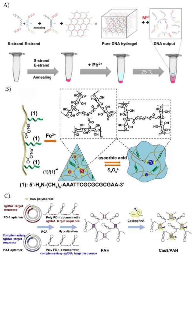
图4 金属离子响应、氧化还原响应和酶响应型核酸水凝胶的应用。A)金属离子响应型纯DNA水凝胶的制备及金属离子检测原理[29]。B)铁3+的组装/铁2+-羧甲基纤维素(CMC)水凝胶和氧化还原触发的水凝胶的受控交联[31]。C)构建Cas9/sgRNA编辑的免疫检查点阻断DNA多核酸适配体水凝胶[35]

Fig. 4 Applications of metal-ion-responsive, redox-responsive, and enzyme-responsive nucleic acid hydrogels. A) Preparation of metal ion-responsive pure DNA hydrogels and the principle of metal ion detection[29]. Copyright 2021, Elsevier B) Assembly of Fe3+/controlled cross-linking of Fe2+-carboxymethylcellulose (CMC) hydrogels and redox-triggered hydrogels[31]. Copyright 2021, Elsevier C) Construction of Cas9/sgRNA-edited immune checkpoint-blocking DNA polynucleic acid aptamer hydrogels[35]. Copyright 2019, Elsevier

3.4.2 Redox Response

Redox groups can also be used to prepare DNA hydrogels, achieving controlled drug release within cells. As a reductant in cells, glutathione can disrupt disulfide redox groups, leading to a phase change in the hydrogel. Tan Weihong and others from Hunan University[30] constructed a glutathione-responsive DNA hydrogel using two Y-shaped monomers A and B with sticky ends and a DNA linker. Notably, monomer A is composed of three DNA single strands, each having three sticky ends; monomer B consists of one sticky end and an aptamer, designed for the control and specific recognition of the DNA hydrogel towards certain cells; the DNA linker is formed by a double-stranded DNA with two sticky ends, which can complementarily bind to the ends of monomers A and B. Ultimately, all components are combined through disulfide bonds, forming spherical nanohydrogels. Moreover, altering the ratio of components A and B can control the DNA nanohydrogel, and secondly, disulfide bonds can be cleaved into fragments under the stimulation of glutathione. In another study, amino-functionalized self-complementary nucleic acids were used to modify carboxymethyl cellulose polymer chains, and under the presence of Fe2+, a stable nucleic acid hydrogel was further obtained, formed by nucleic acid duplexes and Fe3+-tricarboxylate interchain bridging complexes (as shown in Figure 4B)[31]. Ascorbic acid plays a crucial role here, reducing Fe3+ to Fe2+, thus enabling interchain Fe2+-dicarboxylate bridging, significantly lowering the stiffness of the hydrogel. However, the oxidation of the hydrogel with persulfate can regenerate a high-stiffness hydrogel. By cycling the reduction of Fe3+-bridged hydrogel to Fe2+-bridged hydrogel using ascorbic acid and then re-oxidation with persulfate, the hydrogel can switch between low and high stiffness.

3.4.3 Enzyme-Responsive

Programmable stimulus-responsive DNA hydrogels have great potential in the biomedical field, however, due to the relatively slow mass transfer speed and poor mechanical strength within the hydrogel matrix, the response of DNA hydrogels to biomacromolecules is particularly slow, which hinders their development. Guo Weiwei et al.[32]constructed a hierarchically structured DNA hydrogel in a frozen environment, demonstrating significant responsiveness and mechanical properties. By programming the sequence of DNA crosslinkers in the hydrogel, an enzyme-responsive model hydrogel was constructed, which can also synergistically produce biocatalytic cascade reactions with enzymes. In addition, after nucleic acids cleave the phosphodiester bonds of DNA, they can be made enzyme-reactive. Liu Dongsheng et al.[33]introduced an enzyme-responsive hydrogel formed by DNA-functionalized polypeptide chains, where restriction endonucleases can cleave double-stranded DNA at specific bases on the restriction site. By combining the restriction site with the DNA sequence, the DNA hydrogel can specifically respond to restriction endonucleases, thereby triggering a sol-to-solution transition.
In recent years, CRISPR technology has received widespread attention. It is composed of two parts: guide RNA (gRNA) and CRISPR-associated (Cas) proteins, existing in the form of a complex. In this system, gRNA can not only recognize target DNA sequences but also guide Cas nucleases to precisely cut the target DNA at specific sites. Collins et al.[34] designed and prepared a multifunctional CRISPR-Cas12a responsive DNA hydrogel composed of PEG, polyacrylamide, and carbon black. In it, base sequence complementation occurs between gRNA and the target DNA, then activating the Cas12a nuclease to digest the target DNA selectively, ultimately leading to the non-selective hydrolysis of single-stranded DNA crosslinkers. Moreover, under the presence of Cas12a enzyme and gRNA, both PEG-DNA hydrogel and polyacrylamide-DNA hydrogel can achieve customized release of nanoparticles, molecules, and live cells. Additionally, Oh et al.[35] reported a multi-aptamer DNA hydrogel that can be precisely cleaved by Cas9/sgRNA, achieving programmable release of PD-1 aptamers (as shown in Figure 4C). When coexisting with Cas9/sgRNA, double-stranded DNA crosslinkers are cut, causing the PD-1 aptamer hydrogel to lose its original gel properties, leading to its release at specific tumor sites, demonstrating strong anti-tumor potential.

4 Stimuli-Responsive Nucleic Acid Hydrogel for Cell Capture and Release

Stimulus-responsive nucleic acid hydrogels have good biocompatibility and biodegradability, and they can respond to various external physical and chemical stimuli, while exhibiting controllable dynamic behavior. Firstly, by co-culturing cells with the gel matrix, it is possible to capture and fix cells, allowing for further observation of the dynamic changes in cells under the influence of drugs, thereby achieving cell imaging. Notably, stimulus-responsive nucleic acid hydrogels can also be combined with cell therapy, embedding targeted functional sequences or stimulus-responsive structures into DNA hydrogels to enhance their ability to capture or release cells, encapsulating specific types of cells (such as immune cells or stem cells) and delivering them to target tissues, which is conducive to solving problems related to cell localization and survival. In addition, drugs can be encapsulated within nucleic acid hydrogels, and with the help of the stimulus-responsive capability of the hydrogel, the drugs can be precisely released at the target site. This delivery method not only significantly improves the targeting ability and therapeutic effect of the drug but also reduces its side effects in the body. Similarly, nucleic acid hydrogels can also deliver small molecule substances to the body in a targeted manner, achieving therapeutic effects (as shown in Figure 5).
图5 刺激响应型核酸水凝胶在细胞捕获和释放方面的应用

Fig. 5 Application of stimulus-responsive nucleic acid hydrogels for cell capture and release. Created with BioRender.com.

4.1 Cell Imaging

Stimulus-responsive DNA hydrogels can be used for cell imaging. However, to observe the morphology and function of cells in situ (including the processes of fixation, labeling, and imaging), DNA hydrogels need to exhibit good stability during soaking and washing. Liu Dongsheng et al. successfully constructed a dual network through in-situ polymerization, forming a stable, three-dimensional, transparent supramolecular polyacrylamide-DNA hybrid hydrogel that can be used for culturing and observing cells[36]. This DNA hydrogel has a rigid structure that inhibits changes in the distance between crosslinking points in the three-dimensional DNA network, significantly enhancing its tensile and shear strength. The enhanced mechanical properties improve the ability of the DNA hydrogel to undergo staining and washing cycles and allow for the marking of specific areas within cells with multiple colors. The study demonstrated that the controllability of cell localization and density by the DNA hydrogel is beneficial for cell imaging and cell culture. Another study pointed out that an appropriate volume of DNA hydrogel is a necessary condition for it to become an excellent imaging probe or drug delivery carrier. Based on this, Jiang Jianhui et al.[37] reported a nanoscale DNA hydrogel with a protein scaffold, formed through DNA self-assembly based on streptavidin, which can be used for targeted therapy and responsive imaging of tumor cells in clinical settings. The study showed that high levels of adenosine triphosphate (ATP) in the tumor microenvironment activate the hydrogel to produce specific fluorescence signals and release the loaded therapeutic agents, achieving both imaging and treatment effects on cancer cells.
ATP and biothiols play crucial roles in biological systems, with their expression levels closely related to the occurrence of many severe diseases. To more accurately and intuitively detect the concentrations of ATP and biothiols in organisms, Cai-Feng Ding et al.[38] reported a DNA cross-linked hydrogel coated with porous carbon nanospheres (PCN) and dual-color fluorescent probes, which can be used for simultaneous detection and imaging of ATP and glutathione in living cells. After the fluorescent probe is absorbed by living cells, it can specifically bind to the targets and simultaneously present two kinds of fluorescence. Xue-Ji Zhang et al.[39] constructed a porous 3D Au-DNA hydrogel network using different DNAzymes and active metal ions, for the simultaneous imaging of miRNAs within living cells. After cell transfection, specific miRNAs can further trigger strand displacement and sequentially activate the DNAzyme-assisted cyclic reactions, ultimately leading to a significant enhancement in the fluorescence intensity after cell imaging.
In addition, to meet the requirements of structural complexity, precision, cell viability, and scalable manufacturing, Liu Dongsheng et al[40] designed a novel "brick wall" for the fabrication of three-dimensional tissue structures with multiple cell types based on DNA supramolecular hydrogels with excellent biocompatibility and outstanding self-healing properties. The study evaluated the signal response of the DNA hydrogel and found that the hydrogel allows cells to migrate within the three-dimensional structure. This construction strategy can not only accommodate different cell types but also easily remove damaged cells.

4.2 Cell Therapy

Currently, immunotherapy is a relatively ideal method for tumor treatment, as it can overcome the immune tolerance of tumors and simultaneously trigger a strong immune response. However, how to design a system with antigen adaptability, operability, and biodegradability to recruit and activate antigen-presenting cells (APCs) still puzzles many researchers. Li Yanmei et al.[41] designed and prepared an injectable DNA supramolecular hydrogel vaccine (DSHV) inoculation system that demonstrated good biological activity in recruiting and activating APCs both in vitro and in vivo (as shown in Figure 6A). Furthermore, APCs can be activated by locally high concentrations of CpG, thereby triggering a strong immune response and achieving significant antitumor efficacy. In addition, to reduce cell damage caused by conventional methods of capturing circulating tumor cells (CTCs) during tumor treatment, Guo Zijian from Nanjing University[42] utilized aptamer-triggered hybrid chain reactions to prepare 3D porous DNA hydrogels, whose porous structure greatly reduces cell damage. This hydrogel can further encapsulate CTCs and, under ATP-triggered conditions, decompose and release, achieving the capture and release of live CTCs. Moreover, it has been found that functionalized hydrogels constructed using nucleic acid nano-assembly technology can effectively capture T cells and achieve tunable activation of T cell receptors (TCRs)[43]. When the system simultaneously presents three-dimensional force momentum and two-dimensional shear force signals, the activation of TCRs becomes more effective. Immune cells and antigens encapsulated in DNA hydrogels can be delivered into mice together, inducing specific immune responses, and intradermal administration before and after tumor inoculation can delay the cycle of tumor growth in mice without obvious severe adverse reactions. Studies have shown that CTCs contain molecular information about primary tumors and can be used for predicting cancer diagnosis in clinical settings. To overcome the difficulties in capturing live CTCs and their quantification in whole blood, Zuo Xiaolei et al.[44] employed an aptamer-triggered cross-linking reaction to prepare a porous DNA hydrogel for in situ identification, which allows for covering or uncovering CTCs for live cell analysis. This hydrogel can recognize a small number of CTCs in whole blood, enabling high-sensitivity and high-specificity clinical diagnosis, and also provides a basis for cell therapy.
图6 刺激响应核酸水凝胶在细胞治疗方面的应用。A)DSHV 系统诱导的宿主 APC 在体内募集和激活以产生强大的免疫反应和抗肿瘤作用的示意图[41]。B)光反应型核酸水凝胶用于肿瘤治疗的示意图[45]。C)多功能DNA敷料促进烧伤创面愈合的机制[48]

Fig. 6 Application of stimulus-responsive nucleic acid hydrogels in cell therapy. A) Schematic diagram of DSHV system-induced host APCs recruiting and activating in vivo to produce robust immune responses and antitumor effects[41]. Copyright 2018, American Chemical Society B) Schematic diagram of embedded photoreactive nucleic acid hydrogel for tumor treatment[45]. Copyright 2023, Springer Nature C) Mechanism of multifunctional DNA dressings to promote burn wound healing[48]. Copyright 2022, JohnWiley and Sons

It is noteworthy that Zhang Kaixiang et al[45]reported a DNA hydrogel containing a PDL1 aptamer, which can effectively capture and enrich tumor cells, increasing the local ATP concentration to achieve timely signal warning (as shown in Figure 6B). Furthermore, when a positive signal is detected in the body, localized laser irradiation can be used to trigger photodynamic therapy (PDT), a process that not only kills the captured tumor cells but also releases tumor-associated antigens. In addition, PDT is a promising method for cancer treatment; however, due to the limited penetration of external radiation and the complexity of the tumor microenvironment, the antitumor efficiency of photodynamic therapy is greatly restricted. To improve tumor treatment outcomes, Yang Dayong et al from Tianjin University[46]constructed an energy storage hydrogel based on two ultra-long single-stranded DNA chains, which can selectively sensitize PDT in tumor areas without external radiation, thereby producing tumor antigens to stimulate tumor immune responses. This study represents a new paradigm in anticancer photodynamic immunotherapy.
In addition, the efficient separation of high-purity and low-cell-damage immune cells is very important for immunotherapy, but it still faces significant challenges. Based on this, Dàyǒng Yǎng et al[47] designed a cell-capture DNA network for the specific separation of T cells. In this, two ultra-long DNA strands synthesized through an enzymatic amplification process served as cell anchors and immune adjuvants. Moreover, the mutually complementary sequences promoted the formation of the DNA network and the encapsulation of T cells, facilitating the reactive release of T cells and immune adjuvants. At the same time, this network improved the purity of captured tumor-infiltrating T cells, enhanced the biological activity of T cells, and contributed to the realization of immunotherapy.
Apart from tumor treatment, nucleic acid hydrogels also have important applications in skin therapy and tendon repair. Currently, the primary clinical method for treating severe burns is skin grafting, but the overall effectiveness in treating burn symptoms remains unsatisfactory. Wen Yongqiang et al.[48] synthesized a multifunctional DNA hydrogel based on dynamically crosslinked DNA units, L-ascorbic acid 2-phosphate, and polyacrylamide with dense hydrogen bonding to treat skin burns (as shown in Figure 6C). This hydrogel provides a suitable environment for the growth of stem cells in vivo, promoting cell proliferation and maintaining their original activity, while also enabling effective release of the stem cells. Meanwhile, the DNA hydrogel system can also promote macrophage transformation, angiogenesis, and neurogenesis, which are beneficial for skin tissue regeneration. Notably, by incorporating borneol, this system can alleviate pain and itching at the injury site, providing auxiliary benefits for patient treatment. Additionally, stimuli-responsive nucleic acid hydrogels also exhibit specific responses to the environmental conditions (such as pH, temperature, etc.) of diabetic chronic wound areas[49]. The hydrogel shows excellent therapeutic potential in the healing of diabetic wounds. However, due to the fact that cells or cytokines loaded in wound dressings typically function only at specific stages of healing, strict time control is required in practical applications. Furthermore, for tendon injury repair, the use of tendon stem/progenitor cells (TSPCs) is currently the most promising treatment strategy, as it can regulate the metabolic microenvironment of the tendon and promote the synthesis of cellular matrix and collagen. However, the therapeutic effect of injected TSPCs may be suboptimal due to the poor microenvironment of the tendon, sliding shear, and inadequate nutrient supply around the tendon. Therefore, Wang Guanglin et al.[50] reported a DNA hydrogel delivery system encapsulating TSPCs, which not only provides a favorable extracellular matrix microenvironment necessary for proliferation and shear resistance, but also significantly prolongs the retention time of TSPCs in the tendon.
In addition, hydrogels can also serve as synthetic models of the extracellular matrix to study how mechanical signals regulate cell behavior. Based on this, Dongsheng Liu and others from Tsinghua University[51] designed a hydrogel with univariate stiffness regulation using DNA and its enantiomers. The research shows that the degradation of hydrogels may affect the differentiation of neural cells by enhancing intercellular interactions, but the stiffness of the hydrogel itself does not have an impact. Furthermore, Yuanchen Dong and others[52] used a series of DNA nanomodules to adjust the stiffness of pure DNA supramolecular hydrogels, studying the relationship between the stiffness of DNA molecules and macroscopic stiffness, as well as the effect of the connection mode of network cross-linking points on the stiffness of DNA hydrogels. This research lays a theoretical foundation for the potential biological applications of programmable intelligent materials.

4.3 Collaborative Drug Delivery

The complex in vivo environment and the low concentration of inducers have largely limited the development of delivering therapeutic drugs to target sites. Nucleic acid hydrogels possess excellent mechanical properties, but their large volume and irregular shape affect their ability for in vivo drug delivery. With the continuous advancement of research, the controlled drug release performance of DNA hydrogels has been continuously improved. Floxuridine (F) is an important cytotoxic nucleoside analog that plays an anticancer role. Its structure is similar to that of natural nucleosides, making it easy to integrate into DNA or RNA. Zhang Chuan et al.[53] self-assembled DNA and RNA hydrogels and effectively integrated F into the nucleic acid chains through solid-phase synthesis or enzyme-mediated methods. This drug delivery system retains the advantages of original molecular recognition, while the nucleic acid gel can be captured and absorbed by tumor cells, thereby releasing the therapeutic drug and exhibiting excellent antitumor activity. In addition, the chemotherapeutic drug DOX also plays a significant role in tumor treatment, but it also has the disadvantage of non-specific delivery leading to side effects. Recently, to construct a highly efficient targeted drug delivery therapeutic platform, Chen Tingmei et al.[54] prepared a dual-target and multivalent aptamer-modified DNA hydrogel capable of loading the anticancer drug DOX. The DNA hydrogel integrates two aptamers, HER2 and AS1411, enhancing the overall targeting of the system. After reaching the target cells, it not only releases DOX and aptamer nucleic acid drugs but also induces the degradation of the HER2 protein, which is helpful for the treatment of HER2-positive breast cancer. Furthermore, compared with other synthetic drugs, peptide drugs have lower immunogenicity and production costs, but their clinical application is greatly limited due to off-target delivery and unnecessary leakage-induced side effects. To address these issues, researchers[55] functionalized nanoscale DNA hydrogels and designed a peptide drug delivery carrier. On one hand, the study loaded the cell-penetrating anticancer peptide drug Buforin IIb into the hydrogel network via electrostatic interactions, and then assembled it with photothermal agent AnNPs, which can serve as a photo-triggered peptide drug release. On the other hand, integrating the cancer-targeting YNGRT sequence into the DNA hydrogel enables targeted delivery to cancer cells. This study lays a good experimental foundation for achieving safe, cancer-specific targeting, and efficient peptide drug delivery.
In addition, as an FDA-approved oral hypoglycemic drug, metformin (MET) has also shown good therapeutic potential in the treatment of osteoarthritis (OA). However, MET cannot overcome the severe inflammatory environment within the joint cavity, leading to its clearance before reaching the therapeutic site. Therefore, Changhai Ding et al.[56] used DNA supramolecular hydrogel as a carrier for sustained delivery of MET to treat OA, which extended the retention time of MET in the joint cavity to 14 days and significantly enhanced the anti-inflammatory effect. Moreover, due to the repeatability and biodegradability of DNA hydrogels, they have great potential as carriers for periodontal diseases. In particular, physically cross-linked DNA hydrogels without any chemical modifications exhibit minimal cytotoxicity and satisfactory biocompatibility. Thus, Xiaofeng Zheng et al.[57] utilized physically cross-linked DNA hydrogels as scaffolds to construct a biodegradable, anti-inflammatory, and osteogenic interleukin hydrogel for the long-term sustained release of the cytokine interleukin-10, thereby accelerating the reconstruction of diabetic alveolar bone. The results showed that this hydrogel could promote M2 macrophage polarization, alleviate periodontal inflammation, and accelerate the defect healing rate of diabetic alveolar damage.
In recent years, as a new type of therapeutic tool, injectable nucleic acid hydrogels have demonstrated excellent self-healing and sustained release properties. Paul et al.[58] designed and prepared DNA composite hydrogels based on reversible imine bonds and oxidized alginate crosslinking, which can serve as an injectable carrier for the sustained and effective delivery of the drug simvastatin. If charged silicate nanoparticles interact electrostatically with the phosphate groups in the DNA hydrogel, the shear strength of the DNA composite hydrogel is further enhanced, and the release time of the drug simvastatin can last for more than one week. At the same time, hydrogels are also ideal dressings for wound healing, providing a microenvironment similar to the extracellular matrix, which is conducive to cell proliferation and adhesion. Therefore, Wang Pengfei et al.[59] constructed a multifunctional DNA hydrogel (as shown in Figure 7A) with good durability and slow-release performance by utilizing the C—C bridge formed through the mismatch complexation of cytosine (cytosine, C)—Ag+—C and Ag+. Notably, the FKN aptamer carried by this hydrogel can recruit M2 macrophages via G-coupled protein receptors, causing the inflammatory process to prematurely transition into a proliferative process, significantly reducing the time required for wound healing. In vivo experiments in rats have once again confirmed that the bifunctional DNA hydrogel has the ability and potential to accelerate skin tissue regeneration and wound healing.
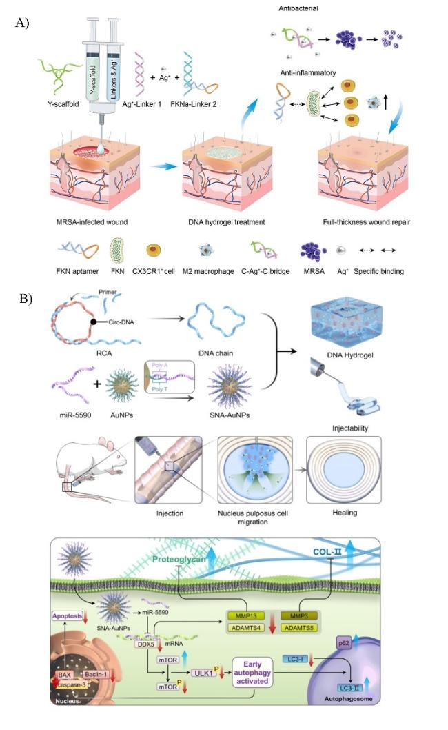
图7 核酸水凝胶在协同药物递送方面的应用。A)DNA-FKNa/Ag水凝胶修复MRSA感染伤口示意图[59]。B)miR-5590-SNA@DNAgel的制备和应用[66]

Fig. 7 Application of nucleic acid hydrogels in synergistic drug delivery. A) Schematic diagram of DNA-FKNa/Ag hydrogel repair of MRSA infected wound[59]. Copyright 2023, Elsevier B) Preparation and application of miR-5590- SNA@DNAgel[66]. Copyright 2023, Springer Nature

In addition, DNA hydrogels containing cytosine-phosphate-guanine (CpG) motifs can play a significant role as vaccine adjuvants. However, these hydrogels cannot pass through the gastrointestinal tract normally, leading to greatly limited efficacy after oral administration. Chitosan, with its adhesive properties, can significantly improve the stability and retention time of hydrogels in the stomach when used as a coating. Based on this, Nishikawa et al.[60] prepared chitosan in advance using an emulsification/coating method and then coated the hydrogel microspheres to enhance the overall stability of the delivery system. Moreover, chitosan exhibits cationic properties in the acidic environment of the stomach, which to some extent reduces its own degradation loss and promotes the oral delivery of CpG DNA. Additionally, Cryj1, a cedar pollen antigen, can be loaded into stimuli-responsive CpG DNA hydrogels, giving Cryj1 a sustained-release property[61]. Furthermore, to prepare DNA hydrogels that are also stable in acidic environments, A*STAR Senior Researcher Ying et al.[62] designed copolymers containing adenine-rich-cytosine-rich oligonucleotides and then prepared acid-resistant and pH-responsive DNA hydrogels through crosslinking. By coating insulin with DNA hydrogel, it can reach the stomach (pH 1.2), duodenum (pH 5.0), and small intestine (pH 7.2) of diabetic rats via oral administration, proving the potential application of this DNA in oral drug delivery for treatment.
Moreover, therapeutic RNA molecules play a crucial role in tumor treatment. However, naked RNA is extremely unstable and is easily degraded by nucleases in the body, making it difficult to enter cells and exert its function. Based on this, Bum et al. from the University of Seoul[63] prepared DNA-RNA hybrid hydrogels through the hybridization of short hairpin RNAs (shRNAs) with DNA aptamers. These hydrogels possess flexibility, robustness, and injectability. The hydrogel can mimic the microstructure within the body and be designed to sequentially release siRNA-aptamer complexes (SACs). In this study, by encoding specific sites for restriction endonucleases, the hybrid hydrogel could release SACs, achieving effective delivery of RNA, which suggests the application potential of RNA therapy. Additionally, single-stranded RNA rich in uridine and guanosine can activate Toll-like receptors (TLR7 and TLR8), triggering a strong immune response in the body. To continuously and effectively deliver uridine-rich RNA to immune cells, another research team constructed an RNA/DNA hydrogel by mixing two sets of hexapod-like uridine-rich RNA/DNA nanostructures, achieving sustained release of uridine-rich RNA[64]. Using mouse experiments, the study demonstrated that uridine-rich RNA loaded into the hydrogel could induce a significant release of tumor necrosis factor-α from DC 2.4 dendritic cells in mice.
Secondly, miRNA can also be used for anti-tumor therapy. In 2021, to address the problem of tumor recurrence caused by hypoxia-induced tumor metastasis, Li Xuemei and others from Linyi University[65]prepared an RNA hydrogel based on two types of anti-tumor molecules: tumor-suppressive miRNA (miRNA-205) and anti-tumor metastasis miRNA (miRNA-182). This hydrogel can co-deliver CpG DNA and shRNA adjuvants, MnO2-loaded photosensitizer Ce6 (MnO2@Ce6), and DOX to MDA-MB-231 cells. After being degraded by enzymes in the body, the hydrogel releases a large amount of therapeutic molecules, triggering the decomposition of H2O2in the tumor, further alleviating the hypoxic condition of the tumor, and simultaneously triggering an anti-tumor immune response in the body. In addition, microRNAs, especially miRNA-5590, show significant differential expression in degenerative nucleus pulposus, which directly affects the expression of the 3′ UTR of DEAD (Asp-Glu-Ala-Asp) box helicase 5, and in turn, leads to the phosphorylation of mammalian target of rapamycin, greatly affecting cell autophagy and apoptosis. However, how to successfully deliver miRNA to the target site remains a challenge. Song Jie and others from the Cancer Hospital of the Chinese Academy of Medical Sciences[66]designed a multifunctional DNA hydrogel loaded with spherical nucleic acid carrying miR-5590 for the treatment of intervertebral disc degeneration (as shown in Figure 7B). Direct injection of the hydrogel at the injury site would ensure consistent and prolonged release of miR-5590, while also inducing autophagy in nucleus pulposus cells, thereby inhibiting cell apoptosis.
In addition, based on the gelation process and the designable responsive properties, DNA hydrogels have been used for encapsulating and releasing nanoscale substances. However, encapsulating and releasing microscale objects remains a significant challenge. Due to its simplicity and versatility, capturing single cells in microwells has been widely utilized. Nevertheless, single cells in microwells are prone to cross-contamination, and the membranes sealing the microwells, mostly impermeable, may negatively impact cell behavior. Therefore, Dongsheng Liu et al. from Tsinghua University[67] prepared an enzyme-triggered permeable DNA hydrogel as a cover, through which nutrients and waste can pass, allowing the survival of cells encapsulated in microwells. Moreover, the network of the DNA hydrogel can be specifically digested by restriction endonucleases, enabling controlled release of the encapsulated cells. This study provides a platform for the culture, detection, and manipulation of single cells and may find potential applications in cell communication research.

5 Conclusions and Future Prospects

In summary, this review elaborately introduces the preparation principles and synthesis methods of pure nucleic acid hydrogels and polymer-nucleic acid hybrid hydrogels, followed by a discussion on the design and application of various stimulus-responsive nucleic acid hydrogels, such as those responsive to pH, light, temperature, and chemical triggers (metal ions, redox, etc.). The article focuses on the applications of stimulus-responsive nucleic acid hydrogels in cell capture and release, including cell imaging, drug delivery, and cell therapy (as shown in Table 1).
表1 刺激响应型核酸水凝胶在生物医学方面的应用

Table 1 Application of stimulus responsive nucleic acid hydrogel in biomedicine

Areas of application Type of stimulus Principle of action Ref
Cell
imaging
Chemical trigger ATP activates fluorescent signalling 36
pH response Fluorescent marker
release
37
Cell
therapy
Chemical trigger CpG activates APC and the immune response 40
ATP triggers the breakdown and release of CTCs 41
Increased local ATP concentration and signalling early warning 44
light
response
Trigger Cell Photodynamic Therapy 44
pH, temperature response
Reacts specifically with the wound wound area 48
Drug
delivery
light
response
Delivery of cell-penetrating anticancer peptide drugs 54
pH response Loaded drug reaches the site of acidic pathology 61
chemical trigger Specific sites encoding
restriction
endonucleases
62
Decomposed by biological enzymes in the body to release therapeutic drugs 64
At present, researchers are adjusting the physical and chemical properties of hydrogels by altering their nanostructures, such as through the self-assembly of DNA, the incorporation of carbon nanotubes, and so on. In addition, they are also exploring how to combine different nanomaterials with nucleic acid hydrogels to achieve more efficient energy conversion and superior mechanical properties, as well as their application in the biomedical field. This article focuses on cell capture and release, which is conducive to an in-depth discussion of the applications of nucleic acid hydrogels in the biomedical field and provides valuable research ideas for the development of new biomaterials. However, there are still many issues regarding the preparation and application of nucleic acid hydrogels. It is hoped that future researchers will pay more attention to improving the stability and mechanical properties of nucleic acid hydrogels, enhancing the controllable degradation of nucleic acids, and achieving effective drug delivery and release. This paper aims to discuss advanced preparation methods for nucleic acid hydrogels and their application progress based on cell capture and release, hoping to provide references and inspiration for relevant research in the future.
[1]
Li J, Zhang Y Z, Zhu L J, Chen K R, Li X Y, Xu W T. Int. J. Mol. Sci., 2022, 23(3): 1068.

[2]
Wang F J, Xie M, Huang Y Y, Liu Y H, Liu X L, Zhu L J, Zhu X Y, Guo Y Y, Zhang C. Angew. Chem. Int. Ed., 2024, 63(4): 2315282.

[3]
Jian X T, Feng X Y, Luo Y N, Li F J, Tan J Y, Yin Y L, Liu Y. Front. Bioeng. Biotechnol., 2021, 9: 661409.

[4]
Chu T W, Feng J Y, Yang J Y, Kopeček J. J. Control. Release. 2015, 220: 608.

[5]
Ahmed E M. J. Adv. Res., 2015, 6(2): 105.

[6]
Wei Y H, Wang K Z, Luo S H, Li F, Zuo X L, Fan C H, Li Q. Small, 2022, 18(36): e2107640.

[7]
Um S H, Lee J B, Park N, Kwon S Y, Umbach C C, Luo D. Nat. Mater., 2006, 5(10): 797.

[8]
Zhou L, Sun N, Xu L J, Chen X, Cheng H, Wang J N, Pei R J. RSC Adv., 2016, 6(115): 114500.

[9]
Schneider L, Richter M, Oelschlaeger C, Rabe K S, Domínguez C M, Niemeyer C M. Chem. Commun., 2023, 59(81): 12184.

[10]
Tang J, Liang A, Yao C, Yang D. Chem. Eur. J., 2023, 29 (9): e202202673.

[11]
Yao C, Tang H, Wu W J, Tang J P, Guo W W, Luo D, Yang D Y. J. Am. Chem. Soc., 2020, 142(7): 3422.

[12]
Wang X Y, Wang H Y, Zhang H M, Yang T X, Zhao B, Yan J. Biosensors, 2023, 13(7): 755.

[13]
Ma X, Zhang Y Z, Xu W T. Curr. Biotechnol., 2019, 9(6): 554.

(马翾, 张洋子, 许文涛. 生物技术进展, 2019, 9(6): 554.)

[14]
Huang Y S, Xu W L, Liu G Y, Tian L L. Chem. Commun., 2017, 53(21): 3038.

[15]
Lu S S, Wang S, Zhao J H, Sun J, Yang X R. Chem. Commun., 2018, 54(36): 4621.

[16]
Lu C H, Guo W W, Qi X J, Neubauer A, Paltiel Y, Willner I. Chem. Sci., 2015, 6(11): 6659.

[17]
Cheng E J, Xing Y Z, Chen P, Yang Y, Sun Y W, Zhou D J, Xu L J, Fan Q H, Liu D S. Angew. Chem. Int. Ed., 2009, 48(41): 7660.

[18]
Xu W L, Huang Y S, Zhao H R, Li P, Liu G Y, Li J, Zhu C S, Tian L L. Chem., 2017, 23(72): 18276.

[19]
Jeong J Y, Do J Y, Hong C A. Microchim. Acta, 2022, 189: 34.

[20]
Lu S S, Shen J L, Fan C H, Li Q, Yang X R. Adv. Sci., 2021, 8(13): 2100328.

[21]
Kang H Z, Liu H P, Zhang X L, Yan J L, Zhu Z, Peng L, Yang H H, Kim Y, Tan W H. Langmuir, 2011, 27(1): 399.

[22]
Yang B, Sun L M, Pan Y F, Dong Y C, Sun Y W, Liu D S. Acta Polymerica Sinica, 2021, 52 (8): 996.

(杨勃, 孙立梅, 潘玙璠, 董原辰, 孙亚伟, 刘冬生. 高分子学报, 2021, 52 (8):996.)

[23]
Kandatsu D, Cervantes-Salguero K, Kawamata I, Hamada S, Nomura S I M, Fujimoto K, Murata S. ChemBioChem, 2016, 17(12): 1118.

[24]
Cheng P, Zhang Y Z, Ma X, Chen X, Zhu B Q, Xu W T. China Biotechnol., 2020, 40 (3):132.

(程平, 张洋子, 马翾, 陈旭, 朱保庆, 许文涛. 中国生物工程杂志, 2020, 40 (3):132. )

[25]
Xing Y Z, Cheng E J, Yang Y, Chen P, Zhang T, Sun Y W, Yang Z Q, Liu D S. Adv. Mater., 2011, 23(9): 1117.

[26]
Song J, Hwang S, Im K, Hur J, Nam J, Hwang S, Ahn G O, Kim S, Park N. J. Mater. Chem. B. 2015, 3(8): 1537.

[27]
He P P, Du X X, Cheng Y, Gao Q, Liu C, Wang X W, Wei Y H, Yu Q L, Guo W W. Small, 2022, 18(40): 2200263.

[28]
Guo W W, Qi X J, Orbach R, Lu C H, Freage L, Mironi-Harpaz I, Seliktar D, Yang H H, Willner I. Chem. Commun., 2014, 50(31): 4065.

[29]
Chu J, Chen C, Li X J, Yu L B, Li W J, Cheng M X, Tang W, Xiong Z H. Anal. Chim. Acta, 2021, 1157: 338400.

[30]
Li J, Zheng C, Cansiz S, Wu C C, Xu J H, Cui C, Liu Y, Hou W J, Wang Y Y, Zhang L Q, Teng I T, Yang H H, Tan W H. J. Am. Chem. Soc., 2015, 137(4): 1412.

[31]
Wang C, Willner B, Willner I. React. Funct. Polym., 2021, 166: 104983.

[32]
Du X X, Bi Y H, He P P, Wang C Y, Guo W W. Adv. Funct. Mater., 2020, 30(51): 2006305.

[33]
Li C, Faulkner-Jones A, Dun A R, Jin J, Chen P, Xing Y Z, Yang Z Q, Li Z B, Shu W M, Liu D S, Duncan R R. Angew. Chem., 2015, 127(13): 4029.

[34]
English M A, Soenksen L R, Gayet R V, de Puig H, Angenent-Mari N M, Mao A S, Nguyen P Q, Collins J J. Science, 2019, 365(6455): 780.

[35]
Lee J, Le Q V, Yang G, Oh Y K. Biomaterials, 2019, 218: 119359.

[36]
Cao T Y, Jia H Y, Dong Y C, Gui S B, Liu D S. ACS Appl. Mater. Interfaces, 2020, 12(4): 4185.

[37]
Li N, Wang X Y, Xiang M H, Liu J W, Yu R Q, Jiang J H. Anal. Chem., 2019, 91(4): 2610.

[38]
Ji X T, Wang Z B, Niu S Y, Ding C F. Chem. Commun., 2020, 56(39): 5271.

[39]
Meng X D, Zhang K, Dai W H, Cao Y, Yang F, Dong H F, Zhang X J. Chem. Sci., 2018, 9(37): 7419.

[40]
Wang Y J, Shao Y, Ma X Z, Zhou B N, Faulkner-Jones A, Shu W M, Liu D S. ACS Appl. Mater. Interfaces, 2017, 9(14): 12311.

[41]
Shao Y, Sun Z Y, Wang Y J, Zhang B D, Liu D S, Li Y M. ACS Appl. Mater. Interfaces, 2018, 10(11): 9310.

[42]
Guo Z J. Chem. Res. Chin. Univ., 2020, 36(5): 981.

[43]
Umeki Y, Saito M, Kusamori K, Tsujimura M, Nishimura M, Takahashi Y, Takakura Y, Nishikawa M. J. Control. Release. 2018, 288: 189.

[44]
Song P, Ye D K, Zuo X L, Li J, Wang J B, Liu H J, Hwang M T, Chao J, Su S, Wang L H, Shi J Y, Wang L H, Huang W, Lal R, Fan C H. Nano Lett., 2017, 17(9): 5193.

[45]
Wang D Y, Liu J W, Duan J, Yi H, Liu J J, Song H W, Zhang Z Z, Shi J J, Zhang K X. Nat. Commun., 2023, 14: 4511.

[46]
Zhao H X, Wang Z Y, Yang S, Zhang R, Guo J F, Yang D Y. Biomaterials, 2024, 309: 122620.

[47]
Yao C, Zhu C X, Tang J P, Ou J H, Zhang R, Yang D Y. J. Am. Chem. Soc., 2021, 143(46): 19330.

[48]
Zhou L P, Zeng Z H, Liu S Y, Min T T, Zhang W M, Bian X C, Du H W, Zhang P X, Wen Y Q. Adv. Funct. Mater., 2022, 32(46): 2207466.

[49]
Pan G Y, Zhang J A D, Liang Y P, Guo B L. Journal of Sichuan University (Medical Sciences), 2023, 54 (4):726.

(潘国莹, 张吉傲笛, 梁永平, 郭保林. 四川大学学报(医学版), 2023, 54 (4):726.)

[50]
Ge Z L, Li W, Zhao R L, Xiong W, Wang D, Tang Y F, Fang Q, Deng X T, Zhang Z, Zhou Y J, Chen X T, Li Y, Lu Y R, Wang C S, Wang G L. Small, 2023, 19(32): 2207231.

[51]
Zhou B N, Yang B, Liu Q, Jin L, Shao Y, Yuan T Y, Zhang Y N, Wang C, Shi Z W, Li X, Pan Y F, Qiao N, Xu J F, Yang Y R, Dong Y C, Xu L J, Gui S B, Liu D S. J. Am. Chem. Soc., 2023, 145(16): 8954.

[52]
Shi Z W, Li Y J, Du X J, Liu D S, Dong Y C. Nano Lett., 2024, 24(28): 8634.

[53]
Ma Y, Liu H X, Mou Q B, Yan D Y, Zhu X Y, Zhang C. Nanoscale, 2018, 10(18): 8367.

[54]
Jin F F, Zeng Q, Qian H S, Zhang D, Wei Y, Wang Y G, Chai C S, Cheng W, Ding S J, Chen T M. Front. Pharmacol., 2022, 13: 807498.

[55]
Quazi M Z, Park N. Biomacromolecules, 2023, 24(5): 2127.

[56]
Zhang C, Huang H, Chen J M, Zuo T T, Ou Q H, Ruan G F, He J, Ding C H. ACS Appl. Mater. Interfaces, 2023, 15(13): 16369.

[57]
Li W, Wang C S, Wang Z H, Gou L P, Zhou Y, Peng G, Zhu M, Zhang J Y, Li R Q, Ni H F, Wu L, Zhang W L, Liu J Y, Tian Y L, Chen Z, Han Y P, Tong N W, Fu X H, Zheng X F, Berggren P O. ACS Appl. Mater. Interfaces, 2022, 14(22): 25173.

[58]
Basu S, Pacelli S, Paul A. Acta Biomater., 2020, 105: 159.

[59]
Gao F, Ma X W, Wang F K, Zhou F, Ye J, Yang D L, Li M, Wang P F. Chem. Eng. J., 2023, 470: 144347.

[60]
Nomura D, Saito M, Takahashi Y, Takahashi Y, Takakura Y, Nishikawa M. Int. J. Pharm., 2018, 547(1-2): 556.

[61]
Tanifuji T, Nishimura M, Kusamori K, Nishikawa M. J. Control. Release. 2023, 354: 429.

[62]
Hu Y W, Gao S J, Lu H F, Ying J Y. J. Am. Chem. Soc., 2022, 144(12): 5461.

[63]
Han S, Park Y, Kim H, Nam H, Ko O, Lee J B. ACS Appl. Mater. Interfaces, 2020, 12(50): 55554.

[64]
Komura F, Okuzumi K, Takahashi Y, Takakura Y, Nishikawa M. Molecules, 2020, 25(3): 728.

[65]
Wang W C, Liu X F, Ding L R, Jin H J, Li X M. Front. Chem., 2021, 9: 797094.

[66]
Song Q X, Jiang K, Zheng D D, Jin L Y, Chen X Y, Feng Y B, Wang K, Han Y C, Chen H, Song J, Chen Z, Shen H X. J. Nanobiotechnol., 2023, 21: 350.

[67]
Jin J, Xing Y Z, Xi Y L, Liu X L, Zhou T, Ma X X, Yang Z Q, Wang S T, Liu D S. Adv. Mater., 2013, 25(34): 4714.

Outlines

/