Home Journals Progress in Chemistry
Progress in Chemistry

Abbreviation (ISO4): Prog Chem      Editor in chief: Jincai ZHAO

About  /  Aim & scope  /  Editorial board  /  Indexed  /  Contact  / 
Review

Precision Detection of Tumor Small Extracellular Vesicles

  • Huijing Wang ,
  • Die Sun ,
  • Ruiying Wang ,
  • Hui Zhang , *
Expand
  • College of Chemistry and Materials Science, Nanjing Normal University, Nanjing 210023, China
* e-mail:

Received date: 2024-03-11

  Revised date: 2024-06-21

  Online published: 2024-06-30

Supported by

National Natural Science Foundation of China(22274077)

Abstract

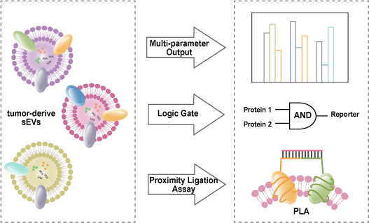
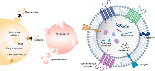
Tumor small extracellular vesicles (sEVs) are membranous vesicles, released by tumor cells, with a particle size less than 200 nm. They carry diverse biomolecular information on their surface and inside, participating in intercellular communication and are recognized as one of the most crucial liquid biopsies for cancer. Because sEVs’ surface contains a variety of proteins that can bind to corresponding antibodies or nucleic acid aptamers, quantitative detection of sEVs can be achieved through optical or electrochemical methods. However, due to the high heterogeneity and complexity of sEVs, relying on a single protein for recognition may lead to false positive or false negative signals. Therefore, accurate detection of tumor-derived sEVs requires simultaneous analysis of multiple biomarkers. Simultaneous analysis of multiple biomarkers can effectively address interference caused by phenotypic heterogeneity in sEVs and provide more accurate guidance for cancer diagnosis and prognosis. This paper focuses on the detection methods of sEVs based on surface proteins using fluorescence, colorimetry, electrochemical methods, and electrochemiluminescence techniques. It emphasizes the importance of achieving high sensitivity and accuracy in detecting sEVs through multi-protein multi-signal proportional output approaches, employing multi-protein logic gates and multi-protein proximity linking reactions.

Contents

1 Introduction

2 General analysis method of small extracellular vesicles

2.1 Fluorometry

2.2 Colorimetry

2.3 Electrochemical method

2.4 Electrochemical luminescence

3 Precision detection of small extracellular vesicles based on surface multiproteins

3.1 Multiprotein proportional output

3.2 Multiprotein Logical Computation Output

3.3 Multiprotein proximity ligation reaction

4 Conclusion and outlook

Cite this article

Huijing Wang , Die Sun , Ruiying Wang , Hui Zhang . Precision Detection of Tumor Small Extracellular Vesicles[J]. Progress in Chemistry, 2024 , 36(12) : 1972 -1982 . DOI: 10.7536/PC240318

1 Introduction

Extracellular vesicles (EVs) are a general term for various vesicles with phospholipid bilayer structures released by cells. Based on their size and formation method, they are classified into three types: exosomes (50-150 nm), microvesicles (MVs, 100-1000 nm), and apoptotic bodies (500-5000 nm)[1]. Among them, exosomes are produced through the continuous invagination of the cytoplasmic membrane[2], as shown in Figure 1. Because it is difficult to completely separate exosomes from microvesicles of similar size, researchers in recent years have generally referred to extracellular vesicles smaller than 200 nm as small extracellular vesicles (sEVs)[3]. Compared to traditional methods of detecting circulating tumor cells and cell-free DNA (cfDNA) in blood, sEVs are more abundant and contain a greater variety and quantity of genetic material. They carry various biomarkers provided by parent cells, participating in intercellular communication, and their expression levels are closely related to the occurrence, development, and metastasis of tumors[4-6]. Secondly, the phospholipid bilayer on the surface of these small vesicles can effectively protect the genetic information they carry from degradation, providing better stability[7-8]. Additionally, they can not only be collected from the blood but also obtained from other body fluids (such as urine, milk, and sweat), making the process more convenient. Therefore, sEVs are currently recognized as one of the best liquid biopsy markers for cancer[9-11]. Studies have shown that sEVs secreted by cancer cells not only have common transmembrane proteins (such as CD9, CD63, and CD81, etc.)[12] but also feature multiple characteristic proteins derived from parent cells (such as EPCAM, EGFR, and PD-L1, etc.)[13]. These proteins can bind with corresponding antibodies or aptamers, and then, using a variety of detection methods, quantitative detection of sEVs can be achieved[14].
图1 小细胞外囊泡的生成示意图及结构图[8]

Fig. 1 Schematic diagram and structure diagram of small extracellular vesicles formation[8]. Copyright 2022, American Chemical Society

As is well known, proteins are the primary molecules responsible for cellular functions, and the types and expression levels of surface proteins on sEVs are closely related to the occurrence, development, and metastasis of tumors. Different types of cancer cells exhibit significant heterogeneity in the sEVs they secrete during their generation, progression, and treatment[15-16]. Therefore, in the actual sample testing, using only one type of antibody or aptamer for the quantitative detection of tumor sEVs is difficult to achieve subtyping and cannot accurately detect the grading and staging of tumors. Based on this, developing a highly sensitive detection platform for studying the types, content, and distribution of surface proteins on sEVs is of greater significance for identifying sEV subtypes, precisely analyzing tumor sEVs, early diagnosis, and monitoring of cancer. Simultaneously analyzing multiple biomarkers can effectively address the interference caused by the phenotypic heterogeneity of sEVs and provide more accurate cancer diagnosis and prognosis guidance[17-19]. This paper, focusing on the recognition role of aptamers and antibodies for surface proteins on sEVs, summarizes the commonly used spectroscopic methods (fluorescence and colorimetric) and electrochemical methods (traditional electrochemical methods and electrochemiluminescence) for sEV detection in recent years, and emphasizes the high-sensitivity precise detection of sEVs based on multiple surface proteins.

2 General Analytical Methods for Small Extracellular Vesicles Based on Surface Proteins

sEVs can be extracted through ultracentrifugation, microfluidic devices, and commercially available isolation kits[20]. The morphological characteristics of sEVs can be directly observed using transmission electron microscopy, scanning electron microscopy, nanoparticle tracking analysis, and atomic force microscopy[21]. Techniques such as Western blot analysis, enzyme-linked immunosorbent assay, polymerase chain reaction (PCR), and sequencing technologies can be used to analyze the surface proteins and nucleic acids of sEVs[22]. Quantitative detection of sEVs can be achieved through methods such as fluorescence detection[23-25], electrochemical analysis[26-28], and colorimetric detection[29] by recognizing surface proteins of sEVs with antibodies or aptamers.

2.1 Fluorescence Method

Fluorescence-based detection of sEVs primarily relies on the use of fluorescent groups, fluorescent dyes, and fluorescent nanoparticles, combined with strategies such as signal amplification and fluorescence resonance energy transfer, to achieve highly sensitive detection.
DNA sequences are programmable and can be used to construct various DNA nanostructures through hybridization reactions between DNAs, thereby achieving signal amplification to enhance detection sensitivity[30]. For example, Zhang et al.[23] designed a fluorescence sensor based on the specific recognition of PD-L1 aptamer combined with magnetic nanoparticles for capturing and separating sEVs, aimed at detecting sEVs from lung cancer. In this study, the PD-L1 aptamer could specifically bind to the PD-L1 protein and utilize sEVs to trigger a DNA hybridization chain reaction (HCR), forming ultra-long dsDNA. By incorporating GelRed dye into the dsDNA structure, the fluorescence signal intensity was measured to achieve quantitative detection of sEVs. Rolling circle amplification (RCA) can also produce high molecular weight DNA products for signal amplification. Xu et al.[31] designed a proximity ligation assay-induced RCA strategy, using proximity ligation probes targeting sEV proteins and specific glycosylation sites to sensitively identify specific glycoproteins on the surface of sEVs. Benefiting from efficient separation, scalable dual recognition, and proximity-triggered RCA amplification, the proposed strategy can convert different levels of protein-specific glycans into changes in fluorescence signals, thus enabling accurate quantification of specific glycosylated sEV proteins. Additionally, catalytic hairpin assembly (CHA)[32], strand displacement reaction (SDR)[33], DNAzyme reactions[34-35], and enzyme-linked signal amplification[36-37] have been widely applied in fluorescent biosensors. Terminal deoxynucleotidyl transferase (TdT) can synthesize extremely long poly-nucleotide tails, catalyzing the signal amplification process. Ren et al.[38] captured PD-L1 positive sEVs directly from biological samples using PD-L1 antibodies immobilized on magnetic beads, then the captured sEVs were bound to cholesterol-modified DNA probes, initiating TdT to amplify the signal of PD-L1 sEVs. Zhao et al.[39] designed an allosteric probe to specifically recognize and bind to target sEVs, initiating subsequent dual-cycle amplification. As shown in Figure 2, the amplification product is transcribed to produce a large amount of single-stranded RNA, which can serve as CRISPR RNA guiding the CRISPR-Cas12a enzyme to recognize and cleave the target single-stranded DNA. This method is easy to operate, does not require ultracentrifugation for sEV extraction, can complete detection within 1.5 h, and dual-cycle amplification-assisted CRISPR-Cas12a can effectively improve sensitivity, with a detection range of 102~106 particles/μL.
图2 变构核酸适配体结合CRISPR-Cas12a酶用于小细胞外囊泡荧光检测的工作原理图[39]

Fig. 2 Working principle of allosteric aptamer binding CRISPR-Cas12a enzyme for fluorescence detection of small extracellular vesicles[39]. Copyright 2020, American Chemical Society

Modifying the 3′ or 5′ end of a DNA strand with a fluorescent group or quencher dye does not affect the hybridization between DNA strands. Therefore, it is helpful for constructing FRET biosensors based on distance-dependent fluorescence quenching. Feng et al[24] developed a novel dual-selective sEVs fluorescent sensor by combining magnetic molecularly imprinted polymers (MIP) with the MUC1 aptamer/graphene oxide FRET system, which was used for selective capture and analysis of sEVs, demonstrating strong resistance to background interference. Yu et al[40], on the other hand, utilized the quenching effect of gold nanoparticles on the fluorophore to detect sEVs. The action of sEVs could trigger the walking of the swing arm DNA on the gold nanoparticles to cleave the substrate strand, thereby inducing the separation of the fluorophore from the gold nanoparticles and restoring fluorescence. The increase in fluorescence intensity is proportional to the number of walking swing arm strands, which in turn is related to the concentration of sEVs. The proposed method can achieve a wide detection range spanning five orders of magnitude, with a detection limit as low as 8.2 particles/μL in phosphate-buffered saline (PBS).
Fluorescence methods have advantages in sEVs detection such as fast response, high sensitivity, simple labeling, and multi-channel detection. However, in practical applications, they still face challenges in the stability and anti-interference of fluorescence labeling. To overcome these limitations, future research should focus more on improving stability, sensitivity, and reliability.

2.2 Colorimetry

The greatest advantage of colorimetry lies in the fact that the results obtained from colorimetric biosensors can be directly observed with the naked eye without the need for any complex sensing instruments. The change in absorbance caused by the color change of the solution to be tested can be detected through UV absorption spectroscopy, thereby achieving quantitative analysis of sEVs, with advantages such as low cost and rapid, simple detection.
Horseradish peroxidase (HRP) can catalyze the colorimetric reactions of 3,3′,5,5′-tetramethylbenzidine (TMB) or 2,2'-azino-bis(3-ethylbenzothiazoline-6-sulfonic acid) diammonium salt (ABTS) mediated by hydrogen peroxide. This property is widely used in the colorimetric sensing of sEVs[41-42]. He et al.[41] introduced HRP into the reaction system as a signal transducer through the streptavidin-biotin interaction with DNA probes. After the target sEVs were anchored, it triggered an HCR reaction, producing HRP-labeled repeating units that, in the presence of hydrogen peroxide, catalyzed the oxidation of TMB, changing the solution color from colorless to blue, indicating successful detection of sEVs. Li et al.[43] combined HRP-modified gold nanoparticles (GNPs-HRP) with signal probes, inducing a significant color change in TMB, and constructed a colorimetric biosensor based on RCA-induced dual signal amplification for visual analysis of leukemia-derived sEVs, with a detection limit of 100 particles/μL. Nanozymes are a class of nanomaterials with enzyme-like catalytic activity. Compared to natural enzymes, they have unique advantages such as high stability, tunable catalytic activity, and low cost. Based on this, Xia et al.[44] prepared single-walled carbon nanotubes (SWCNTs) with excellent water solubility and enhanced the peroxidase activity of SWCNTs by attaching DNA to oxidize TMB, constructing a label-free, rapid, and visual aptamer sensor for detecting sEVs, thereby reducing the need for technology and instrumentation. Zhang et al.[45] encapsulated TMB-loaded graphene quantum dot nanozymes (TMB-GQDzymes) into DNA nanoflowers (DFs) via RCA, developing a colorimetric/photothermal dual-mode sensing platform for human breast cancer cell (MCF-7)-derived sEVs, with detection limits of 1027 particles/μL (colorimetry) and 2170 particles/μL (photothermal detection).
Jiang et al.[46] developed a colorimetric sensing tool that captures and analyzes sEVs protein information in a simple manner. The sensor utilizes the dual nature of aptamers, which serve as recognition elements for sEVs and control the aggregation of gold nanoparticles (Au NPs). As a plasmonic nanomaterial, solutions of Au NPs with different degrees of aggregation exhibit significant color differences. The complexation of aptamers with Au NPs can protect Au NPs from aggregating in high-concentration salt solutions. When target sEVs are present, the specific binding of aptamers to surface proteins on sEVs induces the aggregation of Au NPs, changing the solution color from red to blue. Based on the protective effect of sDNA on the aggregation of Au NPs, Li et al.[47] also constructed a multifunctional spherical nucleic acid and terminal deoxynucleotidyl transferase-induced dual signal amplification colorimetric sensing platform for the rapid and sensitive detection of sEVs, with a detection limit of 45 particles/μL. Additionally, changes in plasmonic nanoparticles during growth or etching processes are accompanied by corresponding color changes in the solution. Zhang et al.[48] proposed a sensitive multicolor visual detection method for sEVs based on enzyme-induced silver deposition on gold nanorods (Au NRs) (Figure 3). In this detection, different concentrations of sEVs lead to varying thicknesses of the silver shell on Au NRs, causing a blue shift in the longitudinal localized surface plasmon resonance peak and producing vivid color changes, thereby enabling the detection of sEVs.
图3 基于酶诱导的金纳米棒上银沉积的外泌体多色视觉检测原理图[48]

Fig. 3 Schematic diagram of exosomes multicolor vision detection based on silver deposition on gold nanorods induced by enzyme[48]. Copyright 2019, American Chemical Society

Colorimetric methods have advantages such as fast response and low cost in the detection of sEVs. However, since colorimetric methods require visual comparison of samples, they can be influenced by the subjective judgment of the operator. Although instrumental detection can be used, it is affected by various factors, resulting in low sensitivity and large errors. Future research should focus more on improving sensitivity and the limit of detection.

2.3 Electrochemical Methods

Electrochemical methods have the advantages of high sensitivity, strong adaptability, fast response speed, miniaturization, and low cost and easy operation. In recent years, different types of electrochemical methods have been used for the analysis and detection of sEVs[49-55].
Electrochemical methods for detecting sEVs are mainly divided into direct and indirect detection. Direct detection refers to the process of fixing sEVs on electrodes through aptamers, antibodies, or cholesterol probes. Huang et al.[56] directly used antibodies to capture sEVs onto the electrode surface, followed by an RCA reaction to produce a large amount of G-quadruplexes, which can catalyze the electrochemical reaction of H2O2, thus quantitatively detecting sEVs. Yu et al.[57] fixed sEVs on the electrode via the action of CD63 aptamers, then triggered HCR for signal amplification through click chemistry reactions, with HRP adsorbed on the HCR product via biotin to catalyze H2O2 to generate an electrochemical signal for indicating sEVs. Lu et al.[58] utilized methylene blue (MB) and DNA probe-functionalized covalent organic frameworks (MB@DNA-COF), binding to sEVs fixed on the electrode, forming a sandwich structure for detection using electrochemical signals. The functionalization of DNA allows COFs to recognize and capture sEVs, and encapsulating a large amount of MB in COFs helps to amplify the signal, thereby enhancing the sensitivity of the biosensor. Simply assembling sEVs on the electrode surface using aptamers, the entanglement and aggregation of aptamers, as well as the spatial hindrance effect, can affect their recognition efficiency. Based on this, Jiang et al.[59] constructed an electrochemical biosensor using DNA nanotetrahedra (NTH) modified with aptamers. DNA NTH has a specific orientation and highly rigid mechanical structure, ensuring that the aptamers are distributed on the electrode in a precise direction and density to improve the capture efficiency of the detection system. Using DNA NTH as the recognition and capture unit, Au NPs-DNA conjugates were coupled with HRP to achieve signal amplification. The detection limit of this detection system is as low as 1.66×10⁴ particles/mL.
As sEVs do not have conductivity, directly loading sEVs onto electrodes for detection would face the issue of excessive resistance, thereby affecting the sensitivity of the detection. Therefore, some studies have transformed the detection of sEVs into the detection of DNA, indirectly detecting the target sEVs through DNA detection, and utilizing DNA reactions and enzyme actions during this process for more convenient signal amplification to enhance detection sensitivity. As shown in Figure 4, Dong et al.[60] converted the detection of sEVs into the detection of multiple DNAs with identical fragments, employing the action of exonucleases for cyclic cleavage to achieve signal amplification; under optimal conditions, the detection limit was as low as 70 particles/μL. Sun et al.[61] utilized the cleavage effect of the endonuclease Nb.BbvCI, connecting substrates after the interaction between sEVs and aptamers, performing in-situ cyclic cleavage of the substrates to generate a large number of new single-stranded DNAs, which were then immobilized on the electrode to trigger HCR. The DNA products from HCR adsorbed a large amount of electroactive substances Ru3+ through electrostatic interactions, generating an electrochemical signal, and achieving sensitive detection of sEVs through dual amplification. Wang et al.[55] used sEVs as templates for forming spherical nucleic acids (SNAs), with each sEV corresponding to multiple primer strands. After extension by TdT, the primer strands served as template chains binding to signal molecule-carrying DNA probes (Probe A), leading to the digestion of Probe A and a decrease in the electrochemical signal. The digested Probe A could act as a new primer strand, continuing to trigger the cyclic digestion process of the signal chain, achieving triple signal amplification based on SNAs and enzyme-mediated processes. Zeng et al.[62] exploited the specific recognition of aptamers for surface proteins on sEVs, using unbound aptamers specific to sEV surface proteins as initiator strands to trigger subsequent reactions, simultaneously detecting two specific proteins (MUC1 and PD-L1) on sEVs. Among these, free MUC1 aptamers that did not bind to MUC1 protein triggered RCA to produce G-rich sequences folding into G-quadruplexes, embedding MB molecules. Thus, when MUC1 protein was absent, the electrochemical signal of MB was very low, but it recovered when MUC1 protein was present. Meanwhile, free PD-L1 aptamers that did not bind to PD-L1 protein could disassemble DNA nanospheres, releasing doxorubicin molecules enriched within the DNA nanospheres. Therefore, in the absence of PD-L1 protein, the electrochemical signal of DOX was significant, whereas it quenched in the presence of PD-L1 protein.
图4 用于外泌体蛋白质分析的电化学核酸适配体传感器的工作原理图[60]

Fig. 4 Working principle of electrochemical aptamer sensor for exosomes protein analysis[60]. Copyright 2018, American Chemical Society

Electrochemistry has advantages in sEVs detection such as simple operation, low cost of instruments, and easy miniaturization. However, due to the influence of conductivity, the direct detection sensitivity is not high. Although indirect detection can effectively improve the detection sensitivity, the introduction of multiple DNA strands can affect the reproducibility of the detection. With the advancement of technology, electrochemical analytical methods are also continuously developing. Currently, some emerging electrochemical analytical techniques, such as microelectrodes and nanoelectrodes, have enabled in-situ detection of sEVs, thereby effectively improving their detection accuracy and sensitivity.

2.4 Electrochemiluminescence

Electrochemiluminescence (ECL) methods combine the advantages of electrochemical methods and luminescence technology, incorporating a luminescence process during the redox reaction of reactants. To date, ECL emitters such as quantum dots, graphitic carbon nitride nanosheets (g-C3N4 NSs), tris(2,2′-bipyridyl)ruthenium(II) (Ru(bpy)32+) derivatives, and luminol have been applied to the detection and analysis of sEVs[63-69]. For example, Qiao et al.[70] used mercaptopropionic acid (MPA)-modified Eu3+-doped CdS quantum dots (MPA-CdS:Eu NCs) as ECL emitters, with H2O2 as a co-reactant, and CD63 aptamers to capture sEVs onto the electrode surface; the presence of sEVs was detected by the quenching of the ECL signal due to the consumption of H2O2. Feng et al.[71] embedded and encapsulated Ru(bpy)32+ molecules in silica nanoparticles (RuSi NPs) to modify the electrode; after sEVs triggered DNA Walker walking and cleavage, the glucose oxidase-DNA conjugate hybridized to the electrode surface to oxidize H2O2, leading to the quenching of the ECL signal from RuSi NPs for sensitive detection of sEVs. Xiong et al.[72] constructed an ECL immunosensor based on localized surface plasmon resonance (LSPR) between Au NPs and polymer dots (Pdots) for the determination of pancreatic cancer sEVs. Based on the LSPR effect, the prepared sandwich-type ECL immunosensor exhibited high sensitivity with a detection limit of 400 particles/mL, a wide linear detection range, and could be used to detect sEVs derived from PANC-01 cells. Additionally, it enabled multiplexed sEV protein profiling, providing a versatile platform to evaluate the surface protein expression levels of sEVs from various cell lines.
Compared to traditional electrochemical methods, ECL measurements can effectively avoid the interference of background currents and have advantages such as a wide concentration response range, high sensitivity, and simple optical setup. However, it also has higher requirements, necessitating the synthesis of excellent electrochemiluminescent materials. In recent years, electrochemiluminescence microscopy, which combines an optical microscope with an electron-multiplying image sensor, has garnered significant attention, becoming an innovative technical means in electrochemical measurements. In the future, electrochemiluminescence imaging can be applied to the detection of sEVs, allowing for more intuitive and sensitive detection.

3 Precise Detection of Small Extracellular Vesicles Based on Surface Multiproteins

In summary, by recognizing the surface proteins of sEVs through antibodies or nucleic acid aptamers, and using methods such as spectroscopy and electrochemistry, quantitative detection of sEVs can be achieved, and numerous articles on this topic have already been published. However, sEVs originating from cancer exhibit high heterogeneity, and the recognition of a single biomarker is easily interfered with by external factors, leading to erroneous signals and affecting the accuracy of detection. For example, sEVs released from normal cells and tumor cells have similar sizes and show similar properties, but their protein expression levels are slightly different. Therefore, it is not sufficient to distinguish them solely by recognizing one surface protein; it is necessary to improve current methods and develop innovative technologies for analyzing multiple proteins on the surface of individual sEVs. To achieve precise detection of sEVs, in recent years, scientists have proposed strategies such as multi-protein ratio output, logic gate strategies, and multi-protein proximity ligation.

3.1 Multiprotein Ratio Output

Multiprotein ratio output refers to the comprehensive detection of multiple biomarkers on sEVs, which can effectively improve the sensitivity, specificity, and accuracy of molecular diagnosis[73-75]. Therefore, clinical diagnostics greatly need comprehensive analysis based on multiparametric sEV membranes[17,76-77].
Zhang et al.[78]selected three cancer-related protein-specific aptamers (CD63, EpCAM, and HER2), using Au NPs as the core, and linking nanoparticles (UCNPs) doped with three different elements (Y, Eu, and Tb) as satellites, thus forming three nano-satellite components. As shown in Figure 5, specific aptamers recognize surface proteins on sEVs and release the corresponding UCNPs, which can be simultaneously detected by inductively coupled plasma mass spectrometry. This system can simultaneously detect three surface proteins of sEVs within 40 min, with a detection limit of 4.7×10³ particles/mL. Through this method, it is possible to accurately distinguish sEVs from seven different cell lines (L-02, HepG1, GES-803, MGC7, AGS, HeLa, and MCF-7), playing a significant role in detecting multiple sEV surface proteins and identifying early-stage cancers.
图5 用于外泌体分析和癌症鉴定的纳米卫星组件的工作原理图[78]

Fig. 5 Working principle of Nanosatellite Assemblies for exosomes analysis and cancer identification[78]. Copyright 2021, American Chemical Society

Cheng et al.[79]selected CD63 (a ubiquitous sEVs transmembrane protein) and MUC1 (a glycoprotein overexpressed in tumor cells) as detection targets for cancer sEVs. They developed a dual-color DNA nanodevice based on toe-mediated DNA strand displacement signal amplification and synchronous fluorescence technology for the simultaneous high-sensitivity analysis of multiple proteins on the surface of sEVs. First, the nanoconjugates of aptamer magnetic beads can recognize the surface proteins of sEVs and lead to the release of single-stranded DNA. Then, the released DNA can trigger toe-mediated DNA strand displacement for signal amplification, achieving ultrasensitive determination of surface proteins on sEVs. Zhang et al.[80]constructed an electrochemical aptasensor for one-step multiplex analysis based on a multi-probe recognition strategy to detect and analyze breast cancer sEVs. This system chose sEVs from HER2-positive breast cancer cells (SK-BR-3) as model targets, using CD63 aptamers, MB-labeled HER2 aptamers, and ferrocene (Fc)-labeled EpCAM aptamers as specific recognition molecules, to achieve precise detection of HER2-positive cancer sEVs. Two functionalized aptamers with different electrochemically active substances (i.e., MB-HER2-Au NPs and Fc-EpCAM-Au NPs) were mixed with the target sEVs and then added to the surface of electrodes modified with CD63 aptamers. By monitoring the current signals of the two electroactive substances, the multiplex analysis of surface proteins on SK-BR-3 sEVs could be achieved in one step on the same electrode. This strategy has the advantages of high sensitivity, low detection limit, and easy operation, and by evaluating sEVs from different cell subtypes of breast cancer through multi-probe identification, it improves the detection accuracy, holding potential application value in the screening and prognosis of breast cancer.

3.2 Multiprotein Logic Computation Output

DNA logic computing produces a precise single output from multiple inputs. Compared with the strategy of single protein detection, the "AND" logic in DNA biocomputing can reduce the impact of phenotypic heterogeneity, thereby improving the accuracy of detection. Gao et al.[81]developed a tri-aptamer-mediated proximity ligation method on the cell membrane to accurately identify cell subtypes and separate specific cells. Peng et al.[82]prepared 3D-DNA logic gate nanomachines for the dual-specific recognition and quantification of surface proteins on target cells, significantly enhancing the accuracy of cell recognition. Zhao et al.[83]used CD63 aptamer and EpCAM aptamer to detect sEVs secreted by MCF-7 cells; only when both proteins were present did it trigger the DNA Walker, and under the dual signal amplification effect of the DNA Walker and exonuclease Ⅲ, the detection limit was 1.3×10⁴ particles/mL, indicating that this detection method has excellent selectivity and high sensitivity. Yu et al.[84]proposed a detection strategy combining two aptamers with an AND logic gate for the highly sensitive and accurate detection of tumor-derived sEVs. As shown in Figure 6,in this assay, two related proteins, tyrosine kinase-like 7 (PTK7) and prostate-specific membrane antigen (PSMA), which are abundantly expressed on the membranes of CCRF-CEM sEVs, serve as the inputs to the DNA logic gate. Only when both proteins are present does the AND logic gate produce an output signal. Four different types of tumor-derived sEVs were used to validate the feasibility of the AND logic gate, and through this assay, CCRF-CEM sEVs were successfully detected, suggesting that this strategy has potential application prospects in accurate cancer diagnosis.
图6 双核酸适配体结合AND逻辑门用于肿瘤衍生小细胞外囊泡检测的电分析测定的工作原理图[84]

Fig. 6 Working principle of dual aptamer combined with AND logic gate for electroanalytical determination of tumor-derived small extracellular vesicles[84]. Copyright 2021, American Chemical Society

Hu et al.[85] developed a polymerase-driven logic signal amplification system (PLSAS) for the highly sensitive detection of surface proteins on sEVs and for breast cancer identification. First, nucleic acid aptamers were introduced as sensing modules to specifically recognize target proteins. By altering the input DNA sequences, two polymerase-driven primer exchange reaction systems were rationally designed to perform DNA logic computations. This allowed the use of "OR" and "AND" logic to autonomously target a limited number of targets, achieving specific and ultrasensitive detection of surface proteins on sEVs. Feng et al.[86] selected three extended nucleic acid aptamers targeting membrane proteins of colorectal cancer sEVs, including EpCAM, CD63, and PTK7, named T1-SYL3C, T2-CD63-1, and T3-Sgc8c, respectively. As shown in Figure 7, the three fixed extended DNA nucleic acid aptamers served as specific inputs to execute an AND logic gate to differentiate between healthy and cancerous EVs. An initial signal is output only when all three targets are present; otherwise, the signal is negative. To maintain the thermal stability of double-stranded DNA in this system, entropy-driven amplification (EDA) was subsequently employed to amplify the target signal strand. This strategy has been successfully used to analyze sEVs in clinical samples from colorectal cancer patients and healthy donors.
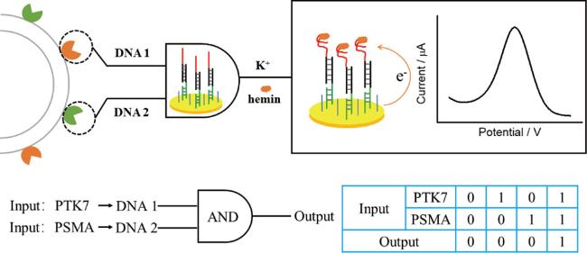
图7 用于小细胞外囊泡分析的多参数输入逻辑门。(a)小细胞外囊泡三种表面蛋白对应的核酸适配体。(b)核酸适配体结合小细胞外囊泡的AND逻辑门检测系统,用于处理基于EpCAM、CD63和PTK7三种蛋白的分子信号。只有当所有目标蛋白都存在时,才能释放初始信号进行等温扩增[86]

Fig. 7 Multi-parameter input logic gate for small extracellular vesicles analysis. (a) Nucleic acid aptamers corresponding to three surface proteins of small extracellular vesicles. (b) The AND logic gate detection system of aptamer binding to small extracellular vesicles is used to process molecular signals based on EpCAM, CD63 and PTK7 proteins. Only when all the target proteins are present can the initial signal be released for isothermal amplification[86]. Copyright 2022, American Chemical Society

3.3 Proximity Ligation Reaction of Multiple Proteins

sEVs are highly heterogeneous in their molecular characteristics; therefore, their surface proteins reflect their cells of origin and determine the type of recipient cells. Based on previous detection methods, the interference caused by the nonspecific binding of non-target sEVs and soluble target proteins cannot be ignored. Although the proximity ligation assay based on dual antibodies can determine the simultaneous expression of two markers on the same sEV to improve detection specificity, it still faces the problem of complex spatial steric hindrance. Therefore, a method is needed to accurately quantify specific biomarkers from tumor-derived sEVs. Compared with antibodies, aptamers can be programmatically designed, without the need for complex DNA-antibody conjugation, have less spatial steric hindrance, and can effectively recognize sEVs. In addition, nucleic acid aptamer types can be simply replaced or increased to analyze the desired sEV subtypes or achieve multiplex analysis[87-91]. Zhao et al.[92] designed an aptamer-cholesterol-mediated proximity ligation assay for the highly specific identification and quantification of sEVs. Zhang et al.[93] developed a proximity-induced DNAzyme chain autonomous assembly for accurate sEV identification. By combining HCR and DNAzyme for dual isothermal amplification, the detection sensitivity was improved. High specificity of detection was achieved by simultaneously recognizing surface proteins and lipid membranes. Sun et al.[94] proposed a recognition pattern based on the simultaneous binding of double-positive EV membrane proteins. An accurate, non-purified, low-cost, and visualizable sEV recognition strategy was developed through the combination of dual aptamer proximity ligation-mediated toe-hold activation G-quadruplex DNAzyme catalyzed Au@Ag nanorods (Au@Ag NRs) etching. Peng et al.[95] designed two aptamer hairpin probes capable of binding to two adjacent proteins on the cell membrane, respectively, to activate the associative interweaving of strand displacement reactions, which then triggers dimeric rolling circle amplification, ultimately producing a significantly amplified electrochemical signal for the sensitive quantification of target CTCs. Lin et al.[96] to achieve specific recognition of tumor-derived sEVs, two sEV-targeting aptamers, MJ5C17 and SYL3C18, were used as affinity probes for PD-L1 and EpCAM, respectively. As shown in Figure 8, both probes were designed with two functional domains: one aptamer domain for target recognition; and an extension part with an additional sequence at the 5′ or 3′ end for proximity ligation. The dual aptamers in this system can recognize two biomarkers expressed simultaneously on the same sEV, effectively eliminating the interference of sEVs from non-tumor cells and soluble proteins, thereby greatly improving the selectivity of detection.
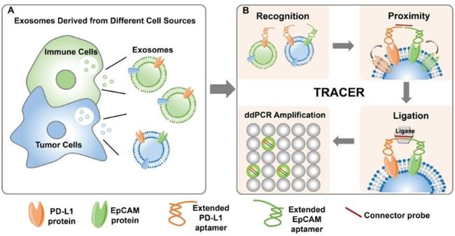
图8 基于核酸适配体的邻近连接反应高灵敏定量检测肿瘤来源外泌体的工作原理。利用设计的两种分别针对EpCAM和PD-L1的核酸适配体探针同时标记小细胞外囊泡上的两种蛋白标记物。由于外泌体膜的流动性,两个核酸适配体的延伸部分接近。连接后,使用dd PCR对连接产物进行定量[96]

Fig. 8 Working principle of the highly sensitive quantification of tumor-derived Exo-PD-L1 using aptamer-based PLA. Two types of designed aptamer probes against EpCAM and PD-L1 were utilized to simultaneously label both types of protein markers on the exosomes. The extended parts of two aptamers are in close proximity because of the fluidity of the exosomes membrane. After ligation, ddPCR is performed to quantify the ligation products [96]. Copyright 2020, Wiley Online Library

Li et al[97] used multiple protein biomarkers on single sEVs as inputs and thermophoretic accumulation to amplify the output signal, achieving high sensitivity and specificity in the analysis of sEVs. This strategy selects two breast cancer-related proteins, HER2 and epithelial cell adhesion molecule (EpCAM), as inputs for a proximity ligation reaction on the sEVs membrane. The designed two ssDNA strands (Apt-S-T) each consist of an aptamer sequence (Apt), a spacer sequence (S), and a bound toe activation sequence (T). After the Apt-S-T probes bind to the target proteins on the EVs membrane, a bound toe nanostructure is formed with the help of a connector sequence. Then, H1 and H2 hairpin probes are used to initiate the toe-activated HCR for signal amplification. To improve detection sensitivity, sEVs are first captured using CD63 aptamer-modified microbeads. After HCR, the microbeads are enriched by thermophoresis to amplify the output signal. This simultaneous detection of EpCAM and HER2 on individual sEVs can effectively enhance the identification of sEVs closely related to breast cancer.
Shi et al.[98] designed a diagnostic tool for breast cancer diagnosis and immunotherapy monitoring, manufactured by assembling nanocatalysts on the sEVs membrane encoded by HCR triggered by dual nucleic acid aptamers. This dual nucleic acid aptamer recognition strategy can effectively eliminate interference from sEVs and soluble proteins secreted by non-tumor cells, achieving excellent selectivity. In addition, the HCR-encoded Pt@AuNPs component with high catalytic activity allows signal amplification and color signal output, which can be read by the naked eye or a mobile phone camera. Deng et al.[88] used a one-step thermophoresis combined with proximity ligation operation on the surface of sEVs to directly detect tumor-derived sEVs simultaneously expressing EpCAM and PSMA in serum, in a uniform and separation-free form.
Lu et al.[99] demonstrated the separation of sEV subpopulations by combining dual nucleic acid aptamer recognition-induced proximity ligation with microfluidic serial separation. This PLA-mediated microfluidics can sequentially separate tumor-derived PD-L1 sEVs and non-tumor-derived PD-L1 sEVs. In addition to previous quantitative analysis, this platform also allows for high-efficiency detection and low-destructive recovery of sEV subpopulations through chip disconnection and DNA hydrolysis. Wang et al.[100] constructed a sensing ratio strategy based on a colocalization-dependent system (Co-DNA-Locker) and applied it to the ultrasensitive detection of sEVs using self-assembled Au NRs vertical arrays. Cy5-labeled EpCAM aptamers were attached to Au NRs for capturing sEVs, while aptamers for CD63 and HER2 proteins were used to form Co-DNA-Locker through proximity ligation, endowing the sEV SERS sensor with excellent specificity. Here, the Cy5-labeled at the end of the EpCAM aptamer served as an internal standard signal molecule, constructing a ratiometric sensing platform that reduced background signal interference, making the prepared SERS sensor have better accuracy, providing an innovative technical platform for early cancer detection.

4 Conclusions and Prospects

sEVs play a crucial role in regulating cell behavior through intercellular communication. The concentration of tumor-derived sEVs is much higher than that of CTC, and their stability is better than cfDNA. They can be easily obtained from body fluids and have been shown to exhibit significant expression differences in various disease models, making them one of the most important liquid biopsy substances. Precise evaluation of sEVs biomarkers and analysis of their surface protein patterns can significantly enhance the progress of treatment and research for diseases such as cancer. Currently, high-sensitivity detection of sEVs has been achieved by combining multiple detection technologies, but most of these methods involve bulk detection of sEVs. Bulk analysis methods often overlook the variability of sEVs during disease progression, thus limiting in-depth understanding of individual sEVs and affecting the accuracy of diagnosis and detection for various diseases. An increasing number of studies have shown that comprehensive detection of multiple biomarkers on individual sEVs can effectively improve the sensitivity, specificity, and accuracy of molecular diagnostics, leading to more research focusing on their precise detection. Additionally, some studies are focused on in situ imaging and tracking of individual sEVs. This approach holds promise for providing deeper insights into cellular dynamics, including the biogenesis of EVs, intercellular transport, uptake, and cargo release. In the future, there will be a need to develop more efficient and powerful multiparametric and multiplexed analysis methods for the detection of individual sEVs, such as simultaneously detecting target proteins and micro RNAs with high sensitivity and specificity, which will help advance the application of sEVs in liquid biopsies.
[1]
Doyle L and Wang M. Cells, 2019, 8.

[2]
Ha D, Yang N N, Nadithe V. Acta Pharm. Sin. B, 2016, 6(4): 287.

[3]
Yan H, Li Y T, Cheng S B, Zeng Y. Anal. Chem., 2021, 93(11): 4739.

[4]
Jeppesen D K, Zhang Q, Franklin J L, Coffey R J. Trends Cell Biol., 2023, 33(8): 667.

[5]
Kowal J, Arras G, Colombo M, Jouve M, Morath J P, Primdal-Bengtson B, Dingli F, Loew D, Tkach M, Théry C. Proc. Natl. Acad. Sci. U. S. A., 2016, 113(8):968.

[6]
Xie H, Di K L, Huang R R, Khan A, Xia Y Y, Xu H P, Liu C, Tan T T, Tian X Y, Shen H, He N Y, Li Z Y. Chin. Chemical Lett., 2020, 31(7): 1737.

[7]
Wu L L, Wang Y D, Zhu L, Liu Y L, Wang T, Liu D, Song Y L, Yang C Y. ACS Appl. Bio Mater., 2020, 3(5): 2743.

[8]
Qian F Y, Huang Z N, Zhong H K, Lei Q, Ai Y R, Xie Z H, Zhang T H, Jiang B W, Zhu W, Sheng Y, Hu J M, Brinker C J. ACS Nano, 2022, 16(12): 19980.

[9]
Martín-Gracia B, Martín-Barreiro A, Cuestas-Ayllón C, Grazú V, Line A, Llorente A, de la Fuente J M, Moros M. J. Mater. Chem. B, 2020, 8(31): 6710.

[10]
Vaidyanathan R, Soon R H, Zhang P, Jiang K, Lim C T. Lab a Chip, 2019: 11.

[11]
Hsu S K, Jadhao M, Liao W T, Chang W T, Lin I L, Chiu C-C, Cancers, 2023, 15.

[12]
Song F T, Wang C, Wang C H, Gao J W, Liu H, Zhang Y, Han L. Anal. Chem., 2021, 93(11): 4697.

[13]
Xiong H W, Huang Z P, Yang Z J, Lin Q Y, Yang B, Fang X E, Liu B H, Chen H, Kong J L. Small, 2021, 17(35): 1.

[14]
Wang Q, Zou L Y, Yang X H, Liu X F, Nie W Y, Zheng Y, Cheng Q, Wang K M. Biosens. Bioelectron., 2019, 135: 129.

[15]
Guo Y N, Tao J, Li Y R, Feng Y M, Ju H X, Wang Z F, Ding L. J. Am. Chem. Soc., 2020, 142(16): 7404.

[16]
Cao Y, Yu X M, Zeng T Y, Fu Z Y, Zhao Y Y, Nie B B, Zhao J, Yin Y M, Li G X. J. Am. Chem. Soc., 2022, 144(30): 13475.

[17]
Zhang P, Zhou X, Zeng Y. Chem. Sci., 2019, 10(21): 5495.

[18]
Wu J C, Lin Z, Zou Z Y, Liang S P, Wu M H, Hu T Y, Zhang Y Q. J. Am. Chem. Soc., 2022, 144(51): 23483.

[19]
Niu R Y, Chen X H, Sun Z X, Wang L, Wang Z N, Zhang C, Ding D, Yang J C, Wang Y Z, Luo Y. Anal. Chim. Acta, 2023, 1276: 341636.

[20]
Wang S, Khan A, Huang R, Ye S, Di K, Xiong T, Li Z. Biosens. Bioelectron., 2020, 154: 112056.

[21]
Altıntaş Ö, Saylan Y. Anal. Chem., 2023, 95(44): 16029.

[22]
Ma Z W, Xu H Y, Ye B C. Anal., 2023, 148(20): 4954.

[23]
Zhang H, Zhou Y J, Luo D, Liu J J, Yang E, Yang G Y, Feng G J, Chen Q H, Wu L. RSC Adv., 2021, 11(9): 4983.

[24]
Feng D W, Ren M X, Miao Y F, Liao Z R, Zhang T J, Chen S, Ye K D, Zhang P J, Ma X L, Ni J T, Hu X Q, Li H J, Peng J R, Luo A Q, Geng L N, Deng Y L. Biosens. Bioelectron., 2022, 207: 114112.

[25]
Chen J, Tang J L, Meng H M, Liu Z, Wang L, Geng X, Wu Y N, Qu L B, Li Z H. Chem. Commun., 2020, 56(63): 9024.

[26]
Sha L J, Bo B, Yang F, Li J Y, Cao Y, Zhao J. Anal. Chem., 2022, 94(24): 8748.

[27]
Shi L, Cai H Y, Wang H, Wang Q W, Shi L L, Li T. Anal. Chem., 2023, 95(48): 17662.

[28]
Bagheri Hashkavayi A, Cha B S, Lee E S, Kim S, Park K S. Anal. Chem., 2020, 92(19): 12733.

[29]
Zhang Y Z, Jiao J Y, Wei Y Y, Wang D N, Yang C G, Xu Z R. Anal. Chem., 2020, 92(22): 15244.

[30]
Huang R, He L, Jin L, Li Z, He N, Miao W. Chin. Chem. Lett., 2023,34.

[31]
Xu L J, Lu S Y, Wang H, Xu H Y, Ye B C. Anal. Chem., 2023, 95(42): 15745.

[32]
Zhou J Q, Lin Q Y, Huang Z P, Xiong H W, Yang B, Chen H, Kong J L. Anal. Chem., 2022, 94(15): 5723.

[33]
Wang X K, Shang H Z, Ma C P, Chen L X. Anal. Chem., 2021, 93(24): 8493.

[34]
Ding L H, Wu Y, Liu L E, He L L, Yu S C, Effah C Y, Liu X, Qu L B, Wu Y J. Biosens. Bioelectron., 2023, 219: 341098.

[35]
Yang Z P, She D, Sun C H, Gong M W, Rong Y. Anal. Methods, 2022, 14(15): 1534.

[36]
Hu W, Su H, Zeng X, Duan X, Li Y, Li L, Appl. Biochem. Biotechnol., 2022, 195: 861.

[37]
Li P, Wang J, Gao M Q, Wang J, Ma Y, Gu Y Q. Anal. Chem., 2021, 93(28): 9860.

[38]
Ren Y, Ge K, Lu W, Peng W, Che H, Ma J, Jia C, Yao B, Sens. Actuators, B, 2023, 377.

[39]
Zhao X X, Zeng L L, Mei Q, Luo Y. ACS Sens., 2020, 5(7): 2239.

[40]
Yu Y, Zhang W S, Guo Y, Peng H, Zhu M, Miao D, Su G. Biosens. Bioelectron., 2020,167: 112482.

[41]
He F, Liu H, Guo X G, Yin B C, Ye B C. Anal. Chem., 2017, 89(23): 12968.

[42]
Zeng R J, Wang J, Wang Q S, Tang D P, Lin Y. Talanta, 2021, 221: 121600.

[43]
Li C, Zhou M Y, Wang H Y, Wang J, Huang L. Talanta, 2022, 245: 123444.

[44]
Xia Y, Liu M, Wang L, Yan A, He W, Chen M, Lan J, Xu J, Guan L, Chen J. Biosens. Bioelectron., 2017, 92: 8.

[45]
Zhang X Y, Zhu X Y, Li Y F, Hai X, Bi S. Talanta, 2023, 258: 124456.

[46]
Jiang Y, Shi M L, Liu Y, Wan S, Cui C, Zhang L Q, Tan W H. Angew. Chem., 2017, 129(39): 12078.

[47]
Li C, Wang H Y, Wei R, Ren J Z, Zhou M Y, Yan C, Huang L. Sens. Actuat. B Chem., 2023, 380: 133361.

[48]
Zhang Y Z, Wang D N, Yue S, Lu Y B, Yang C G, Fang J, Xu Z R. ACS Sens., 2019, 4(12): 3210.

[49]
Xu L Z, Shoaie N, Jahanpeyma F, Zhao J J, Azimzadeh M, Al-Jamal K T. Biosens. Bioelectron., 2020, 161: 112222.

[50]
Li S, Ma Q. Biosens. Bioelectron., 2022, 214: 114554.

[51]
Liang T T, Qin X L, Xiang Y H, Tang Y J, Yang F. Trac Trends Anal. Chem., 2022, 148: 116532.

[52]
Chang K L, Fang Y, He P, Zhu C N, Liu X J, Zheng D Y, Chen D J, Liu C. Anal. Chem., 2023, 95(44): 16194.

[53]
Su J, Chen S X, Dou Y Z, Zhao Z H, Jia X L, Ding X T, Song S P. Anal. Chem., 2022, 94(7): 3235.

[54]
Zhang W F, Tian Z H, Yang S J, Rich J, Zhao S G, Klingeborn M, Huang P H, Li Z S, Stout A, Murphy Q, Patz E, Zhang S S, Liu G Z, Huang T J. Microsyst. Nanoeng., 2021, 7: 63.

[55]
Wang L, Deng Y, Wei J, Huang Y, Wang Z X, Li G X. Biosens. Bioelectron., 2021, 191: 113465.

[56]
Huang R R, He L, Xia Y Y, Xu H P, Liu C, Xie H, Wang S, Peng L J, Liu Y F, Liu Y, He N Y, Li Z Y. Small, 2019, 15(19): 1900735.

[57]
An Y, Jin T, Zhu Y, Zhang F, He P. Biosens. Bioelectron., 2019, 142.

[58]
Lu J Y, Wang M H, Han Y W, Deng Y, Zeng Y J, Li C, Yang J, Li G X. Anal. Chem., 2022, 94(12): 5055.

[59]
Jiang J, Yu Y, Zhang H, Cai C. Anal. Chim. Acta, 2020, 1130: 1.

[60]
Dong H, Chen H, Jiang J, Zhang H, Cai C, Shen Q. Anal. Chem., 2018,90:4507.

[61]
Sun D, Guo Q Q, Zhang H, Cai C X. Chemosensors, 2023, 11(7): 415.

[62]
Zeng X H, Wu C Y, Xiong Y, Zhan Z X, Shen C C, Lin F, Zhang J L, Chen P P. Biosens. Bioelectron., 2024, 256: 116273.

[63]
Nie Y M, Yuan X D, Zhang P, Chai Y Q, Yuan R. Anal. Chem., 2019, 91(5): 3452.

[64]
Wang H, Song Y, Chai Y, Yuan R. Biosens. Bioelectron., 2019, 140: 111344.

[65]
Zhang H, Wang Z, Zhang Q, Wang F, Liu Y. Biosens. Bioelectron., 2019, 124-125: 184.

[66]
Zhang Y M, Wang F, Zhang H X, Wang H Y, Liu Y. Anal. Chem., 2019, 91(18): 12100.

[67]
Liu X, Wang Q, Chen J, Chen X, Yang W. Talanta, 2021, 221.

[68]
Bezuneh T T, Fereja T H, Kitte S A, Li H, Jin Y. Talanta, 2022, 248.

[69]
Chen Y H, Kong D R, Qiu L P, Wu Y G, Dai C H, Luo S, Huang Z P, Lin Q Y, Chen H, Xie S T, Geng L, Zhao J, Tan W H, Liu Y Q, Wei D C. Anal. Chem., 2022: 1446.

[70]
Qiao B, Guo Q Q, Jiang J Q, Qi Y L, Zhang H, He B S, Cai C X, Shen J. Anal., 2019, 144(11): 3668.

[71]
Feng Q M, Ma P, Cao Q H, Guo Y H, Xu J J. Chem. Commun., 2020, 56(2): 269.

[72]
Xiong H W, Huang Z P, Lin Q Y, Yang B, Yan F, Liu B H, Chen H, Kong J L. Anal. Chem., 2022, 94(2): 837.

[73]
Lyu Y, Cui D, Huang J, Fan W, Miao Y and Pu K, Angew. Chem., Int. Ed., 2019, 58: 4983.

[74]
An Y, Li R, Zhang F, He P G. Anal. Chem., 2020, 92(7): 5404.

[75]
Chen X H, Deng Y, Niu R Y, Sun Z X, Batool A, Wang L, Zhang C, Ma N Y, Yang Q T, Liu G X, Yang J C, Luo Y. Anal. Chem., 2022, 94(38): 13019.

[76]
Zhou S S, Hu T, Zhang F, Tang D Z, Li D K, Cao J, Wei W, Wu Y F, Liu S Q. Anal. Chem., 2020, 92(1): 1574.

[77]
Su X M, Liu X Y, Xie Y, Chen M Y, Zheng C, Zhong H, Li M. ACS Nano, 2023, 17(4): 4077.

[78]
Zhang X W, Liu M X, He M Q, Chen S, Yu Y L, Wang J H. Anal. Chem., 2021, 93(16): 6437.

[79]
Cheng S, Kong Q, Hu X, Zhang C, Xian Y. Anal. Chem., 2022, 94: 1085.

[80]
Zhang M W, Xia L, Mei W J, Zou Q Q, Liu H, Wang H Q, Zou L Y, Wang Q, Yang X H, Wang K M. Anal. Chim. Acta, 2023, 1254: 341130.

[81]
Gao Q Q, Zhao Y M, Xu K L, Zhang C, Ma Q, Qi L Q, Chao D D, Zheng T T, Yang L L, Miao Y Y, Han D. Angew. Chem. Int. Ed., 2020, 59(52): 23564.

[82]
Peng R Z, Zheng X F, Lyu Y F, Xu L J, Zhang X B, Ke G L, Liu Q L, You C J, Huan S Y, Tan W H. J. Am. Chem. Soc., 2018, 140(31): 9793.

[83]
Zhao L, Sun R J, He P, Zhang X R. Anal. Chem., 2019, 91(22): 14773.

[84]
Yu Y Q, Guo Q Q, Jiang W L, Zhang H, Cai C X. Anal. Chem., 2021, 93(32): 11298.

[85]
Hu X Y, Cheng S S, Luo X Z, Xian Y Z, Zhang C L. Anal. Chem., 2023, 95(27): 10330.

[86]
Feng Y W, Yang Y S, Xiao Y T, Fu T, He L, Qi L B, Yang Q X, Peng R Z, Tan W H. Anal. Chem., 2023: 1132.

[87]
Feng C, Chen T S, Mao D S, Zhang F, Tian B, Zhu X L. ACS Sens., 2020, 5(10): 3116.

[88]
Deng J, Zhao S, Li J, Cheng Y, Liu C, Liu Z, Li L, Tian F, Dai B, Sun J. Angew. Chem., Int. Ed., 2022, 61.

[89]
Hao J, Wang J Y, Dong Y, Yang J Y, Wang Z, Zhao X X, Zeng T, Zhao X, Liang H J, Li J J. Anal. Chem., 2023, 95(17): 6854.

[90]
Yin Y, Xie W, Xiong M Y, Gao Y Y, Liu Q, Han D, Ke G L, Zhang X B. Angew. Chem. Int. Ed., 2023, 62(44): 2309837.

[91]
Tan W Q, Zhang C L, Cheng S S, Hu X Y, Wang M, Xian Y Z. Anal. Chem., 2024, 96(3): 1328.

[92]
Zhao X X, Luo C J, Mei Q, Zhang H M, Zhang W Q, Su D W, Fu W L, Luo Y. Anal. Chem., 2020, 92(7): 5411.

[93]
Zhang D, Qiao L, Xu S W, Peng L, Yang Y, Zhang P S, Song Z L, Chen J, Zhang C H. Sens. Actuat. B Chem., 2023, 383: 133581.

[94]
Sun W Y, Wang Y, Zhu Z X, Wang Y R, Zhang M R, Jiang L, Liu S, Yu J H, Huang J D. Anal. Chem., 2021, 93(36): 12383.

[95]
Peng Y, Lu B, Deng Y, Yang N, Li G. Biosens. Bioelectron., 2022, 201: 11393.

[96]
Lin B Q, Tian T, Lu Y Z, Liu D, Huang M J, Zhu L, Zhu Z, Song Y L, Yang C Y. Angew. Chem. Int. Ed., 2021, 60(14): 7582.

[97]
Li Y K, Deng J Q, Han Z W, Liu C, Tian F, Xu R, Han D, Zhang S H, Sun J S. J. Am. Chem. Soc., 2021, 143(3): 1290.

[98]
Shi H, Zeng T, Liu Y, Liang Q, Yang F, Liang Y, Fu Z, Li W, Li G, Yin Y. ACS Mater. Lett., 2022, 5: 282.

[99]
Lu Y, Lin B, Liu W, Zhang J, Zhu L, Yang C, Song Y. Small Methods, 2023, 7.

[100]
Wang J, Xie H Y, Ding C F. ACS Appl. Mater. Interfaces, 2021, 13(28): 32837.

Outlines

/