Home Journals Progress in Chemistry
Progress in Chemistry

Abbreviation (ISO4): Prog Chem      Editor in chief: Jincai ZHAO

About  /  Aim & scope  /  Editorial board  /  Indexed  /  Contact  / 
Review

Fabrication and Biomedical Application of Hyaluronic Acid Based Micro- and Nanogels

  • Xuedan Qian , * ,
  • Weijiang Yu ,
  • Junzhe Fu ,
  • Youxiang Wang * ,
  • Jian Ji
Expand
  • MOE Key Laboratory of Macromolecular Synthesis and Functionalization, Department of Polymer Science and Engineering, Zhejiang University,Hangzhou 310027, China
* Corresponding author e-mail:

Received date: 2022-10-09

  Revised date: 2023-01-11

  Online published: 2023-02-20

Supported by

National Key Research and Development Project(2020YFE0204400)

National Natural Science Foundation of China(51873186)

Abstract

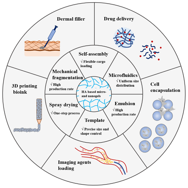
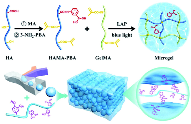
Hyaluronic acid is one of the primary components of the natural extracellular matrix, which has outstanding biocompatibility, biodegradability and excellent water retention capacity. Hyaluronic acid based micro- and nanogels have many desirable features of size adjustability, easy modification, deformability and structural integrity. They can not only meet most application requirements of bulk hydrogels, but also possess unique advantages due to small dimension, thus have been widely researched and used in biomedical field. This paper reviews the preparation methods and biomedical applications of hyaluronic acid based micro- and nanogels, and the future development of it is prospected.

Cite this article

Xuedan Qian , Weijiang Yu , Junzhe Fu , Youxiang Wang , Jian Ji . Fabrication and Biomedical Application of Hyaluronic Acid Based Micro- and Nanogels[J]. Progress in Chemistry, 2023 , 35(4) : 519 -525 . DOI: 10.7536/PC221001

Contents

1 Introduction
2 Fabrication of hyaluronic acid based micro-and nanogels
2.1 Mechanical fragmentation
2.2 Emulsion
2.3 Microfluidics
2.4 Self-assembly
2.5 Spray drying
2.6 Template
3 Biomedical applications of hyaluronic acid based micro-and nanogels
3.1 Dermal filler
3.2 Drug delivery
3.3 Cell encapsulation
3.4 3D printing bioink
3.5 Imaging agents loading
4 Conclusion and outlook

1 Introduction

Hyaluronic acid (HA) is a natural linear macromolecular acidic mucopolysaccharide. Its molecular chain is composed of disaccharide units composed of D-glucuronic acid and N-acetylglucosamine. It was first isolated from bovine vitreous body by Carl Meyer, a professor of ophthalmology at Columbia University, in 1934. HA molecular chain contains a large number of carboxyl, hydroxyl and acetamido, which endows it with strong hydrophilic ability. It can absorb water equivalent to 1000 times its own weight, which is called "biomacromolecule moisturizer". At the same time, HA molecular chain is easy to modify, which can combine with various functional molecules by covalent or non-covalent interaction, and has a wide range of applications in the biomedical field[1~5].
Micro-nanogels are nano- or micron-sized crosslinked polymer particles with three-dimensional network structure, which can swell in water. Compared with bulk gels, micro-nanogels have more diverse designs and wider applications[6~10]. As one of the components of natural extracellular matrix, HA has good biocompatibility, non-immunogenicity and biodegradability, so micro-and nanogels based on HA have been widely studied in drug delivery carriers, tissue engineering scaffolds, medical diagnosis, medical cosmetology and other fields[11][12][13][11~15].

2 Preparation of hyaluronic acid-based micro-nanogel

There are various methods to prepare hyaluronic acid-based micro-nanogels, and the appropriate preparation method can be selected according to the application requirements. The microgels prepared by the template method and the microchannel method have uniform size and regular morphology, but the preparation efficiency is low[16]; Mechanical crushing is a common method for rapid preparation of microgels in large quantities, but it can not be applied in the field of high requirements for size uniformity and morphology regularity. The micro-nanogel for embedding bioactive molecules or cells can be prepared by self-assembly in an aqueous environment, an emulsion method or a microchannel method[17][18][19]. Common preparation methods are described below.

2.1 Mechanical crushing

Microgels with a size of more than 20 μm can be prepared by applying external pressure to the crosslinked HA bulk hydrogel through a fine steel wire mesh or by rotating and stirring to break it up[6]. The size of the microgel can be controlled by changing the pore size of the fine steel wire mesh or the stirring speed. This mechanical breaking method breaks the bulk hydrogel into tiny gels by external force, which is direct, fast and simple, and the early cross-linked HA gel facial filler is made by this method. The structure of the microgel prepared by this method is the same as that of the bulk gel, and it will not be destroyed by physical action. However, the shape, particle size and particle size distribution of the microgel prepared by this method can not be accurately controlled, so this method has great limitations.

2.2 Emulsion method

Taking advantage of the incompatibility between the aqueous phase and the oil phase, the aqueous solution of HA was dispersed in the oil phase to form small droplets, which were separated, washed and dried after crosslinking to obtain crosslinked microspheres of HA. The adjustment of parameters in the emulsion preparation process, such as emulsification speed, water-oil ratio, surfactant content, etc., plays a decisive role in the formation of stable emulsion and the final preparation of microspheres with uniform size and good morphology. If the emulsifying speed is too low, it is not conducive to the full mixing of oil phase and water phase, and the effective emulsion system can not be formed. If the emulsification speed is too high, the size of droplets will be reduced, the system disturbance will be increased, and the collision probability between droplets will be increased, which will cause the adhesion and aggregation between droplets. The ratio of water to oil is another important factor affecting the stability of the emulsion. The increase of the ratio of water phase is beneficial to the formation of microspheres with uniform particle size distribution. Surfactants contain hydrophilic and lipophilic groups, which can reduce the surface tension of the dispersed phase and facilitate the formation of stable emulsions. If the surfactant content is too low and the droplet volume is too large, it is easy to cause adhesion or demulsification between droplets. If the content of surfactant is too high, the excess surfactant will be free near the interface to increase the collision probability and cause the instability of the system[20,21]. It can be seen that the preparation of HA microgel by emulsion method is a process of continuous optimization of parameters.
Zhou et al. Prepared glutaraldehyde-crosslinked HA/gelatin (Gel) composite microgel by emulsion method, and the drug loading was achieved by adding 5-fluorouracil in the aqueous phase[21]. It was found that the ratio of HA to Gel was an important factor in controlling the size of microspheres. Keeping the total concentration of HA and Gel constant, the dry particle size of microspheres decreased from 50 ~ 150 μm to 10 ~ 100 μm when the concentration of HA increased from 2 wt% to 10 wt%. However, due to the increase of HA concentration, the viscosity of aqueous phase increases, and the adhesion and agglomeration of microgels occur. In addition, the stirring speed, the concentration of crosslinking agent and the ratio of water to oil also affect the morphology and size distribution of microgels.
Emulsion method can be used to prepare microgels in batches, but the wide particle size distribution has always been an unavoidable disadvantage of emulsion method. By optimizing the preparation conditions and sieving the product, the particle size can be made more uniform to a certain extent. In addition, biosafety has always been one of the basic requirements for the application of biomaterials, so the use of organic solvents and surfactants in emulsion method puts forward higher requirements for the purification of microgels.

2.3 Microchannel method

Microchannel method is a widely used method for preparing microspheres in scientific research in recent years. Due to the incompatibility of oil and water phases, when the aqueous phase and the oil phase containing HA converge at the intersection point through different microchannels, the surface tension and the shear effect of the oil phase on the aqueous phase cause the aqueous phase to form microdroplets dispersed in the oil phase. Microgels with different sizes and structures can be obtained by adjusting the flow rate and the design of the microchannel structure[22]. The microspheres prepared by the microchannel method have uniform particle size distribution, and are particularly suitable for research with high requirements on particle size uniformity, for example, the drug-loaded microspheres prepared by the microchannel method can avoid the problem of drug burst release caused by different sizes of the microspheres. However, the microchannel device is usually designed and manufactured by photolithography, which increases the complexity and cost of this method, and the preparation efficiency of this method is usually not high, which limits its further large-scale production and application.

2.4 Self-assembly

Self-assembly is a common method to prepare nanogels, and the particle size of nanogels can be controlled by changing the assembly conditions, such as pH value, polymer concentration, etc[23,24]. HA molecular chain contains a large number of carboxyl and hydroxyl groups, which can be easily modified. In aqueous phase, HA can assemble with small biological functional molecules or polymers to form nanostructures through hydrophobic interaction, host-guest interaction, electrostatic interaction, hydrogen bonding, metal ion coordination bonds and other supramolecular interactions, but it is difficult to combine with electronegative molecules[25]. For example, a hydrophobic alkane long chain is attached to a HA molecular chain through an EDC/NHS reaction, and the obtained amphiphilic molecule can be self-assembled in water to form a nanogel[26]; HA modified with β-cyclodextrin and adamantane can be used to obtain supramolecular microgels[25].
Qi, Ding, and Zhang et al. Prepared a cisplatin/doxorubicin co-loaded HA crosslinked nanogel, as shown in Figure 1. The nanogel utilized the electrostatic interaction between the amino group contained in doxorubicin (DOX) and the carboxyl group on the HA molecular chain, and the chelation between cisplatin (CDDP) and the carboxyl group on the HA molecular chain to achieve crosslinking[27]. It was found that the molar ratio of HA, DOX and CDDP affected the stability of nanogels. If the content of CDDP is too low, the stability of nanogel will decrease due to insufficient crosslinking. Excessive CDDP content will lead to the precipitation of nanogels. When the molar ratio of CDDP to the carboxyl group on the molecular chain of HA increased from 1/90 to 1/10, the particle size decreased from 86 nm to 57 nm with the increase of the crosslinking degree of the nanogel, and the nanogel showed good therapeutic effect in the mouse osteosarcoma xenograft tumor model.
图1 顺铂/阿霉素共负载的HA交联纳米凝胶示意图[27]

Fig.1 Schematic illustration of cisplatin/doxorubicin co-loaded crosslinked HA nanogels[27]

2.5 Spray drying method

Spray drying is a preparation method in which the polymer precursor solution is sprayed from a nozzle into a collection container, and the microgel is obtained by dehydration and solidification. This method can prepare the microgel with the minimum size of 1 ~ 2 μm by adjusting the nozzle size, ejection speed and other conditions. The preparation process does not use surfactant and oil phase, but the particle size uniformity of the prepared microgel is not good[28]. Electrostatic spraying is often used to prepare alginate microgels, and hyaluronic acid microgels can be prepared by spray drying[29]. Gomes et al. Prepared HA microgels by spraying a mixed solution of HA and adipic dihydrazide (ADH) from a nozzle, precipitating and dehydrating it in an isopropanol solution containing carbodiimide (EDCI), and diffusing EDCI into the matrix to promote the crosslinking reaction between HA and ADH[30]. By changing the molecular weight and concentration of HA, the diameter of the nozzle, the pressure in the nozzle chamber, and the distance between the nozzle and the crosslinking bath, the particle size of the microgel was adjusted from 7 μm to 56 μm.

2.6 Template method

The HA precursor solution is placed in the cavity of the template and cross-linked to form a micrometer-sized gel, which is often used to prepare particles larger than 100 μm in size. The template method can strictly control the shape and size of the microgel and avoid the use of surfactant in the preparation process, so it has good biocompatibility. However, due to the limitation of precision in the template manufacturing process, the preparation of microgels with unique and complex structures is still a major problem. With the development of precision machining technology, especially the development of lithography technology, the template or photomask with resolution up to nanometer level has been gradually applied, which makes it possible to prepare microgels with complex internal structures[31]. The combination of 3D hydrogel microstructure fabricated by lithography with cell culture and regenerative medicine has broad prospects for development[32].

3 Application of hyaluronic acid-based micro-and nanogels in biomedical field

Hyaluronic acid-based micro-nanogels have the advantage of flexible and adjustable size, which can load various functional molecules through weak interaction and covalent bonding such as hydrogen bonding or electrostatic interaction. Compared with micelles, vesicles, liposomes and other nanoparticles, micro-nanogels have higher drug loading rate. Due to the high water content, the micro-nano gel can be deformed when subjected to an external force, and due to the existence of an internal crosslinking structure, the micro-nano gel has certain mechanical strength and can maintain the integrity of the structure, so the hyaluronic acid-based micro-nano gel has wide application in the biomedical field.

3.1 Facial filler

Cross-linked sodium hyaluronate gel for injection, as the second largest non-surgical medical cosmetic injection after botulinum toxin, has been widely favored in the world. It can repair and make up for the problems of facial skin aging and loss of elasticity caused by aging. Cross-linked HA gel for filling injection is usually in the form of microgel, because microgel has good injectability and plasticity, which can make the treatment effect more natural and smooth. 1,4-butanediol diglycidyl ether (BDDE) is a commonly used crosslinking agent for this kind of HA microgel. The crosslinked HA microgel can be obtained by the reaction of the hydroxyl groups on the HA molecular chain with the epoxy groups of BDDE molecules. The degree of crosslinking and the concentration of the compound will affect the maintenance time of the filler.
Because HA can be gradually degraded and absorbed in the human body over time, and with the continuous progress of technology and the continuous improvement of people's requirements for products, the status of HA microgel fillers which only play a physical filling role is gradually impacted by some regenerative fillers.Uch as a filler product coated with polylactic acid microspheres or polycaprolactone microspheres, but HA plays an essential role in immediate filling in both fillers.

3.2 Drug delivery

Hyaluronic acid-based micro-and nanogels have been widely studied as drug delivery carriers[33]. Micro-nano gel can play a role in protecting drug molecules and local slow release, which is conducive to maintaining drug activity, reducing drug dosage and avoiding side effects. Drug release can be achieved by the change of the microenvironment of the lesion site to weaken the force between the drug molecule and the carrier material, or by the degradation of the micro-nanogel itself. According to the type of drug molecules, it can be divided into small molecule drug delivery and polypeptide drug delivery[34,35].

3.2.1 Small molecule drug delivery

HA nanogels are often used for targeted delivery of tumor drugs because HA can specifically bind to CD44 receptors overexpressed on the surface of various tumor cells and improve the enrichment of drugs in tumor sites[36,37]. However, it is very important to balance the stability of HA nanogels during circulation in the body and the release of drugs at the lesion site.
Li and Wang et al. Prepared a hyaluronic acid nanogel integrating imaging, photodynamic therapy, stimulus response and targeting, as shown in Figure 2. First, histidine-grafted hyaluronic acid (HA-His) was synthesized.HA was cross-linked to form nanogels by the electrostatic interaction between the Mn2+ of MRI agent and the carboxylate group of HA-His and the coordination of imidazole, while the chemotherapeutic drug doxorubicin hydrochloride (DOX · HCl) and the photosensitizer chlorin E6 (Ce6) were embedded in the nanogels through non-covalent interaction, and the particle size of the gel was about 240 nm[38]. When the nanogel is injected into a melanoma mouse body, the nanogel is targeted to a tumor site by the specific recognition of HA and a CD44 receptor highly expressed in the melanoma of the mouse, electrostatic interaction and coordination are weakened in a slightly acidic environment of the tumor, and the nanogell is decomposed to release a drug to achieve the purpose of killing tumor cells.
图2 集成像、化疗、光动力治疗于一体的HA纳米凝胶制备示意图[38]

Fig.2 Representative scheme of preparation of HA nanogels for image-guided combined chemo and photodynamic therapy[38]

3.2.2 Polypeptide drug delivery

Peptide drugs have a short half-life and are easily degraded and inactivated, so frequent administration is often required to achieve the desired therapeutic effect. On the one hand, the micro-nanogel based on HA can combine with positively charged peptides through electrostatic interaction, on the other hand, it can protect the activity of peptides in vivo, so it can be used as a carrier of polypeptide drugs[6,39~41].
Govender et al. Prepared a HA nanogel co-delivering NO donor molecule SNAP and antimicrobial peptide (AMP)[42]. Divinyl sulfone (DVS) was added to an alkaline aqueous solution of HA under vigorous stirring to form a cross-linked nanogel, which was subsequently added to a mixed solution of SNAP and AMP. It was found that the microporous structure of the nanogel was beneficial to the loading of small molecule SNAP, while the positively charged AMP was efficiently loaded through electrostatic interaction, and the gel particles with a particle size of about 600 nm were obtained. In vitro experiments show that the co-delivered nanogel can prolong the half-life of SNAP and increase the release time of NO; It has antibacterial effect on both Gram-negative and Gram-positive bacteria, and can resist biofilm formation.
Burdick et al. Prepared a supramolecular bulk gel embedding HA cross-linked microgel[43]. As shown in Fig. 3, norbornene-grafted HA (NorHA) was first synthesized, and positively charged interleukin-10 (IL-10) -loaded HA crosslinked microgel with a particle size of about 60 μm was obtained by microchannel method combined with ultraviolet light in-situ crosslinking. Then adamantane modified HA (AdHA), β-cyclodextrin modified HA (CDHA) and microgel aqueous solution were mixed to obtain supramolecular bulk gel embedded with microgel driven by host-guest interaction. The composite structure not only utilizes the injectability of the supramolecular hydrogel, but also combines the controlled release of the HA crosslinked microgel for drugs, and the material plays a good therapeutic effect in a rat myocardial infarction model.
图3 微凝胶/本体凝胶复合制备示意图[43]

Fig.3 Schematics of preparation of microgel/hydrogel composites[43]

3.3 Cell encapsulation

Hyaluronic acid, as one of the components of natural extracellular matrix, has good biocompatibility. Emulsion method, microchannel method, template method and other preparation methods are compatible with cell encapsulation, and cells can be directly encapsulated inside the microgel to achieve cell delivery during the preparation process[44]. Microgels can be further stacked in three-dimensional space to form scaffolds, and the existing microporous structure is conducive to cell spreading and nutrient delivery, so microgel scaffolds have good application prospects in three-dimensional cell culture and cell behavior research. The particle size, porosity and mechanical properties of microgels can affect the migration and growth of cells.
Dong and Cao et al. Used the microchannel method to mix bone marrow mesenchymal stem cells, vinyl sulfonated hyaluronic acid and thiolated gelatin, and prepared an internal stem cell-loaded microgel by Michael addition reaction[45]. This material can not only simulate the environment of natural polysaccharides and proteins of extracellular matrix to improve the viability, proliferation and chondrogenic differentiation potential of stem cells, but also cross-link the microgel in situ to form a macroporous scaffold through cell-cell interconnection, which has been proved to promote cartilage repair in vitro and in vivo.
Segura et al. Broke the cross-linked HA bulk gel into microgel through a sieving filter, and then mixed it with different cells such as mesenchymal stem cells, dermal fibroblasts and neural progenitor cells, respectively[46]. Under physiological conditions, the K peptide and Q peptide modified on HA form amide bonds mediated by activation factor ⅩⅢ, thus realizing the crosslinking between microgels and forming microporous scaffolds. It was found that the larger the mesh size, the larger the size of the microgel, and the porosity of the microporous scaffold increased accordingly. Cells could penetrate, diffuse and proliferate in the microgel scaffold to achieve three-dimensional cell culture. The microgel scaffold can also be injected into the tissue repair site to achieve the purpose of treatment through local cell infiltration into the scaffold.

3.4 3D printing bio-ink

The injectability of HA microgel makes it a good material for 3D printing bioink, which can be applied to drug delivery, tissue regeneration and other studies. The properties of microgel itself and printing characteristics, such as extrusion continuity, shape fidelity, and mechanical stability, are important factors affecting subsequent applications[47].
Cao and Dong et al. Proposed a novel strategy to prepare microgel-assembled bioink[48]. As shown in Fig. 4, HA modified by methacrylate and phenylboronic acid (HAMA-PBA) and gelatin modified by methacrylate (GelMA) were first synthesized, and HA/Gel composite microgel was further prepared by microchannel method. The microgel was then mixed with dopamine-modified HA, and the interaction between the microgels was enhanced by the dynamic cross-linking bonds formed by the phenylboronic acid groups and dopamine. This bioink based on microgel assembly has better printing performance and shape fidelity in 3D printing without sacrificing the viability of encapsulated cells.
图4 微凝胶组装生物墨水[48]

Fig.4 Bioink assembled by microgels[48]

Previous studies have shown that bioink requires high size uniformity of microgels, which can improve printing accuracy. The microgels are combined by physical interaction or dynamic covalent bonds, which endows the microgels with shear-thinning characteristics, which is conducive to 3D printing, while maintaining shape stability by the interaction between particles. Because HA microgel is a good carrier for cell encapsulation, combining 3D printing with cell encapsulation, HA microgel has great application prospects in the field of regenerative medicine[49,50].

3.5 Developer loading

The microgel has a relatively regular shape and can be used for tumor diagnosis in vivo in combination with a contrast agent. How to load molecules or particles with developing function into HA micro-nanogels is the key to such applications.
Lim et al. Prepared a nanogel composed of HA, poly (β-amino ester) (PBAE) and near-infrared fluorescent indocyanine green (ICG) through non-covalent interaction, with a particle size of about 70 nm[51]. As a nanoprobe for detecting tumor cells, the nanogel is accumulated in tumor cells through endocytosis mediated by HA and CD44 receptor, PBAE is rapidly degraded in lysosomal acidic environment, and ICG is released to emit fluorescence, which provides a possibility for tumor diagnosis.
Kong et al. Prepared an in vivo visible embolization microsphere based on HA[52]. HA dilute hydrochloric acid solution containing crosslinker 1,4-butanediol diglycidyl ether and barium sulfate (BaSO4) nanoparticles was dripped from a syringe pump into isobutanol, and after drying, HA microspheres encapsulated with BaSO4 nanoparticles were obtained. The size of the microspheres could be adjusted from 435 μm to 782 μm according to the inner diameter of the needle. The BaSO4 is used as a contrast medium to realize the real-time tracking of the microsphere in vivo, and the HA microsphere has good mechanical strength and can realize distal delivery when used as an embolization microsphere. Animal experiments show that the microspheres can be seen under X-ray in vivo, can effectively block blood flow for more than 4 weeks, and show good therapeutic effect.

4 Conclusion and prospect

To sum up, hyaluronic acid-based micro-nanogels have been widely used in drug delivery carriers, tissue engineering scaffolds, medical diagnosis, medical cosmetology and other fields. However, up to now, there are still some challenges in the preparation and application of hyaluronic acid-based micro-nanogels. Hyaluronic acid gel itself has low mechanical strength and is easily degraded by hyaluronidase and free radicals. Although the performance is improved by crosslinking, how to coordinate the mechanical properties and controlled degradation deserves special attention. Especially in the field of tissue regeneration, how to coordinate tissue growth and scaffold degradation is a problem that researchers need to consider. Compounding hyaluronic acid with other polymers, such as collagen and chitosan, can further improve the mechanical properties of the gel, which is expected to improve the properties of the material. In addition, there is still a gap between hyaluronic acid-based micro-nanogels and practical applications in the biomedical field, and the complexity of physiological conditions puts forward stricter requirements for material design and higher quality control requirements. The development of more precise stimuli-responsive materials and the design of precise and controllable structures for specific application scenarios are the research directions for the practical application of hyaluronic acid-based micro-and nano-gels. With the development of preparation methods and related polymer theoretical knowledge, hyaluronic acid-based micro-and nano-gels will have more extensive research and application in the biomedical field.
[1]
Snetkov P P, Zakharova K, Morozkina S, Olekhnovich R, Uspenskaya M. Polymers, 2020, 12: 1800.

[2]
Tiwari S, Bahadur P. Int. J. Biol. Macromol., 2019, 121: 556.

[3]
Wu L, He Y, Mao H, Gu Z. J Mater Chem B., 2022, 10(37):7148.

[4]
Knopf-Marques H, Pravda M, Wolfova L, Velebny V, Schaaf P, Vrana N E, Lavalle P. Adv. Healthcare Mater., 2016, 5(22): 2841.

[5]
Highley C B, Prestwich G D, Burdick J A. Curr. Opin. Biotechnol., 2016, 40: 35.

[6]
Daly A C, Riley L, Segura T, Burdick J. Nat. Rev. Mater., 2019, 5: 20.

[7]
Lee S, Lee Y, Kim E M, Nam K, Choi I. ACS Appl. Polym. Mater. 2020, 2, 2, 342.

[8]
Zhang X J, Malhotra S, Molina M, Haag R. Chem. Soc. Rev., 2015, 44(7): 1948.

[9]
Neam瘙塅u I, Rusu A, Diaconu A, Nita L, Chiriac A, Drug Deliv., 2017, 24: 539.

[10]
Sung B, Kim M H, Abelmann L. Bioeng. Transl. Med., 2021, 6(1): e10190.

[11]
Junzhe Lou, Ryan Stowers, Sungmin Nam, Yan Xia, Ovijit Chaudhuri. Biomaterials, 2018, 154: 213.

[12]
Seong Y J, Lin G, Kim B J, Kim H E, Kim S, Jeong S H. ACS omega, 2019, 4: 13834.

[13]
Ki Y C, Hwa S H, Eun S L, Jung M S, Benjamin D. A, Doo S L, Jae H P. Advanced Materials, 2019, 31: 1803549.

[14]
Darling N J, Sideris E, Hamada N, Carmichael S T, Segura T. Adv. Sci., 2018, 5(11): 1801046.

[15]
Yin Y L, Hu B, Yuan X, Cai L, Gao H L, Yang Q. Pharmaceutics, 2020, 12: 290.

[16]
Khademhosseini A, Eng G, Yeh J, Fukuda J, Blumling J, Langer R, Burdick J A. J. Biomed. Mater. Res., 2006, 79A(3): 522.

[17]
Ma W, Chen Q L, Xu W G, Yu M, Yang Y Y, Zou B H, Zhang Y S, Ding J X, Yu Z Q. Nano Res., 2020, 14: 846.

[18]
Zhang C H, Li Q H, Wu C H, Wang J L, Su M, Deng J J. Nanotechnology, 2021, 32: 095107.

[19]
Sideris E, Griffin D R, Ding Y C, Li S R, Weaver W M, Di Carlo D, Hsiai T, Segura T. ACS Biomater. Sci. Eng., 2016, 2(11): 2034.

[20]
Kupper S, Kłosowska-Chomiczewska I, Szumała P. Carbohydr. Polym., 2017, 175: 347.

[21]
He S, Zhou Z H, Peng C, Huang H, Xiang L, Cao D, Zhou H, Chen J, Ou B, Liu Q, Zeng W, Liu L H. Mater. Technol., 2016, 31: 145.

[22]
Donald R. G, Westbrook M. W, Philip O. S, Dino D C, Tatiana S. Nature Materials, 2015, 14: 737.

[23]
Wang H M, Deng H, Gao M H, Zhang W Q. Front. Bioeng. Biotechnol., 2021, 9: 703559.

[24]
Kabanov Alexander V, Vinogradov Serguei V. Angewandte Chemie, 2009, 48: 5418.

[25]
Wang K, Wang Z Y, Hu H J, Gao C Y. Supramol. Mater., 2022, 1: 100006.

[26]
Santos Pedrosa S, Gonçalves C, David L, Gama M. Macromol. Biosci., 2014, 14(11): 1556.

[27]
Zhang Y, Wang F, Li M Q, Yu Z Q, Qi R G, Ding J X, Zhang Z Y, Chen X S. Advanced science, 2018, 5: 1700821.

[28]
Gansau J, Kelly L, Buckley C. Biofabrication, 2018, 10: 035011.

[29]
Lee B B, Bhandari B, Howes T. Chem. Eng. & Technol., 2016, 39: 2355.

[30]
Babo P S, Reis R L, Gomes M E. J. Biomater. Appl., 2016, 31(5): 693.

[31]
Loebel C, Broguiere N, Alini M, Zenobi-Wong M, Eglin D. Biomacromolecules, 2015, 16(9): 2624.

[32]
Qin X H, Gruber P, Markovic M, Plochberger B, Klotzsch E, Stampfl J, Ovsianikov A, Liska R. Polym. Chem., 2014, 5: 6523.

[33]
Soni K S, Desale S S, Bronich T K. J. Control. Release, 2016, 240: 109.

[34]
Hajebi S, Rabiee N, Bagherzadeh M, Ahmadi S, Rabiee M, Roghani-Mamaqani H, Tahriri M, Tayebi L, Hamblin M R. Acta Biomater., 2019, 92: 1.

[35]
Raghupathi K, Skinner M, Chang G, Crawley C, Yoshida-Moriguchi T, Pipenhagen P, Zhu Y X, Avila L Z, Miller R J, Dhal P K. ACS Biomater. Sci. Eng., 2018, 4(2): 558.

[36]
Zhang M, Asghar S, Tian C H, Hu Z Y, Ping Q N, Chen Z P, Shao F, Xiao Y Y. Carbohydr. Polym., 2021, 253: 117194.

[37]
Li B B, Xu Q N, Li X F, Zhang P, Zhao X, Wang Y X. Carbohydrate Polymers, 2019, 203: 378.

[38]
Pan Y T, Ding Y F, Han Z H, Yuwen L H, Ye Z, Greta S.P. Mok, Li S K, Wang L H. Carbohydrate Polymers, 2021, 268: 118257.

[39]
Nakai T, Hirakura T, Sakurai Y, Shimoboji T, Ishigai M, Akiyoshi K. Macromol. Biosci., 2012, 12(4): 475.

[40]
Jooybar E, Abdekhodaie M J, Karperien M, Mousavi A, Alvi M, Dijkstra P J. Int. J. Biol. Macromol., 2020, 144: 837.

[41]
Jooybar E, Abdekhodaie M J, Mousavi A, Zoetebier B, Dijkstra P J. Eur. Polym. J., 2019, 115: 234.

[42]
Fasiku V O, Omolo C A, Kiruri L W, Devnarain N, Faya M, Mocktar C, Govender T. Int. J. Biol. Macromol., 2022, 206: 381.

[43]
Chen M H, Chung J J, Mealy J E, Zaman S, Li E C, Arisi M F, Atluri P, Burdick J A. Macromol. Biosci., 2019, 19(1): 1800248.

[44]
Nguyen T P T, Li F Y, Shrestha S, Tuan R S, Thissen H, Forsythe J S, Frith J E. Biomaterials, 2021, 279: 121214.

[45]
Feng Q, Li Q T, Wen H J, Chen J X, Liang M H, Huang H H, Lan D X, Dong H, Cao X D. Advanced Functional Materials, 2019, 29: 1906690.

[46]
Kurt E, Segura T. Adv. Healthc. Mater., 2022, 11(3): 2101867.

[47]
Highley C, Song K H, Daly A C, Burdick J. Advanced Science, 2019, 6(1): 1801076.

[48]
Feng Q, Li D G, Li Q T, Li H F, Wang Z T, Zhu S L, Lin Z F, Cao X D, Dong H. ACS Appl. Mater. Interfaces, 2022, 14(13): 15653.

[49]
Ma Q M, Song Y, Sun W T, Cao J, Yuan H, Wang X Y, Sun Y, Shum H. Advanced Science, 2020, 7(7): 1903359.

[50]
Mendes B B, Daly A C, Reis R L, Domingues R M A, Gomes M E, Burdick J A. Acta Biomater., 2021, 119: 101.

[51]
Park H S, Lee J E, Cho M Y, Hong J H, Cho S H, Lim Y T. Macromol. Rapid Commun., 2012, 33(18): 1549.

[52]
Yi Z H, Sun Z C, Shen Y, Luo D D, Zhang R, Ma S T, Zhao R B, Farheen J, Iqbal M Z, Kong X D. J. Mater. Chem. B, 2022, 10(21): 4105.

Outlines

/