Home Journals Progress in Chemistry
Progress in Chemistry

Abbreviation (ISO4): Prog Chem      Editor in chief: Jincai ZHAO

About  /  Aim & scope  /  Editorial board  /  Indexed  /  Contact  / 
Review

Development of Protein Separation Membranes and Their Progress

  • Junping Miao ,
  • Zhaoqian Zhang ,
  • Shaopeng Xin ,
  • Yunxia Hu , *
Expand
  • State Key Laboratory of Separation Membranes and Membrane Processes, School of Material Science and Engineering, Tiangong University, Tianjin 300387, China

These authors contributed equally to this work.

Received date: 2024-03-08

  Revised date: 2024-05-27

  Online published: 2025-03-28

Supported by

National Natural Science Foundation of China(21476249)

Natural Science Foundation of Tianjin(18JCZDJC37100)

Abstract

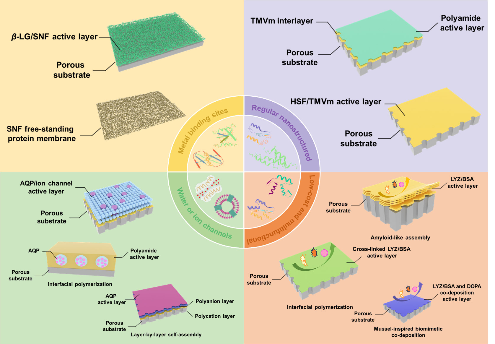
Membrane separation technology has been intensively used in numerous applications such as seawater desalination, water treatment and reuse, fine separation and product concentration, biomedical treatment and so forth owing to its low operation temperature, easy operation process, modularity, and high separation efficiency. However, due to membrane materials, membrane structures, and membrane manufacturing technology, the trade-off behavior between the water flux and the rejection rate of conventional separation membranes has become a technical bottleneck. The preparation of high-performance separation membranes using proteins as membrane materials is expected to break the trade-off behavior of conventional separation membranes. Protein separation membrane works super-efficiently for the target separation and transport, as well as the antibacterial and antifouling properties, where an emerging membrane material of proteins can transport the solute due to their inherent specific water or ion channels, rich binding sites with metal ions, regular nanostructures or low-cost and multifunctional. In this review, the widely implemented membrane materials and fabrication strategies for protein separation membranes are summarized in detail, and the research progress of the various protein separation membranes is described. Furthermore, the challenges faced by protein separation membranes are comprehensively reviewed. This review provides some insights into the construction and prospect of protein separation membranes.

Contents

1 Introduction

2 Novel protein materials for membrane fabrication

2.1 Protein containing channels

2.2 Protein containing rich binding sites with metal ions

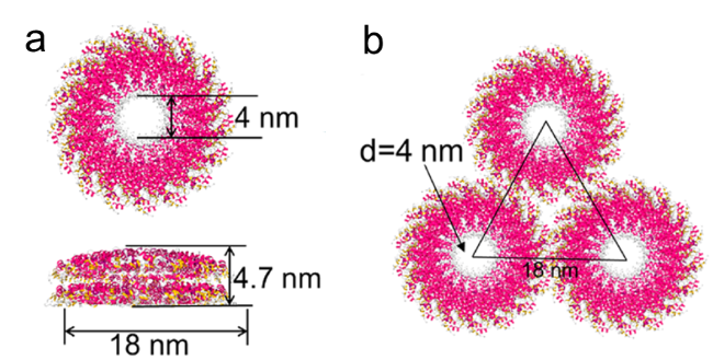
2.3 Protein containing regular nanostructures

2.4 Low-cost and multifunctional protein

3 Fabrication methods of protein separation membranes

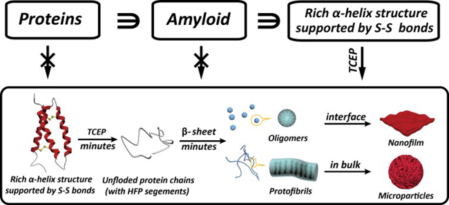
3.1 Amyloid-like assembly

3.2 Interfacial polymerization

3.3 Layer-by-layer self-assembly

3.4 Mussel-inspired biomimetic co-deposition

3.5 Other methods

4 Research progress of protein separation membranes

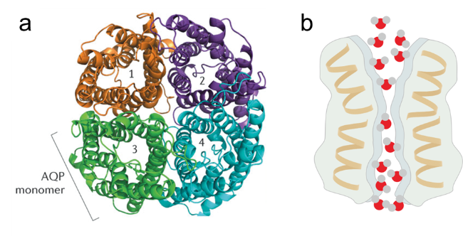
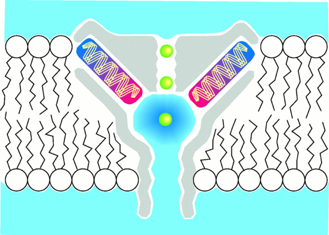
4.1 Aquaporin biomimetic separation membranes

4.2 Lysozyme separation membranes

4.3 Protein separation membrane for chelating metal ions

4.4 Other protein separation membrane

5 Conclusion and outlook

Cite this article

Junping Miao , Zhaoqian Zhang , Shaopeng Xin , Yunxia Hu . Development of Protein Separation Membranes and Their Progress[J]. Progress in Chemistry, 2025 , 37(2) : 195 -210 . DOI: 10.7536/PC240312

1 Introduction

Membrane separation technology is widely used in human production and life due to its advantages of high efficiency, energy saving, environmental protection, and small footprint, such as gas separation (carbon capture, O2/N2 separation, removal of CO2 and H2S from natural gas, air dehydration, etc.), water treatment (seawater desalination, drinking water purification, urban and industrial sewage treatment and reuse, etc.), bioseparation and purification (sterile filtration, buffer exchange, protein concentration, vaccine production and purification, drug purification, etc.), food processing and concentration (clarification of beer and wine, concentration of whey, juice, and sugar, etc.), chemical production and recovery (production of chlorine and sodium hydroxide in the chlor-alkali process, organic solvent recovery, catalyst recovery, etc.), healthcare (dialysis, blood oxygenation, drug delivery, etc.), energy production and storage (lithium extraction from salt lakes, hydrogen production by electrolysis, fuel cells, etc.)[1-6].
Separation membranes are the core components of membrane separation technology, directly determining the separation efficiency of the membrane[7]. However, traditional separation membranes exhibit a trade-off effect between permeability and selectivity, and existing strategies often fail to achieve simultaneous improvements in both membrane permeability and selectivity[8]. In recent years, protein materials with multifunctionality and good film-forming properties have gained increasing attention in membrane fabrication applications. Particularly, using proteins with specific molecular/ion transport channels and rich metal ion binding sites as membrane materials to develop high-flux and high-selectivity separation membranes holds promise for achieving selective transmission or retention of specific molecules and ions, potentially overcoming the Trade-off effect of traditional membrane materials.
Therefore, the preparation of high-performance separation membranes using proteins as materials has become one of the research hotspots in the field of separation membranes. This article reviews the progress of protein separation membranes at home and abroad over the past decade, detailing the structure and function of protein molecules used for membrane formation, the preparation methods and research progress of existing protein separation membranes, and points out the current issues in protein separation membrane research and future research directions.

2 Protein Materials

Based on the functional advantages of proteins in membrane fabrication applications, protein materials used for membrane fabrication can be divided into channel proteins (such as aquaporins, ion channel proteins, etc.), proteins rich in metal ion binding sites, proteins with regular nanostructures, and cost-effective multifunctional proteins. This section focuses on introducing the structure, function, and potential advantages of protein materials that can be used for membrane fabrication.

2.1 Channel Proteins

The cell membranes of living organisms contain channel proteins that can bind with specific water molecules or ions and efficiently and specifically transport water molecules, functional molecules (such as glycerol, urea), or specific ions (such as Na+, K+, Ca2+, etc.). For instance, aquaporins (Aquaporin, AQP), which are widely present in the cell membranes of animals, plants, and microorganisms, have the ability to efficiently and specifically transmit water molecules. Except for a few types of AQP that are permeable to glycerol[9], most AQPs have a rejection rate of 100% for other molecules, ions, and protons[10]. One AQP molecule can specifically transport 3 billion water molecules per second[11], and its water transmission capacity is much higher than that of existing commercial separation membranes. Studies have shown that AQPs on the cell membrane exist in tetrameric form, and each AQP monomer can functionally act as an independent channel to transmit water molecules. Each AQP monomer consists of a single peptide chain containing six transmembrane α-helices, connected by five loops (loops A-E), with two short α-helical chains embedded inside the membrane but not spanning it (i.e., loops B and E). Loops B and E fold back into the lipid bilayer, and these two loops contain a highly conserved asparagine-proline-alanine (NPA) amino acid signature sequence. The six transmembrane α-helices surround the two NPAs, together constructing a channel that specifically transmits water molecules, with a diameter of approximately 0.28 nm (Figure 1)[12].
图1 (a)AQP四聚体的俯视图[29];(b)AQP的纳米通道

Fig. 1 (a) Overhead view of AQP tetramer[29]. Copyright 2020, Taylor & Francis (b) Schematic diagram of AQP nanochannel.

The efficient water permeability of AQP is mainly attributed to the unique transport mechanism of water molecules within AQP. Since the narrow pore diameter of the AQP channel is only 0.28 nm, which is similar to the size of a water molecule, water molecules form a single column when entering the tortuous and narrow channel[13]. Subsequently, the dipole forces and polarity inside the channel help rotate the water molecules, allowing them to pass through the narrow channel at an appropriate angle. As the narrow pore diameter is smaller than the minimum requirement for continuous water flow (1.6 nm), water molecules cannot be continuously transported within the channel, reducing energy loss due to collisions between water molecules. This results in low transmission resistance inside the channel, thereby endowing AQP with high water permeability[14]. Given that AQP can efficiently and specifically transport water molecules, researchers have utilized AQP to develop biomimetic AQP membranes[15-28]. Section 4.1 of this paper provides a detailed introduction to the research progress of biomimetic AQP membranes.
The main ion channel proteins commonly found on cell membranes include sodium ion channel proteins, potassium ion channel proteins, calcium ion channel proteins, and chloride ion channel proteins. These ion channel proteins have the following characteristics[30]: (1) Specific selectivity for certain ions. Generally speaking, each type of ion channel protein can specifically allow one type of ion to pass through, which is also the basis for naming ion channel proteins. (2) Efficient transport of specific ions. Studies have found that after the ion channel opens, it can selectively transport about 107 specific ions per second. (3) Controllability of the switch. The channels of such proteins have open and closed states, and these states are regulated by transmembrane voltage, mechanical stimuli (such as gravity stimuli, vibrations, etc.), and certain signaling molecules (such as ligands). The specificity and efficiency of ion channel proteins for specific ions are mainly related to the spatial structure inside the channel and the distribution of charged groups[31-32]. Taking potassium ion channel proteins as an example[33], the pore size of potassium ion channel proteins is about 0.1 nm, which is equivalent to the size of potassium ions, and its channel consists of a barrel-shaped selective filter and an inverted cone-shaped cavity. Potassium ions enter the channel in a hydrated state, then dehydrate when passing through the barrel-shaped selective filter, and are subsequently surrounded by oxygen atoms on the selective filter. This interaction with oxygen atoms is unique to potassium ions, meaning that the selective filter can achieve specific transmission of potassium ions. After potassium ions pass through the selective filter, they enter the inverted cone-shaped cavity where they are rehydrated. The rehydrated potassium ions reduce electrostatic repulsion from other ions, thus increasing the transport rate of potassium ions in the channel (Figure 2). Although sodium ions are smaller than potassium ions, they cannot interact with oxygen atoms when passing through the selective filter of the potassium ion channel, causing sodium ions to be unable to effectively pass through the potassium ion channel. Utilizing ion channel proteins for membrane fabrication allows for specific ion sieving.
图2 钾离子在钾离子通道蛋白中传输的示意图[32]。绿色球为钾离子

Fig. 2 Schematic diagram of potassium ion transport in potassium channel proteins[32]. The green sphere represents potassium ions. Copyright 1998, American Association for the Advancement of Science

In addition, there is an ion channel protein, gramicidin A (GA), which only allows water molecules and monovalent cations (such as Na+, K+) to pass through, and is impermeable to anions (such as Cl-, SO42-) and divalent cations (such as Mg2+, Ca2+). Its water permeability is comparable to that of AQP[31,34-35]. Using GA as a biomimetic channel, membranes with highly efficient ion separation performance can also be prepared.

2.2 Protein Rich in Metal Ion Binding Sites

Currently, proteins rich in metal ion binding sites have important application prospects in membrane fabrication. The metal ion binding sites in these proteins mainly originate from functional groups (donors) such as hydroxyl, amino, carboxyl, mercapto, and imidazole groups contained in the amino acids that make up the protein[36-40], which can provide unpaired electrons and bond with metal ions (acceptors) that have empty valence electron orbitals to form coordination compounds. Table 1 summarizes the complexation constants K between common functional groups in amino acids and metal ions[41~46]. The larger the value, the stronger the complexation ability between the amino acid functional groups and metal ions. It can be seen that compared with amino and imidazole groups, hydroxyl, carboxyl, and mercapto groups have higher complexation constants K with metal ions, meaning that the above-mentioned amino acid functional groups are more likely to undergo complexation reactions with metal ions. Research[47] shows that amino acid functional groups can complex with specific metal ions under mild conditions, thereby forming stable coordination compounds. Therefore, when removing metal ions, amino acids can be screened according to the complexation constants of functional groups with metal ions, and protein materials rich in suitable amino acids can be selected to increase the number of metal ion binding sites in the protein. Based on this, using proteins rich in metal ion binding sites to prepare separation membranes can selectively adsorb metal ions, achieving the removal or recovery of metal ions[48-49].
表1 氨基酸中常见官能团与金属离子之间的结合常数K[41-46]

Table 1 The binding constant K between functional groups and metal ions[41-46]

Ions -OH -NH2 -COOH -SH Imidazolyl
Mn2+
Co2+
Ni2+
Cu2+
Zn2+
Cd2+
Pb2+
4.35
6.08
9.72
9.72
8.77
2.47
10.12
4.66
5.12
5.45
4.42
5.39
4.90
3.51
1.56
10.89
0.89
4.45
11.93
6.65
14.65
3.20
9.04
5.00
2.43
3.94
5.53
7.98
3.10
1.66
0.78
0.85
5.50
1.10
1.48
For example, β-lactoglobulin (β-LG) protein derived from bovine milk contains five cysteines (Cys), fifteen glutamates (Glu), and two histidines (His). These amino acids can undergo complexation reactions with metal ions to form complexes[50-56]. Among them, 121-Cys has a heavy metal ion binding site[56], which can specifically chelate arsenic or lead ions; Glu can specifically chelate iron ions; His can specifically chelate copper ions. Additionally, the β-LG protein is inexpensive, has excellent mechanical properties, and good film-forming ability, making it suitable for preparing functional membranes that efficiently chelate metal ions.
In addition, silk fibroin extracted from silk contains approximately 10% of its amino acid composition as phenylalanine (Phe), tyrosine (Tyr), and tryptophan (Trp), which contain binding sites capable of complexing metal ions[57]. Researchers constructed SNF separation membranes using silk fibroin nanofibers (SNF) formed after the regeneration of silk fibroin as the membrane material[58~70] for the efficient removal of metal ions from water bodies. It is worth noting that silk fibroin generally needs to be regenerated before it can be used for membrane formation and efficient complexation of metal ions.
In addition to β-LG and silk fibroin, insulin (whose amino acids are Cys and lysine) [71-72] and zein (whose amino acid is His) [73-74] can also form complexes by chelating with metal ions, which can be used for the preparation of metal ion adsorption membranes.

2.3 Proteins with Regular Nanostructures

There is a class of proteins with regular nanostructures in nature, such as viral coat proteins, ferritin, β-barrel proteins, etc. By utilizing the regular nanostructures of these proteins and assembling them into membranes, separation membranes with high porosity and uniform pore size can be obtained, thereby endowing the membranes with advantages of high flux and high selectivity[75-78]. Among them, Tobacco mosaic virus mutant (TMVm) is a cylindrical protein with an outer diameter of 18.0 nm, an inner diameter of 4.0 nm, and a height of 4.7 nm (Figure 3). Ferritin is also a protein with a regular nanostructure, generally obtained from the liver and spleen, with a wide range of sources. Specifically, horse spleen ferritin (HSF) extracted from horse spleen has found more applications in membrane fabrication; it consists of dodecamers with a diameter of 12.0 nm and has a cage-like structure with a hollow cavity (diameter of 6.0 nm) in the center[79-81]. Compared with other protein materials, HSF can withstand alkaline aqueous solutions and organic solvents at 60 °C, so the protein separation membranes prepared from it can be used in harsh environments[82].
图3 (a)单层TMVm纳米片;(b)由TMVm纳米片堆积形成的孔示意图[75]

Fig. 3 (a) The single-layer TMVm nanosheets. (b) Schematic illustration of pores formed by TMVm nanosheets[75]. Copyright 2018, American Chemical Society

In addition, β-barrel proteins are a class of barrel-shaped porous proteins that can allow the passage of small molecules (such as monosaccharides and antibiotics) but prevent the passage of large molecules (such as proteins and polysaccharides)[83], including outer membrane protein α-hemolysin, outer membrane protein F, and bacterial outer membrane iron transport proteins, etc. The polar amino acid residues on the pore surface contain small molecule binding sites, thus they can be used for the selective screening of specific molecules.

2.4 Economical Multifunctional Proteins

Among numerous protein materials, there is a class of multifunctional proteins with good economic value, such as lysozyme (LYZ) protein, which is widely sourced and can be obtained from egg whites, animal and plant cells. It is not only inexpensive but also has excellent antibacterial properties and biocompatibility[84]. LYZ can be used for hydrophilic antibacterial anti-fouling modification of membrane surfaces, endowing the membrane with anti-fouling properties, and can also be used to assemble separation membranes with different pore sizes for various applications such as hemodialysis, dye removal, organic solvent purification, oil droplet emulsification, and toxic chemical separation[85-91]. In addition, bovine serum albumin (BSA) is a multifunctional protein derived from bovine serum, characterized by low cost and good anti-fouling properties, which can be used for membrane fabrication and also for anti-fouling modification of membrane surfaces[92-100].

3 Preparation Method of Protein Separation Membrane

The main methods used to prepare protein separation membranes include amyloid-like assembly, interfacial polymerization, layer-by-layer self-assembly, mussel-inspired co-deposition, and other methods. This section mainly reviews the principles of various membrane-making methods, key membrane-making factors affecting membrane structure and performance, etc.

3.1 Amyloid-like Assembly Method

Amyloid aggregates of proteins are special aggregates found in diseased animal tissues, characterized by typical features of insolubility and high adhesiveness, and are named amyloid structures because they can be stained with iodine[101]. In 2012, Yang et al.[102] proposed a method for film formation based on traditional amyloid protein aggregation, using the disulfide bond reducing agent tris(2-carboxyethyl)phosphine hydrochloride (TCEP) to cleave disulfide bonds between protein secondary structures under mild conditions of room temperature and near-neutral pH. This process causes the α-helical structure of proteins to unwind and triggers the spontaneous transition of high-energy α-helices to low-energy β-sheet aggregates, thereby forming a large number of nano-scale aggregates in solution[103]. This process occurs within minutes, and driven by the reduction of interfacial free energy, a large number of nano-scale aggregates formed in a short time gather at the gas/liquid or solid/liquid interface to form a dense nanofilm, while further forming microparticles in solution (Figure 4)[102,104]. Subsequently, using LYZ as a model protein, the team analyzed the kinetics of amyloid-like assembly in solution using the amyloid-specific dye thioflavin T. The study found that under the action of TCEP, after the disulfide bonds within LYZ molecules were broken, the originally internal hydrophobic amino acids of the α-helix were exposed to the solution, inducing hydrophobic aggregation of proteins, leading to a rapid transition of proteins from their native soluble state to insoluble amyloid-like aggregates. A large number of nano-aggregate particles formed in the solution in a short time would further aggregate at the gas/liquid or solid/liquid interface to form a dense nanofilm. Additionally, amyloid-like protein nanofilms can be attached to different substrate surfaces either by directly immersing the substrate into the reaction solution or by transferring the nanofilm onto the substrate surface using a hydrogel as a medium[105-108]. Since proteins contain a large variety of functional groups (—NH2, —COOH, —OH, —SH, —(CH)n—CH3, aromatic rings), they can form various interactions with different substrate materials through metal-sulfur bonds, hydrogen bonds, hydrophobic forces, and electrostatic attractions, providing strong adhesion for the stable loading of protein nanofilms on different material surfaces, thus constructing protein composite films[102-103,109-112].
图4 类淀粉样蛋白组装机理[103]

Fig. 4 Mechanism of amyloid-like assembly of proteins[103]. Copyright 2013, The Royal Society of Chemistry

The amyloid-like assembly method has certain limitations, and not all proteins can form protein nanofilms through amyloid-like protein aggregation. Common proteins that can undergo amyloid-like aggregation have the following characteristics[102]: (1) Containing high-fibrillation-prone segments, these proteins themselves have a high tendency for amyloid aggregation. (2) Relying on disulfide bonds to stabilize the secondary structure of proteins, it is possible to unfold the α- helical structure of proteins by disrupting disulfide bonds. (3) Once the disulfide bonds are disrupted, unfolded or partially unfolded proteins aggregate from the native α- helix to β- sheet conformation. A series of globular proteins, such as LYZ, insulin, BSA, α-lactalbumin, lactoferrin, β-LG, etc., have been proven to meet the above requirements and can be used for preparing protein nanofilms via the amyloid-like assembly method[113-114].
The physical properties of amyloid-like protein-made nanofilms can be regulated by adjusting the assembly kinetics in the solution and the growth kinetics at the interface, specifically through the following reaction conditions: (1) pH value. Although TCEP has a good effect on reducing disulfide bonds over a wide pH range, because it can significantly affect the charge properties of proteins in the solution environment, the pH value plays an important regulatory role in the structure during the formation process of protein nanofilms. When the pH value of the solution environment changes, the ionization and hydrolysis of functional groups in protein molecules also change, thereby altering their exhibited charge properties. The farther the pH value is from the isoelectric point of the protein molecule, the more charge the protein molecule carries, the stronger the electrostatic repulsion between them, and the denatured protein chains remain stably dispersed without aggregation. Therefore, the formed protein nanofilm is looser, and may not even form a complete continuous film; conversely, when the pH value is closer to the isoelectric point of the protein molecule, the assembled protein nanofilm becomes denser[87]. (2) Protein concentration. Proteins are the basic units for assembling nanofilms, and their concentration significantly affects the physical structure of the protein nanofilm. As the protein concentration increases, the concentration of assembly units increases, leading to faster and denser protein packing during amyloid-like assembly, resulting in increased membrane thickness and reduced pore size. Taking LYZ as an example, when its concentration increases from 1 mg·mL−1 to 30 mg·mL-1, the membrane thickness increases from 30 nm to 250 nm, and the pore size decreases from 3.6 nm to 1.8 nm[85]. (3) Film-forming time. The Xu research group[87] monitored the size change of aggregate particles formed by the self-assembly of LYZ treated with TCEP in a solution at pH 8.4 and found that when the reaction time was before 10 minutes, the average size of LYZ aggregate particles rapidly increased with prolonged reaction time, whereas beyond 10 minutes, the average size would decrease, possibly due to the deposition of overly large aggregates formed by LYZ assembly. Other studies[113] also indicate that only those smaller aggregate particles (no larger than 50 nm) contribute to the formation of LYZ nanofilms, while larger particles do not participate in the formation of LYZ nanofilms. Correspondingly, the growth of nanofilm thickness also shows that with the extension of growth time, the thickness of the LYZ nanofilm continuously increases until reaching 50 nm, and after reaching 50 nm, the thickness basically does not continue to increase with prolonged growth time[113].
Amyloid-like protein assembly-prepared nanofilms can be stably loaded onto the surfaces of hydrophobic porous base membranes such as polyethylene, polypropylene (PP), polyvinylidene fluoride, polytetrafluoroethylene, and polyethylene terephthalate (PET) [87], and onto the surfaces of hydrophilic porous base membranes such as hydrolyzed polyacrylonitrile (hPAN) [86], to prepare ultrafiltration (UF) and nanofiltration (NF) membranes. Section 4.2 of this paper, which covers the LYZ separation membrane, will provide a detailed introduction to the research progress in preparing LYZ protein membranes using amyloid-like assembly, and will not be repeated here.
The amyloid-like assembly method is a simple, low-cost, green, and efficient way to prepare protein separation membranes. It has strong universality and mild conditions, does not destroy any amide bonds in the protein backbone, and uses protein as the main film-forming material, which can maximize the functional properties of the prepared membrane[112]. However, the current understanding of the film-forming mechanism and pore-forming mechanism of amyloid-like protein assembly is still unclear and requires further research.

3.2 Interfacial Polymerization Method

Interfacial polymerization (IP) was first proposed by Mogran et al[115], and then widely used in the preparation of separation membranes. It is a method that dissolves two monomers capable of undergoing polymerization reactions in two immiscible solvents (usually aqueous phase and organic phase), and through the reaction between the aqueous phase monomer (mostly diamines) and the organic phase monomer (mostly triacyl chlorides) at the interface of the two phases, a thin and dense polymer membrane is formed on the surface of the porous substrate[116]. Currently, there are three main methods for preparing protein separation membranes using IP (Figure 5): (1) Directly using protein as the aqueous phase monomer, utilizing the functional groups of the protein to react with the acyl chloride in the organic phase monomer, a cross-linked functional layer is generated at the water-oil interface on the substrate surface, forming a protein separation membrane[92,117]. The separation membrane prepared by this method not only has good permeability and selectivity but also exhibits excellent protein functional properties such as antibacterial and anti-fouling properties. However, compared with the aqueous phase monomers used in traditional IP reactions, proteins have larger molecular weights and lower reactivity of functional groups, resulting in low cross-linking degree and loose structure of the membrane functional layer, which can efficiently retain dye molecules but is difficult to retain inorganic salts. (2) Loading proteins with regular nanostructures onto the substrate surface to construct a high-porosity intermediate layer, and then forming a composite NF membrane embedded with proteins on its surface via IP[118]. Among them, the high-porosity protein intermediate layer provides a good interface for the IP reaction, making the distribution of monomers more uniform and generating a defect-free IP functional layer, thereby improving the permeability of the separation membrane, but this method is difficult to highlight the influence of the protein's own structure on the separation characteristics. (3) Introducing vesicles containing proteins into the aqueous phase monomer solution as additives, so that the vesicles containing proteins are encapsulated within the generated functional layer during the IP reaction, producing protein composite reverse osmosis membranes and forward osmosis (FO) membranes[25-26,119]. This method helps maintain the biological activity of proteins and fully utilizes the additional water channels provided by proteins, thus making the membrane have high water flux and high NaCl retention rate.
图5 (a)以蛋白为水相单体[117]、(b)以具有规则纳米结构的蛋白构建中间层[118]和(c)以含功能蛋白的囊泡为水相添加剂[26],通过IP法制备蛋白分离膜的示意图

Fig. 5 Schematic illustration of the fabrication process of the protein separation membranes via IP using protein as (a) an aqueous monomer [117], (b) an interlayer [118], and (c) an aqueous additive [26]. Copyright 2017, Elsevier; Copyright 2020, American Chemical Society; Copyright 2015, Elsevier

Studies have shown that by controlling IP reaction conditions such as the concentration of reaction monomers, reaction time, and aqueous phase additives, the structure and physicochemical properties of the functional layer, such as porosity, thickness, surface charge, and crosslinking degree, can be regulated, thereby achieving performance regulation of the prepared protein separation membranes. (1) Concentration of reaction monomers. Since only a small amount of protein aqueous phase monomer reacts with the organic phase monomer at the interface, the concentration of protein aqueous phase monomer should be higher than that of the organic phase monomer. During the IP reaction process, the concentration of monomers directly affects the concentration ratio between protein aqueous phase monomers and organic phase monomers, significantly influencing the thickness and crosslinking degree of the crosslinked functional layer. When the concentration ratio between protein aqueous phase monomers and organic phase monomers is low, more oligomers with acyl chloride groups remain on the surface of the crosslinked functional layer, and due to the exhaustion of protein aqueous phase monomers during the IP reaction, some residual acyl chlorides hydrolyze to form linear polymers, resulting in reduced crosslinking degree of the functional layer and poorer selectivity of the composite membrane; when the concentration ratio between protein aqueous phase monomers and organic phase monomers is high, the organic phase monomers are quickly consumed in the IP reaction, generating oligomers containing amino groups, increasing the thickness of the functional layer but reducing the crosslinking degree, leading to enhanced permeability and decreased selectivity of the composite membrane[92,117]. (2) Reaction time. The reaction time also affects the thickness and crosslinking degree of the functional layer. Under conventional reaction conditions, if the reaction time is too short, the reaction between the two-phase monomers is insufficient, forming a thin functional layer with low crosslinking degree, resulting in high flux and poor separation performance of the composite membrane; extending the reaction time allows for more complete crosslinking between the two-phase monomers, yielding a higher crosslinking degree and reduced pore size of the prepared functional layer, decreasing the water permeability coefficient and increasing the rejection rate of the composite membrane[92,117]. (3) Aqueous phase additives. Proteins used as aqueous phase monomer additives generally reduce the diffusion rate of monomers to the interface, forming a relatively loose and thinner functional layer. Depending on the properties of the additives, they may have additional effects on the performance of the composite membrane. Research[25-26,119] has found that using vesicles containing AQP as aqueous phase additives can slow down the diffusion rate of aqueous phase monomers to the interface, reduce the reaction rate of two-phase monomers at the interface, and vesicles containing AQP can provide additional water molecule transport channels, promoting efficient water molecule penetration in the functional layer and enhancing the water permeability coefficient of the protein composite membrane.

3.3 Layer-by-Layer Self-Assembly Method

As one of the commonly used methods for preparing protein separation membranes, the layer-by-layer self-assembly (LBL) method is a technique that utilizes interactions between polyelectrolytes and proteins (such as electrostatic interactions, van der Waals forces, covalent bonds, hydrogen bonds, etc.) to drive the co-assembly of polyelectrolytes and proteins on the surface of a porous substrate to form separation membranes with permeation selectivity[120-121]. The preparation of protein separation membranes by driving the alternate assembly of polyelectrolytes and proteins on the surface of the base membrane through electrostatic interactions is more widely applied. This film-forming method needs to meet the following three conditions: (1) The substrate must be charged to facilitate the deposition of the first layer of polyelectrolyte. (2) The electric properties of polyelectrolytes and proteins are different. (3) The concentration of polyelectrolytes and proteins should be higher than the minimum critical value, which is determined by the solubility and charge density of the polyelectrolyte or protein, because an appropriate concentration can ensure their successful adsorption on the membrane and reverse the charge on the membrane surface. Currently, according to the type of charge carried by the polymer chain, commonly used polyelectrolytes mainly include cationic polyelectrolytes (such as poly(allylamine hydrochloride) (PAH), polyethyleneimine (PEI), poly(dimethyldiallylammonium chloride) (PDADMAC)) and anionic polyelectrolytes (such as polystyrene sulfonate (PSS), polyacrylic acid (PAA)), etc.[120]. Based on the degree of ionization of polyelectrolytes in aqueous solutions, they can also be divided into strongly ionized strong polyelectrolytes (such as PEI, PDADMAC, PSS) and partially ionized weak polyelectrolytes (such as PAH, PAA)[120].
To prepare protein separation membranes by assembling polyelectrolyte materials and protein materials through electrostatic interactions, the permeability and selectivity of the protein separation membrane can be controlled by adjusting the number of assemblies of polyelectrolytes and proteins, the concentration of proteins, and the type of polyelectrolytes[23,27-28,122-123]. (1) Number of assemblies of polyelectrolytes and proteins. It was found that the number of assemblies of polyelectrolytes and proteins is positively correlated with the thickness of the membrane functional layer. As the number of assemblies increases, the defects on the membrane surface can be reduced, the membrane pores can be decreased, thereby improving the separation performance of the membrane[27,122-123]. Wang et al.[27] studied the effect on membrane performance by controlling the number of assemblies of polyelectrolytes and vesicles containing AQP on the surface of the base membrane. They found that compared to the membrane prepared after one alternate assembly of polyelectrolyte and vesicle containing AQP on the base membrane, the membrane prepared after two alternate assemblies had fewer surface defects. The water permeability coefficient decreased from 6.1 to 5.5 L·m−2·h−1·bar−1, and the rejection rate of NaCl increased from 62% to 75%. (2) Protein concentration. The protein concentration is proportional to the thickness of the membrane functional layer. In addition, considering the properties of the protein itself, increasing the protein concentration may affect the membrane performance. For example, taking vesicles containing AQP as an example, increasing the concentration of vesicles containing AQP can enhance the loading amount of AQP in the membrane, increase the water passage channels in the membrane, thereby improving the permeation flux of the membrane[23,27-28]. (3) Type of polyelectrolyte. Adjusting the types of different polyelectrolytes can influence the compactness (e.g., pore size, porosity) and surface properties (e.g., hydrophilicity, charge) of the polyelectrolyte layer, thereby affecting the number of defects generated on the membrane surface and regulating membrane selectivity. For example, a polyelectrolyte layer formed by strong polyelectrolytes with excellent hydrophilicity has a high degree of compactness and good hydrophilicity, effectively compensating for membrane surface defects and making the distribution of hydrophilic proteins in the membrane more uniform, significantly enhancing the separation performance of the membrane[23,27,122-123].
The layer-by-layer self-assembly method is simple and easy to operate, with mild conditions, fast assembly speed, and the ability to finely control the structure of protein separation membranes, making it an effective method for preparing protein separation membranes. Currently, the main approach involves the layer-by-layer assembly of polyelectrolytes and vesicles containing AQP protein, encapsulating vesicles with AQP protein on the surface of the base membrane to produce protein separation membranes, while there is less research on the co-assembly of polyelectrolytes and proteins into membranes. It is worth further studying to utilize proteins with different charges, as well as polyelectrolytes and proteins with different charges, for layer-by-layer self-assembly into membranes, which is expected to significantly increase the protein content in the membranes, thereby enhancing the functionality of the proteins.

3.4 Mussel-inspired Co-deposition Method

The mussel-inspired co-deposition method utilizes the adhesive effect produced after the oxidative polymerization of tea polyphenol derivatives (such as dopamine (DOPA), tannic acid, gallic acid, etc.) [124-126], which co-deposits tea polyphenol derivatives and target proteins onto the surface of the base membrane through chelation, electrostatic, hydrogen bonding, and π-π stacking interactions to prepare protein separation membranes [127-128]. Among them, the DOPA co-deposition method for membrane preparation has been more extensively studied. By adding protein to a DOPA solution, it co-deposits onto the porous base membrane surface during the oxidative polymerization process of DOPA, which can adjust the surface charge of the UF membrane through the protein's own charge properties, serving as a coating that imparts anti-fouling properties to the UF membrane. Additionally, through Michael addition or Schiff base reactions between the protein and dopamine, a dense layer of polydopamine/protein can be generated to directly prepare separation membranes.
By varying the deposition time and the mass ratio of DOPA to protein, the oxidative polymerization process of DOPA and protein, solution assembly kinetics, membrane pore size, and membrane surface properties can be controlled. (1) Deposition time. The deposition time affects the polymerization mechanism of DOPA and protein and the assembly kinetics in the solution. During the co-deposition process of DOPA and protein, different types of protein materials exhibit different oxidative polymerization mechanisms with DOPA at various deposition times. For instance, when DOPA co-deposits with LYZ under alkaline conditions, the negatively charged polydopamine (PDA) particles formed by DOPA oxidative polymerization first form mixed micelles with positively charged LYZ through electrostatic interactions, and as the deposition time prolongs, further polymerize to form PDA-LYZ particles, effectively suppressing the oxidative polymerization degree of DOPA. Subsequently, the aggregation of positively charged LYZ on the surface of PDA-LYZ particles increases the positive charge of the particles and enhances the electrostatic repulsion between particles, thereby limiting the further growth of PDA-LYZ particles with deposition time and existing in a form much smaller than PDA particle size while closely packing on the surface of the porous substrate membrane, ultimately forming a thin and dense functional layer[129]. Hemoglobin (BHb) contains four +2 ferrous ions that can complex with nitrogen and oxygen atoms in PDA to form complexes, thereby accelerating the oxidative polymerization process of DOPA[130]. Additionally, the deposition time affects the assembly kinetics process of DOPA and protein. Taking the co-deposition process of DOPA and LYZ as an example, within the first 5 minutes of co-deposition, the size of LYZ particles in the solution is 3.58 nm, and the size of the mixed micelles formed by PDA particles and LYZ is 529 nm; as the deposition time increases to 10 minutes, there are no free LYZ particles in the solution, and the size of the mixed micelles increases to 635 nm; further extending the deposition time to 30 minutes, the size of the mixed micelles becomes 697 nm, and PDA-LYZ polymer particles with a size of 24.06 nm appear; continuing to extend the deposition time, the size of the mixed micelles gradually decreases, while the size of PDA-LYZ polymer particles increases[129]. (2) The mass ratio of DOPA to protein. The mass ratio of DOPA to protein significantly affects membrane surface properties, membrane thickness, membrane density, and membrane pore size. Taking BHb as an example, since PDA contains a large number of amino groups and BHb contains a large number of carboxyl groups, the separation membrane prepared by co-deposition of DOPA and BHb has good hydrophilicity, and as the mass ratio of DOPA to protein decreases, the content of BHb increases, and the dynamic water contact angle value on the membrane surface gradually decreases[130]; when pH < the isoelectric point of the protein, the protein is positively charged while PDA is negatively charged, the negative charge on the membrane surface weakens, and as the mass ratio of DOPA to protein decreases, the protein content increases, and the electronegativity of the membrane surface weakens. When pH > the isoelectric point of the protein, the protein is negatively charged, the negative charge on the membrane surface enhances and is positively correlated with the mass ratio of DOPA to protein[129-130]; taking LYZ as an example[129], as the DOPA/LYZ mass ratio decreases from 1:0, 1:0.25, 1:0.5 to 1:1, the mass of LYZ increases, and the thickness of the functional layer formed by co-deposition of DOPA and LYZ increases. At a mass ratio of 1:0.5 and a co-deposition time of 30 minutes, the density of the functional layer formed by co-deposition of DOPA and LYZ reaches its maximum value (1.53 g·cm−3), which is 95% higher than the density of the functional layer formed by PDA under the same conditions, thus compared with the separation membrane prepared by DOPA deposition (pore size of 7.4 nm), the pore size of the separation membrane prepared by co-deposition of DOPA and LYZ shrinks to 1.5 nm.
The protein separation membranes prepared by this method have good hydrophilicity, biocompatibility, stability, and multifunctionality (such as antibacterial, anti-fouling, etc.). Moreover, this membrane-making method is versatile and suitable for various base membranes such as polyethersulfone, polysulfone, and polyvinylidene fluoride, but it is time-consuming. Generally, oxidants (ammonium sulfate, sodium periodate, sodium perchlorate, and metal ions), UV irradiation, and electrochemical driving are required to accelerate the co-deposition process of tea polyphenol derivatives and proteins, thereby shortening the membrane-making duration[126].

3.5 Other Methods

In addition to the widely used amyloid-like assembly method, IP method, LBL method, and mussel-inspired co-deposition method, novel membrane fabrication methods (such as molecular imprinting technology) are also crucial for the development of protein separation membranes[131]. Molecular imprinting technology is designed based on the working principle of "molecular keys and locks," which is a polymer membrane preparation technique capable of specific recognition for a particular target molecule. When using molecular imprinting technology to prepare protein separation membranes, it is necessary to covalently immobilize vesicles rich in proteins on the surface of the substrate membrane and coat initiators on the vesicle surface. Then, using monomers with specific recognition for the initiator and the cross-linking polymerization reaction between the initiator on the vesicle surface, an imprinted polymer functional layer is formed on the vesicle surface to fabricate the protein separation membrane (Fig. 6)[132-133]. However, the low density of the imprinted polymer functional layer leads to defects on the membrane surface, making it difficult to achieve high rejection performance.
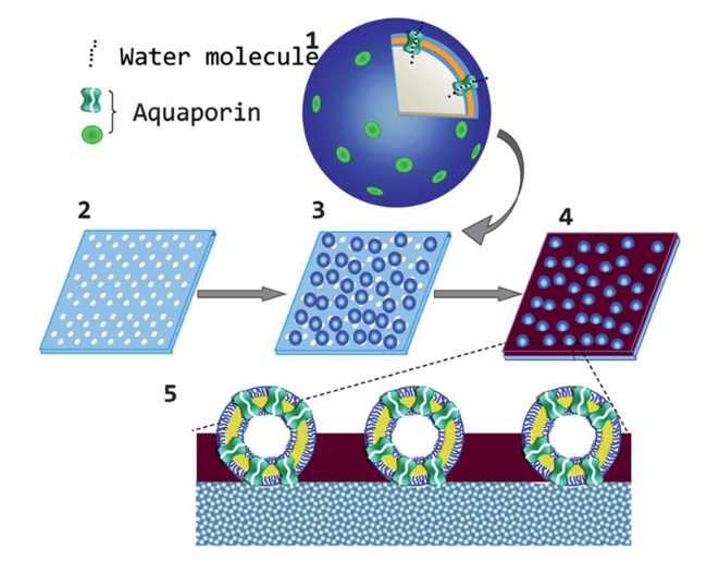
图6 AQP囊泡印迹膜的制备过程示意图[133]。(1)AQP囊泡,(2)多孔醋酸纤维素膜基底,(3)固定在多孔膜上的AQP囊泡,(4)AQP囊泡印迹膜和(5)AQP囊泡印迹膜的断面结构

Fig. 6 The schematic fabrication process of the AQP-vesicle-imprinted membrane[133]. (1) vesicles containing AQP, (2) porous cellulose acetate membrane substrate, (3) vesicles containing AQP immobilized on the porous membrane e, (4) AQP-vesicle-imprinted membrane, and (5) cross-section of the AQP-vesicle-imprinted membrane. Copyright 2013, The Royal Society of Chemistry

4 Research Progress on Protein Separation Membranes

In view of the specific channel of AQP with exclusive water permeability, the advantages of LYZ such as low cost, high stability, antibacterial and antifouling properties, and the ability of metal ion binding site-rich proteins to specifically adsorb metal ions and other specific functionalities of proteins, these types of proteins have been most studied for membrane fabrication. This chapter focuses on reviewing the research progress of these types of protein separation membranes.

4.1 Aquaporin Biomimetic Separation Membrane

The unique sieving mechanism and high water permeability of AQP make it an ideal membrane material for preparing biomimetic membranes, and AQP biomimetic separation membranes have become one of the current research hotspots. The water permeability of the membrane is enhanced by embedding AQP protein into the membrane. To ensure the biological activity of AQP, researchers embed AQP into lipid or block copolymer vesicles to simulate the cellular membrane microenvironment required for AQP membrane proteins to function, then spread the AQP-rich vesicles after adsorption, rupture, and fusion, or encapsulate the AQP-rich vesicles onto the substrate membrane surface to form an AQP functional layer, thus preparing AQP biomimetic separation membranes[15-26].
When AQP is embedded in lipids, the compatibility between AQP and lipids is crucial to ensure the biological activity of AQP and the stability of vesicles. The source of lipids used in preparing AQP-containing vesicles affects the water permeability performance of the vesicles produced. Among them, vesicles containing AQP prepared using natural lipids from the Escherichia coli cell membrane as carriers exhibit a significantly higher water permeability rate (>1.6×10⁻³ m·s−1) than the permeability level of vesicles containing AQP prepared using synthetic lipids as carriers[134]. However, compared with natural lipids, artificially synthesized block copolymers have higher physicochemical stability[135], and the AQP vesicles prepared using them show excellent stability and well-maintained functional activity of AQP. Among these, the poly(2-methyl oxazoline)-poly(dimethylsiloxane)-poly(2-methyl oxazoline) (PMOXA-PDMS-PMOXA) block copolymer is widely applied.
Currently, one of the common methods for preparing AQP biomimetic separation membranes is to directly spread vesicles containing AQP on the surface of a porous substrate membrane after adsorption, rupture, and fusion to prepare AQP biomimetic separation membranes (Fig. 7a), including vesicle rupture method[18], polymer cushion method[16], and micelle fusion method[20]. The vesicle rupture method involves directly adsorbing, rupturing, and fusing vesicles containing AQP on the substrate surface to form a phospholipid bilayer membrane, thereby obtaining an AQP biomimetic separation membrane. For instance, Chung et al.[18] embedded AQP into PMOXA-PDMS-PMOXA block copolymers to prepare vesicles containing AQP, and then prepared AQP biomimetic FO membranes using the vesicle rupture method, achieving a water flux of up to 142 L m−2·h−1 and a reverse salt flux below 10 g m−2·h−1. This method is simple to operate and results in fewer defects on the membrane surface, but the mechanical strength of the phospholipid bilayer is not high, leading to poor stability during membrane operation. To address this, researchers have proposed adding a polymer cushion (e.g., carboxylated polyethylene glycol [16]) between the substrate membrane and the vesicles containing AQP to enhance the binding force between the vesicles and the substrate surface, improving the mechanical stability of the membrane. Besides the vesicle system, the micelle system can also be used to prepare AQP biomimetic membranes. This method involves spreading micelles containing AQP on the surface of a porous substrate membrane after adsorption and fusion to obtain a biomimetic membrane[20]. Compared with amphiphilic phospholipid molecules, the functional layer formed by the fusion of AQP-containing phospholipid micelles on the substrate surface is more continuous, reducing membrane surface defects; the electrostatic interaction between positively charged AQP phospholipid micelles and negatively charged substrate membranes improves the membrane's stability.
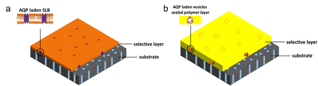
图7 (a)含AQP囊泡通过吸附、破裂融合后铺展到基膜表面制备AQP仿生膜;(b)封装含AQP囊泡到基膜表面制备AQP仿生膜[10]

Fig. 7 Schematic diagram of preparing AQP biomimetic membranes by (a) adsorbing, rupturing, fusing, and then spreading AQP containing vesicles onto the surface of the substrate or (b) encapsulating vesicles containing AQP onto the surface of the substrate[10]. Copyright 2015, Elsevier

To enhance the stability of AQP biomimetic membranes, researchers encapsulated vesicles containing AQP on the surface of the base membrane to prepare biomimetic membranes and used vesicles to protect AQP (Fig. 7b). Among them, the main methods of vesicle encapsulation are cross-linking encapsulation[133], IP encapsulation[26], and LBL encapsulation[23,28]. Cross-linking encapsulation involves loading molecules with reactive groups onto the outer surface of vesicles containing AQP, then depositing the vesicles onto the porous base membrane under pressure, followed by a cross-linking reaction to form chemical bonds, which fixes the vesicles containing AQP on the base membrane surface, thereby enhancing the stability of the biomimetic membrane[21]. IP encapsulation introduces vesicles containing AQP as additives into the aqueous monomer phase and encapsulates the vesicles containing AQP in the cross-linked functional layer through an IP reaction to prepare biomimetic membranes. Due to the maturity of IP technology, it can be used for large-area membrane preparation, and currently, this encapsulation technology is widely applied in the preparation of AQP biomimetic membranes. LBL encapsulation embeds vesicles into the polyelectrolyte layer through electrostatic interactions between polyelectrolytes and vesicles containing AQP to prepare biomimetic membranes[28]. Encapsulating vesicles containing AQP into the membrane can significantly reduce membrane surface defects, enhance membrane mechanical strength, and help protect the biological activity of AQP. Currently, Aquaporin A/S in Denmark uses IP encapsulation technology to prepare AQP biomimetic membranes, achieving scaled-up membrane preparation, successfully developing the InsideTM series of commercial spiral-wound and hollow fiber membranes, such as InsideTM A and InsideTM B, which are used in reclaimed water recovery and seawater desalination fields[10,136-138].
In addition, researchers have increased the embedding amount of AQP in vesicles[27] or increased the content of AQP-rich vesicles in biomimetic membranes[28,139] and other strategies to enhance the loading amount of AQP in membranes, thereby improving the permeability of the membrane. Sun et al.[28] used the LBL method to encapsulate AQP-rich magnetic vesicles in polyelectrolyte layers, and utilized magnetic field-driven adsorption to deposit more AQP-rich magnetic vesicles on the surface of the base membrane, thereby increasing the loading amount of AQP in the membrane. Compared with the control membrane without a magnetic field, the water flux of this membrane increased by 70%, and the reverse salt flux was lower than 2.4 g·m−2·h−1. Wang et al.[27] prepared AQP vesicles using strongly cationic phospholipids, enhancing the embedding amount of AQP in vesicles by strengthening the electrostatic interaction with negatively charged AQP, thus creating high-flux AQP biomimetic NF membranes. Compared with the control membrane without added AQP, the water flux of the AQP biomimetic membrane increased 20-fold, while the rejection rate of MgCl2 remained at 97%.
However, the water flux of existing AQP biomimetic FO membranes is far from its theoretical value (600 L·m−2·h−1)[140], and the reverse salt flux is also higher than that of traditional thin-film composite desalination membranes made by the IP method (0.4 g·m−2·h−1)[141], showing no outstanding advantages in water flux and salt rejection performance. The possible main reasons are as follows: (1) The process of membrane preparation may reduce the bioactivity of AQP. Current preparation methods for AQP biomimetic separation membranes involve processes such as extraction, purification, and reassembly of AQP into vesicles, which are complicated and prone to cause denaturation and aggregation of AQP[140], thereby losing some functional activity and deteriorating the selective separation function of AQP biomimetic membranes. (2) The loading amount of AQP in the membrane is low. Studies have found that the molar ratio of AQP to lipids or block copolymers in vesicles is less than 1:50[24,26,142], and further increasing the content of AQP will lead to defects in the vesicles. Therefore, how to increase the embedding amount of AQP in biomimetic membranes and ensure its water permeability activity has become the key to developing high-performance AQP biomimetic membranes.

4.2 Lysozyme Separation Membrane

LYZ, due to its low cost and advantages such as good antibacterial properties and biocompatibility, is used to prepare multifunctional protein separation membranes. Yang et al[89,109,143-144] found that LYZ can form a film on the surface of various substrates through amyloid-like assembly, with its pore size being tunable within the range of 0.5~3.6 nm. The prepared LYZ protein membrane not only has excellent mechanical properties but also can withstand various organic solvents (such as ethanol and hexane) and extreme pH (pH=1~12), and it also has good blood/cell compatibility, showing great application potential in the field of separation membranes. By adjusting key membrane-making parameters such as pH of the reaction solution, LYZ concentration, number of assemblies, and reaction time, the thickness and pore size of the protein membrane can be controlled to achieve efficient separation of different substances. For example, Xu et al[87] prepared an organic solvent NF membrane using amyloid-like LYZ under the action of TCEP at different pH values and found that when the pH increased from 5.4 to 8.4, the average pore size of the membrane increased from 1.15 nm to 3.35 nm. Yang et al[85] prepared membranes with different concentrations of LYZ. As the LYZ concentration increased from 1 mg·mL-1 to 30 mg·mL-1, the thickness of the membrane selective layer linearly increased from 30 nm to 250 nm, and the membrane pore size decreased from 3.6 nm to 1.8 nm. As the number of assemblies increased from 1 to 15 times, the thickness of the membrane selective layer linearly increased from 50 nm to 800 nm[113]. Currently, the area of separation membranes prepared by amyloid-like LYZ assembly can reach 20 inches[113], with adjustable protein functional layer thickness (30~250 nm) and pore size (0.5~3.6 nm)[85-87], and the membrane also has excellent adhesion, chemical stability, and thermal stability, which can stably adhere to the surface of various porous substrates under multiple forces (Fig. 8), forming a filtration membrane with selective separation performance[90,145].
图8 类淀粉样组装制备的LYZ蛋白膜表面和各类基材的相互作用力[109]

Fig. 8 The interaction forces between the surface of LYZ nanofilms prepared by amyloid-like assembly and various substrates[109]. Copyright 2017, Elsevier

Moreover, separation membranes prepared by utilizing amyloid-like LYZ assembly can be applied in fields such as hemodialysis, small molecule removal, and organic solvent purification[85-91]. Yang et al.[85] grew 50 mM TCEP and 2 mg·mL-1 LYZ on a PET microfiltration (MF) base membrane with a pore size of 10 μm for 2 h at a solution pH of 7.0 to prepare a UF membrane, which achieved clearance rates of 82.2%, 50.0%, and 81.3% for urea, LYZ, and creatinine within 4 h, respectively, and the clearance rate of uremic toxins per unit membrane area is 5~6 times that of current traditional hemodialysis membranes (such as cellulose membranes and polymer membranes). Chang et al.[90] utilized LYZ to create separation membranes with water permeability coefficients as high as 899 L·m−2·h−1·bar−1 and rejection rates of up to 100% for various organic dyes such as rhodamine B and methylene blue (MB), which can be used for printing and dyeing wastewater treatment. Additionally, Xu et al.[87] reacted 50 mM TCEP and 4 mg·mL-1 LYZ on the surface of a PP MF base membrane with a pore size of 0.2 μm for 1 h at a solution pH of 5.4 to prepare NF membranes with pore sizes around 1 nm; these membranes have permeability coefficients exceeding 29 L·m−2·h−1·bar−1 for organic solvents such as methanol, outperforming most organic solvent NF membranes. Moreover, the prepared LYZ protein membranes exhibited rejection rates of up to 98% for organic solvent dyes such as Acid Black 10B and recovery rates of 92.5% for organometallic catalysts such as cumene hydroperoxide. Zhu et al.[86] reacted 40 mM TCEP and 2 mg·mL-1 LYZ on an hPAN UF base membrane for 6 h at a pH of 5.3 to prepare LYZ protein NF membranes with a pore size of 0.54 nm, which showed antibacterial rates exceeding 92% against E. coli and Staphylococcus aureus, demonstrating excellent antibacterial properties. Xu et al.[87] confirmed that the LYZ protein membranes remained stable after being soaked in HCl (pH=1) and NaOH (pH=12) solutions for 2 h.
Moreover, during the amyloid-like assembly of LYZ, small molecules (such as dye molecules), fluorescently labeled peptides, nanoparticles (such as gold nanoparticles), and proteins (such as insulin) can be embedded into the LYZ nanofilm through simple physical mixing, endowing the film with corresponding functional properties[143,146]. For instance, dye molecules, fluorescently labeled peptides, and nanoparticles can serve as detection signals for ultra-sensitive biomolecule detection. The LYZ protein film can also be used for drug sustained-release in disease treatment. For example, the LYZ protein film has been utilized for the sustained release of insulin, which can be applied to blood glucose control in diabetic patients[106].
In addition, the LYZ protein membrane can also be prepared through mussel-inspired co-deposition. By adjusting the mass ratio of DOPA and LYZ and the deposition time, the oxidative polymerization and adhesion mechanism of DOPA and LYZ, the deposition kinetics of the solution, and the physicochemical properties of the deposition layer can be influenced, thereby preparing LYZ protein membranes with tunable pore sizes. An[129] The LYZ protein NF membrane was prepared by co-depositing DOPA and LYZ on the surface of the hPAN UF membrane. It was found that as the DOPA/LYZ mass ratio decreased from 1:0, 1:0.25, 1:0.5 to 1:1, the rejection rate of the membrane for dye MB first increased and then decreased, while its water permeability coefficient showed a gradually decreasing trend; as the co-deposition time increased, the rejection rate of the membrane for MB continuously increased and stabilized at 98.1%, while the water permeability coefficient continuously decreased to 23 L·m−2·h−1·bar−1; the antibacterial rate of the membrane against E. coli exceeded 91.7%, showing excellent antibacterial performance. In addition, through mussel-inspired co-deposition, LYZ protein can also be used for anti-fouling and antibacterial modification of the membrane. Hu et al.[125] used tea polyphenol derivatives and protein co-deposition method to modify the substrate membrane for anti-fouling. An[129] DOPA and LYZ were co-deposited on the surface of the PSf UF membrane to prepare an anti-fouling UF membrane, the positive charge on the membrane surface was enhanced, and when positively charged LYZ was used as a model pollutant, the flux attenuation rate of the LYZ-modified membrane was only 18.7%, and the flux recovery rate was as high as 91.8%, showing excellent anti-fouling performance.
In view of the economic and multifunctional advantages of LYZ, it can not only be assembled into a film by amyloid-like assembly, but also prepared into a film by mussel-inspired co-deposition. It can be used for the sieving of molecules of different sizes and for the anti-fouling modification of the membrane surface, which shows high application value in film preparation.

4.3 Protein Separation Membrane for Adsorbing Metal Ions

The β-LG protein, SNF, and zein contain a large number of amino acid binding sites that can specifically complex metal ions, which can be used to prepare protein membranes for adsorbing metal ions to efficiently remove harmful metal ions from water bodies. Among them, 121-Cys in β-LG protein can specifically adsorb arsenic or lead ions, and His can specifically adsorb copper ions. Bolisetty et al.[55] vacuum-filtered β-LG protein fibers onto the surface of a cellulose MF base membrane with 0.22 μm pore size to prepare β-LG protein membranes, which have a water permeability coefficient as high as 3397 L·m−2·h−1·bar−1, can completely remove arsenic ions from groundwater, with an adsorption capacity for +5 valent arsenic ions reaching 266 mg·g-1, and for +3 valent arsenic ions exceeding 1133 mg·g-1, being 7.4 and 18 times that of the control membrane without β-LG protein respectively. The β-LG protein separation membrane prepared by Peng et al.[51] has a removal rate for lead ions as high as 99.9%, with an adsorption capacity reaching 212 mg·g-1, and after undergoing the adsorption-desorption-resorption cycle process, the removal rate for lead ions still remains at 95.7%. Lan et al.[147] found that the adsorption capacity of β-LG protein membranes for copper ions exceeds 226 mg·g-1. Additionally, Mezzenga et al.[148] prepared mixed matrix membranes by blending β-LG protein fibers with activated carbon, which can remove 100% of heavy metal ions (such as technetium-99m and gallium-68) and various clinically relevant radionuclides (such as lutetium-177) from actual hospital nuclear wastewater, and the adsorption rate for heavy metal ions still does not decrease after five cycles of filtration. Moreover, Mezzenga et al.[52] also found that the prepared mixed matrix membranes of β-LG protein fibers and activated carbon have a removal rate for chromium, nickel, silver, and platinum in groundwater all exceeding 99%, with a water permeability coefficient four times that of current traditional NF membranes, and the removal rate for metal ions remains stable after ten cycles of filtration. Because Phe, Tyr, and Trp contained in SNF can specifically adsorb metal ions, Buehler et al.[69] grew needle-like hydroxyapatite (HAP) nanocrystals on the surface of SNF through in-situ biomineralization, then vacuum-filtered SNF/HAP onto the surface of a polycarbonate MF membrane, preparing SNF/HAP mixed matrix membranes with a separation layer thickness of 200 nm, achieving efficient adsorption removal of gold ions, where the adsorption removal rate for gold ions exceeds 92%, with an adsorption capacity of about 164.2 mg·g-1, and its water permeability coefficient is as high as 8355 L·m-2·h-1·bar−1. Due to different complexation constants K between SNF and different metal ions, SNF protein membranes can achieve selective adsorption of different metal ions. Ping et al.[68] prepared SNF/MoS2 separation membranes by adjusting the volume ratio of molybdenum disulfide (MoS2) nanosheets and SNF using a vacuum filtration method, this membrane has an adsorption removal rate for gold ions and platinum ions both >99%, with adsorption capacities as high as 759.1 and 425.1 mg g-1 respectively; while the adsorption removal rates for copper ions, lead ions, and silver ions are 44%, 44%, and 54% respectively, with adsorption capacities of 22.2, 153.5, and 246.2 mg g-1 respectively. In addition, the adsorption rate of SNF protein membranes for metal ions is 1.2 times that of the control membrane without SNF. Furthermore, composite nanofiber membranes of zein/nylon-6 prepared using electrospinning technology have a removal rate for chromium ions as high as 87%[149].
The preparation of membranes using proteins rich in metal ion binding sites can efficiently adsorb metal ions, not only removing metal ions from water bodies but also being applicable for the recovery and reuse of metal ions, showing potential applications in the field of precious metal ion recovery.

4.4 Other Protein Separation Membranes

In addition to the several protein separation membranes that have been extensively studied, some proteins with specific functions have also been used for membrane fabrication and exhibit special selectivity. Research has found[150]that among the two enantiomers of chiral drugs, one often has efficacy while the other is ineffective or even has toxic side effects. Therefore, the efficient separation of the two enantiomers is crucial for patient medication safety. Consequently, chiral separation membranes have emerged. These membranes use chiral recognition units (i.e., chiral selectors) as the driving force due to the differential binding affinity between different enantiomers, efficiently separating the two enantiomer molecules. They are typically composed of a porous polymer substrate and a functional layer containing chiral selectors[151]. L-Glu, L-Cys, and sodium poly-L-glutamate can serve as chiral selectors to covalently modify the membrane pores for fabricating chiral separation membranes, achieving efficient sieving of enantiomer molecules[151-154]. Researchers have employed molecular docking methods in molecular dynamics simulations to model the recognition process between two or more molecules and calculate the affinity generated between them[155]. It was discovered that the affinity of BSA for the chiral molecule L-Phe is better than for D-Phe, meaning L-Phe can preferentially adsorb onto BSA and transfer rapidly, thereby enabling the specific recognition of these two chiral molecules[98]. Hou et al.[98]first functionalized graphene oxide (GO) nanosheets using carbodiimide/N-hydroxysuccinimide coupling with BSA, then deposited them on the surface of mixed cellulose ester (MCE) MF membranes to prepare chiral separation membranes. Both experimental and simulation results confirmed that BSA can expand the interlayer spacing of GO membranes and act as a chiral selector to preferentially adsorb L-phenylalanine onto the membrane surface, thus achieving efficient transport of L-phenylalanine and imparting high flux and high selectivity to the chiral membrane. The enantiomeric excess value of the membrane (i.e., the content of one enantiomer minus that of the other) is as high as 97%, and the flux is 5 to 200 times that of previously reported chiral membranes.
Wang et al[75] deposited single-layer TMVm nanosheets with a size of 5-10 μm onto the surface of an anodic aluminum oxide MF substrate membrane through vacuum filtration, obtaining a high-flux UF membrane with a regular pore size of 4 nm and a separation layer thickness of only 40 nm. Due to the orderly structure of TMVm nanosheets and uniform inherent nanopores, the fabricated membrane has regular pore sizes and high porosity, with a rejection rate of 100% for nanoparticles with a diameter of 4 nm, while the water permeability coefficient of the membrane is as high as 7000 L·m−2·h−1·bar−1. Jin et al[118] first filtrated and deposited TMVm protein nanosheets on the surface of an MCE MF substrate membrane, then used IP technology to form a polyamide functional layer on its surface, creating an NF membrane. This highly porous and uniformly sized TMVm intermediate layer enables monomers to be evenly distributed and rapidly diffuse at the water-oil interface, thereby generating a defect-free functional layer through the IP reaction. The water permeability coefficient of this NF membrane is as high as 84 L·m−2·h−1·bar−1, while the rejection rate for MgSO4 exceeds 98%.
In summary, a great number of proteins have been used to prepare high-performance separation membranes. The prepared protein membranes exhibit unique pore structures and sieving properties, which can be used in high value-added separation systems and special separation fields. In the future, it is expected to develop protein separation membranes with efficient separation functions by selecting functional proteins with good economy and excellent film-forming properties, breaking through the application fields of existing membrane materials.

5 Conclusion and Prospect

The preparation of protein membranes with high flux and good selectivity by utilizing the functional properties of proteins can be applied in the fields of bioseparation, metal ion removal, and drug separation. Currently, the literature has reported various methods for preparing biomimetic separation membranes of functional proteins (especially aquaporin AQP). However, the developed biomimetic membranes of functional proteins have not yet reached the expected high flux and high selectivity, mainly due to: (1) The functional proteins are prone to lose their activity during the process of embedding them into the biomimetic membrane. (2) More defects are formed in the membrane, through which target molecules can easily pass, causing the functional proteins unable to exert efficient selective performance. (3) More importantly, the loading amount of functional proteins embedded in the biomimetic membrane is small, forming fewer selective channels, which limits the improvement of water flux and selectivity of the biomimetic membrane.
To address the above issues, future research should focus on learning to construct biomimetic cell membranes, providing the required biological microenvironment for functional proteins to exert their functional activity, increasing the effective loading of functional proteins, simplifying membrane fabrication steps, and suppressing defect formation. Additionally, by studying the assembly and folding behavior of proteins in nature and regulating intermolecular forces between protein molecules, the aggregation structure and pore-forming characteristics of proteins can be effectively controlled, which is expected to develop protein membranes with unique pore structures and separation performances. Therefore, studying the influence rule of amino acid composition of proteins and intermolecular forces between protein molecules, identifying the influence mechanism of intermolecular forces between protein molecules - protein aggregation structure - membrane pore structure - separation performance, and screening out proteins with unique pore-forming characteristics and sieving properties for membrane fabrication applications are of great research value.
[1]
Elimelech M, Phillip W A. Science, 2011, 333(6043): 712.

[2]
Logan B E, Elimelech M. Nature, 2012, 488(7411): 313.

[3]
Fane A G, Wang R, Hu M X. Angew. Chem. Int. Ed., 2015, 54(11): 3368.

[4]
Lin L G, Zheng T T, Qiang R R. China Text. Lead., 2018(S1): 99.

(林立刚, 郑甜甜, 强荣荣. 纺织导报, 2018(S1): 99.).

[5]
Godiya C B, Park B J. Environ. Chem. Lett., 2022, 20(3): 1801.

[6]
Park H B, Kamcev J, Robeson L M, Elimelech M, Freeman B D. Science, 2017, 356(6343): eaab0530.

[7]
Hu Y. Front. Membr. Sci. Technol., 2023, 2: 1177528.

[8]
Zhang R, Tian J, Gao S, Van der Bruggen B. J. Mater. Chem. A, 2020, 8(18): 8831.

[9]
Ishibashi K. Aquaporins, 2009, 190: 251.

[10]
Tang C, Wang Z, Petrinić I, Fane A G, Hélix-Nielsen C. Desalination, 2015, 368: 89.

[11]
Li X, Wang R, Wicaksana F, Tang C, Torres J, Fane A G. J. Membr. Sci., 2014, 450: 181.

[12]
Porter C J, Werber J R, Zhong M, Wilson C J, Elimelech M. ACS Nano, 2020, 14(9): 10894.

[13]
Gu Z Y, Gong C, Yang W Z, Yu S L, Yao Q H. Chem. Ind. Eng. Prog., 2018, 37(3): 1037 (in Chinese).

(顾正阳, 龚超, 杨望臻, 于水利, 姚启翰. 化工进展, 2018, 37(3): 1037.

[14]
Zeng Y J, Zhang L, Chen H L. Strategic Study of CAE., 2014, 16(7): 10.

(曾艳军, 张林, 陈欢林. 中国工程科学, 2014, 16(7): 10.).

[15]
Kaufman Y, Grinberg S, Linder C, Heldman E, Gilron J, Shen Y, Kumar M, Lammertink R G H, Freger V. J. Membr. Sci., 2014, 457: 50.

[16]
Wang H, Chung T S, Tong Y W, Meier W, Chen Z, Hong M, Jeyaseelan K, Armugam A. Soft Matter, 2011, 7(16): 7274.

[17]
Duong P H H, Chung T S, Jeyaseelan K, Armugam A, Chen Z, Yang J, Hong M. J. Membr. Sci., 2012, 409: 34.

[18]
Wang H, Chung T S, Tong Y W, Jeyaseelan K, Armugam A, Chen Z, Hong M, Meier W. Small, 2012, 8(8): 1185.

[19]
Ding W, Cai J, Yu Z, Wang Q, Xu Z, Wang Z, Gao C. J. Mater. Chem. A, 2015, 3(40): 20118.

[20]
Kaufman Y, Grinberg S, Linder C, Heldman E, Gilron J, Freger V. Langmuir, 2013, 29(4): 1152.

[21]
Sun G, Chung T S, Jeyaseelan K, Armugam A. Colloids Surf. B, 2013, 102: 466.

[22]
Lee C S, Kim I, Jang J W, Yoon D S, Lee Y J. Adv. Sci., 2021, 8(20): e2101882.

[23]
Sun G, Chung T S, Jeyaseelan K, Armugam A. RSC Adv., 2013, 3(2): 473.

[24]
Qi S, Wang R, Chaitra G K M, Torres J, Hu X, Fane A G. J. Membr. Sci., 2016, 508: 94.

[25]
Zhao Y, Qiu C, Li X, Vararattanavech A, Shen W, Torres J, Hélix-Nielsen C, Wang R, Hu X, Fane A G, Tang C Y. J. Membr. Sci., 2012, 423: 422.

[26]
Li X, Chou S, Wang R, Shi L, Fang W, Chaitra G, Tang C Y, Torres J, Hu X, Fane A G. J. Membr. Sci., 2015, 494: 68.

[27]
Wang M, Wang Z, Wang X, Wang S, Ding W, Gao C. Environ. Sci. Technol., 2015, 49(6): 3761.

[28]
Sun G, Chung T S, Chen N, Lu X, Zhao Q. RSC Adv., 2013, 3(24): 9178.

[29]
Li J M, Liu Z H, Shang Z L. Journal of Hebei Normal University, 2005, 29(5): 519.

(李俊敏, 刘朝晖, 尚忠林. 河北师范大学学报, 2005, 29(5): 519.).

[30]
Saeki D, Yamashita T, Fujii A, Matsuyama H. Desalination, 2015, 375: 48.

[31]
Doyle D A, Cabral J M, Pfuetzner R A, Kuo A, Gulbis J M, Cohen S L, Chait B T, MacKinnon R. Science, 1998, 280(5360): 69.

[32]
Wang K W. Science, 2006, 4: 13.

(王克威. 科学, 2006, 4: 13.)

[33]
Noskov S Y, Roux B. Biophys. Chem., 2006, 124(3): 279.

[34]
Finkelstein A, Andersen O S. J. Membr. Biol., 1981, 59(3): 155.

[35]
Jensen M O, Mouritsen O G. Biophys. J., 2006, 90(7): 2270.

[36]
Uzun L, Türkmen D, Yılmaz E, Bektaş S, Denizli A. Colloids Surf. A Physicochem. Eng. Aspects, 2008, 330(2-3): 161.

[37]
Yang S, Fu S, Liu H, Zhou Y, Li X. J Appl. Polym. Sci., 2011, 119(2): 1204.

[38]
Qin L, Yan L, Chen J, Liu T, Yu H, Du B. Ind. Eng. Chem. Res., 2016, 55(27): 7344.

[39]
Chen Y, Zhao W, Yang X, Li Y. Polym. Bull., 2019, 76(3): 1081.

[40]
Song X, Niu Y, Zhang P, Zhang C, Zhang Z, Zhu Y, Qu R. Fuel, 2017, 199: 91.

[41]
Yang F, Yang P. Macromol. Rapid Commun., 2022, 43(3): 2100669.

[42]
Al-Sogair F M, Operschall B P, Sigel A, Sigel H, Schnabl J, Sigel R K O. Chem. Rev., 2011, 111(8): 4964.

[43]
Sigel A, Operschall B P, Sigel H. Coordin. Chem. Rev., 2012, 256(1-2): 260.

[44]
Wan S, Ma Z, Xue Y, Ma M, Xu S, Qian L, Zhang Q. Ind. Eng. Chem. Res., 2014, 53(9): 3629.

[45]
Anirudhan T S, Shainy F, Deepa J R. Chem. Ecol., 2019, 35(3): 235.

[46]
Hua R, Li Z. Chem. Eng. J., 2014, 249: 189.

[47]
Murphy J M, Powell B A, Brumaghim J L. Coord. Chem. Rev., 2020, 412: 213253.

[48]
Sun Y Y, Tang K M, Yang M Y, Zhu L J. Bulletin of Sericulture, 2016, 47(2): 26.

(孙越宜, 唐科梦, 杨明英, 朱良均. 蚕桑通报, 2016, 47(2): 26.)

[49]
Schneider C, Rajmohan R S, Zarebska A, Tsapekos P, Hélix-Nielsen C. Sci. Total Environ., 2019, 647: 1021.

[50]
Hao M H. Master Dissertation of Shandong Normal University, 2019.

(郝明浩. 山东师范大学硕士论文, 2019.).

[51]
Zhang Q, Zhang S, Zhao Z, Liu M, Yin X, Zhou Y, Wu Y, Peng Q. J. Clean. Prod., 2020, 255: 120297.

[52]
Peydayesh M, Bolisetty S, Mohammadi T, Mezzenga R. Langmuir, 2019, 35(11): 4161.

[53]
Peydayesh M, Pauchard M, Bolisetty S, Stellacci F, Mezzenga R. Chem. Commun., 2019, 55(74): 11143.

[54]
Pan S, Zhai Z, Yang K, Xiang Y, Tang S, Zhang Y, Jiao T, Zhang Q, Yuan D. Sep. Purif. Technol., 2022, 289: 120806.

[55]
Bolisetty S, Reinhold N, Zeder C, Orozco M N, Mezzenga R. Chem. Commun., 2017, 53(42): 5714.

[56]
Bolisetty S, Mezzenga R. Nat. Nanotechnol., 2016, 11(4): 365.

[57]
Wu C X. Master Dissertation of Soochow University, 2020.

(吴晨星. 苏州大学硕士论文, 2020.).

[58]
Gore P M, Naebe M, Wang X, Kandasubramanian B. Chem. Eng. J., 2019, 374: 437.

[59]
Chen T. Master Dissertation of Southwest Petroleum University, 2015.

(陈婷. 西南石油大学硕士论文, 2015).

[60]
Sun D, Meng G, Sun S, Guo W, Hai J, Su J, Song Y, Chen F, Wang B. Environ. Sci. Nano, 2021, 8(11): 3408.

[61]
Li Z, Zhou G, Dai H, Yang M, Fu Y, Ying Y, Li Y. J. Mater. Chem. A, 2018, 6(8): 3402.

[62]
Mruthunjayappa M H, Kotrappanavar N S, Mondal D. J. Hazard. Mater., 2022, 424: 127561.

[63]
Rastogi S, Kandasubramanian B. Environ. Sci. Pollut. Res., 2020, 27(1): 210.

[64]
Gao A, Xie K, Song X, Zhang K, Hou A. Ecol. Eng., 2017, 99: 343.

[65]
Ling S, Jin K, Kaplan D L, Buehler M J. Nano Lett, 2016, 16(6): 3795.

[66]
Wang Y, Guo J, Zhou L, Ye C, Omenetto F G, Kaplan D L, Ling S. Adv. Funct. Mater., 2018, 28(52): 1805305.

[67]
Lv L, Han X, Zong L, Li M, You J, Wu X, Li C. ACS Nano, 2017, 11(8): 8178.

[68]
Zhao F, Peydayesh M, Ying Y, Mezzenga R, Ping J. ACS Appl. Mater. Interfaces, 2020, 12(21): 24521.

[69]
Ling S, Qin Z, Huang W, Cao S, Kaplan D L, Buehler M J. Sci. Adv., 2017, 3(4): e1601939.

[70]
Tan X, Zhao W, Mu T. Green Chem., 2018, 20(15): 3625.

[71]
Vinayagam V, Murugan S, Kumaresan R, Narayanan M, Sillanpää M, Vo D V N, Kushwaha O S. Chemosphere, 2022, 301: 134635.

[72]
Chanana M, Correa-Duarte M A, Liz-Marzán L M. Small, 2011, 7(18): 2650.

[73]
An B, Wu X, Li M, Chen Y, Li F, Yan X, Wang J, Li C, Brennan C. Int. J. Food Sci. Technol., 2016, 51(12): 2621.

[74]
Kong B, Xiong Y L. J. Agric. Food Chem., 2006, 54(16): 6059.

[75]
Zhang S, Zhang J, Fang W, Zhang Y, Wang Q, Jin J. Nano Lett, 2018, 18(10): 6563.

[76]
Zhang J, Zhou K, Wang Q. Small, 2016, 12(36): 4955.

[77]
Zhou K, Li F, Dai G, Meng C, Wang Q. Biomacromolecules, 2013, 14(8): 2593.

[78]
Bruckman M A, Soto C M, McDowell H, Liu J L, Ratna B R, Korpany K V, Zahr O K, Blum A S. ACS Nano, 2011, 5(3): 1606.

[79]
Andrews S C, Arosio P, Bottke W, Briat J F, Darl M v, Harrison P M, Laulhkre J P, Levi S, Lobremx S, Yewdall S J. J. Inorg. Biochem., 1992, 41: 161.

[80]
van Rijn P, Tutus M, Kathrein C, Mougin N C, Park H, Hein C, Schürings M P, Böker A. Adv. Funct. Mater., 2014, 24(43): 6762.

[81]
Peng X, Jin J, Nakamura Y, Ohno T, Ichinose I. Nat. Nanotechnol., 2009, 4(6): 353.

[82]
Abe S, Maity B, Ueno T. Chem. Commun., 2016, 52(39): 6496.

[83]
Wimley W C. Curr. Opin. Struct. Biol., 2003, 13(4): 404.

[84]
Lian Z X, Ma Z S, Wei J, Liu H. Process. Biochem., 2012, 47(2): 201.

[85]
Yang F, Tao F, Li C, Gao L, Yang P. Nat Commun, 2018, 9: 5443.

[86]
Fang J, Liu G, Chen C, Lin C, Zhang B, Jin H, Chen Y, Lu J, Zhu L. Sep. Purif. Technol., 2021, 254: 117585.

[87]
Wu M, Yang F, Yang J, Zhong Q, Körstgen V, Yang P, Müller-Buschbaum P, Xu Z K. Nano Lett., 2020, 20(12): 8760.

[88]
Yang F, Yan Z, Zhao J, Miao S, Wang D, Yang P. J. Mater. Chem. A, 2020, 8(6): 3438.

[89]
Gu J, Su Y, Liu P, Li P, Yang P. ACS Appl. Mater. Interfaces, 2017, 9(1): 198.

[90]
Huang Y N, Yang P, Yang F C, Chang C Y. J. Membr. Sci., 2021, 635: 119537.

[91]
Remanan S, Samantaray P K, Bose S, Das N C. Microporous Mesoporous Mater., 2021, 316: 110945.

[92]
Zhao J, Zhang Y, Su Y, Liu J, Zhao X, Peng J, Jiang Z. J. Membr. Sci., 2013, 445: 1.

[93]
Chen X, Hu Y, Wilson G S. Biosens. Bioelectron., 2002, 17(11-12): 1005.

[94]
Salahshoori I, Montazeri N, Yazdanbakhsh A, Golriz M, Farhadniya R, Khonakdar H. ACS Appl. Mater. Interfaces, 2023, 15(26): 31185.

[95]
Rastogi L, Arunachalam J. Colloids Surf. B Biointerfaces, 2013, 108: 134.

[96]
Zhu L P, Jiang J H, Zhu B K, Xu Y Y. Colloids Surf B Biointerfaces, 2011, 86(1): 111.

[97]
Ma F, Cai C, Zhao X, Ahmad H, Liu C. Desalination, 2020, 496: 114732.

[98]
Liu J, Chu T, Cheng M, Su Y, Zou G, Hou S. J. Membr. Sci., 2023, 668: 121198.

[99]
Homaeigohar S, Tsai T Y, Zarie E S, Elbahri M, Young T H, Boccaccini A R. Mater. Sci. Eng. C, 2020, 116: 111248.

[100]
Yu X, Sun S, Zhou L, Miao Z, Zhang X, Su Z, Wei G. Nanomaterials, 2019, 9(2): 276.

[101]
Sipe J D, Cohen A S. J. Struct. Biol., 2000, 130(2-3): 88.

[102]
Li C, Qin R, Liu R, Miao S, Yang P. Biomater Sci, 2018, 6(3): 462.

[103]
Li C, Xu L, Zuo Y, Yang P. Biomater Sci, 2018, 6(4): 836.

[104]
Han Q, Tao F, Yang P. Macromol Biosci, 2023, 23(11): 2300172.

[105]
Tao F, Han Q, Liu K Q, Yang P. Angew. Chem. Int. Ed., 2017, 56(43): 13440.

[106]
Xu Y, Liu Y, Hu X, Qin R, Su H, Li J, Yang P. Angew. Chem. Int. Ed., 2020, 132(7): 2872.

[107]
Hu X, Tian J, Li C, Su H, Qin R, Wang Y, Cao X, Yang P. Adv. Mater., 2020, 32(23): e2000128.

[108]
Gao A, Wu Q, Wang D, Ha Y, Chen Z, Yang P. Adv. Mater., 2016, 28(3): 579.

[109]
Gu J, Miao S, Yan Z, Yang P. Colloid Interface Sci Commun., 2018, 22: 42.

[110]
Yang P. Macromol Biosci, 2012, 12(8): 1053.

[111]
Liu R, Zhao J, Han Q, Hu X, Wang D, Zhang X, Yang P. Adv. Mater., 2018, 30(38): 1802851.

[112]
Liu Y C, Tao F, Miao S T, Yang P. Acc. Chem. Res., 2021, 54(15): 3016.

[113]
Wang D, Ha Y, Gu J, Li Q, Zhang L, Yang P. Adv. Mater., 2016, 28(34): 7414.

[114]
Saif B, Zhang W, Zhang X, Gu Q, Yang P. ACS Nano, 2019, 13(7): 7736.

[115]
Wittbecker E L, Morgan P W. J. Polym. Sci., 1959, 40(137): 289.

[116]
Cao Y, Ren Y L, Guo S W, Wan Y H, Luo J Q. Chemical Industry and Engineering progress, 2020, 39(6): 2125.

(曹阳, 任玉灵, 郭世伟, 万印华, 罗建泉. 化工进展, 2020, 39(6): 2125.)

[117]
Wang J, Qin L, Lin J, Zhu J, Zhang Y, Liu J, Van der Bruggen B, Chem. Eng. J., 2017, 323: 56.

[118]
Gui L, Dong J, Fang W, Zhang S, Zhou K, Zhu Y, Zhang Y, Jin J. Nano Lett., 2020, 20(8): 5821.

[119]
Liang Z, Yun Y, Wang M, Liu G, Lu P, Yang W, Li C. RSC Adv., 2019, 9(19): 10715.

[120]
Yang X L. Master Dissertation of Hebei University of Technology, 2015.

(杨秀丽. 河北工业大学硕士论文, 2015).

[121]
He S, Liang J M, Hu H L, Yuan J N, He J B, Ni Z F. Materials Review, 2016, 30(6): 1.

(何帅, 梁靖媚, 胡惠兰, 袁嘉男, 何剑波, 倪照飞. 材料导报, 2016, 30(6): 1.).

[122]
Reurink D M, Du F, Górecki R, Roesink H D W, de Vos W M. Membranes, 2020, 10(5): 103.

[123]
Wang S, Cai J, Ding W, Xu Z, Wang Z. Membranes, 2015, 5(3): 369.

[124]
Liang C, Zhao L, Qiao L, Du K. J Hazard. Mater., 2022, 425: 127886.

[125]
Hu Y X, An H X, Zhu H C, Gong G H. EP 115920649A, 2021.

(胡云霞, 安红匣, 朱寒超, 龚耿浩. EP 115920649A, 2021.).

[126]
Tian X X, Wang X, Peng W, Lu X L, Yu Y, Yuan X T. Membrane Science and Technology, 2021, 41(1): 152.

(田欣欣, 王暄, 彭维, 吕晓龙, 于越, 袁晓彤. 膜科学与技术, 2021, 41(1): 152.)

[127]
Lee H, Dellatore S M, Miller W M, Messersmith P B. Science, 2007, 318(5849): 426.

[128]
Liu Y, Bai Z, Lin G, Xia Y, Tong L, Li T, Liu C, Liu S, Jia K, Liu X. Appl. Surf. Sci., 2022, 602: 154264.

[129]
An H X. Master Dissertation of Tiangong University, 2023.

(安红匣. 天津工业大学硕士论文, 2023.).

[130]
Zhu H C. Master Dissertation of Tiangong University, 2022.

(朱寒超. Master Dissertation of Tiangong University, 2022.).

[131]
Tan C J, Tong Y W. Anal Bioanal Chem, 2007, 389(2): 369.

[132]
Tan C J, Chua H G, Ker K H, Tong Y W. Anal. Chem., 2008, 80(3): 683.

[133]
Xie W, He F, Wang B, Chung T S, Jeyaseelan K, Armugam A, Tong Y W. J. Mater. Chem. A, 2013, 1(26): 7592.

[134]
Zhao Y, Vararattanavech A, Li X, HélixNielsen C, Vissing T, Torres J, Wang R, Fane A G, Tang C Y. Environ. Sci. Technol., 2013, 47(3): 1496.

[135]
Discher B M, Won Y Y, Ege D S, Lee J C M, Bates F S, Discher D E, Hammer D A. Science, 1999, 284(5417): 1143.

[136]
Zhang W, Qin Y, Shi W, Hu Y. J. Phys. Chem. B, 2020, 124(52): 11939.

[137]
Chen Y, Ren X, Huang M, Li Y. Chemosphere, 2023, 333: 138983.

[138]
Li Y, Qi S, Tian M, Widjajanti W, Wang R. Desalination, 2019, 467: 103.

[139]
Fuwad A, Ryu H, Lee J H, Kim D, Yoo Y E, Kim Y R, Kim S M, Jeon T J. Desalination, 2019, 452: 9.

[140]
Grzelakowski M, Cherenet M F, Shen Y, Kumar M. J. Membr. Sci., 2015, 479: 223.

[141]
Zhou Z, Wang Q, Qin Y, Hu Y. Environ. Sci. Technol., 2023, 57(14): 5999.

[142]
Li X, Loh C H, Wang R, Widjajanti W, Torres J. J. Membr. Sci., 2017, 525: 257.

[143]
Li C, Lu D, Deng J, Zhang X, Yang P. Adv. Mater., 2019, 31(46): e1903973.

[144]
Ha Y, Yang J, Tao F, Wu Q, Song Y, Wang H, Zhang X, Yang P. Adv. Funct. Mater., 2018, 28(4): 1704476.

[145]
Kushner A M, Guan Z. Angew. Chem. Int. Ed., 2011, 50(39): 9026.

[146]
Liu Y, Miao S, Ren H, Tian L, Zhao J, Yang P. Nat Protoc, 2024, 19(2): 539.

[147]
Fan Y, Lan H, Qi Z, Liu R, Hu C. Chemosphere, 2022, 297: 134241.

[148]
Bolisetty S, Coray N M, Palika A, Prenosil G A, Mezzenga R. Environ Sci Water Res. Technol, 2020, 6(12): 3249.

[149]
Ansari S, Ahmed N, Mahar R B, Khatri Z, Khatri M. Environ. Sci. Pollut. Res., 2022, 29(1): 653.

[150]
Wang Y, Wu N, Wang Y, Ma H, Zhang J, Xu L, Albolkany M K, Liu B. Nat. Commun., 2019, 10: 2500.

[151]
Meng C, Chen Q, Tan H, Sheng Y, Liu H. J. Membr. Sci., 2018, 555: 398.

[152]
Meng C, Sheng Y, Chen Q, Tan H, Liu H. J. Membr. Sci., 2017, 526: 25.

[153]
Meng C, Chen Q, Li X, Liu H. J. Membr. Sci., 2019, 582: 83.

[154]
Liu J, Yuan W, Li C, Cheng M, Su Y, Xu L, Chu T, Hou S. ACS Appl. Mater. Interfaces, 2021, 13(41): 49215.

[155]
Sun M, Liu S, Wei X, Wan S, Huang M, Song T, Lu Y, Weng X, Lin Z, Chen H, Song Y, Yang C. Angew. Chem. Int. Ed., 2021, 133(18): 10354.

Outlines

/