
Construction and Application of ONOO- Small Molecule Fluorescent Probes in Pathophysiological Processes
Received date: 2024-09-04
Revised date: 2025-01-20
Online published: 2025-03-20
Supported by
the National Natural Science Foundation of China(22307104)
Sichuan Science and Technology Program(2023NSFSC0637)
Sichuan Science and Technology Program(2023NSFSC1977)
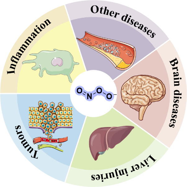
ONOO-,produced by the diffusion-controlled reaction of nitric oxide and superoxide radicals,is a strong oxidizing and nitrating agent that causes damage to DNA,proteins,and other biomolecules in cells. Since ONOO- is characterized by its short lifetime,high reactivity,and low concentration under physiological conditions,and the pathophysiological roles it plays in biological systems are not yet fully understood,it is of great significance to develop highly sensitive and selective detection technologies to achieve real-time dynamic monitoring of ONOO-. In this paper,we review the research progress of ONOO- fluorescent probes in disease-related processes in the recent 5 years,revealing the potential role of ONOO- in various diseases,such as inflammation,tumors,liver injury,and brain diseases. Finally,the bottlenecks in the development of ONOO- probes and future trends are discussed,which will promote the application of ONOO- probes in chemistry,biology,and pharmacology.
Contents
1 Introduction
2 Design strategies of ONOO- fluorescent probe
3 Detection and imaging of ONOO- by fluorescent probes in disease-related processes
3.1 Detection and imaging of ONOO- in inflammation
3.2 Detection and imaging of ONOO- in tumors
3.3 Detection and imaging of ONOO- in liver injuries
3.4 Detection and imaging of ONOO- in brain diseases
3.5 Detection and imaging of ONOO- in other disease models
4 Conclusion and outlook
Key words: fluorescence imaging; peroxynitrite; tumor; liver injury; neurodegenerative disease
Ting Ma , Chunyu Deng , Jie Li , Zhouyu Wang , Qian Zhou , Xiaoqi Yu . Construction and Application of ONOO- Small Molecule Fluorescent Probes in Pathophysiological Processes[J]. Progress in Chemistry, 2025 , 37(4) : 519 -535 . DOI: 10.7536/PC240815
表1 ONOO-荧光探针的相关参数Table 1 Related parameters of ONOO- fluorescent probes |
| Probe | Year | Test solution | λex/λem (nm) | Response time (s) | Detection range (μM) | LOD (nM) | Ref |
|---|---|---|---|---|---|---|---|
| 1 | 2016 | DMSO/PBS = 1:1 (v/v,pH = 7.4) | 670/742 | / | 0~40 | 400 | 33 |
| 2 | 2019 | DMSO/PBS = 1:19 (v/v,pH = 7.4) | 500/660 | / | 0~110 | 600 | 34 |
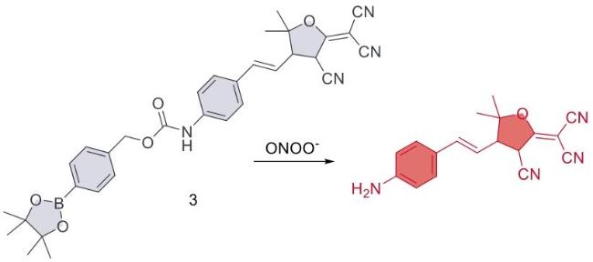
| 3 | 2020 | ACN/PBS = 3:20 (v/v,pH = 7.4) | 540/604 | 40 | 0~10 | 21 | 35 |
| 4 | 2021 | PBS (pH = 7.4,1% DMSO,3 mM CTAB) | 525/590 | 600 | 0~35 | 238 | 36 |
| 5 | 2022 | PBS (pH = 7.4,1% DMSO,3 mM CTAB) | 525/590 | < 300 | 0~30 | 220 | 37 |
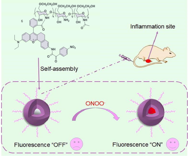
| 6 | 2023 | PBS (pH = 7.4) | 570/628 | 60 | 0~1 | 33 | 38 |
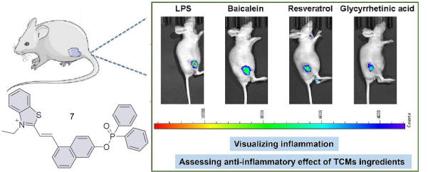
| 7 | 2024 | PBS (pH = 7.4,1% DMSO) | 381/518 | 120 | 0~5×10⁷ | 8.03 | 39 |
| 8 | 2021 | PBS (pH = 7.4,1% DMSO) | 548/662 | < 5 | 0~26 | 2.6×10⁴ | 43 |
| 9 | 2021 | PBS (pH = 7.4) | 530/545-750 | 5 | 0~10 | 7 | 44 |
| 10 | 2020 | PBS (pH = 7.4,10% DMSO) | 560/652 | < 180 | 0.5~20 | 72 | 45 |
| 11 | 2018 | PBS (pH = 7.4) | 675/712 | / | 50~100 | 53 | 46 |
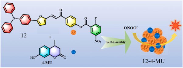
| 12 | 2023 | ACN/PBS = 4:6 (v/v,pH = 7.4) | 450/555 | / | 0~14,0~90 | 123,40 | 47 |
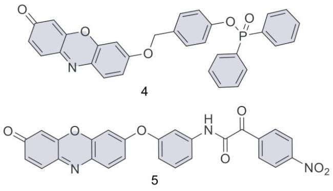
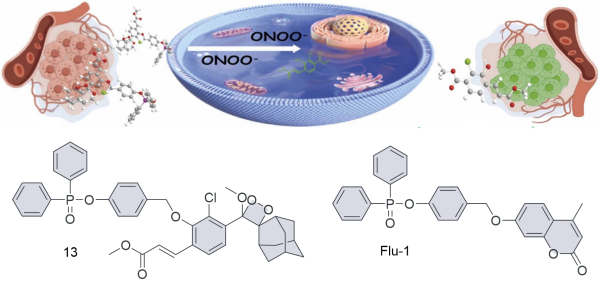
| 13 | 2024 | DMSO/PBS = 1:9 (v/v,pH = 7.4) | 400/550 | 600 | 0~2.5×10⁵ | 9.8,98 | 48 |
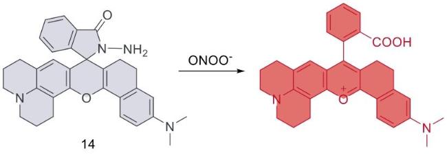
| 14 | 2021 | PBS (pH = 7.4) | 620/660 | 10 | 0.05~10 | 15 | 53 |
| 15 | 2016 | PBS (pH = 8.2) | 405/553 | / | 0~10 | 6.2 | 54 |
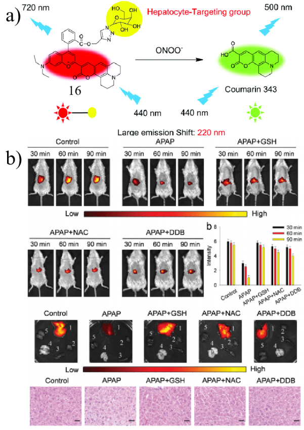
| 16 | 2019 | PBS (pH = 7.4) | 440/500,720 | / | 500~1.5×10⁴ | 1.7×10⁵ | 55 |
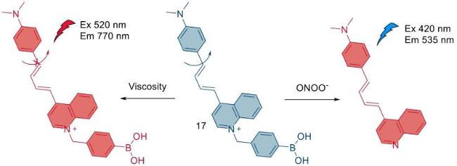
| 17 | 2020 | PBS (pH = 7.4,40% DMSO) | 420/635 | 300 | 0~1 | 1.69 | 56 |
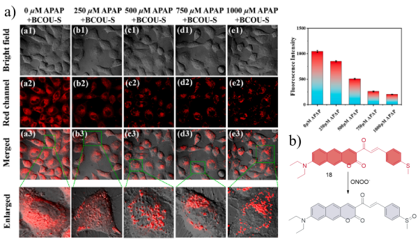
| 18 | 2024 | PBS (pH = 7.4,30% DMSO) | 518/655 | 2 | 0~28 | 27 | 57 |
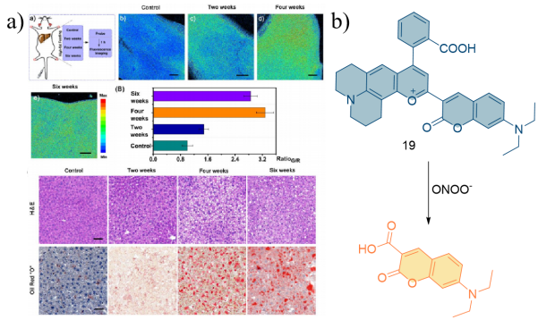
| 19 | 2020 | EtOH/PBS = 2:9 (v/v,pH = 7.4) | 405/469,703 | 25 | 0~7 | 4.1 | 58 |
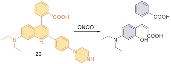
| 20 | 2023 | Purified water | 580/638 | 19 | / | 9.36 | 59 |
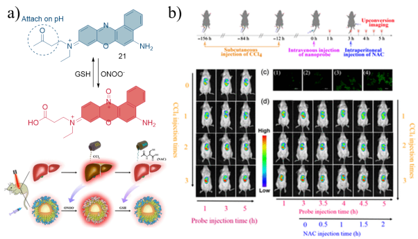
| 21 | 2022 | PBS (pH = 7.4,1% Ethanol) | 550/600 | < 10 | / | / | 60 |
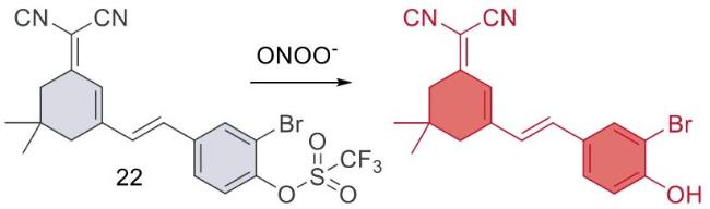
| 22 | 2024 | PBS (pH = 7.4,20% DMSO) | 500/655 | 600 | 20~40 | 65.8 | 61 |
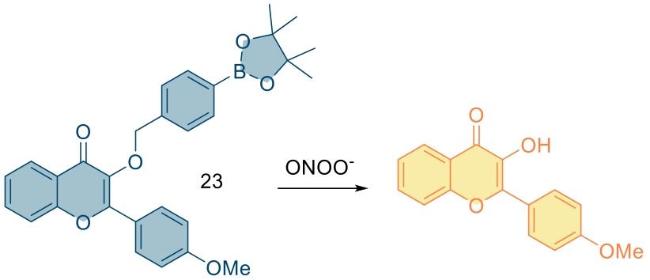
| 23 | 2018 | Tris-HCl (pH = 7.4) | 365/420,530 | / | 0~2 | 65.5 | 64 |
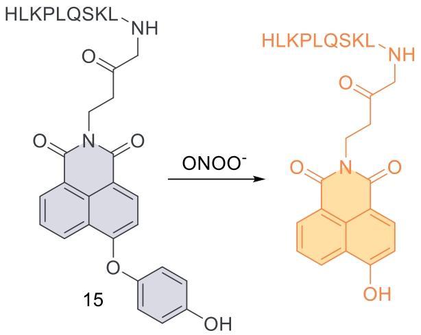
| 24 | 2024 | PBS (pH = 7.4,1% DMSO) | 474/544 | 3 | 0~38 | 11 | 65 |
| 25 | 2020 | PBS (pH = 7.4,0.4% Tween 80) | 511/670 | < 5 | 0~15 | 3.3 | 66 |
| 26 | 2021 | PBS (pH = 7.4,2% DMSO,0.4% Tween 80) | 450/540 | 25 | 0~5 | 8.3 | 67 |
| 27 | 2023 | PBS (pH = 7.4) | 350/490 | 60 | 0~3 | 9.89 | 68 |
| 28 | 2021 | PBS (pH = 7.4,30% DMSO) | 520/685 | 600 | 0~20 | 96 | 69 |
| 29 | 2023 | PBS (pH = 7.4,1% DMSO) | 425/548 | < 2 | 0~10 | 900 | 70 |
| 30 | 2021 | PBS (pH = 7.4,10% CH3CN) | 580/770 | < 120 | 0~10 | 330 | 71 |
| 31 | 2023 | PBS | 365/474,574 | 120 | 4~10 | 1.33 | 72 |
表2 ONOO-荧光探针的病理信息Table 2 Pathological information of ONOO- fluorescent probes |
| Probe | Cell imaging | Tissue imaging | Disease models | Disease | Ref |
|---|---|---|---|---|---|
| 1 | RAW 264.7 | / | Mice were intraperitoneally injected with LPS and PMA | Inflammation | 33 |
| 2 | MCF-7 | / | Mice were intraperitoneally injected with LPS and PMA | Inflammation | 34 |
| 3 | CHO-KE | / | Zebrafish larvae were immersed in LPS | Inflammation | 35 |
| 4 | HepG2,RAW 264.7 | / | LPS was injected subcutaneously into the left leg of nude mice | Inflammation | 36 |
| 5 | HepG2,RAW 264.7,HeLa | / | LPS was injected subcutaneously into the right leg of nude mice | Inflammation | 37 |
| 6 | RAW 264.7 | / | LPS was injected subcutaneously into the left leg of mice | Inflammation | 38 |
| 7 | LO2,MCF-7 | / | LPS was injected subcutaneously into the hind legs of nude mice | Inflammation | 39 |
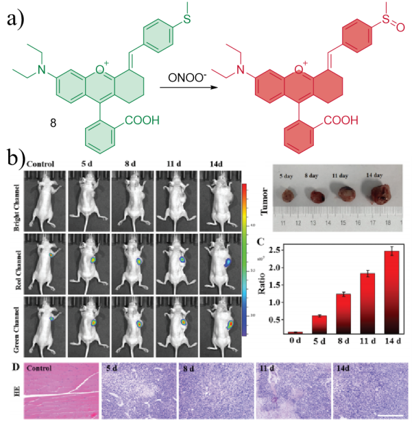
| 8 | RAW 264.7 | Tumor tissues | LPS induced acute peritonitis in mice Xenotransplantation of HepG2 tumor cells in nude mice | Tumor | 43 |
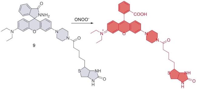
| 9 | Cal-27,HSC-2,MC3 | / | Xenotransplantation of HSC-2 tumor cells in nude mice | Tumor | 44 |
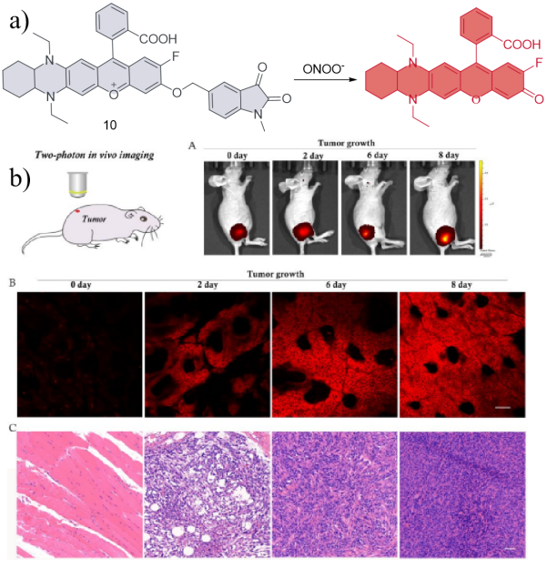
| 10 | HeLa,RAW 264.7 | Zebrafish pancreatic tissue | Subcutaneous transplantation of 4T1 tumor cells in nude mice | Tumor | 45 |
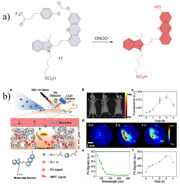
| 11 | RAW 264.7 | / | Subcutaneous transplantation of 4T1 tumor cells in nude mice | Tumor | 46 |
| 12 | RAW 264.7 | / | / | Tumor | 47 |
| 13 | HepG2,A549, SH-SY5Y,LO2 | Human liver cancer tissue | Xenotransplantation of HepG2 tumor cells into nude mice | Tumor | 48 |
| 14 | HeLa,RAW 264.7 | Mice liver tissue | APAP induced hepatotoxicity in mice | Liver injury | 53 |
| 15 | SMMC-7721,HL-7702 | Mice liver tissue | CCl4 induced acute liver injury in mice | Liver injury | 54 |
| 16 | HepG2,HCT116,HeLa ,MCF-7 | Mice liver tissue | APAP induced hepatotoxicity in mice | Liver injury | 55 |
| 17 | HeLa | Mice liver tissue | APAP induced liver injury in mice LPS and IFN-γ stimulated zebrafish Subcutaneous injection of exogenous ONOO- mice | Liver injury | 56 |
| 18 | HepG2 | / | / | Liver injury | 57 |
| 19 | HepG2,LO2 | / | HFD induced nonalcoholic fatty liver mice | Liver injury | 58 |
| 20 | HepG2 | Mice liver tissue | APAP induced liver injury in mice | Liver injury | 59 |
| 21 | HepG2,RAW 264.7 | Mice liver tissue | CCl4 induced acute liver injury in mice | Liver injury | 60 |
| 22 | LO2 | Mice liver tissue | APAP and INH induced ferroptosis-mediated drug induced liver injury in zebrafish and mice | Liver injury | 61 |
| 23 | / | Brain tissue of AD | AD transgenic mice | Neurodegenerative diseases | 64 |
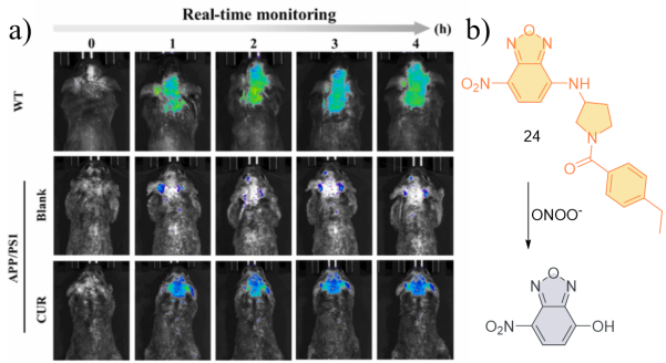
| 24 | SH-SY5Y | Murine brain | APP/PS1 transgenic mice | Neurodegenerative diseases | 65 |
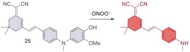
| 25 | PC12 | Drosophila brain tissues | MPTP induced Parkinson 's disease in mice LRRK2 overexpression constructed WLZ3 C.elegans Parkinson's disease | Neurodegenerative diseases | 66 |
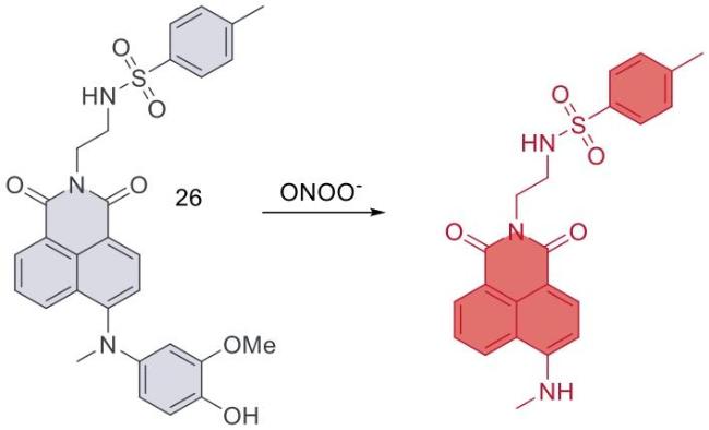
| 26 | PC12 | / | LRRK2 overexpression constructed WLZ3 C.elegans Parkinson's disease | Neurodegenerative diseases | 67 |
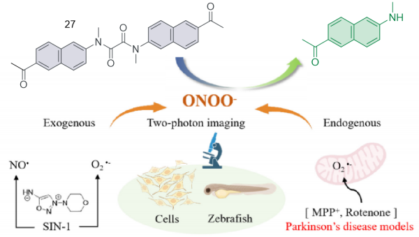
| 27 | SH-SY5Y | / | SIN-1 and MPP+ stimulated zebrafish | Neurodegenerative diseases | 68 |
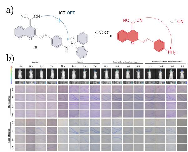
| 28 | RAW 264.7,HT22 | Brain sections of epileptic rats | KA induced epilepsy in rats | Neurodegenerative diseases | 69 |
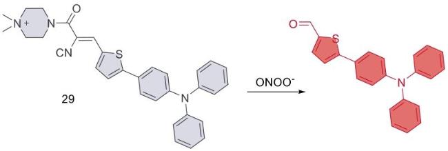
| 29 | U87MG | Glioblastoma tissue | Xenotransplantation of U87MG cells into the brain of nude mice | Neurodegenerative diseases | 70 |
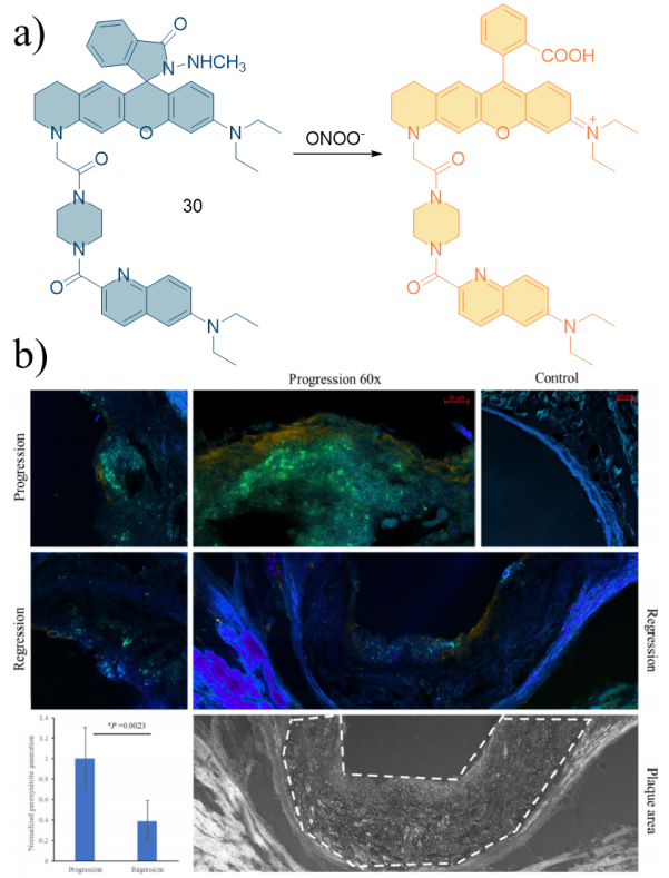
| 30 | RAW 264.7 | Mouse aortic tissue | High fat fed low-density lipoprotein receptor knockout (Ldlr-/-) mice | Cardiovascular | 71 |
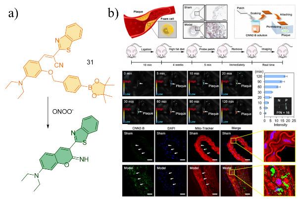
| 31 | A549,RAW 264.7 | / | Carotid artery ligation combined with high-fat diet feeding ApoE gene-deficient mice | Cardiovascular | 72 |
| [1] |
|
| [2] |
|
| [3] |
|
| [4] |
|
| [5] |
|
| [6] |
|
| [7] |
|
| [8] |
|
| [9] |
|
| [10] |
|
| [11] |
|
| [12] |
|
| [13] |
|
| [14] |
|
| [15] |
|
| [16] |
|
| [17] |
|
| [18] |
|
| [19] |
|
| [20] |
|
| [21] |
|
| [22] |
|
| [23] |
|
| [24] |
|
| [25] |
|
| [26] |
(景晓彤, 于法标, 陈令新. 化学进展., 2014, 26(5): 866.).
|
| [27] |
|
| [28] |
|
| [29] |
|
| [30] |
(王振, 李曦, 栗园园, 王其, 卢晓梅, 范曲立. 化学进展., 2022, 34(1): 198.).
|
| [31] |
|
| [32] |
|
| [33] |
|
| [34] |
|
| [35] |
|
| [36] |
|
| [37] |
|
| [38] |
|
| [39] |
|
| [40] |
|
| [41] |
|
| [42] |
|
| [43] |
|
| [44] |
|
| [45] |
|
| [46] |
|
| [47] |
|
| [48] |
|
| [49] |
|
| [50] |
|
| [51] |
|
| [52] |
|
| [53] |
|
| [54] |
|
| [55] |
|
| [56] |
|
| [57] |
|
| [58] |
|
| [59] |
|
| [60] |
|
| [61] |
|
| [62] |
|
| [63] |
|
| [64] |
|
| [65] |
|
| [66] |
|
| [67] |
|
| [68] |
|
| [69] |
|
| [70] |
|
| [71] |
|
| [72] |
|
/
| 〈 |
|
〉 |
