Home Journals Progress in Chemistry
Progress in Chemistry

Abbreviation (ISO4): Prog Chem      Editor in chief: Jincai ZHAO

About  /  Aim & scope  /  Editorial board  /  Indexed  /  Contact  / 
Review

Clinical Application Value of Exosomes and Research Progress on Exosome Detection based on Surface-Enhanced Raman Spectroscopy

  • Xinyu Liu ,
  • Xinyue Gu ,
  • Xiaoyuhao Jin ,
  • Jingjing Zhang ,
  • Lianhui Wang , * ,
  • Chunyuan Song , *
Expand
  • Institute of Advanced Materials, Nanjing University of Posts and Telecommunications, Nanjing 210023, China
* (Chunyuan Song);
(Lianhui Wang)

Received date: 2024-08-19

  Revised date: 2024-11-09

  Online published: 2025-06-15

Supported by

National Natural Science Foundation of China(62235008)

Abstract

Since exosomes were discovered in sheep reticulocytes, more and more studies have shown that the function and characteristics of exosomes are closely related to the occurrence and development of diseases. The analysis and detection of exosomes have clinical significance for the diagnosis, treatment and prognosis of diseases. In recent years, researchers have taken advantage of surface-enhanced Raman spectroscopy (SERS) technology and developed a variety of strategies for high-sensitive, specific and multivariate detection of various biological information of exosomes. The SERS-based exosome detection technology shows a good application prospect in clinical medical diagnosis and treatment. This review summarizes the basic characteristics and main physiological mechanisms of exosomes, and discusses their clinical significance, correlation with diseases, related indicators for characterizing and difficulties in detection, and then focuses on the research progress of SERS detections of exosomes in the aspects of concentration, phenotype, content analysis, etc., as well as the summary and prospect at the end.

Contents

1 Introduction

2 Exosome

2.1 Clinical significance

2.2 Correlation with disease

2.3 Clinical diagnostic significance and difficulties of concentration analysis, surface phenotype and contents detection

3 SERS detection for exosomes

3.1 Overview of SERS

3.2 Concentration analysis

3.3 Phenotype analysis

3.4 SERS combined with other analytical techniques

4 Conclusion and outlook

Cite this article

Xinyu Liu , Xinyue Gu , Xiaoyuhao Jin , Jingjing Zhang , Lianhui Wang , Chunyuan Song . Clinical Application Value of Exosomes and Research Progress on Exosome Detection based on Surface-Enhanced Raman Spectroscopy[J]. Progress in Chemistry, 2025 , 37(6) : 812 -826 . DOI: 10.7536/PC240804

1 Introduction

In 1983, Pan et al.[1] first discovered the presence of membranous small vesicles in sheep reticulocytes and observed the fusion of large multivesicular bodies with the plasma membrane and the release of their contents during the maturation of transferrin receptors on the surface of sheep reticulocytes[2]. In 1987, the concept of exosomes was formally proposed, and Johnstone[3] named them "Exosome." Since then, pioneers have continuously advanced exosome research, and to this day, human exploration of exosomes has never ceased. Studies have revealed that exosomes serve as an excellent medium for material delivery within the body, opening the door to a new world of biology, and analyzing exosomes provides new directions for biomedical research. Surface-enhanced Raman scattering (SERS) sensing technology is an ultrasensitive method for substance detection. In recent years, researchers have combined SERS technology with exosome analysis, offering high sensitivity, high specificity, and multiplex analytical techniques for exosome research[4]. Exosome analysis methods based on SERS technology have demonstrated promising applications in clinical medicine diagnosis and treatment[5-8]. In recent years, researchers have conducted extensive studies on exosome detection using SERS technology. Current reviews have classified and elaborated on SERS-based exosome analysis from various aspects, including the microstructure and functional modification of SERS-active metal materials, labeled and label-free detection methods, and analytical strategies for different exosome biomarkers[9-13]. As of October 2024, over the past decade, research interest in SERS-based exosome detection technology has significantly increased, with a total of 213 research papers published and 4051 citations accumulated, including as many as 1794 citations in 2023 alone. As shown in Figure 1, this trend clearly illustrates the rapid development and immense potential of this technology. This article will summarize the basic characteristics and main physiological mechanisms of exosomes, their association with diseases, clinical significance, and detection challenges, and will focus on recent advances in SERS technology for exosome concentration analysis, vesicle surface phenotyping, quantitative detection of intramembrane substances, and multiplex detection and integration with multiple techniques. Finally, the article will provide a summary and outlook on the research and clinical applications of SERS-based exosome analysis technologies.
图1 2015-2024年逐年发表SERS外泌体检测文章及年引用情况

Fig.1 The published articles and total citations of SERS-based exosomes detections

2 Exosomes

Exosomes are extracellular membranous vesicles with a lipid bilayer, approximately 30-100 nm in diameter[14], containing specific proteins, mRNA, miRNA, active lipids, and other biomolecules (i.e., exosomal cargo). They can mediate intercellular substance transfer, information exchange, cell proliferation and differentiation through signal-mediated mechanisms[15]. Exosomes are also involved in various pathological processes such as immune regulation, antigen presentation, endothelial cell migration and inflammation, tumor invasion, and metastasis[16-17]. Exosomes can be released by various cell types and stably exist in body fluids. The specific proteins, nucleic acids, and lipid components they carry reflect the cellular origin of the exosomes[18-23]. There are two mechanisms through which exosomes exert biological effects[24]: the first involves direct fusion or endocytosis to deliver their cargo into target cells, thereby regulating cellular biological behaviors; the second mechanism involves activation of signaling pathways through specific binding of surface ligands to receptors, as shown in Figure 2.
图2 外泌体的发生、分泌机制以及受体细胞的摄取过程[27]

Fig.2 Exosomes biogenesis and release mechanism, as well as the uptake of recipient cell[27]. Copyright 2021, Multidisciplinary Digital Publishing Institute

2.1 Clinical Significance of Exosomes

The clinical applications of exosomes are primarily divided into two aspects: disease diagnosis and treatment. Because exosomes released by malignant tumor cells contain specific substances that are crucial for communication between cancer cells and their environment, exosomes can be used to monitor patients' conditions in real time. Furthermore, they can indirectly inhibit the deterioration of diseased areas, thereby effectively controlling the disease and improving cure rates[28].
Exosomes are widely distributed in the body and carry disease biomarkers that reflect physiological abnormalities; analyzing these biomarkers can enhance diagnostic accuracy and sensitivity while improving detection efficiency[29]. Many cancers are typically diagnosed only at advanced stages[30], but studies have found that early diagnosis of gastric cancer can be achieved by detecting exosomal circRNAs in body fluids with high specificity and sensitivity (Figure 3a)[31]. Compared with conventional histopathological examinations, exosomes are easier to obtain, allowing for repeated sampling and reducing the risks associated with invasive procedures. Compared with traditional serum markers, the vesicular membrane structure of exosomes enables their contents to remain more concentrated and stable in complex body fluids without being easily damaged by external factors[32-33], so their quantitative and qualitative detection results can provide more accurate diagnostic information. Existing studies have shown that the long non-coding RNA lncUEGC1 in exosomes from early-stage gastric cancer patients is significantly upregulated and remains stable and resistant to degradation[34], and its diagnostic accuracy in distinguishing early gastric cancer patients from healthy individuals and patients with precancerous chronic atrophic gastritis is significantly higher than that of the serological markers CEA, CA72-4, and CA19-9.
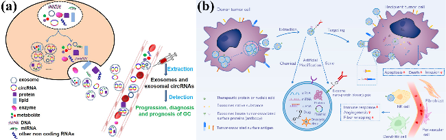
图3 外泌体用于疾病的诊断与治疗。(a) 检测circRNA和外泌体circRNA用于胃癌的诊断、进展和预后分析[31];(b)肿瘤细胞来源外泌体的工程化策略以增强靶向性[35]

Fig.3 Exosomes for the diagnosis and treatment of diseases. (a) CircRNAs and exosomal circRNAs play a very important role in the diagnosis, progression and prognosis of GC[31]. Copyright 2021, Dove Medical Press Ltd. (b) Schematic illustration of the tumor-derived exosomes with modification strategies for enhancing targeting[35]. Copyright 2022, Multidisciplinary Digital Publishing Institute

Exosomes possess excellent biocompatibility and can serve as drug delivery carriers in targeted therapy, effectively increasing the drug dose reaching target cancer cells while reducing damage to tissues and organs[36-37]. Currently, researchers have directly encapsulated drugs within tumor-derived exosomes, generating engineered exosomes carrying tumor-associated antigens (Fig. 3b), which facilitate drug uptake by target cells and offer potential solutions to the problem of tumor drug resistance[35]. Additionally, Liang et al.[38] modified exosomes with specific ligands through bioengineering techniques, enabling them to act as stimulus-responsive factors and thereby providing an innovative approach for tumor treatment.

2.2 Exosomes and Their Relevance to Diseases

Exosomes act as messengers between cells, and when abnormalities occur in the human body, their content or composition changes accordingly. Compared with healthy individuals, the characteristics of exosomes in patients become more pronounced, providing feature information for the occurrence and progression of diseases[24]. For example, in kidney disease research[39], researchers found that increased expression levels of urinary exosomal hsa_circ_000892 are closely related to renal fibrosis. In cardiovascular disease studies, Zhu et al.[40] utilized regression analysis and found a linear correlation between the expression of platelet endothelial cell adhesion molecule-1 protein in exosomes and systolic blood pressure in patients with hypertension. In addition, numerous studies in the medical field have shown that exosomes are highly associated with obstetric[41], neurological[42], and digestive system-related diseases[43].

2.3 Clinical Diagnostic Significance and Challenges of Detecting Exosome Concentration, Surface Phenotype, and Cargo Content

Concentration, surface phenotype, content and its composition are important parameters of exosomes, which have disease-specific indications in clinical diagnosis and can help accurately identify the causes. The concentration of exosomes in cancer patients differs significantly from that in healthy controls, and it also changes accordingly at different stages of the disease. Researchers have found a linear negative correlation between exosome concentration and the lymph node status of lung cancer patients, with the concentration decreasing as the disease progresses[44].
The surface phenotypes of exosomes, referring to their external molecular and functional characteristics, can also serve as indicators for monitoring physiological and pathological processes in the body[45]. Bonhoure et al.[46] utilized specific antibodies to identify melanotransferrin overexpressed on the exosomal membrane, thereby enabling the monitoring of disease onset and progression. Additionally, researchers assessed various surface markers of serum exosomes using flow cytometry[47], finding that the fluorescence intensities of CD13, CD34, and HLA-DR were higher in patients with myelodysplastic syndrome compared to healthy individuals. Moreover, the fluorescence intensities of CD13, CD34, HLA-DR, CD33, and CD117 were significantly higher in patients with acute myeloid leukemia than in those with chronic myeloid leukemia. These findings indicate that differences in the types and levels of membrane proteins can not only aid in disease diagnosis but also help distinguish between different diseases.
Exosomes carry specific bioactive components, mainly including proteins and RNAs (miRNAs, lncRNAs, etc.), which play important roles in regulating gene expression of diseased cells and mediating cell signaling pathways in various biological processes[42,48-49], and are closely associated with disease development[50]. Researchers have found that miR-155 levels are extremely high in plasma exosomal miRNAs, whereas miR-21 and miR-122 are expressed at low levels, making them important indicators for diagnosing systemic inflammation (Figure 4a); the expression levels of exosomal miRNAs in patients with chronic pancreatitis (CP) are significantly different from those in patients with pancreatic ductal adenocarcinoma (PDAC), making them useful as criteria for diagnosing and differentiating between these two diseases (Figure 4b)[51].
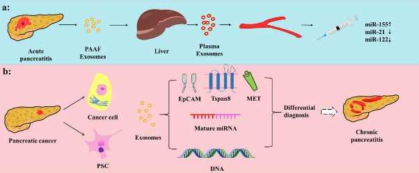
图4 基于外泌体分析的胰腺炎检测。(a)血浆外泌体高表达miRNA用于诊断急性胰腺炎症;(b)外泌体miRNA诊断与区分胰导管腺癌和慢性胰腺炎疾病时体现高特异性[51]

Fig.4 Application of exosomes in the diagnosis of pancreatic diseases.(a)miRNAs expressed in plasma-derived exosomes with greater diagnostic value in acute pancreatitis.(b)Exosomes for identification of PDAC and CP[51]. Copyright 2022, BioMed Central

In summary, the potential of exosomal characteristic indicators in clinical medical diagnosis has become increasingly prominent, but exosome detection still faces many challenges[52-53]. Currently, traditional exosome detection methods, including transmission electron microscopy (TEM), nanoparticle tracking analysis (NTA)[54], Western blot[55], and mass spectrometry (MS)[56-59], primarily measure exosome particle size and protein expression indicators, but have limitations in sensitivity, specificity, or throughput, making it difficult to comprehensively cover multidimensional exosome information. In recent years, technological innovations have ushered in a new era of highly sensitive exosome concentration and molecular information analysis techniques, such as surface-enhanced Raman spectroscopy (SERS)[60-61], electrochemistry[62], fluorescence[63], and colorimetric[64] detection methods, which have rapidly developed, continuously optimizing exosome detection efficiency and accuracy. Among them, SERS analysis technology has attracted significant attention due to its unique advantages, such as ultra-high sensitivity at the single-molecule level and high spectral resolution for multiplexed detection, and is expected to become a powerful tool for highly sensitive and multi-index detection. Table 1 lists the advantages and disadvantages of traditional exosome detection methods and SERS-based exosome detection methods.
表1 外泌体传统检测方法与SERS检测方法的优缺点

Table 1 Advantages and disadvantages of traditional and SERS-based exosome analysis techniques

Detecting techniques Advantages Disadvantages
TEM Intuitive presentation of exosome morphological and structural details of exosomes. Complex sample preparation, destructive to the sample, and time-consuming.
NTA Direct measurement of exosome size and concentration. Inability to provide molecular composition information, and easily affected by environmental factors.
Western blot High specificity, suitable for identification of specific proteins. Analyze one or a few proteins in a single blot per assay, and time-consuming.
MS High sensitivity and specificity, suitable for protein identification. Complex sample preparation; Inability to provide real-time monitoring.
FC Rapid analysis of cell surface markers, suitable for large-scale screening and point-of-care detection. Inability to provide molecular composition information.
SERS Rapid, high specificity and sensitivity, and non-destructive to the sample; Provide molecular composition information; Suitable for multiple exosome detection; Suitable for large-scale screening and point-of-care detection. Inability to directly provide the morphological and structural details of exosomes.

3 SERS-Based Exosome Detection

Surface-enhanced Raman scattering technology utilizes the electromagnetic field enhancement effect of noble metal (gold or silver) nanostructures[65] to amplify the Raman signals of target molecules adsorbed on the SERS substrate by more than a million times, enabling highly sensitive trace detection with only a small sample amount. Its detection concentration range is typically at the ppb to ppm level[66]. Additionally, due to its rapid, sensitive, and non-destructive characteristics, it has the potential to address challenges in exosome analysis such as long processing time, low abundance, and low accuracy, making exosomes promising for clinical detection applications. In the following sections, this paper will focus on recent research and application advancements in SERS biosensors for exosome detection.

3.1 Overview of SERS

In 1974, Fleischmann et al.[67] observed a significantly enhanced Raman spectrum of a monolayer of pyridine adsorbed on a rough silver electrode under light irradiation. Van Duyne et al.[68] and Albrecht et al.[69] both confirmed Fleischmann's findings, suggesting that this was a Raman enhancement effect related to the rough metal surface, later named the surface-enhanced Raman scattering (SERS) effect. Studies have shown that SERS enhancement arises from two mechanisms: physical enhancement and chemical enhancement. Physical enhancement, also known as the electromagnetic enhancement mechanism (EM), refers to the excitation of localized surface plasmon resonance (Localized-surface plasma resonance, LSPR) when incident light irradiates the surface of a nanostructured metal, forming a significantly enhanced electromagnetic field that increases the Raman scattering intensity of Raman molecules adsorbed on the metal surface.[70] The enhancement factor can reach up to 1011.[71] EM exhibits a long-range effect, meaning that the enhanced electromagnetic field still exists away from the metal surface, influencing regions up to hundreds of angstroms (Å) from the surface, making this effect the dominant contributor to SERS signal enhancement. At nanoscale gaps between metal nanoparticles in nanoparticle assemblies, the coupling of LSPR can generate a significantly enhanced electromagnetic field, known as SERS hotspots. The chemical mechanism has a more complex effect on the Raman signal and often coexists with the physical mechanism, further enhancing the signal. The chemical enhancement mechanism (CM) involves charge transfer between Raman molecules adsorbed on the rough metal surface and the metal surface itself, increasing the effective polarizability and significantly enhancing the Raman signal when the excitation wavelength resonates with the metal's electronic states, with an enhancement factor of 102 to 103.[72] CM exhibits a short-range effect; once the molecules move away from the metal surface, the charge transfer process is limited, thereby restricting the enhancement effect. Therefore, this mechanism contributes relatively less to the overall enhancement effect. It is generally believed that the SERS effect results from the combined action of both physical and chemical enhancement mechanisms.[73]
SERS-based biosensing detection methods are divided into direct and indirect detection. Label-free SERS direct detection achieves analysis of the analyte by directly obtaining its SERS signal. The SERS signal of the analyte is susceptible to interference from surrounding impurities, thus requiring measures to avoid the impact of other interfering substances in the analytical environment on the detection specificity. In label-free SERS detection, the analyte is directly dropped onto a SERS-active substrate for SERS signal measurement (Fig. 5a)[74], offering convenient operation, but challenges remain in improving sensitivity and reproducibility, as well as efficiently extracting useful information[75]. Indirect detection based on SERS tags does not directly obtain the Raman information of the analyte itself; instead, it utilizes the Raman signal characteristics of SERS tags to specifically sense the analyte, thereby improving detection sensitivity and specificity by measuring the sensing signal of the SERS tags rather than that of the analyte itself. SERS tagging techniques have been widely applied in the detection of biomarkers such as nucleic acids, proteins, cells, and exosomes[76], with typical detection methods including sandwich assays and target-induced SERS tag aggregation (Fig. 5b, c)[77-78].
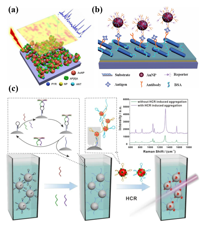
图5 典型SERS检测方法示意图:(a) 无标签SERS检测[74];(b) 三明治结构SERS免疫分析法[77];(c) 多级核酸扩增诱导形成三维SERS热点聚集体用于循环miRNA的检测[78]

Fig.5 Typical SERS detection methods. (a) Label-free SERS detection[74]. Copyright 2017, American Chemical Society. (b) Schematic of sandwich structure SERS immunoassay[77]. Copyright 2022, Elsevier. (c) Target-induced nano-aggregation for 3D hotspots-improved SERS detection of circulating miRNAs[78]. Copyright 2022, BioMed Central

SERS, as a non-destructive analytical technique, requires very low concentrations/volumes of analytes due to its potential for surface-enhanced signal generation[79], which can effectively address the current issues of low sensitivity in exosome detection and the requirement for large sample volumes. The following section will focus on the research progress of SERS technology in the analysis of various characteristic information of exosomes.

3.2 SERS Exosome Concentration Analysis

Accurately and sensitively analyzing changes in the concentration of pathogenic exosomes can help understand the occurrence and development of diseases. Therefore, in recent years, researchers have continuously explored SERS-based methods for exosome concentration analysis. Through the construction of SERS substrates and probes, as well as innovative design of sensing strategies, various SERS detection methods have been developed for exosome concentration analysis.
The preparation of SERS substrates, which involves the formation of microstructures of substrate materials and functionalization modification of sensing interfaces, is crucial for achieving rapid, sensitive, and specific detection of exosomes through liquid biopsy. Common substrate materials include noble metals such as gold, silver, and copper; in recent years, other metals such as iron, cobalt, and nickel, as well as their oxides, have also been explored. Low-dimensional nanostructures, such as nanoparticles, nanoparticle arrays, and nanohole arrays, have been widely applied in the fabrication of SERS substrates. To increase the surface area of metals and thereby generate more hotspots, substrates with special three-dimensional nanostructures have been developed, including arrays of metal nanorods and micro-pyramids. Wang et al.[80-81] fabricated vertically aligned hexagonally close-packed gold nanorod arrays on an ITO substrate. Compared with horizontally or randomly arranged gold nanorod arrays, the vertically aligned arrays can generate a stronger electromagnetic field enhancement effect, as shown in Figure 6a. The researchers found that the SERS signal exhibited a good linear relationship with exosome concentration in the range of 1.0×10⁴ to 5.0×10⁶ particles/mL, with a detection limit as low as 5.3×10³ particles/mL, enabling highly sensitive detection of exosomes.
图6 外泌体SERS定量检测。(a) ITO基底上六方紧密堆积的金纳米棒竖直阵列SERS基底[80];(b) 双探针的SERS乳腺癌外泌体检测策略[84];(c) 基于三种纳米探针的SERS外泌体传感器[85];(d) 基于酶促沉积银结构和杂交链式反应SERS信号放大策略[93]

Fig.6 Quantitative SERS detection for exosome. (a) Vertical array of hexagonal closely packed gold nanorods on ITO substrate[80]. Copyright 2021, American Chemical Society. (b) Dual-probe assisted detection strategy for SERS breast cancer exosomes[84]. Copyright 2023, American Chemical Society. (c) SERS sensor for detecting exosomes based on three types of nanotags[85]. Copyright 2020, American Chemical Society. (d) Alkaline phosphatase and HCR -induced Ag-shell nanostructure for SERS detection for exosomes[93]. Copyright 2023, American Chemical Society

Since metal substrates lack bioaffinity, functionalized interfaces are typically required to achieve specific capture of exosomes. Antigen-antibody recognition based on immunoaffinity principles is a common method for exosome separation and enrichment. By modifying antibodies on the substrate to specifically recognize transport proteins or fusion proteins on the exosome surface, target exosomes can be captured for quantitative detection. For example, Zhang et al.[82] functionalized the interface of a 3D nano-herringbone structured chip through antibody conjugation using (3-mercaptopropyl)trimethoxysilane, enabling highly sensitive detection of ovarian cancer exosomes with a detection limit as low as 10 EVs/μL, and allowing for quantitative detection of low-level exosome subpopulations in plasma. However, in recent years, aptamers have gradually become a research hotspot due to their high specificity, low immunogenicity, high affinity, and excellent chemical stability, making them promising alternatives to traditional antibodies, with CD63 aptamers being commonly used[83].
SERS probes are typically composed of metal nanoparticles, signal reporter molecules, and nucleic acids or proteins. In SERS labeling detection, the probes can measure specific Raman signals, thereby indirectly reflecting information about exosomes. Tian et al.[81] prepared a SERS nanosubstrate (gold nanostars@4-mercaptobenzoic acid@gold shell) that inserts into the exosomal phospholipid bilayer via cholesterol and self-assembles onto exosomes captured by magnetic microspheres, forming a "sandwich" structure that generates a SERS signal positively correlated with exosome concentration. As shown in Figure 6b, Zhang et al.[84] synthesized a calibration probe, 4-nitrobenzenethiol, immobilized on a substrate to maintain a constant output signal, and another dual-aptamer V-shaped probe modified with rhodamine at its terminus for exosome detection from breast cancer cells. Due to the stronger affinity of the V-shaped probe toward exosomes, it detaches from the SERS-active substrate, causing the rhodamine signal to decrease with increasing exosome concentration, enabling ultrasensitive detection with a detection limit as low as 1.5×10² particles/mL. As shown in Figure 6c, Wang et al.[85] selected three proteins—Glypican-1, EpCAM, and CD44V6—present on the surface of pancreatic cancer Panc-1 cell exosomes as biomarkers and used different specific antibodies and Raman signal molecules to prepare three SERS probes—MIL38-DTNB, EpCAM-MBA, and CD44V6-TFMBA—for multiplex labeling detection of exosomes with high specificity and accuracy.
In addition to the signal amplification effect of metallic nanostructures, signal amplification strategies in sensing detection also play a crucial role in achieving highly sensitive SERS detection of exosomes at low concentrations. These strategies typically include enzyme-mediated isothermal amplification reactions and enzyme-free chain amplification reactions. Well-established enzyme-catalyzed isothermal amplification techniques include loop-mediated isothermal amplification (LAMP)[86], strand displacement amplification (SDA)[87], rolling circle amplification (RCA)[88], and tyramide signal amplification (TSA)[89]. These methods eliminate the need for thermal cycling in traditional PCR techniques, significantly reducing both time and cost[90]. However, due to the stringent transportation and storage requirements of biological enzymes, enzyme-free isothermal chain amplification methods such as catalyzed hairpin assembly (CHA)[91] and hybridization chain reaction (HCR)[92] have emerged as innovative alternatives to enzyme-mediated approaches. As shown in Figure 6d, Cun et al.[93] combined an alkaline phosphatase-induced silver-coated nanostructure amplification strategy with a hybridization chain reaction to enhance the sensitivity of quantitative exosome detection, achieving a detection limit of 19 particles/μL. To further optimize signal amplification strategies, researchers often employ cascade amplification approaches that combine multiple amplification methods to overcome the limitations of single-stage signal amplification.
Based on the construction of the aforementioned sensing platform and the selection of detection strategies, direct capture of exosome particles can be achieved and converted into a quantifiable signal output. This approach can effectively replace traditional indirect quantitative analysis methods that target specific exosome surface proteins, avoiding inaccuracies and low sensitivity caused by the co-expression of exosomal proteins and subtle expression differences among different cancer subtypes, thus holding great potential for clinical disease monitoring of exosomes.

3.3 SERS Exosome Phenotyping

Exosomes, as biomacromolecules with specific functionalities, possess distinct genotypes and biological phenotypes. Exosome phenotyping is a technology that combines optical techniques with immunology, enabling a relatively comprehensive quantitative analysis of the characteristics of target exosomes from multiple perspectives, including size, distribution, surface markers, and carried contents, after being immunologically identified and specifically captured[94]. NTA is commonly used to determine the size distribution and absolute quantity of circulating exosomes, providing quantitative information on the physical features of exosomes. However, its limitation lies in the inability to provide details regarding specific exosome components, requiring combination with other techniques for compositional analysis. SERS, as a highly sensitive and selective spectroscopic technique, can detect the Raman scattering signals of nanoscale molecules, and its high resolution enables accurate identification of the complex biological components of vesicles. Currently, it can serve as an ideal technical approach for phenotypic analysis of substances on and within exosome membranes[95].

3.3.1 SERS Phenotyping of Exosome Surfaces

Exosomes are composed of lipids, proteins, nucleic acids, and carbohydrates on their surface, among which membrane proteins carry critical information and are often used as the primary targets for SERS-based exosome surface phenotyping. These include common proteins that distinguish exosomes from other extracellular vesicles (e.g., CD9, CD63, CD81), as well as characteristic proteins that identify exosomes from different cellular origins (e.g., BSG, PSMA, A33). Membrane proteins serve as liquid biopsy biomarkers for exosome detection, carrying protein components originating from their parent cells and tissues. The protein composition of exosomes correlates with tumor molecular subtypes and reflects the characteristics of tumor cells. Detecting the expression of specific proteins on exosomes can enable precise classification and differential diagnosis of tumors across different molecular subtypes.[96-97]
Common SERS-based exosome phenotypic analysis mainly relies on labeled and label-free approaches. SERS nanotags can connect to abundant modification sites on the exosome surface through biomolecular recognition, thereby concentrating numerous reporter molecules within the enhanced electromagnetic field region, generating amplified characteristic SERS signals. Quantitative relationships can be established based on SERS signals from functionalized nanoparticles at different concentrations, enabling sensitive and specific identification and quantitative phenotypic analysis of exosomes. For example, Muhammad et al.[98] prepared two SERS probes for capturing circulating exosomes and forming a "sandwich" structure to quantitatively detect the PD-L1 protein in exosomes, achieving a detection limit of 4.31 ag/mL. Su et al.[99] developed a paper-based SERS-vertical flow biosensor (iREX), as shown in Figure 7a, which utilizes three different aptamer-modified substrates for multiplexed quantitative analysis of exosomal proteins MUC1, HER2, and CEA from patient clinical serum samples. Distinct expression profiles of these three exosomal proteins were obtained, enabling molecular-specific and highly accurate subtyping and personalized diagnosis of breast cancer.
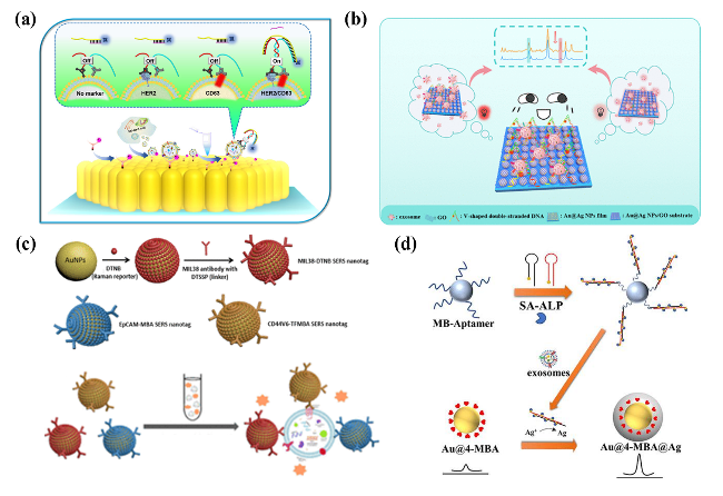
图7 SERS外泌体表型分析策略:(a) 乳腺癌细胞亚型外泌体蛋白MUC1、HER2和CEA的多重定量分析示意图[99];(b) 基于Au@Ag纳米粒子的SERS外泌体无标记表型分析示意图[100],(c)一个用于鉴定不同来源外泌体的无标记SERS平台[101]

Fig.7 SERS exosomal phenotype analysis strategy. (a) Breast cancer subtypes in cells secrete body protein (MUC1, HER2 and CEA) multiple quantitative analysis diagram[99]. Copyright 2023, American Chemical Society. (b) Marker-free phenotype analysis of SERS exosomes based on Au@Ag nanoparticles[100]. Copyright 2019, American Chemical Society. (c) A label-free SERS platform for identification of exosomes from different sources[101]. Copyright 2019, American Chemical Society

Compared with other methods, label-free detection can be widely applied to various types of samples without the need for designing specific markers for targets, thereby avoiding the complicated operations and time inefficiency associated with labeling. Additionally, it enables direct acquisition of richer and more accurate exosome phenotypic information, preventing deactivation caused by markers and potential damage to samples due to labeling processes, making it a promising detection strategy. As shown in Figure 7b, Fraire et al.[100] proposed a novel strategy involving in situ generation of Au@Ag nanoparticles on the surface of exosomes. Using this strategy, molecular information from the exosomal surface can be directly obtained, enabling simple and high-throughput exosome phenotypic analysis without relying on specific antibodies or labels. Yan et al.[101] developed a SERS-based multiplexed exosome analysis system employing an innovative graphene-enhanced gold substrate, achieving highly sensitive Raman spectral analysis of individual exosomes, as shown in Figure 7c. This platform does not require specific biomarkers; instead, it identifies biological origins based solely on the relative intensities of characteristic peaks from exosomal lipids, nucleic acids, and other bands. Zhang et al.[95] conducted rapid and accurate label-free SERS phenotyping of exosomes secreted by eight different types of cancer cells (human esophageal cancer cells EC109, EC9706, and Kyse150; breast cancer epithelial cells M231 and MCF-7; hepatocellular carcinoma cells HepG2; human normal liver cells L02; non-tumorigenic breast epithelial cells MCF-10A), obtaining distinctive Raman peaks and relative peak intensities within the fingerprint region of 500-1600 cm-1, thereby differentiating exosomes from various cancer origins. In 2016, Stremers et al.[102] coated vesicles derived from B16F10 melanoma and erythrocytes with a gold shell and used SERS technology to characterize the Raman spectra of individual vesicles, enabling phenotypic analysis of exosomal surface proteins and lipids through characteristic biomolecular peaks.

3.3.2 Phenotypic Contents of SERS Exosomes

Exosomes contain abundant biomolecules including specific nucleic acids (such as DNA, mRNA, lncRNA, miRNA), proteins (such as heat shock proteins involved in protein folding, metabolic enzymes, and signaling transduction factors), and active lipids[103], whose composition varies with health status[104]. Compared to healthy individuals, the characteristics of exosomes in patients become more pronounced. Accurate analysis of exosomal contents can provide feature information regarding the occurrence and progression of diseases[95]. For example, in 2012, Tirinato et al.[105] utilized a superhydrophobic surface to detect the contents of exosomes from different cell sources. By analyzing the intensity differences in Raman characteristic peaks of lipids and nucleic acids, they successfully distinguished exosomes derived from healthy colon cells from those derived from tumor colon cells, achieving label-free, non-invasive, and highly sensitive cancer diagnosis.
Among the aforementioned exosome content detection targets, nucleic acids are widely regarded as important biomarkers for early cancer diagnosis, with miRNA being the most common. Due to the protection provided by the exosomal phospholipid bilayer, miRNA can stably exist in body fluids. Additionally, it participates in the regulation of gene expression, and its expression levels are strongly correlated with diseases. Therefore, developing highly sensitive quantitative detection methods for exosomal miRNA expression levels is crucial[106].
In existing research, labeled SERS techniques have been widely applied in the detection of cellular miRNAs. As shown in Figure 8a, Kang et al.[107] fabricated densely packed, ordered arrays of gold octahedra as SERS-active substrates for single-base-specific quantitative analysis of miRNA from MCF-7 exosomes, achieving a detection range of 10 aM to 10 nM with a low detection limit of 5.3 aM. Similarly, Lee et al.[108] utilized highly uniform plasmonic gold nanorod substrates to detect exosomal miRNAs from breast cancer cell lines captured by probes, enabling multiplexed miRNA detection with a low detection limit (1 μM) and a wide detection range (1 aM to 100 nM) without requiring additional SERS signal amplification methods, as illustrated in Figure 8b. Wu et al.[109] stabilized the assembly of gold nanoparticles at the cyclohexane-water interface through a DNA structure, reducing particle gap distances to enhance both SERS and LSPR effects. They developed a liquid-phase biosensor specialized for highly sensitive detection of miRNA-155 down to 1.45 fmol/L, significantly improving the reliability and feasibility of biosensors for biomolecular detection and offering new applications for liquid-liquid SERS detection, as shown in Figure 8c. Ma et al.[110] designed a ternary SERS-switch probe combined with a double-strand-specific nuclease-aided cyclic amplification of miRNA targets, greatly enhancing detection sensitivity. This method could be directly applied to detect exosomal miRNA in trace patient blood samples, demonstrating potential for clinical diagnostic applications.
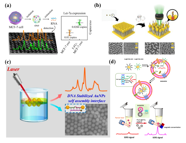
图8 SERS外泌体核酸内容物检测。(a) 基于金八面体阵列用于SERS外泌体miRNA传感检测的示意图[107];(b) 基于等离子磁头植绒金纳米柱的表面增强拉曼散射传感器定量检测乳腺癌外泌体miRNA[108];(c) DNA结构稳定的液-液自组装金纳米颗粒有序界面用于miRNA-155的SERS检测[109];(d) 基于Fe3O4 @TiO2富集、靶标触发的SERS检测外泌体miRNA[111]

Fig.8 SERS detections of exosomal nucleic acid content. (a) Schematic diagram of gold octahedra array for SERS sensing exosomal miRNAs[107]. Copyright 2021, American Chemical Society. (b) Quantitative detection of exosomal miRNAs of breast cancer using a SERS sensor based on plasmonic head-flocked gold nanopillars[108]. Copyright 2019, Wiley. (c) DNA structure-stabilized liquid-liquid self-assembled ordered Au nanoparticle interface for SERS detection of miRNA-155[109]. Copyright 2021, American Chemical Society. (d) In situ exosomal miRNA determination by target-triggered SERS and Fe3O4@TiO2-based exosome accumulation[111]. Copyright 2021, American Chemical Society

However, the processes of exosome isolation, lysis, and cargo extraction and amplification hinder the accurate quantification of exosomal miRNA. To address this challenge, researchers have proposed corresponding solutions, such as synthesizing specific SERS probes that enter the exosomes and bind with the contained miRNA to form SERS hotspots. As shown in Figure 8d, Jiang et al.[111] used Fe3O4@TiO2 to capture and enrich exosomes, and subsequently introduced locked nucleic acid-modified Au@DTNB into the exosomes to assemble with the target miRNA, inducing numerous SERS signal hotspots with the ability to distinguish single-base mismatch. Based on this platform[112], target miRNAs can be highly sensitively identified directly in situ, with a detection limit as low as 0.21 fM, outperforming quantitative reverse transcription polymerase chain reaction and other in situ methods.

3.3.3 SERS Multi-Index Detection

Single biomarkers have limitations in assessing disease progression and are prone to misinterpretation due to non-disease factors. In contrast, multiplexed testing approaches, particularly the comprehensive analysis of multiple molecules within exosomes, provide a more complete understanding of cancer progression and significantly enhance diagnostic specificity and accuracy. The complementarity among different biomarkers, such as the association between exosomal surface proteins and cellular signaling or between miRNAs and gene regulation, collectively improves the complexity and precision of diagnosis. By analyzing multiple markers, not only can disease states be identified more accurately, but also the ability to detect subtle differences is enhanced, thereby advancing precision medicine and ensuring more accurate disease identification and tailored treatment. [99,113-114]
Li et al.[115] performed multiplexed detection of different receptors (LRG1 and GPC1) on the exosome surface bound to specific probes by comparing the characteristic peak intensities of different Raman signal molecules, DTNB and MBA, at specific Raman shifts (1332 and 1078 cm-1). This method not only offers high sensitivity but also enables simultaneous detection of multiple exosomal markers, thereby expanding the scope and accuracy of exosome analysis, as shown in Figure 9a. Similarly, Lu et al.[116] achieved simultaneous detection of miRNA-21, miRNA-122, and miRNA-24 using SERS technology, innovatively employing the full spectrum of specific SERS reporter molecules as fingerprint codes for miRNAs and simplifying the analysis through pseudocolor mapping, as illustrated in Figure 9b. Different functionalized gold nanoparticles specifically recognize each miRNA, and detection is completed on a single microsphere through a one-step binding reaction. As the concentration of target miRNA increases, the SERS signal correspondingly enhances, and the constructed calibration curve ensures precise quantitative analysis of multiple targets, providing an efficient tool for miRNA detection in complex samples.
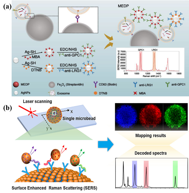
图9 SERS多指标检测:(a) 双SERS探针多重检测外泌体受体LRG1和GPC1原理图[115];(b) 基于miRNA-21、miRNA-122和miRNA-24的平均光谱和SERS图谱的多重SERS检测[116]

Fig.9 SERS multiple index detection for exosomes. (a) Schematic diagram of dual SERS probes for multiple detection of exosome receptors LRG1 and GPC1[115]. Copyright 2022, Wiley. (b) Multiplex SERS detection of exosomal microRNAs by average spectra and SERS mapping images of miRNA-21, miRNA-122, and miRNA-24[116]. Copyright 2021, American Chemical Society

3.4 SERS Combined with Other Analytical Techniques

In recent years, SERS has been combined with various techniques such as multispectral technology, mass spectrometry, microfluidic technology, and machine learning for application in exosome sensing and detection, demonstrating significant potential for application.
Multimodal detection can enhance the accuracy of detection. For example, Lin et al.[117] developed an exosome detection technique that outputs colorimetric/SERS/temperature tri-modal signals, with detection limits of 2.6×10³, 4.1×101, and 4.6×10² exosomes/μL for the colorimetric, SERS, and temperature modes, respectively, which is superior to the commonly used gold nanoparticle-based multimodal lateral flow assay (∼105 exosomes/μL). Carney et al.[118] reported a multispectral analysis technique combining SERS spectroscopy and fluorescence imaging for analyzing individual exosomes, enabling precise identification and characterization of exosome subpopulations. In addition, researchers combined SERS and matrix-assisted laser desorption/ionization time-of-flight mass spectrometry (MALDI-TOF) for the detection of exosomes in plasma from osteosarcoma patients and healthy individuals, where SERS provides characteristic vibrational modes of exosomal nucleic acids, lipids, and amino acids, reflecting comprehensive molecular composition information, while MALDI-TOF provides mass spectrometric fingerprints of exosomal proteins and peptides. Leveraging the complementary nature of these two techniques, high-precision differential diagnosis of osteosarcoma was achieved through comprehensive fingerprint analysis of exosome sample components[119], demonstrating the diagnostic potential of combined detection for exosome liquid biopsy.
Microfluidic technology, characterized by miniaturization and integration, enables precise control over liquid sample flow, facilitating steps such as sample mixing, enrichment, and capture. By leveraging the physical or biological properties of microfluidic chips to perform sample separation and enrichment, and integrating them with the high-sensitivity detection capability of surface-enhanced Raman spectroscopy (SERS), high-throughput and highly sensitive detection can be achieved while reducing both detection time and consumption of samples and reagents. The entire detection process is integrated onto a chip, making it conducive to on-site point-of-care testing (POCT)[120]. As shown in Figure 10a, Wang et al.[121] developed a microfluidic chip that combines immunomagnetic capture with surface-enhanced Raman spectroscopy. The device features a staggered triangular pillar array within a microchannel to enhance collisions between magnetic microspheres and exosomes, thereby improving the efficiency of exosome capture. Using EpCAM antibody and Raman molecule-modified polymer nanospheres as SERS probes, this system integrates the immunoenrichment capability of magnetic microspheres, the high-throughput advantage of microfluidic chips, and the specificity and anti-interference detection properties of Raman probes, enabling rapid and highly sensitive in situ detection of exosomes in serum from patients with prostate cancer, demonstrating the synergistic enhancement effect of combining SERS with microfluidic chip technology. Similarly, Ho et al.[61] proposed a SERS sensor based on a microfluidic chip for detecting HER2-positive exosomes, as illustrated in Figure 10b. This strategy achieves efficient discrimination of droplets containing HER2-positive exosomes by precisely controlling the specific binding of gold nanoparticles with HER2-positive exosomes and salt-induced aggregation, utilizing enhanced SERS signals to identify target exosomes, resulting in an automated and highly sensitive detection process.
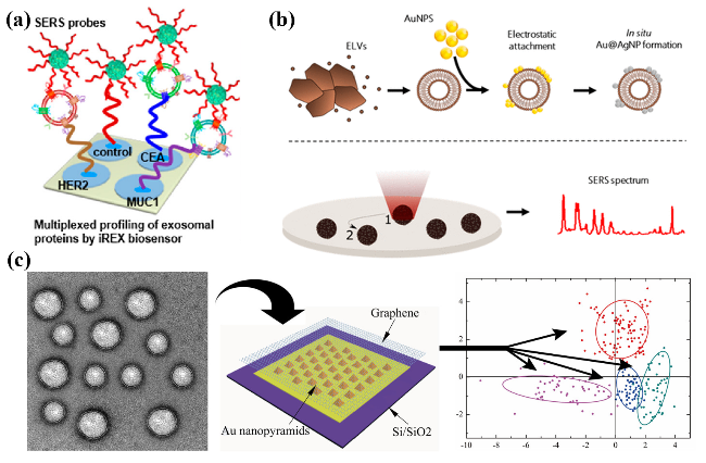
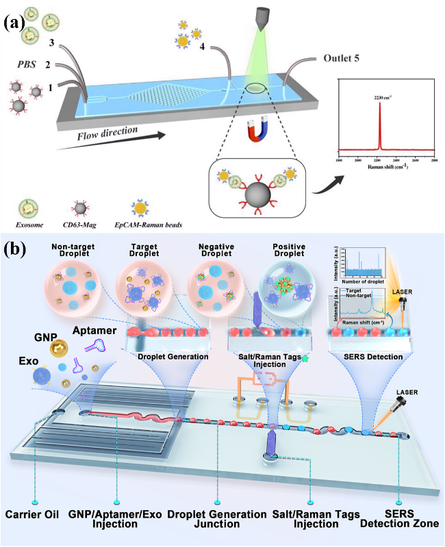
图10 SERS与微流控技术结合检测外泌体。(a) 微流控拉曼生物芯片检测前列腺癌外泌体示意图[121];(b) 基于SERS的液滴微流控平台检测HER2阳性外泌体的过程说明[61]

Fig.10 SERS combined with the microfluidic technology for exosomes detections. (a) Microfluidic SERS biochip for detection of prostate cancer-derived exosomes[121]. Copyright 2020, Royal Society of Chemistry. (b) Illustration of the process of SERS-based droplet microfluidic platform for detecting HER2-positive exosomes[61]. Copyright 2024, American Chemical Society

With the development of machine learning techniques, various data analysis methods have been effectively applied to SERS-based exosome detection. For instance, in label-free SERS detection of exosomes, the differences among SERS spectra of various exosome samples are often very subtle and typically difficult to distinguish based solely on certain characteristic peaks. Principal component analysis (PCA), as a multivariate statistical analysis method, can effectively reduce data dimensionality by explaining most of the variation across samples through a few principal components, thereby simplifying the analysis process. This approach yields relatively robust results that are less affected by outliers. Park et al.[122] isolated and enriched exosomes from lung cancer cell lines (H1299 and H522) and normal alveolar cells, directly detected them using SERS spectroscopy, and analyzed the full spectral data via PCA. This method clearly distinguished exosomes derived from lung cancer cells and normal cells, achieving a sensitivity of 95.3% and a specificity of 97.3%. Additionally, 11 Raman shifts corresponding to characteristic peaks of exosomes from lung cancer cells and 6 Raman shifts corresponding to those from normal cells were identified, indicating that SERS combined with PCA enables label-free, highly sensitive differentiation of exosomes from cancerous and normal cells. Shin et al.[123] successfully extracted unique Raman peaks of exosomes derived from non-small cell lung cancer cells using PCA analysis of SERS spectra, as shown in Figure 11a. Furthermore, Stremersch et al.[102] utilized partial least squares regression discriminant analysis to analyze SERS spectra for distinguishing exosomes of different cellular origins.
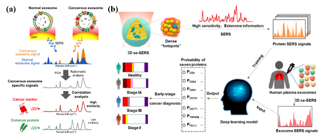
图11 SERS结合机器学习的外泌体分析。(a) SERS结合PCA分析癌性外泌体[123];(b) SERS结合深度学习进行血浆外泌体表面蛋白的多重检测[126]

Fig.11 Exosome analysis of SERS with machine learning. (a) Exosomes analysis based on SERS and PCA[123]. Copyright 2018, American Chemical Society. (b) SERS combined with deep learning was used for multiple detection of plasma exosome surface proteins[126]. Copyright 2024, American Chemical Society

The development of artificial intelligence technology has further enhanced data analysis capabilities. Compared to traditional PCA statistical methods, deep learning models are better suited for handling more complex data and achieving higher classification accuracy. Deep learning can automatically learn complex patterns from large-scale data, perform feature extraction and classification prediction without complicated preprocessing. Meanwhile, increasing training data can continuously optimize the model and improve performance. For example, Ma et al.[124] constructed a ResNet-based convolutional neural network model, using SERS spectra of three types of exosomes to train the model for classification. The final model could distinguish between normal and cancer cell exosomes with an accuracy as high as 95%. Other researchers isolated exosomes from plasma samples of 70 healthy controls and 70 patients with major depressive disorder (MDD), and used SERS to obtain approximately 400 Raman spectra per sample. Using deep learning (convolutional neural network) for training and classification, they achieved very high sensitivity and specificity on an independent test set. The prediction score for each sample was positively correlated with the Hamilton Depression Rating Scale (HAMD-17) score, indicating that the prediction results were related to the severity of depression[125]. These results suggest that SERS detection of plasma exosomes combined with deep learning analysis can serve as a biomarker for MDD diagnosis, providing an objective screening method for mental disorders. Another example is Chen et al.[126], who developed a three-dimensional circumferential SERS platform combined with deep learning algorithms, as shown in Fig. 11b. By calculating the total gray value of the color barcodes, they achieved multiplexed quantitative analysis of seven key proteins (CD63, CD81, CD9, CD151, CD171, TSPAN8, and PD-L1) on the surface of exosomes in plasma, with clinical detection sensitivity and specificity reaching 82% and 90%, respectively, while accurately distinguishing different stages of early lung cancer.

4 Conclusion and Prospect

Exosomes are small vesicles released by cells containing various types of information. They exist abundantly and stably in different types of body fluids and play a key role in intercellular material transport, including DNA, RNA, proteins, and lipids, which are used for intercellular communication. Current studies[127-131] indicate that exosomes play a crucial role in cancer growth, angiogenesis, immune regulation, tumor metastasis, and biological drug resistance. As a novel type of circulating biomarker, exosomes are now widely applied in various fields such as early cancer screening, diagnosis, treatment monitoring, and prognosis. Traditional exosome detection techniques, such as enzyme-linked immunosorbent assay[132] and mass spectrometry, are difficult to effectively apply in clinical settings due to multiple drawbacks including low sensitivity, time lag, high cost, and complex procedures. The introduction of SERS spectroscopy technology not only enables ultra-sensitive detection[133], but also, due to its large spectral coding capacity, potentially solves the challenge of simultaneously detecting multiple exosomes[134]. Additionally, using body fluid testing[135], it minimizes physiological damage to patients, allows real-time monitoring of exosome status and drug response, facilitates timely adjustment of treatment plans, and thereby provides a real-time, non-invasive analytical approach for cancer patients[136].
In the future, high-sensitivity detection of exosomes based on SERS technology still requires continuous research and innovation. By integrating machine learning methods and extracting features from SERS signals, important signals associated with exosomes can be efficiently identified, enhancing the processing capability for large-scale samples and analysis of complex data, thus providing strong technical support for the development of precision diagnostic strategies and personalized medical plans. In addition to technological advancements, SERS detection also needs to focus on improving uniformity and reproducibility to overcome the application bottleneck of difficult large-scale clinical practice. For example, personalized detection strategies can be implemented according to the specificity of exosomes. At the same time, due to their specificity and heterogeneity of content, exosomes increase the difficulty of accurate SERS detection. To overcome these issues, the main solutions include: (1) measuring a sufficient number of exosome samples; (2) implementing corresponding personalized SERS detection strategies based on the specificity of different exosomes; (3) controlling the localization of exosomes on the SERS sensors, such as through in situ detection of exosomes[111]. Considering the cancer stage and subtype of patients, current SERS sensing technology still requires large-scale clinical validation for cancer diagnosis, that is, improving adaptability to more complex real-world environments.
In conclusion, the highly sensitive detection of exosomes based on SERS biosensors can serve as a powerful auxiliary tool for cancer screening, diagnosis, and prognosis. Although the development of exosome detection based on SERS technology is still at a relatively early stage, the rapid progress in this field in recent years highlights its significant potential for early cancer diagnosis.
[1]
Trams E G, Lauter C J, Norman Salem J, Heine U.Biochim. Biophys. Acta BBA Biomembr., 1981, 645(1): 63.

[2]
Pan B T, Teng K, Wu C, Adam M, Johnstone R M. J. Cell Biol., 1985, 101(3): 942.

[3]
Johnstone R M, Adam M, Hammond J R, Orr L, Turbide C. J. Biol. Chem., 1987, 262(19): 9412.

[4]
Wang Z, Zong S, Wang Y, Li N, Li L, Lu J, Wang Z, Chen B, Cui Y. Nanoscale, 2018, 10: 905.

[5]
Han Z Z, Peng X Y, Yang Y, Yi J, Zhao D, Bao Q Y, Long S P, Yu S X, Xu X X, Liu B H, Liu Y J, Shen Y H, Qiao L. Biosens. Bioelectron., 2022, 217: 114709.

[6]
Xie Y, Su X M, Wen Y, Zheng C, Li M. Nano Lett., 2022, 22(19): 7910.

[7]
Li T D, Zhang R, Chen H, Huang Z P, Ye X, Wang H, Deng A M, Kong J L. Chem. Sci., 2018, 9(24): 5372.

[8]
Diao X K, Li X L, Hou S P, Li H J, Qi G H, Jin Y D. Anal. Chem., 2023, 95(19): 7552.

[9]
Liu Y J, Li M, Liu H S, Kang C, Wang C. Theranostics, 14(5): 1966.

[10]
Al Mannai A, Haik Y, Elmel A, Qadri S, Saud K M. Emergent Mater., 2022, 5(6): 1579.

[11]
Kim W H, Lee J U, Jeon M J, Park K H, Sim S J. Biosens. Bioelectron., 2022, 205: 114116.

[12]
Li J, Li Y R, Li P L, Zhang Y, Du L T, Wang Y S, Zhang C P, Wang C X. Acta Biomater., 2022, 144: 1.

[13]
Liu X B, Jia Y N, Zheng C. Front. Oncol., 2024, 14: 1400498.

[14]
Ferguson S W, Nguyen J. J.Control. Release, 2016, 228: 179.

[15]
Conigliaro A, Cicchini C. J. Clin. Med., 2019, 8(1): 26.

[16]
Waqas M Y, Javid M A, Nazir M M, Niaz N, Nisar M F, Manzoor Z, Bhatti S A, Hameed S, Khaliq M H. J. Physiol. Biochem., 2022, 78(3): 573.

[17]
Dai M J, Zhang Y, Yu M, Tian W D. Cell Prolif., 2016, 49(5): 561.

[18]
Guo W, Gao Y B, Li N, Shao F, Wang C N, Wang P, Yang Z L, Li R D, He J. Oncol. Rep., 2017, 38(2): 665.

[19]
Zará M, Amadio P, Campodonico J, Sandrini L, Barbieri S S. Diagnostics, 2020, 10(11): 943.

[20]
Alipoor S D, Mortaz E, Garssen J, Movassaghi M, Mirsaeidi M, Adcock I M.Mediat. Inflamm., 2016, 2016: 5628404.

[21]
Liu K, Gao X, Kang B Q, Liu Y P, Wang D D, Wang Y. Front. Oncol., 2022, 12: 836548.

[22]
Zheng Y, Tu C, Zhang J, Wang J. Int. J. Oncol., 2019, 54: 1061.

[23]
Wu Y W, Niu D, Deng S J, Lei X Y, Xie Z Z, Yang X Y. Int. Immunopharmacol., 2022, 106: 108626.

[24]
Moon B, Chang S. Cells, 2022, 11(3): 316.

[25]
Słomka A, Kornek M, Cho W C. Cells, 2022, 11(18): 2913.

[26]
Beit-Yannai E, Tabak S, Stamer W D. J. Cell. Mol. Med., 2018, 22(3): 2001.

[27]
Hade M D, Suire C N, Suo Z C. Cells, 2021, 10(8): 1959.

[28]
Liu Y F, Gu Y, Cao X T. OncoImmunology, 2015, 4(9): e1027472.

[29]
Tran P H L, Xiang D X, Tran T T D, Yin W, Zhang Y M, Kong L X, Chen K S, Sun M M, Li Y, Hou Y C, Zhu Y M, Duan W. Adv. Mater., 2020, 32(18): 1904040.

[30]
Van Cutsem E, Sagaert X, Topal B, Haustermans K, Prenen H. Lancet, 2016, 388(10060): 2654.

[31]
Lu L, Fang S K, Zhang Y, Jin L T, Xu W R, Liang Z F. Cancer Manag. Res., 2021, 13: 8121.

[32]
Wang X Y, Huang J, Chen W J, Li G P, Li Z H, Lei J Y. Exp. Mol. Med., 2022, 54(9): 1390.

[33]
Mukherjee A, Bisht B, Dutta S, Paul M K. Acta Pharmacol. Sin., 2022, 43(11): 2759.

[34]
Lin L Y, Yang L, Zeng Q, Wang L, Chen M L, Zhao Z H, Ye G D, Luo Q C, Lv P Y, Guo Q W. Mol. Cancer, 2018, 17: 1.

[35]
Li T W, Li X Q, Han G P, Liang M, Yang Z R, Zhang C Y, Huang S Z, Tai S, Yu S. Pharmaceutics, 2023, 15(1): 21.

[36]
Lee J, Lee J H, Chakraborty K, Hwang J, Lee Y K. RSC Adv., 2022, 12(29): 18475.

[37]
Lan B, Zeng S Y, Grützmann R, Pilarsky C. Int. J. Mol. Sci., 2019, 20(18): 4332.

[38]
Liang S F, Xu H Y, Ye B C. Langmuir, 2022, 38(1): 299.

[39]
Cao Y H, Shi Y H, Wang Y W, Yang Y L, Guo W J, Zhang C F, Pei W J, Fu C. Dis. Markers, 2022, 2022: 1899282.

[40]
Zhu Z D, Shen Y Y, Chen Y F, Shi H M, Shi Y. Medicine, 2021, 100(4): e21370.

[41]
Radnaa E, Richardson L S, Sheller M S, Baljinnyam T, de Castro Silva M, Kumar Kammala A, Urrabaz G R, Kechichian T, Kim S, Han A, Menon R. Lab a Chip, 2021, 21(10): 1956.

[42]
Sun H H, Sun R, Song X, Gu W D, Shao Y J. J. Transl. Med., 2022, 20(1): 189.

[43]
Jiang K, Chen H Y, Fang Y M, Chen L B, Zhong C H, Bu T T, Dai S Q, Pan X, Fu D L, Qian Y C, Wei J S, Ding K F. J. Exp. Clin. Cancer Res., 2021, 40(1): 21.

[44]
Dimtrakopoulos F D, Kottorou A, Yang A, Rodgers K, Baylin S, Hulbert A, Brock M.J. Clin. Oncol., 2016, 34(15_suppl): e23016.

[45]
Wu Q, Ding Q, Lin W Z, Weng Y L, Feng S Y, Chen R, Chen C B, Qiu S F, Lin D. Adv. Healthc. Mater., 2023, 12(8): 2370039.

[46]
Bonhoure A, Henry L, Morille M, Aissaoui N, Bellot G, Stoebner P E, Vidal M. Melanoma Res., 2021, 31(4): 338.

[47]
Cerisoli S, Marinelli Busilacchi E, Mattiucci D, Rossi E, Mariani M, Guescini M, Pugnaloni A, Olivieri F, Olivieri A, Poloni A, Br. J. Haematol., 2021, 192(1): e4.

[48]
Tong F J, Mao X H, Zhang S W, Xie H H, Yan B Q, Wang B Z, Sun J, Wei L L. Cancer Lett., 2020, 478: 34.

[49]
Vicentini C, Calore F, Nigita G, Fadda P, Simbolo M, Sperandio N, Luchini C, Lawlor R T, Croce C M, Corbo V, Fassan M, Scarpa A. BMC Gastroenterol., 2020, 20(1): 137.

[50]
Mrowczynski O D, Madhankumar A B, Slagle W B, Lee S Y, Zacharia B E, Connor J R. Biochim. Biophys. Acta BBA Gen. Subj., 2017, 1861(8): 1921.

[51]
Han L, Zhao Z R, Yang K, Xin M, Zhou L C, Chen S P, Zhou S B, Tang Z, Ji H, Dai R W. Stem Cell Res. Ther., 2022, 13(1): 153.

[52]
Wu M X, Ouyang Y, Wang Z Y, Zhang R, Huang P H, Chen C Y, Li H, Li P, Quinn D, Dao M, Suresh S, Sadovsky Y, Huang T J. Proc. Natl. Acad. Sci. U. S. A., 2017, 114(40): 10584.

[53]
Kluszczyńska K, Czernek L, Cypryk W, Pęczek Ł, Düchler M. Curr. Pharm. Des., 2020, 25(42): 4464.

[54]
Bagrov D V, Senkovenko A M, Nikishin I I, Skryabin G O, Kopnin P B, Tchevkina E M. J. Phys.: Conf. Ser., 2021, 1942(1): 012013.

[55]
Gassmann M, Grenacher B, Rohde B, Vogel J. Electrophoresis, 2009, 30(11): 1845.

[56]
Jarrold M F. Chem. Rev., 2022, 122(8): 7415.

[57]
Koller A, Patel P, Kim J K, Chen E I. Methods Mol. Biol., 2017, 1619: 21.

[58]
Peter Q A E, Jacquat R P B, Herling T W, Challa P K, Kartanas T, Knowles T P J. J. Phys. Chem. B, 2022, 126(44): 8913.

[59]
Leeson C, Vigil H, Witherspoon L. Can. Fam. Physician, 2025, 71(2): 105.

[60]
Wang Y R, Gao R K, Ma C, Liu H C, Zhu H J, Jin H, Wang F F, Cheng Z Y, Feng Y J, Yu L D. Chem. Eng. J., 2024, 496: 154270.

[61]
Ho K H W, Lai H, Zhang R L, Chen H T, Yin W, Yan X J, Xiao S, Lam C Y K, Gu Y T, Yan J X, Hu K P, Shi J Y, Yang M. ACS Sens., 2024, 9(9): 4860.

[62]
Bagheri Hashkavayi A, Cha B S, Lee E S, Kim S, Park K S. Anal. Chem., 2020, 92(19): 12733.

[63]
Yu Y Y, Zhang W S, Guo Y H, Peng H P, Zhu M, Miao D D, Su G X. Biosens. Bioelectron., 2020, 167: 112482.

[64]
Sun Z H, Zhang B M, Tu H J, Pan C Y, Chai Y J, Chen W W. Nanoscale, 2024, 16(3): 1005.

[65]
Reznickova A, Slepicka P, Nguyenova H Y, Kolska Z, Dendisova M, Svorcik V. RSC Adv., 2017, 7(37): 23055.

[66]
Lin C L, Liang S S, Peng Y S, Long L, Li Y Y, Huang Z R, Long N V, Luo X Y, Liu J J, Li Z Y, Yang Y. Nano Micro Lett., 2022, 14(1): 75.

[67]
Fleischmann M, Hendra P J, McQuillan A J. J. Chem. Phys. Lett., 1974, 26(2): 163.

[68]
Jeanmaire D L, Van Duyne R P. J. Electroanal. Chem. Interfacial Electrochem., 1977, 84(1): 1.

[69]
Albrecht M G, Creighton J A. J. Am. Chem. Soc., 1977, 99(15): 5215.

[70]
Kirtley J R, Jha S S, Tsang J C. Solid State Commun., 1980, 35(7): 509.

[71]
Park T H, Galperin M. Phys. Rev. B, 2011, 84(7): 075447.

[72]
Doering W E, Nie S M. J. Phys. Chem. B, 2002, 106(2): 311.

[73]
Okeil S, Pashchanka M, Heinschke S, Bruns M, Schneider J J. J. Phys. Chem. C, 2020, 124(24): 13316.

[74]
Montes García V, Gómez González B, Martínez Solís D, Taboada J M, Jiménez Otero N, de Uña Álvarez J, Obelleiro F, García Río L, Pérez Juste J, Pastoriza Santos I. ACS Appl. Mater. Interfaces, 2017, 9: 26372.

[75]
Han L F, Zhu C C, Tan Z, Wang J, Liao X W, Xia X H, Wang C. Chem. Commun., 2023, 59(51): 7967.

[76]
Liu H Q, Gao X, Xu C, Liu D B. Theranostics, 12(4): 1870.

[77]
Song C Y, Chen J, Zhao Y P, Wang L H. J. Mater. Chem. B, 2014, 2(43): 7488.

[78]
Sun Y D, Fang L, Yi Y, Feng A B, Zhang K, Xu J J. J. Nanobiotechnol., 2022, 20(1): 285.

[79]
Xie L P, Zeng H, Zhu J X, Zhang Z L, Sun H B, Xia W, Du Y N. Nano Res., 2022, 15(5): 4374.

[80]
Wang J, Xie H Y, Ding C F. ACS Appl. Mater. Interfaces, 2021, 13(28): 32837.

[81]
Tian Y F, Ning C F, He F, Yin B C, Ye B C. Anal., 2018, 143(20): 4915.

[82]
Zhang P, Zhou X, He M, Shang Y Q, Tetlow A L, Godwin A K, Zeng Y. Nat. Biomed. Eng., 2019, 3(6): 438.

[83]
Song Z G, Mao J, Barrero R, Wang P, Zhang F Q, Wang T. Molecules, 2020, 25(23): 5585.

[84]
Zhang Q, Ma R F, Zhang Y Z, Zhao J, Wang Y, Xu Z R. ACS Sens., 2023, 8(2): 875.

[85]
Zhang W, Jiang L M, Diefenbach R J, Campbell D H, Walsh B J, Packer N H, Wang Y L. ACS Sens., 2020, 5(3): 764.

[86]
Zhao Y, Fang X X, Bai M, Zhang J, Yu H H, Chen F, Zhao Y X. Chin. Chemical Lett., 2022, 33(4): 2101.

[87]
Hu H C, Wu S H, Jin L X, Sun J J. Biosens. Bioelectron., 2022, 210: 114283.

[88]
Tian C, Zhao L, Qi G L, Zhang S S. ACS Appl. Nano Mater., 2023, 6(2): 1386.

[89]
Fu C C, Jin S L, Shi W B, Oh J, Cao H Y, Jung Y M. Anal. Chem., 2018, 90(22): 13159.

[90]
Mohammadniaei M, Zhang M, Ashley J, Christensen U B, Friis-Hansen L J, Gregersen R, Lisby J G, Benfield T L, Nielsen F E, Henning Rasmussen J, Pedersen E B, Olinger A C R, Kolding L T, Naseri M, Zheng T, Wang W, Gorodkin J, Sun Y. Nat. Commun., 2021, 12(1): 5089.

[91]
Wang K, Liu X J, Lian X L, Bao X P, Li K L. Front. Bioeng. Biotechnol., 2023, 11: 1250175.

[92]
Cao X W, Gu Y Y, Li Z Y, Ge S J, Mao Y, Gu Y X, Lu D. Sens. Actuat. B Chem., 2023, 375: 132894.

[93]
Cun F, Huang Z P, Lin Q Y, Yu G P, Chen H, Kong J L, Weng W H. Anal. Chem., 2023, 95(26): 10025.

[94]
Jørgensen M, Bæk R, Pedersen S, Søndergaard E K L, Kristensen S R, Varming K. J. Extracell. Vesicles, 2013, 2(1): 20920.

[95]
Zhang P, Wang L M, Fang Y P, Zheng D W, Lin T F, Wang H Q. Molecules, 2019, 24(16): 2947.

[96]
Avella-Oliver M, Puchades R, Wachsmann-Hogiu S, Maquieira A. Sens. Actuat. B Chem., 2017, 252: 657.

[97]
Lee C, Carney R P, Hazari S, Smith Z J, Knudson A, Robertson C S, Lam K S, Wachsmann-Hogiu S. Nanoscale, 2015, 7(20): 9290.

[98]
Muhammad M, Shao C S, Liu C, Song G, Zhan J, Huang Q. Biosens. Bioelectron. X, 2022, 12: 100177.

[99]
Su X M, Liu X Y, Xie Y, Chen M Y, Zheng C, Zhong H, Li M. ACS Nano, 2023, 17(4): 4077.

[100]
Fraire J C, Stremersch S, Bouckaert D, Monteyne T, De Beer T, Wuytens P, De Rycke R, Skirtach A G, Raemdonck K, De Smedt S, Braeckmans K. ACS Appl. Mater. Interfaces, 2019, 11(43): 39424.

[101]
Yan Z B, Dutta S, Liu Z R, Yu X K, Mesgarzadeh N, Ji F, Bitan G, Xie Y H. ACS Sens., 2019, 4(2): 488.

[102]
Stremersch S, Marro M, Pinchasik B E, Baatsen P, Hendrix A, De Smedt S C, Loza-Alvarez P, Skirtach A G, Raemdonck K, Braeckmans K. Small, 2016, 12(24): 3292.

[103]
Yáñez M M, Siljander P R M, Andreu Z, Bedina Z A, Borràs F E, Buzas E I, Buzas K, Casal E, Cappello F, Carvalho J. J. Extracell. Vesicles, 2015, 4: 27066.

[104]
Griffin J L, Shockcor J P. Nat. Rev. Cancer, 2004, 4(7): 551.

[105]
Tirinato L, Gentile F, Di Mascolo D, Coluccio M L, Das G, Liberale C, Pullano S A, Perozziello G, Francardi M, Accardo A, De Angelis F, Candeloro P, Di Fabrizio E. Microelectron. Eng., 2012, 97: 337.

[106]
Hou M, He D, Wang H, Huang J, Cheng H, Wan K, Li H, Tang Z, He X, Wang K. Chin. Chem. Lett., 2022, 33: 318.

[107]
Kang T L, Zhu J T, Luo X J, Jia W Y, Wu P, Cai C X. Anal. Chem., 2021, 93(4): 2519.

[108]
Lee J U, Kim W H, Lee H S, Park K H, Sim S J. Small, 2019, 15: 1804968.

[109]
Wu C J, Huang S Q, Wang Y Y, Chai Y Q, Yuan R, Yang X. Anal. Chem., 2021, 93(31): 11019.

[110]
Ma D D, Huang C X, Zheng J, Tang J R, Li J S, Yang J F, Yang R H. Biosens. Bioelectron., 2018, 101: 167.

[111]
Jiang S, Li Q, Wang C, Pang Y, Sun Z, Xiao R. ACS Sens., 2021, 6: 85.

[112]
Pang Y, Shi J, Yang X, Wang C, Sun Z, Xiao R. Biosens. Bioelectron., 2020, 148: 111800.

[113]
Hashkavayi A B, Cha B S, Lee E S, Park K S. Anal. Chim. Acta, 2022, 1205: 339762.

[114]
Lee J H, Kim J A, Jeong S, Rhee W J. Biosens. Bioelectron., 2016, 86: 202.

[115]
Li J, Li Y R, Chen S, Duan W L, Kong X, Wang Y S, Zhou L Q, Li P L, Zhang C P, Du L T, Wang C X. Small Meth., 2022, 6(6): 2200154.

[116]
Lu X H, Hu C, Jia D L, Fan W J, Ren W, Liu C H. Nano Lett., 2021, 21(15): 6718.

[117]
Lin X R, Zhou P Y, Li Q, Pang Y F. Anal. Chem., 2024, 96(26): 10686.

[118]
Carney R P, Hazari S, Colquhoun M, Tran D, Hwang B, Mulligan M S, Bryers J D, Girda E, Leiserowitz G S, Smith Z J, Lam K S. Anal. Chem., 2017, 89(10): 5357.

[119]
Han Z Z, Yi J, Yang Y, Li D D, Peng C, Long S P, Peng X Y, Shen Y H, Liu B H, Qiao L. Anal., 2021, 146(21): 6496.

[120]
Nie C H, Shaw I, Chen C P. J. Pharm. Anal., 2023, 13(12): 1429.

[121]
Wang Y, Li Q, Shi H, Tang K, Qiao L, Yu G, Ding C, Yu S. Lab Chip, 2020, 20: 46.

[122]
Park J, Hwang M, Choi B, Jeong H, Jung J H, Kim H K, Hong S, Park J H, Choi Y. Anal. Chem., 2017, 89(12): 6695.

[123]
Shin H, Jeong H, Park J, Hong S, Choi Y. ACS Sens., 2018, 3(12): 2637.

[124]
Ma X, Xiong H L, Guo J H, Liu Z M, Han Y R, Liu M D, Guo Y X, Wang M Y, Zhong H Q, Guo Z Y. J. Innov. Opt. Health Sci., 2023, 16(2): 2244001.

[125]
Shin H, Kang Y B, Choi K W, Kim S, Ham B J, Choi Y. Anal. Chem., 2023, 95(15): 6410.

[126]
Chen M, Wang H Y, Zhang Y B, Jiang H Y, Li T, Liu L X, Zhao Y T. Anal. Chem., 2024, 96(17): 6794.

[127]
Kugeratski F G, Kalluri R. FEBS J., 2021, 288(1): 10.

[128]
Lim G B. Nat. Rev. Cardiol., 2021, 18(6): 386.

[129]
Soung Y, Ford S, Zhang V, Chung J. Cancers, 2017, 9(1): 8.

[130]
Wang D, Zhang W, Zhang C, Wang L, Chen H, Xu J. Mol. Ther. Nucleic Acids, 2022, 29: 16.

[131]
Guo Q R, Wang H, Yan Y D, Liu Y, Su C Y, Chen H B, Yan Y Y, Adhikari R, Wu Q, Zhang J Y. Front. Oncol., 2020, 10: 472.

[132]
Khodashenas S, Khalili S, Forouzandeh Moghadam M. Biotechnol. Lett., 2019, 41(4): 523.

[133]
Krishnan S K, Chipatecua Godoy Y. ACS Omega, 2020, 5(3): 1384.

[134]
Wang Z L, Zong S F, Liu Y, Qian Z T, Zhu K, Yang Z Y, Wang Z Y, Cui Y P. Nanotechnology, 2021, 32(8): 085504.

[135]
Sun Y, Liu S, Qiao Z, Shang Z, Xia Z J, Niu X M, Qian L Q, Zhang Y, Fan L Y, Cao C X, Xiao H. Anal. Chim. Acta, 2017, 982: 84.

[136]
Hu R W, Korutla L, Reddy S, Harmon J, Zielinski P D, Bueker A, Molina M, Romano C, Margulies K, McLean R, Lal P, Vallabhajosyula P. Transplant. Direct, 2020, 6(11): e615.

Outlines

/