
Application of Polyurethane Materials in Bone Defect Repair
† There authors contributed equally.
Received date: 2024-11-04
Revised date: 2025-04-27
Online published: 2025-07-30
Supported by
the National Natural Science Foundation of China(12272253)
the Natural Science Foundation of Shanxi Province,China(202203021212270)
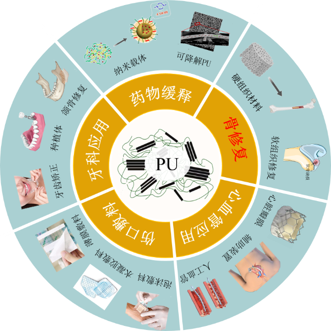
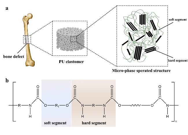
Bone defects caused by accidents or diseases are a common and serious problem in orthopedic surgery. Finding ideal bone repair materials has become a hotspot in current bone tissue engineering. Polyurethane (PU) is a multiblock copolymer with a microphase-separated structure formed by alternating soft and hard segments. Its application properties - such as mechanical performance,biocompatibility,and biodegradability-can be tailored by adjusting the soft segment structure,hard segment ratio,crystallinity,and other factors,demonstrating broad prospects in the field of bone defect repair. This paper reviews recent research on the design,synthesis,modification,and biological performance of PU in bone tissue engineering,with a focus on its application progress in bone regeneration,including implantable scaffolds,injectable materials,and drug carriers. The aim is to provide more insights for the future design and clinical application of PU materials.
1 Introduction
2 Development of polyurethane
3 Synthesis of polyurethane
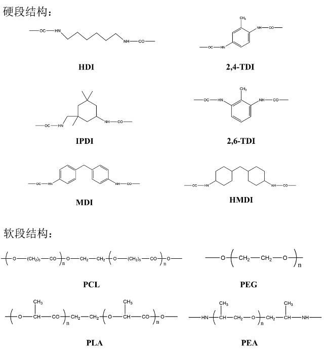
3.1 Main raw material
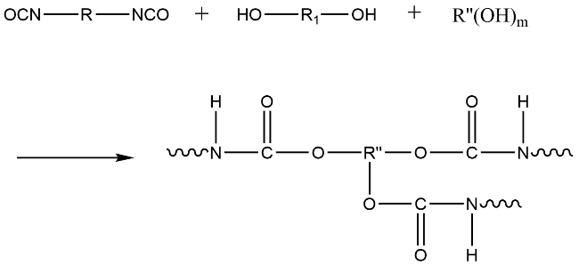
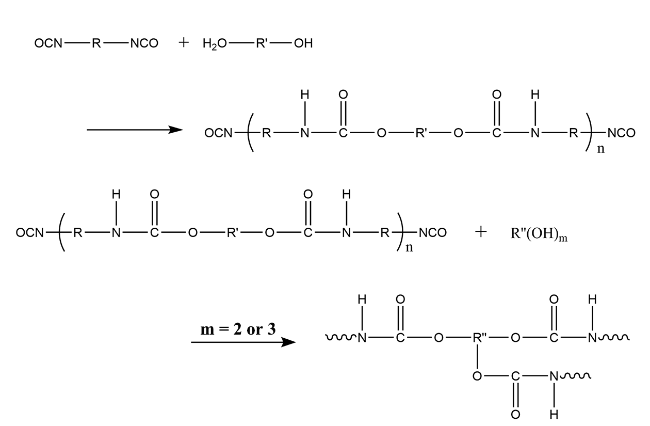
3.2 Main reaction pathways
4 Structure of polyurethane
5 Properties of polyurethane
5.1 Mechanical properties
5.2 Biological activity
5.3 Biodegradation
5.4 Shape memory properties
6 Applications of polyurethane in bone defects repairing
6.1 Implanted scaffold
6.2 Injected polyurethane
6.3 Drug carrier
7 Conclusion and outlook
Key words: polyurethane; bone repair; bone tissue engineering; functionalization; osteogenesis
Weimo Han , Yahui Wang , Yin Li , Jianan Yan , Zhiqin Li , Di Huang . Application of Polyurethane Materials in Bone Defect Repair[J]. Progress in Chemistry, 2025 , 37(8) : 1188 -1203 . DOI: 10.7536/PC241103
表1 骨修复用PUTable 1 PU for bone repair |
| SS | HS | CE | AD | APP | Ref |
|---|---|---|---|---|---|
| PCL | MDI | BDO | HA/rGO | bone adhesive | 28 |
| PPG | MDI | Gl | Bi2O3、Ta2O5、ZrO2 | bone replacement materials | 29 |
| PCL | HDI | H2O | CAP | Shape memory scaffold | 30 |
| GCO | IPDI | BDO | HA | Implanted scaffold | 31 |
| β-CD | HDI | H2O | HA | Implanted scaffold | 32 |
| MAG、PEG | IPDI | - | HA | Implanted scaffold | 33 |
SS:soft segment; HS:hard segment; CE:chain extender; AD:addition; APP:application; GCO:castor oil; β-CD:β-Cyclodextrin; MAG:monoacylglyceride; BDO:1,4-butanediol; CAP:calcium phosphate; HA:hydroxyapatite |
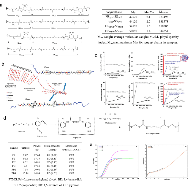
图6 软硬段结构对机械性能的影响:(a) PU链结构图[38];(b) 伸展时HSglass-SScryst和HScryst-SScryst分子链条重排模型[38];(c) 不同软段PU的应力应变曲线[38];(d) 不同扩链剂的PU分子结构示意图[39];(e) 扩链剂不同PU的应力应变曲线[39]Fig. 6 The effect of hard and soft segment structure on mechanical properties. (a) sketches of polyurethane chain architecture[38];(b) model for molecular rearrangement of HSglass-SScryst and HScryst-SScryst chains upon stretching[38]; (c) sress-strains for PU involving in different soft segment[38]; (d) schematic of PU molecular structure using different chain extenders[39]; (e) sress-strains for PU involving in different chain extenders[39] |
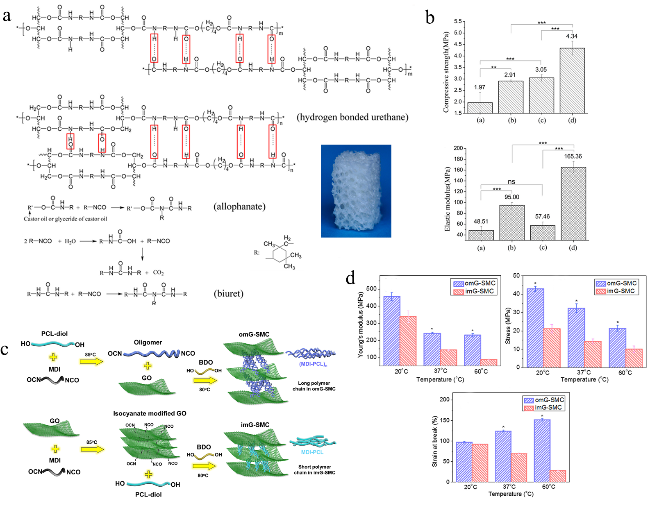
图7 PU与其他材料复合:(a) GCO/PU分子结构示意图[40];(b) 不同HA含量GCO/PU支架的抗压强度与弹性模量[40];(c) omG-SMC和imG-SMC的制备[41];(d) omG-SMC和imG-SMC的杨氏模量及断裂应力与应变[41]Fig. 7 Composition of PU with other materials. (a) schematic of the molecular structure of GCO/PU[40]; (b) compressive strength of GCO/PU with HA in different contents[40]; (c) preparation of omG-SMC and imG-SMC[41]; (d) Young’s modulus,stress and strain of omG-SMC and imG-SMC[41] |
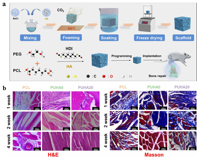
图8 PEG/PCL/HA/PU支架的制备及其促成骨性能表征[44]:(a) PEG/PCL/HA/PU复合多孔支架的制备及原理;(b) 肌肉组织H&E和Masson染色图Fig. 8 Preparation of PEG/PCL/HA/PU scaffolds and characterization of osteogenic properties[44]. (a) preparation and principle of PEG/PCL/HA/PU composite porous scaffold; (b) H&E and Masson staining pictures of muscle tissue |
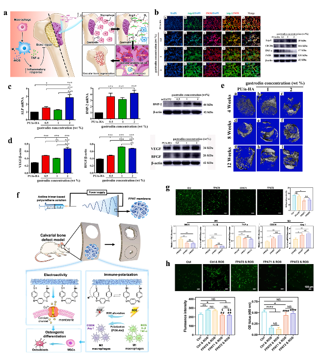
图9 功能化PU支架调控细胞应答反应并促成骨分化:(a) 天麻素-HA/PCL/PU复合支架及其工作机制[48];(b) 巨噬细胞Arg-1、iNOS和细胞核的免疫荧光染色及相关蛋白的表达[48];(c) rBMSCs ALP和BMP-2基因表达[48];(d) HUVECs血管生成基因BFGF和VEGF的表达[48];(e) 新骨的Micro-CT 3D图像[48];(f) FPAT膜制备及及其工作机制[49];(g) 巨噬细胞ROS阳性率、iNOS、IL-1β、TNF-α、和CD206、Arg-1基因表达[49];(h) H2O2孵育2 h后,FPAT膜上MSCs的荧光图像分析及细胞活力表征[49]Fig. 9 Functionalized PU scaffolds regulate cellular response reactions and improve osteogenesis. (a) gastrodin-HA/PCL/PU composite and its working principle[48]; (b) immunofluorescence staining of Arg-1,iNOS and nuclei,and relevant gene expression in RAW 264.7 cells[48]; (c) osteogenic gene expression of ALP and BMP-2 in rBMSCs[48]; (d) angiogenic gene expression of BFGF and VEGF in HUVECs[48]; (e) micro-CT 3D images of new bones[48]; (f) preparation and working principle of FPAT membranes[49]; (g) Quantitative analysis of ROS positive rate,and gene expression of iNOS,IL-1β,TNF-α,and CD206,Arg-1[49]; (h) fluorescence images analysis of fluorescence intensity,and cell viability of MSCs on different membranes after incubating with H2O2 for 2 h[49] |
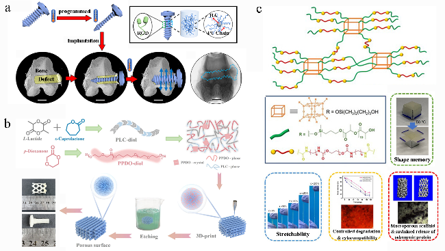
图10 形状记忆生物智能材料:(a) 形状记忆PU骨螺钉的合成及工作原理[78];(b) 3D打印形状记忆可生物降解PU用于多孔支架或骨螺钉制备[80];(c) 具有星型结构PU的制备及其形状记忆特性、拉伸性、可降解性、生物相容性、多孔结构及其成骨性能[81]Fig. 10 Shape memory smart material. (a) preparing and working principle of shape memory PU bone screw[78]; (b) synthetic approach of full-biodegradable shape memory PU for the preparation of porous scaffold and bone screw[80]; (c) preparing of star-branched PU and its stretchability,degradation,cytocompatibility,porous structure and osteogenesis[81] |
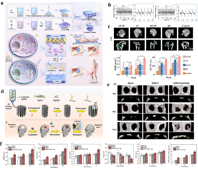
图11 3D打印PU支架:(a) 3D打印原位自供电支架的制备及工作机理[90];(b) 复合支架在连续机械冲击下的电压输出[90];(c) 重建大鼠颅骨三维显微ct图及再生骨的骨体积分数(BV/TV)和骨矿物质密度(BMD)值,AF和RF为排列整齐与无序排列的PVDF纳米纤维,SP为SMPU[90];(d) 3D打印形状记忆Mg/PU支架及其工作原理[91];(e) 重建缺损骨三维显微ct图[91];(f) BMD、BV/TV、小梁数、小梁间距和小梁厚度变化[91]Fig. 11 3D-printed PU scaffold. (a) Preparation and working principle of 3D printed in-situ self-powered scaffold[90]; (b) the voltage output of composite scafolds under continuous mechanical impact[90]; (c) reconstructed three-dimensional micro-CT images of the rat skull and variation of BV/TV and BMD,AF and RF mean the PVDF nanofbers with aligned and random fibers respectively,SP means SMPU[90]; (d) 3D printed shape memory Mg/PU scaffold and its working principle[91]; (e) reconstructed three-dimensional micro-CT images of defective bones[91]; (f) variation of BMD,BV/TV,trabecular number,trabecular separation and trabecular thickness[91] |
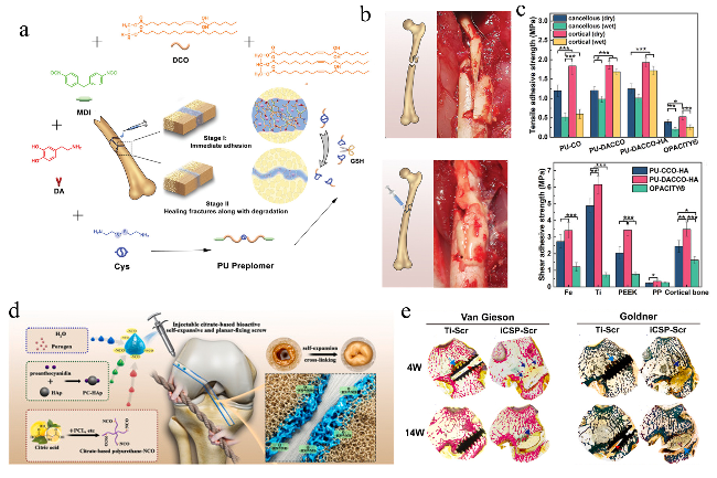
图12 可注射PU:(a) PU-DACCO-HA制备及应用与促进骨愈合原理示意图[92];(b) PU-DACCO-HA用于复杂粉碎性骨折[92];(c) 不透明为对照,观察湿干条件下PU在松质骨和皮质骨上的拉伸黏附强度与剪切结合强度以及以不透明为对照,PU与不同材料的剪切力[92];(d) 可注射ICSP-Scr螺钉的制备及作用原理[93];(e) 家兔前交叉韧带重建的VG染色与Goldner三色染色[93]Fig. 12 Injected PU. (a) fabrication of PU-DACCO-HA and principle for fixing fractures and promoting fracture healing[92]; (b) using of PU-DACCO-HA for complicated comminuted fracture[92]; (c) tensile adhesive strength of PU on the cancellous and cortical bones under wet or dry conditions with opacity as control and shear bonding strength of PU for different materials with opacity as control[92]; (d) the synthesis process and working principle of ICSP-Scr[93]; (e) VG staining and goldner trichrome results of anterior cruciate ligament reconstruction in rabbits[93] |
The contributions made by students Hu Qingbiao, Xi Jiawei, and Li Ruifang during the literature review.
| [1] |
|
| [2] |
|
| [3] |
|
| [4] |
|
| [5] |
|
| [6] |
|
| [7] |
|
| [8] |
|
| [9] |
|
| [10] |
|
| [11] |
|
| [12] |
|
| [13] |
|
| [14] |
|
| [15] |
|
| [16] |
|
| [17] |
|
| [18] |
|
| [19] |
|
| [20] |
|
| [21] |
|
| [22] |
|
| [23] |
|
| [24] |
|
| [25] |
|
| [26] |
|
| [27] |
|
| [28] |
|
| [29] |
|
| [30] |
|
| [31] |
|
| [32] |
|
| [33] |
|
| [34] |
|
| [35] |
|
| [36] |
|
| [37] |
|
| [38] |
|
| [39] |
|
| [40] |
|
| [41] |
|
| [42] |
|
| [43] |
|
| [44] |
|
| [45] |
|
| [46] |
|
| [47] |
|
| [48] |
|
| [49] |
|
| [50] |
|
| [51] |
|
| [52] |
|
| [53] |
|
| [54] |
|
| [55] |
|
| [56] |
|
| [57] |
|
| [58] |
|
| [59] |
|
| [60] |
|
| [61] |
|
| [62] |
|
| [63] |
|
| [64] |
|
| [65] |
|
| [66] |
|
| [67] |
|
| [68] |
|
| [69] |
|
| [70] |
|
| [71] |
|
| [72] |
|
| [73] |
|
| [74] |
|
| [75] |
|
| [76] |
|
| [77] |
|
| [78] |
|
| [79] |
|
| [80] |
|
| [81] |
|
| [82] |
|
| [83] |
|
| [84] |
|
| [85] |
|
| [86] |
|
| [87] |
|
| [88] |
|
| [89] |
|
| [90] |
|
| [91] |
|
| [92] |
|
| [93] |
|
| [94] |
|
| [95] |
|
| [96] |
|
| [97] |
|
| [98] |
|
| [99] |
|
| [100] |
|
/
| 〈 |
|
〉 |