Home Journals Progress in Chemistry
Progress in Chemistry

Abbreviation (ISO4): Prog Chem      Editor in chief: Jincai ZHAO

About  /  Aim & scope  /  Editorial board  /  Indexed  /  Contact  / 
Review

Development of Mirror-Image Protein Drugs: Advances in Chemical Synthesis, Mirror-Image Phage Display, and Computational Design

  • Ren Yuxiang 1 ,
  • Han Dongyang 1 ,
  • Shi Weiwei , 2, *
Expand
  • 1 Department of Chemistry, Tsinghua University, Beijing 100084, China
  • 2 College of Chemistry, Beijing Normal University, Beijing 100875, China

Received date: 2025-03-26

  Revised date: 2025-04-29

  Online published: 2025-09-01

Supported by

The National Natural Science Foundation of China(22307061)

Abstract

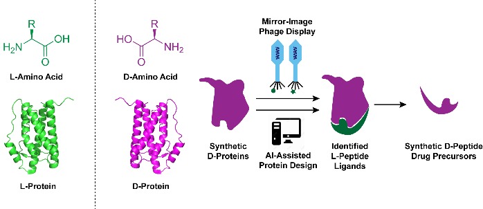
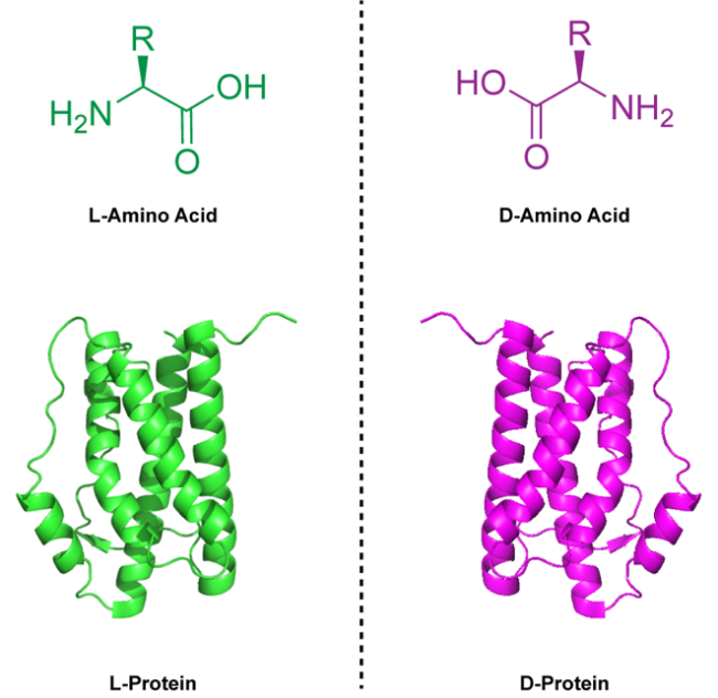
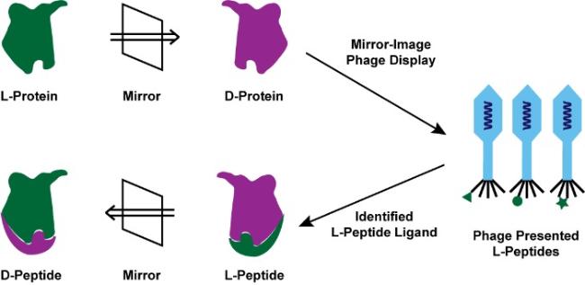
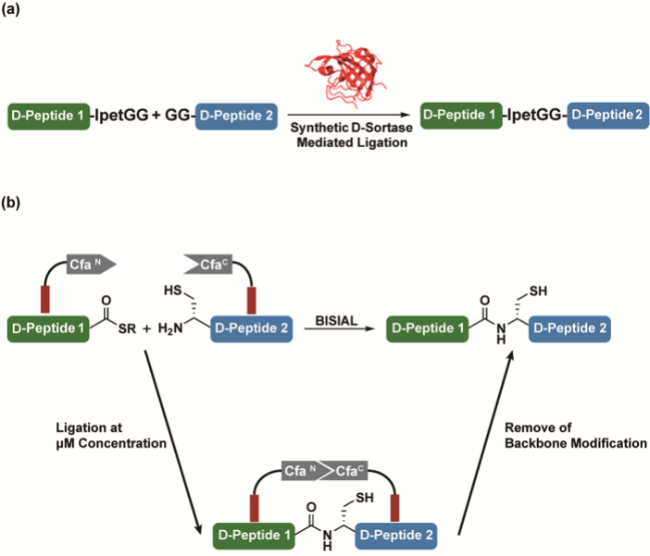
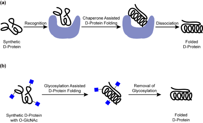
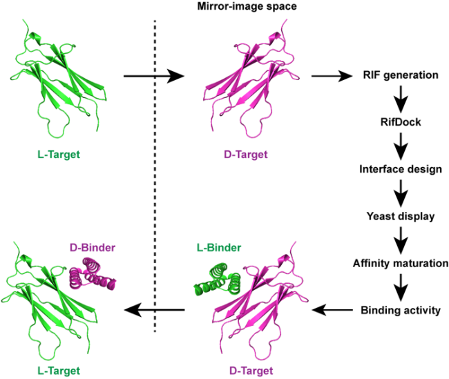
Mirror-image peptides and proteins composed entirely of D-amino acids have emerged as promising therapeutic candidates owing to their resistance to proteolysis and reduced immunogenicity. Mirror-image phage display (MIPD) is currently the main experimental technique for identifying mirror-image peptide ligands targeting disease-related proteins. However, the success of MIPD critically depends on synthetic mirror-image target proteins, which cannot be produced by traditional recombinant methods due to the intrinsic chirality of biological systems. Recent advances in chemical protein synthesis, such as enzyme-cleavable solubilizing tags, backbone-installed split intein-assisted ligation, and removable glycosylation modification-assisted folding strategies, have effectively addressed key challenges in preparing these complex mirror-image proteins. In addition, computational approaches, exemplified by AI-driven protein design, have become powerful complementary tools, accelerating the discovery and optimization of mirror-image protein drug candidates. Although mirror-image protein drugs have not yet reached clinical use, ongoing innovations in chemical synthesis and ligand screening methods are steadily advancing their therapeutic potential toward clinical translation.

Contents

1 Introduction

2 Mirror-image phage display

3 Chemical protein synthesis

3.1 Solid-phase peptide synthesis

3.2 Native chemical ligation

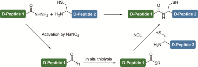
3.3 Peptide hydrazide ligation

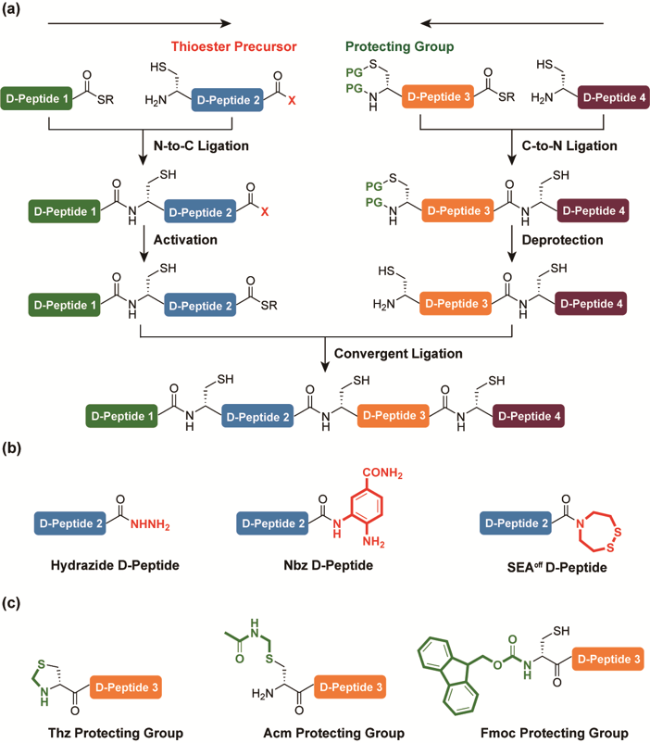
3.4 Multiple-segment ligation

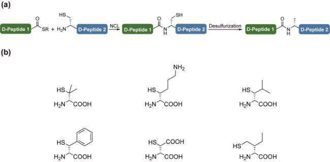
3.5 The ligation-desulfurization strategy

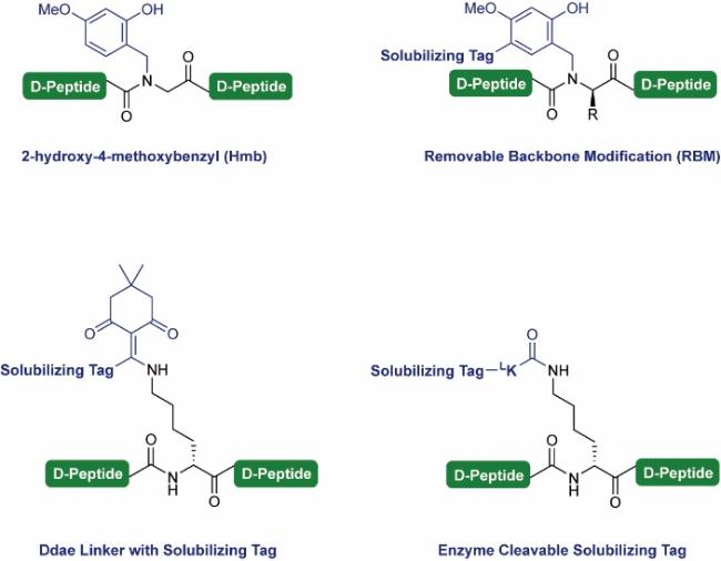
3.6 Solubilizing tags for hydrophobic segment

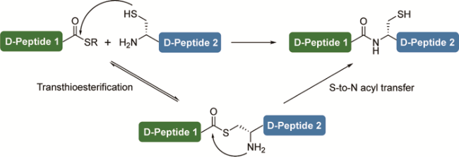
3.7 Chemoenzymatic D-peptide ligation

3.8 The folding of D-protein

4 Applications of mirror-image phage display

5 Computationally assisted discovery of mirror-image protein drugs

6 Conclusion and outlook

Cite this article

Ren Yuxiang , Han Dongyang , Shi Weiwei . Development of Mirror-Image Protein Drugs: Advances in Chemical Synthesis, Mirror-Image Phage Display, and Computational Design[J]. Progress in Chemistry, 2025 , 37(9) : 1261 -1273 . DOI: 10.7536/PC20250315

1 Introduction

Proteins are essential biomolecules required for life processes and serve as key therapeutic targets for various diseases[1-5]. Peptides capable of specifically binding to target proteins have emerged as promising therapeutic molecules, achieving remarkable clinical success in recent years, particularly in the treatment of metabolic disorders and tumors[6-9]. Over the past decade, more than 100 peptide-based therapies have been approved for market, accounting for approximately 8% of drugs approved by the U.S. Food and Drug Administration (FDA)[10]. Compared with traditional small-molecule drugs, peptides can bind to their targets with higher specificity and affinity[11-12]. However, peptide drugs also have inherent limitations: susceptibility to proteolytic degradation leading to a shortened plasma half-life[6]; low oral bioavailability due to poor transmembrane transport efficiency (approximately 90% of peptide and protein drugs require injection administration, significantly limiting treatment convenience)[13-15]; and potential immunogenicity, which may trigger excessive immune activation and result in severe adverse reactions such as cytokine storms[16-17].
To address these limitations, a promising strategy is to develop enantiomers or "mirror" versions of natural proteins, known as mirror proteins. Like other biomolecules, peptides and proteins exhibit homochirality, with naturally occurring proteins composed exclusively of L-amino acids (except glycine). The enantiomer composed entirely of D-amino acids is referred to as a mirror protein (Figure 1)[18]. These mirror proteins possess the same physicochemical properties as their L-enantiomers but exhibit opposite optical activity. The unique structural and physicochemical characteristics of mirror proteins provide significant opportunities for drug development[19]. Due to their opposite D-chirality, mirror proteins cannot be recognized by proteases, enabling them to resist proteolytic degradation and significantly enhancing their stability and serum half-life in biological systems[20-21]. Furthermore, they are less likely to be recognized by the immune system[22-23], reducing the likelihood of triggering an immune response, making them highly suitable for oral therapeutic applications[24-25]. These properties render mirror proteins particularly promising candidates for developing novel therapeutic approaches.
图1 天然蛋白与镜像蛋白

Fig.1 Native L-proteins and mirror-image proteins

Currently, the most widely used method for screening and discovering mirror-image protein drugs is mirror-image phage display (MIPD) technology. To perform MIPD, it is first necessary to obtain a mirror-image version of the target protein. Since mirror proteins cannot be prepared through recombinant expression as natural proteins can, chemical synthesis remains the only viable approach for generating mirror-image target proteins[26]. Advances in chemical synthesis methods for mirror proteins have enabled the successful development of mirror-image drug precursors targeting a wider variety of proteins. In this review, we systematically discuss mirror-protein synthesis methods, as well as the latest techniques and advancements in mirror-image phage display technology. Additionally, emerging artificial intelligence (AI)-based design strategies have opened new avenues for discovering and optimizing mirror proteins, further expanding their therapeutic potential. In summary, mirror proteins offer promising solutions to some of the most challenging limitations currently faced by peptide therapies, providing unprecedented opportunities to enhance drug stability, reduce side effects, and broaden treatment modalities such as oral administration. This article reviews the current state of mirror-protein drug development, including the fundamental principles, workflow, and application examples of screening mirror-protein drug precursors via MIPD technology, the latest advances in mirror-protein chemical synthesis methods, and recent computational approaches for mirror-protein drug discovery. These research cases highlight the promising prospects of mirror peptides and proteins as candidates for developing novel therapeutic agents.

2 Mirror phage display

The earliest mirror-image protein drugs were obtained by directly preparing the mirror-image versions of existing natural protein drugs. Due to the change in chirality, the binding affinity of these mirror-image proteins for their target proteins was significantly reduced, resulting in activities far inferior to those of their corresponding natural protein drug counterparts[21,27]. Another approach to obtaining mirror-image protein drugs is retro-inversion (RI). RI involves reversing the sequence of the parent natural peptide and replacing L-amino acids with D-amino acids to produce a mirror-image peptide drug[28]. The side-chain topology of the RI product is similar to that of its parent natural peptide, but it has an amino acid sequence that is reverse to that of the parent. The RI strategy has achieved some success with unstructured peptide targets[29-32]; however, for peptides with secondary structures, the RI strategy typically fails. This is mainly due to the topological characteristics of α-helices: mirror-image peptides composed of D-amino acids always adopt a left-handed helix, whereas natural peptides composed of L-amino acids form right-handed helices. This topological difference disrupts the binding between the peptide and its target protein[33]. Therefore, alternative methods are needed to discover and identify mirror-image protein drug precursors with high affinity for natural target proteins.
Kim et al. from the Massachusetts Institute of Technology[34]reported the MIPD technology in 1996. This pioneering work remains an important method for screening mirror-image peptides with high specificity and affinity for target proteins, and a series of mirror-image protein drug precursors have already been identified (Figure 2) [18]. Phage display is a common in vitro screening technique used to discover and identify peptide ligands capable of binding to target proteins[35-37]. This method involves genetically encoding the fusion expression of phage coat proteins with exogenous peptide sequences, allowing each phage particle to display a unique peptide on its surface. The phage-displayed peptide library is incubated with the target protein immobilized on a solid-phase carrier, followed by washing away free phages with low affinity for the target protein. The phages bound to the target protein are then eluted, infect host cells to propagate and amplify, and subsequently subjected to another round of "adsorption-washing-amplification." After multiple rounds of cycling, phages exhibiting highly specific binding to the target protein can be significantly enriched. Phage display can screen different peptide sequences with the strongest binding affinity from peptide libraries containing >109members, and decode the amino acid sequences of these candidate peptides based on the correspondence between their genotypes and phenotypes[38-40]. In MIPD, the natural target protein is replaced by a chemically synthesized mirror-image version of the target protein; thus, the screened candidate phage peptides exhibit strong binding affinity for the mirror-image target protein. Subsequently, the mirror-image versions of these peptides are synthesized, and according to the principle of mirror symmetry, the synthesized mirror-image peptides will have strong binding affinity for the native target protein, serving as mirror-image protein drug precursors.
图2 镜像噬菌体展示

Fig.2 Mirror-image phage display

Therefore, obtaining the mirror-image target protein is fundamental for screening mirror-image protein drug precursors using MIPD[41]. Since recombinant expression methods rely on biological mechanisms such as transcription and translation, the proteins obtained are limited to naturally chiral proteins composed of L-amino acids. Recently, advancements in technologies such as genetic code expansion (GCE) have enabled the incorporation of non-natural amino acids, including D-amino acids, into recombinant expression systems[42-44]. However, mirror-image proteins entirely composed of D-amino acids still cannot be obtained through recombinant expression; chemical synthesis of proteins remains the only means to achieve this. In recent years, developments in protein chemical synthesis technologies have not only provided new perspectives for addressing a range of biological mechanism issues[45-48], but have also played a crucial role in preparing the mirror-image target proteins required for MIPD[49].

3 Mirror Protein Chemical Synthesis

3.1 Solid-phase peptide synthesis

Solid-phase peptide synthesis (SPPS), first proposed by Merrifield in 1963, is an important method for synthesizing mirror-image peptides[50]. Currently, the most commonly used method is Fmoc-SPPS, which employs amino acids protected by 9-fluorenylmethyloxycarbonyl (Fmoc) as synthetic building blocks[51-52]. The condensation or deprotection reaction solution is added to a solid-phase resin with an active linker, and after the reaction is complete, the waste liquid is filtered. Through repeated cycles of condensation and deprotection, a peptide chain loaded onto the resin is obtained. Subsequently, the peptide is cleaved from the resin using a trifluoroacetic acid (TFA) mixture, and after purification by high-performance liquid chromatography (HPLC), pure peptide products can be acquired. Recently, advancements in technologies such as automated fast-flow peptide synthesis (AFPS) have enabled faster and more efficient synthesis of mirror-image peptides and proteins, and these techniques have already been successfully applied to subsequent MIPD screening[53-55].

3.2 Natural chemical ligation

Although advances in peptide synthesis technology have significantly increased the length of peptides synthesized in a single run, direct solid-phase synthesis of longer mirror-image proteins still presents challenges. This is because when the peptide chain loaded onto the resin becomes excessively long, the efficiency of reactions such as condensation and deprotection gradually declines, and impurities become more difficult to purify from the product. A method to overcome this issue is to ligate several purified, individually synthesized peptide fragments into a full-length mirror-image protein[56]. The most commonly used ligation method is native chemical ligation (NCL), first reported in 1994 by the Kent team at the University of Chicago[57].
The NCL reaction initially involves a reversible thiol-thioester exchange between a peptide thioester fragment and another peptide fragment with a cysteine (Cys) at the amino terminus, forming an intermediate linked by a thioester bond. Subsequently, this intermediate undergoes an irreversible intramolecular N-S acyl transfer rearrangement, yielding a spliced product connected by an amide bond (Figure 3). NCL is typically performed in a neutral buffer solution containing 6 mol/L guanidine hydrochloride as a denaturing agent, and the purified ligation product is obtained after HPLC purification[58-59].
图3 自然化学连接

Fig.3 Native chemical ligation

3.3 Peptide hydrazide method

NCL reactions rely on peptide thioesters, which are difficult to obtain directly via Fmoc-SPPS. This is because peptide thioesters are unstable under piperidine conditions used for removing the Fmoc protecting group. In recent years, several peptide thioester precursor strategies have been developed for obtaining peptide thioesters. Among these, the peptide hydrazide method developed by Liu Lei's team at Tsinghua University[60]has been widely adopted (Figure 4). Peptide hydrazides can be readily synthesized using Fmoc-SPPS, and then activated with NaNO2(or acetylacetone) to form acyl azide (or acyl pyrazole) intermediates, which subsequently undergo in situ thiolysis to yield peptide thioesters. At this point, another peptide fragment with a Cys at its amino terminus can also be directly added for an in situ NCL reaction[61-71].
图4 多肽酰肼法

Fig.4 Peptide hydrazide

3.4 Multi-segment connection method

Some target mirror proteins need to be obtained by connecting multiple peptide fragments. Convergent ligation is an effective protein chemical synthesis method for assembling multiple peptide fragments, but during the synthesis process, it is necessary to temporarily mask the reactive thioester at the carboxyl terminus and the Cys residue at the amino terminus of intermediate peptide fragments to prevent side reactions such as oligomerization or self-cyclization (Figure 5a). Mirror peptide hydrazides, N-acylureas (Nbz), and various mirror peptide thioester precursors with activatable N-S acyl transfer groups have been successfully applied; these mirror peptide thioester precursors can all be selectively activated into mirror peptide thioesters under mild conditions as needed (Figure 5b)[72-77]. On the other hand, to temporarily mask the reactive Cys at the amino terminus of mirror peptide fragments, various protective groups including acetamidomethyl (Acetamidomethyl, Acm)[78-79], 1,3-thiazolidine-4-carbonyl (Thz)[80-81], and Fmoc have been developed and successfully used in the multi-fragment synthesis of mirror proteins (Figure 5c)[82].
图5 (a) 镜像蛋白四片段收敛法连接;(b) 用于临时掩蔽的多肽硫酯前体;(c) 用于临时掩蔽的Cys保护基

Fig.5 (a) Convergent ligation of four D-peptide segments. (b) D-peptide thioester surrogates. (c) D-Cys protecting groups

3.5 Connection-desulfurization strategy

The NCL reaction relies on Cys residues in peptide fragments, which have a low natural abundance (about 1.4%). The ligation-desulfurization strategy is one approach to address this issue: first, alanine (Ala) residues in the target protein are temporarily mutated to Cys for NCL, and then the ligation product is treated with a desulfurization reagent to convert the Cys back to Ala (Figure 6a). In 2001, Dawson's team at the Scripps Research Institute[83]reported that Pd/Al2O3-metal catalysis was the initial desulfurization condition used. In 2007, Danishefsky's team at the Sloan Kettering Cancer Center reported desulfurization reactions using tris(2-carboxyethyl)phosphine (TCEP), 2,2′-azobis[2-(2-imidazolin-2-yl)propane] (VA-044), and tert-butyl mercaptan (t-Butyl mercaptan, tBuSH), which are currently the most commonly used desulfurization methods[84]. Recently, additional desulfurization methods based on NaBH4, NaBEt4, iron catalysis, ultrasound-assisted processes, and low-energy visible-light catalysis have also been reported[85-93].
图6 (a) 天然化学连接-脱硫策略;(b) D-巯基化氨基酸

Fig.6 (a) The native chemical ligation-desulfurization strategy. (b) D-thiolated amino acids

The same principle can also be applied to amino acid residues other than Ala to create additional conjugation sites. Corresponding thiolated amino acid monomers can be obtained through chemical synthesis and incorporated into peptide fragments via SPPS (Figure 6b)[87]. For example, the team led by Dong Suwei at Peking University[94] reported a simple and efficient enzymatic method for synthesizing thioprolines, which was successfully applied to the chemical synthesis of interferon γ.
In addition, other Cys-independent ligation methods have also been reported, such as serine/threonine ligation (STL), ketoacid-hydroxylamine ligation (KAHA), and diselenide-selenoester ligation (DSL)[95-101]. Auxiliary-mediated ligation has achieved widespread success in preparing natural protein targets with ubiquitination modifications on their side chains and is also expected to be used for the preparation of mirror-image proteins with more diversified structures[102-106].

3.6 Solubilizing tag for hydrophobic mirror-image peptide fragments

The chemical synthesis of mirror-image target proteins often encounters difficulties due to the strong hydrophobicity and low solubility of mirror-image peptide fragments. Adding a solubilizing tag to hydrophobic peptide fragments can increase their solubility and facilitate purification and ligation processes in the synthesis of mirror-image target proteins (Figure 7)[107-108]. For example, Danishefsky et al.[107] incorporated 2-hydroxy-4-methoxybenzyl (Hmb) into hydrophobic mirror-image peptide fragments during the chemical synthesis of the mirror-image version of KRas (G12V), a carcinogenic mutant of the protein expressed by Kirsten rat sarcoma viral oncogene (Kras), thereby improving its solubility and synthetic efficiency.
图7 用于疏水镜像多肽片段的增溶标签

Fig.7 Solubilizing tags for hydrophobic D-peptides

The team led by Lei Liu at Tsinghua University[109-111]has developed a Removable backbone modification (RBM) strategy. By attaching solubilizing tags (such as polylysine or polyarginine) to the amide groups of the peptide backbone, this strategy disrupts the aggregation of peptides and proteins, thereby enhancing the solubility of poorly soluble peptide fragments and proteins. The attached solubilizing tags can be cleaved and removed from the mirror-image target protein using acid (TFA or hydrochloric acid) after synthesis[82,100].This strategy has been applied to the chemical synthesis of several membrane-associated mirror-image proteins, such as Programmed death ligand 1 (PD-L1) and T-cell immunoglobulin and immunoreceptor tyrosine-based inhibitory motif domain (TIGIT)[112-113].
Kay et al. from the University of Utah[114]reported another method for attaching solubilizing tags, using a Ddae linker to temporarily attach the solubilizing tag to the ω-amino group of lysine side chains in poorly soluble peptide fragments, followed by hydrazine treatment to cleave the Ddae linker and remove the solubilizing tag. This method was successfully applied to the synthesis of the mirror-image version of the molecular chaperone GroES.
In addition, our research group developed an enzyme-cleavable solubilization tag strategy, attaching the solubilization tag to the terminal α-amino group of D-lysine/D-serine/D-threonine or using a single L-lysine as a linker. Under denaturing conditions, the Lys-C protease can still maintain its activity and remove these tags, yielding tag-free target protein products. The successful synthesis of mirror-image versions of programmed cell death protein 1 (PD-1) and the immunoglobulin V (IgV) domain of the severe acute respiratory syndrome coronavirus 2 (SARS-CoV-2) envelope protein validated the effectiveness of this strategy[115].

3.7 Chemical Enzymatic Ligation of Mirror Peptide Fragments

Chemical enzymatic methods are another commonly used approach for connecting peptide fragments. By employing naturally sourced ligases such as Sortase A, Butelase-1, and split inteins, selective peptide ligation can be achieved under mild conditions[116-119]. However, the inherent chiral selectivity of natural enzymes prevents them from effectively catalyzing the ligation between mirror-image peptides. To overcome this limitation, our research group has developed mirror-image versions of Sortase-mediated ligation and backbone-installed split intein-assisted ligation (BISIAL) methods[120-121].
The mirror Sortase obtained through fragment ligation based on the peptide hydrazide method can serve as a mirror peptide ligase catalyzing the splicing of mirror peptide fragments (Figure 8a) [120]. The BISIAL method involves installing natural chiral split intein Cfa onto the two mirror peptide fragments to be ligated via the RBM strategy, leveraging Cfa's inherent high affinity to promote a faster and more efficient NCL reaction between the mirror peptides. A key advantage of the BISIAL method is its ability to ligate micromolar concentrations of mirror peptide fragments under highly denaturing conditions (such as 8.0 mol/L urea). After obtaining the ligation product, removal of the RBM modification yields the target mirror protein without any "ligation scars" (Figure 8b) [121].
图8 (a) D-sortase介导的连接;(b) 骨架安装的分裂内含肽辅助连接

Fig.8 (a) D-sortase mediated ligation. (b) Backbone-installed split intein-assisted ligation

3.8 Folding of Mirror Proteins

After the synthesis of the mirror-image target protein is completed, it needs to be folded to restore its native structure. The folding conditions for the mirror-image target protein can be pre-determined using the recombinantly expressed native version of the target protein. Protein folding and restoration of its native structure can be achieved by gradually reducing the concentration of denaturants such as guanidine hydrochloride or urea in the protein solution through gradient dialysis or direct dilution. These conventional strategies are sufficient to fold most artificially synthesized mirror-image target proteins. Nevertheless, several mirror proteins that are difficult to fold have been reported, particularly those containing disulfide bonds, which tend to form oligomers or folding products with incorrectly paired disulfide bonds[122].
Strategies to promote the in vitro folding of mirror-image proteins have already been developed. Kay et al.[123]reported that the natural GroEL/ES chaperone can promote the folding of both native chiral proteins and mirror-image proteins (Figure 9a). The effectiveness of this strategy has been validated in promoting the successful folding of the mirror-image DapA protein.
图9 (a) 天然GroEL/GroES辅助镜像蛋白折叠;(b)天然O-GlcNAc糖基化辅助含二硫键的镜像蛋白折叠

Fig. 9 (a) Native GroEL/ES assisted D-protein folding. (b) Native O-GlcNAc group assisted disulfide-bonded D-protein folding

Protein glycosylation has been shown to inhibit protein aggregation during in vitro protein folding[124-126]. For example, Dong Suwei's team[127]completed the chemical synthesis and in vitro folding of homogeneous glycosylated human interleukin-17A (Interleukin-17A, IL-17A); Wang Ping's team from Shanghai Jiao Tong University[128]achieved the synthesis of a homogeneous glycosylated form of the SARS-CoV-2 spike receptor-binding domain. Our group[129]reported a removable glycosylation modification (Removable glycosylation modification, RGM)-assisted folding strategy for disulfide-containing proteins, where N-acetylglucosamine (N-acetylglucosamine, GlcNAc) is temporarily attached to the native protein to be folded during chemical synthesis, and after folding is complete, O-GlcNAcase (OGA) is used to remove the O-GlcNAc modification from the protein. Subsequently, our group extended this strategy to promote the folding of disulfide-containing mirror-image proteins, demonstrating that the O-GlcNAc modification installed on mirror-image proteins can also facilitate folding, and natural OGA can completely remove O-GlcNAc from the mirror-image protein substrate, yielding correctly folded, non-glycosylated mirror-image target proteins (Figure 9b). The effectiveness of the RGM-assisted mirror-image protein folding strategy was demonstrated through the synthesis and folding of difficult-to-fold mirror-image proteins such as the homotrimeric D-TNFα (Tumor necrosis factor alpha) and the receptor-binding domain of the omicron spike protein (Receptor-binding domain of omicron spike protein, D-RBD)[122]. Strategies like RGM and RBM, which introduce temporary structural support during protein chemical synthesis, hold promise for facilitating the chemical synthesis of more mirror-image target proteins[130].

4 Applications of mirror phage display

Thanks to the development of mirror protein chemical synthesis methods, various mirror peptide inhibitors targeting proteins such as cancer proteins, immune checkpoints, and cell growth factors—proteins associated with tumors—have been screened using MIPD technology[112-113,131-133], as well as other disease-related proteins such as HIV protease and amyloid proteins[134-135]. This demonstrates the broad applicability and promising future of screening technologies like MIPD in the discovery of mirror protein drug precursors.
Take the mirror-image peptide inhibitors targeting tumor-associated proteins as an example. The oncoprotein MDM2 can suppress the activity and function of the tumor suppressor protein p53, making it an attractive target for cancer therapy[136]. The team led by Wu-Yuan Lu at the University of Maryland[131-132]used mirror-image protein chemical synthesis and MIPD to discover mirror-image peptide inhibitors targeting the p53-MDM2 interaction: DPMI-α, DPMI-β, and DPMI-γ, with binding affinities (K D) to MDM2 of 219, 38, and 35 nmol/L, respectively. Subsequently, through structural optimization, they designed a mirror-image peptide inhibitor named DPMI-δ, with a K Das high as 220 pmol/L, which holds promise as a lead candidate for the development of anticancer therapies[133].
Protein-protein interactions of immune checkpoints such as PD-1/PD-L1 and TIGIT/PVR can mediate tumor immune escape, and blocking these interactions represents a promising approach for cancer immunotherapy. Our research group[112]first synthesized an IgV domain mirror-target protein of PD-L1 consisting of a total of 124 residues, named DIgVPD-L1. Through a three-step process involving peptide hydrazide coupling, desulfurization, and Acm deprotection, DIgVPD-L1was obtained at the milligram level. Subsequently, folding was completed via gradient dialysis, and the folded DIgVPD-L1exhibited an antiparallel D-configuration β-fold, with chirality opposite to that of the natural PD-L1 IgV domain. Using the synthesized mirror-image PD-L1, we then screened and identified the first mirror-image peptide inhibitor,DPPA-1, which blocks the PD-1/PD-L1 pathway via MIPD. The optimized DPPA-1 effectively inhibited the PD-1/PD-L1 interaction at the cellular level and in cancer mouse models.
In most indications, the efficacy of PD-1/PD-L1 blockade is less than 30%, indicating that cancer immunotherapy requires new and effective targets. The immune checkpoint TIGIT is expressed in many tumor cells resistant to PD-1 therapy and is considered an emerging target for cancer immunotherapy. Our research group[113]synthesized a mirror-image version of the TIGIT immunoglobulin variable domain and used MIPD to identify the first mirror-image peptide inhibitor targeting TIGIT,DTBP-3.DTBP-3 occupies the binding region of the TIGIT/PVR interaction and inhibits tumor growth in models resistant to anti-PD-1 therapy.DTBP-3 exhibits high proteolytic stability, a longer half-life, and strong tissue penetration ability, making it a potential candidate drug for TIGIT-targeted cancer immunotherapy.
Overexpression of growth factor proteins such as vascular endothelial growth factor (VEGF-A) and epidermal growth factor (EGF) is common in various cancers, and developing inhibitors targeting these factors may potentially suppress tumor growth. Kent et al.[137]successfully synthesized the mirror-image VEGF-A using a one-pot NCL method. Subsequently, they identified the mirror-image peptide inhibitor D-RFX001 of VEGF-A through MIPD, with a binding affinity (K D) to native VEGF-A of 85 nmol/L. In subsequent studies, they modified D-RFX001 to obtain RFX037.D. Compared to D-RFX001, it exhibited improved thermal stability and higher binding affinity for VEGF-A (K D = 6 nmol/L)[138].
Mirror peptide inhibitors generally have lower affinity for native protein targets compared to their corresponding antibodies. However, recently, the Sidhu research group at the University of Toronto[139]developed a heterodimeric mirror protein inhibitor targeting VEGF-A, which holds promise to overcome this limitation. They synthesized a mirror-image VEGF-A and performed orthogonal screening against two different phage display libraries, identifying two mirror peptides, RFX-V1 and RFX-V2, with binding affinities (K D) to VEGF-A of 43 and 200 nmol/L, respectively. Subsequently, they covalently linked these peptides via click chemistry to generate the heterodimer RFX-V1a2a, which exhibited a binding affinity (K D) to VEGF-A of 0.08 nmol/L—twice as high as that of the corresponding antibody bevacizumab (K D = 0.16 nmol/L).

5 Computational-assisted discovery of mirror-image protein drugs

Although MIPD is currently the most effective and widely used technique for identifying mirror-image protein drug precursors, computational methods may offer more advanced strategies for discovering additional novel mirror-image protein drug precursors[140-142].Kim et al.[33]created mirror images of all 111,867 natural protein structures in the Protein Data Bank (PDB) and extracted approximately 2.8 million mirror-image polypeptide helix structures from this repository, resulting in a new database called (D)-PDB. For natural polypeptides that have been reported to be associated with disease treatment, the "hotspot residues," which are amino acid residues that significantly contribute to target recognition, binding, and receptor activation, are first identified through experiments or existing studies. Subsequently, structural alignment scanning of (D)-PDB is performed to search for mirror-image polypeptide helices whose configurations match those of the hotspot residues in the natural polypeptide targets. Using glucagon-like peptide (GLP-1) and parathyroid hormone (PTH) as proof-of-concept test cases, they successfully identified mirror-image polypeptide helices in (D)-PDB that specifically bind to the GLP-1 receptor and the PTH1 receptor.
MIPD and other screening methods can identify mirror-image peptides that bind to the target protein; however, whether the initial library contains potential candidate peptide molecules capable of effectively binding to the target protein is random. Moreover, it remains unknown whether the screened peptide molecules can precisely target the specific surface region of the target protein where it interacts with its binding partners[143].
AI-assisted de novo protein design can precisely address this issue[144-147]. De novo protein design first involves selecting specific regions on the target protein's surface, then docking amino acid residues to these surfaces to identify discrete amino acid residues capable of binding. These amino acid residues are subsequently incorporated into a protein framework and continuously optimized using deep learning, thereby designing peptide binders that specifically bind to particular surfaces of the target protein[148]. De novo protein design has already been widely applied in natural proteins, with many binders for natural target proteins being designed and synthesized[149-153]. RosettaFold, developed by Baker et al. at the University of Washington[155] based on the Rosetta framework[154], is a core module for protein structure prediction. RosettaFold evaluates the conformational stability of designed proteins using an energy function and identifies the most stable structure through energy minimization. RFdiffusion, a diffusion model-based de novo protein design tool built upon RosettaFold, generates novel protein structures that conform to biophysical principles and possess specific functions by gradually adding noise and employing deep learning for denoising.
However, the *de novo* design of D-binders—mirror-image protein binders targeting native proteins—faces a significant challenge: only a limited number of high-resolution structures in the PDB are available as deep learning databases for *de novo* design of chiral-protein complexes[137-138]. Our research group, in collaboration with Lu Peilong's team[156], has developed an integrated computational and experimental approach capable of designing D-binders *de novo* for a given native target protein, followed by synthesis and characterization. The computational design, high-throughput testing, characterization, and directed evolution of D-binders are initially performed in mirror space. First, the mirror-image version of the native target protein is obtained through protein chemical synthesis. Subsequently, RifDock[157]is used to dock 9,606 natural mini-protein scaffolds with five different topologies onto specific surface regions of the mirror-image target protein structure, generating chiral-protein interaction pairs. The scaffold geometry and interaction interface composition of the designed binders are then optimized using the MotifGraft algorithm[158]. Next, yeast display[36,159]is employed for high-throughput screening to identify the binder mutant sequences with the strongest binding affinity to the target protein. The selected binders are then expressed recombinantly, and their binding affinity and other characteristics are tested. For binders with favorable characterization results, their mirror-image counterparts, D-binders, will be chemically synthesized and folded, followed by further binding affinity tests and other characterizations to determine whether they can serve as precursors for mirror-image protein drugs (Figure 10).
图10 镜像蛋白结合物的从头设计

Fig.10 De novo design of D-binder

We first conducted a proof-of-concept validation of this strategy on an artificially synthesized helical peptide (L-Pep-1), successfully designing and screening a D-binder targeting L-Pep-1, named D-19437l-PEP-1(K D = 59 nmol/L). After completing the proof-of-concept validation, two human target proteins, tropomyosin receptor kinase A (TrkA) and interleukin-6 (IL-6), were selected as targets for the design and screening of D-binders. As a result, we successfully identified two D-binders, D-57445-EVOl-TrkAand D-25367-EVOl-IL-6, with K Dvalues of 1 and 89 nmol/L, respectively. Subsequently, we determined the crystal structure of the heterochiral protein complex between D-19437l-PEP-1and L-Pep-1, which was nearly identical to the designed model.
This study is the first to propose and achieve *de novo* design of D-binders, enriching the approaches for obtaining mirror-image protein drug precursors and providing new insights into the interactions between natural and mirror-image proteins.

6 Conclusion and Outlook

Mirror-image protein drugs are considered promising therapeutic agents due to their advantages such as resistance to proteolytic degradation, low immunogenicity, and oral bioavailability. MIPD is the most commonly used method for screening and discovering mirror-image protein drugs, and obtaining a mirror-image version of the target protein is the foundation of MIPD. Currently, mirror-image proteins cannot be produced through recombinant expression; chemical synthesis of proteins is the only method available for obtaining mirror-image proteins.
Advances in protein chemical synthesis have facilitated the discovery of mirror-image protein drug precursors. Recently, new technologies for synthesizing mirror-image peptides, such as AFPS, have made it possible to more rapidly obtain mirror-image proteins for MIPD. Meanwhile, other protein chemical synthesis strategies, including multi-peptide hydrazide-driven mirror-image protein multi-fragment ligation and auxiliary synthesis strategies like RBM and desulfurization, have significantly expanded the range of accessible mirror-image target proteins. Challenging and therapeutically significant mirror-image target proteins, such as PD-L1 and TIGIT, have been successfully obtained, and corresponding mirror-image protein drug precursors have also been successfully screened, paving the way for the development of disease-targeted therapies.
Despite certain progress, challenges remain in the chemical synthesis of mirror-image proteins. For instance, some fragments of mirror-image target proteins still exhibit poor solubility or tend to aggregate and precipitate even under harsh denaturing conditions. Moreover, larger mirror-image target protein fragments (e.g., those exceeding 800–1000 amino acid residues) require peptide fragment ligation at low concentrations. Classical ligation methods such as NCL typically operate at millimolar concentrations, making it difficult to meet the synthetic demands of these protein targets. Additionally, even when some mirror-image target proteins can be successfully synthesized, their subsequent folding may still present certain difficulties. Recent advances in addressing these issues include proximity-promoted ligation techniques, such as BISIAL, as well as auxiliary folding strategies involving the temporary installation of native O-GlcNAc groups. As the demand for mirror-image protein drugs targeting an increasing number of disease-related targets continues to grow, there remains an urgent need to develop more innovative strategies for protein chemical synthesis, enabling the production of many larger, more complex, and synthetically challenging mirror-image target proteins.
MIPD is currently the most commonly used technique for identifying mirror-image protein drug candidates, and several new technologies have recently been successfully applied to discover mirror-image protein drug precursors. AI-assisted de novo protein design has already achieved a series of successes in the field of natural proteins; however, its application to mirror-image proteins is still in the exploratory stage. Further successful applications of AI-assisted de novo protein design may more efficiently facilitate the direct discovery of mirror-image protein drug precursors. Additionally, technologies such as D-amino acid-encoded one-bead one-compound (OBOC) have also been reported for the development of mirror-image protein drugs[160]. Other recently developed high-throughput screening techniques, such as Random nonstandard peptide-integrated discovery (RaPID) designed for screening macrocyclic peptides, are also expected to be used for identifying mirror-image protein drug precursors with strong affinity[161].
Finally, to actually apply mirror protein drugs in diagnosis and disease treatment, many long-standing challenges still need to be overcome. For example, practical drug delivery technologies must be developed. Recently, mirror proteins have been successfully transported into cells using trans-activator of transcription (TAT)-derived peptides and cell-penetrating peptides such as D-Arg9. Additionally, naturally occurring transport mechanisms, such as protective antigen (PA) and the amino-terminal domain of lethal factor (LFN), have also been successfully developed as intracellular delivery platforms for mirror proteins[162]. The emergence of these new strategies enables mirror protein drugs to target intracellular interactions. Another limiting factor in advancing mirror proteins toward practical applications is the high cost of obtaining them. Some simple strategies, such as replacing more expensive monomers with structurally similar mirror monomers, could also play a crucial role. The feasibility of this strategy has already been demonstrated in the synthesis of mirror enzymes like D-Pfu, where replacing the costly D-isoleucine (Ile) with D-valine (Val) or D-leucine (Leu) can significantly reduce overall costs without substantially affecting the activity of the synthesized enzyme[163]. In summary, although there are currently no approved mirror protein drugs on the market, the increasing number of mirror protein synthesis methods and drug precursor screening techniques is opening up exciting application opportunities for mirror proteins as an emerging potential therapeutic approach.
[1]
Xie X, Yu T T, Li X, Zhang N, Foster L J, Peng C, Huang W, He G. Signal Transduct. Target. Ther., 2023, 8: 335.

[2]
Ünlü A, Ulusoy E, Yiğit M G, Darcan M, Doğan T. Curr. Opin. Struct. Biol., 2025, 91: 103017.

[3]
Qi Y, Jiang H, Lun Y, Gang Q W, Shen S K, Zhang H, Liu M Y, Wang Y X, Zhang J. J. Am. Heart Assoc., 2025, 14(5): e037802.

[4]
Conibear A C. Nat. Rev. Chem., 2020, 4(12): 674.

[5]
Ai H S, Sun M S, Liu A J, Sun Z X, Liu T T, Cao L, Liang L J, Qu Q, Li Z C, Deng Z H, Tong Z B, Chu G C, Tian X L, Deng H T, Zhao S W, Li J B, Lou Z Y, Liu L. Nat. Chem. Biol., 2022, 18(9): 972.

[6]
Muttenthaler M, King G F, Adams D J, Alewood P F. Nat. Rev. Drug Discov., 2021, 20(4): 309.

[7]
Lau J L, Dunn M K. Bioorg. Med. Chem., 2018, 26(10): 2700.

[8]
Vrbnjak K, Sewduth R N. Pharmaceutics, 2024, 16(11): 1486.

[9]
Sharma K, Sharma K K, Sharma A, Jain R. Drug Discov. Today, 2023, 28(2): 103464.

[10]
Gare C L, White A M, Malins L R. Trends Biochem. Sci., 2025, 50(6): 467.

[11]
Wang Y F, Yang S Y. Signal Transduct. Target. Ther., 2020, 5: 86.

[12]
Deshaies R J. Nature, 2020, 580(7803): 329.

[13]
Shultz M D. J. Med. Chem., 2019, 62(4): 1701.

[14]
Bockus A T, Lexa K W, Pye C R, Kalgutkar A S, Gardner J W, Hund K C R, Hewitt W M, Schwochert J A, Glassey E, Price D A, Mathiowetz A M, Liras S, Jacobson M P, Lokey R S. J. Med. Chem., 2015, 58(11): 4581.

[15]
Nielsen D S, Shepherd N E, Xu W J, Lucke A J, Stoermer M J, Fairlie D P. Chem. Rev., 2017, 117(12): 8094.

[16]
Pham N B, Meng W S. Int. J. Pharm., 2020, 585: 119523.

[17]
Ratanji K D, Derrick J P, Dearman R J, Kimber I. J. Immunotoxicol., 2014, 11(2): 99.

[18]
Zhao L, Lu W Y. Curr. Opin. Chem. Biol., 2014, 22: 56.

[19]
Qi Y K, Zheng J S, Liu L. Chem, 2024, 10(8): 2390.

[20]
Lander A J, Jin Y, Luk L Y P. ChemBioChem, 2023, 24(4): e202200537.

[21]
Zheng J S, Liang J, Shi W W, Li Y, Hu H G, Tian C L, Liu L. Sci. Bull., 2021, 66(15): 1542.

[22]
Adamala K P, Agashe D, Belkaid Y, de C Bittencourt D M, Cai Y Z, Chang M W, Chen I A, Church G M, Cooper V S, Davis M M, Devaraj N K, Endy D, Esvelt K M, Glass J I, Hand T W, Inglesby T V, Isaacs F J, James W G, Jones J D G, Kay M S, Lenski R E, Liu C L, Medzhitov R, Nicotra M L, Oehm S B, Pannu J, Relman D A, Schwille P, Smith J A, Suga H, Szostak J W, Talbot N J, Tiedje J M, Venter J C, Winter G, Zhang W W, Zhu X G, Zuber M T. Science, 2024, 386(6728): 1351.

[23]
Dintzis H M, Symer D E, Dintzis R Z, Zawadzke L E, Berg J M. Proteins Struct. Funct. Bioinform., 1993, 16(3): 306.

[24]
Lohman R J, Nielsen D S, Kok W M, Hoang H N, Hill T A, Fairlie D P. Chem. Commun., 2019, 55(89): 13362.

[25]
Liu M, Li X, Xie Z X, Xie C, Zhan C Y, Hu X F, Shen Q, Wei X L, Su B X, Wang J, Lu W Y. Chem. Rec., 2016, 16(4): 1772.

[26]
Li Y, Cao X X, Tian C L, Zheng J S. Chin. Chem. Lett., 2020, 31(9): 2365.

[27]
Dooley C T, Chung N N, Wilkes B C, Schiller P W, Bidlack J M, Pasternak G W, Houghten R A. Science, 1994, 266(5193): 2019.

[28]
Li C, Zhan C Y, Zhao L, Chen X S, Lu W Y, Lu W Y. Bioorg. Med. Chem., 2013, 21(14): 4045.

[29]
Wei X L, Zhan C Y, Chen X S, Hou J P, Xie C, Lu W Y. Mol. Pharmaceutics, 2014, 11(10): 3261.

[30]
Wang J, Lei Y, Xie C, Lu W Y, Wagner E, Xie Z X, Gao J, Zhang X Y, Yan Z Q, Liu M. Bioconjugate Chem., 2014, 25(2): 414.

[31]
Xie Z X, Shen Q, Xie C, Lu W Y, Peng C M, Wei X L, Li X, Su B X, Gao C L, Liu M. Cancer Lett., 2015, 369(1): 144.

[32]
Li Y, Lei Y, Wagner E, Xie C, Lu W Y, Zhu J H, Shen J, Wang J, Liu M. Bioconjugate Chem., 2013, 24(1): 133.

[33]
Garton M, Nim S, Stone T A, Wang K E, Deber C M, Kim P M. Proc. Natl. Acad. Sci. U. S. A., 2018, 115(7): 1505.

[34]
Schumacher T N M, Mayr L M, Minor D L Jr, Milhollen M A, Burgess M W, Kim P S. Science, 1996, 271(5257): 1854.

[35]
Smith G P. Angew. Chem. Int. Ed., 2019, 58(41): 14428.

[36]
Jaroszewicz W, Morcinek-Orłowska J, Pierzynowska K, Gaffke L, Węgrzyn G. FEMS Microbiol. Rev., 2022, 46(2): fuab052.

[37]
Chen S Y, Lovell S, Lee S M, Fellner M, Mace P D, Bogyo M. Nat. Biotechnol., 2021, 39(4): 490.

[38]
Chen F J, Pinnette N, Gao J M. ChemBioChem, 2024, 25(9): e202400072.

[39]
Heinis C, Rutherford T, Freund S, Winter G. Nat. Chem. Biol., 2009, 5(7): 502.

[40]
Urban J H, Moosmeier M A, Aumüller T, Thein M, Bosma T, Rink R, Groth K, Zulley M, Siegers K, Tissot K, Moll G N, Prassler J. Nat. Commun., 2017, 8: 1500.

[41]
Díaz-Perlas C, Varese M, Guardiola S, Sánchez-Navarro M, García J, Teixidó M, Giralt E. ChemBioChem, 2019, 20(16): 2079.

[42]
Chin J W. Annu. Rev. Biochem., 2014, 83: 379.

[43]
Wang L, Xie J M, Schultz P G. Annu. Rev. Biophys. Biomol. Struct., 2006, 35: 225.

[44]
Liu C C, Schultz P G. Annu. Rev. Biochem., 2010, 79: 413.

[45]
Pan M, Zheng Q Y, Wang T, Liang L J, Mao J X, Zuo C, Ding R C, Ai H S, Xie Y, Si D, Yu Y Y, Liu L, Zhao M L. Nature, 2021, 600(7888): 334.

[46]
Ai H S, Tong Z B, Deng Z H, Tian J K, Zhang L Y, Sun M S, Du Y X, Xu Z Y, Shi Q, Liang L J, Zheng Q Y, Li J B, Pan M, Liu L. Chem, 2023, 9(5): 1069.

[47]
Deng Z H, Ai H S, Sun M S, Tong Z B, Du Y X, Qu Q, Zhang L Y, Xu Z Y, Tao S X, Shi Q, Li J B, Pan M, Liu L. Mol. Cell, 2023, 83(17): 3080.

[48]
Ai H S, He Z Z, Deng Z H, Chu G C, Shi Q, Tong Z B, Li J B, Pan M, Liu L. Nat. Struct. Mol. Biol., 2024, 31(11): 1745.

[49]
Dong S W, Zheng J S, Li Y M, Wang H, Chen G, Chen Y X, Fang G M, Guo J, He C M, Hu H G, Li X C, Li Y M, Li Z G, Pan M, Tang S, Tian C L, Wang P, Wu B, Wu C L, Zhao J F, Liu L. Sci. China Chem., 2024, 67(4): 1060.

[50]
Merrifield R B. J. Am. Chem. Soc., 1963, 85(14): 2149.

[51]
Behrendt R, White P, Offer J. J. Pept. Sci., 2016, 22(1): 4.

[52]
Zheng J S, Yu M, Qi Y K, Tang S, Shen F, Wang Z P, Xiao L, Zhang L H, Tian C L, Liu L. J. Am. Chem. Soc., 2014, 136(9): 3695.

[53]
Callahan A J, Gandhesiri S, Travaline T L, Reja R M, Lozano Salazar L, Hanna S, Lee Y C, Li K H, Tokareva O S, Swiecicki J M, Loas A, Verdine G L, McGee J H, Pentelute B L. Nat. Commun., 2024, 15: 1813.

[54]
Collins J M, Porter K A, Singh S K, Vanier G S. Org. Lett., 2014, 16(3): 940.

[55]
Hartrampf N, Saebi A, Poskus M, Gates Z P, Callahan A J, Cowfer A E, Hanna S, Antilla S, Schissel C K, Quartararo A J, Ye X, Mijalis A J, Simon M D, Loas A, Liu S, Jessen C, Nielsen T E, Pentelute B L. Science, 2020, 368(6494): 980.

[56]
Zhang B C, Li Y L, Shi W W, Wang T Y, Zhang F, Liu L. Chem. Res. Chin. Univ., 2020, 36(5): 733.

[57]
Dawson P E, Muir T W, Clark-Lewis I, Kent S B H. Science, 1994, 266(5186): 776.

[58]
Agouridas V, El Mahdi O, Diemer V, Cargoët M, Monbaliu J M, Melnyk O. Chem. Rev., 2019, 119(12): 7328.

[59]
Huang D L, Guo W C, Shi W W, Gao Y P, Zhou Y K, Wang L J, Wang C, Tang S, Liu L, Zheng J S. Sci. Adv., 2024, 10(29): eado9413.

[60]
Fang G M, Li Y M, Shen F, Huang Y C, Li J B, Lin Y, Cui H K, Liu L. Angew. Chem. Int. Ed., 2011, 50(33): 7645.

[61]
Li Y M, Yang M Y, Huang Y C, Li Y T, Chen P R, Liu L. ACS Chem. Biol., 2012, 7(6): 1015.

[62]
Fang G M, Wang J X, Liu L. Angew. Chem. Int. Ed., 2012, 51(41): 10347.

[63]
Zheng J S, Tang S, Qi Y K, Wang Z P, Liu L. Nat. Protoc., 2013, 8(12): 2483.

[64]
Pan M, Gao S, Zheng Y, Tan X D, Lan H, Tan X L, Sun D M, Lu L N, Wang T, Zheng Q Y, Huang Y C, Wang J W, Liu L. J. Am. Chem. Soc., 2016, 138(23): 7429.

[65]
Gao S, Pan M, Zheng Y, Huang Y C, Zheng Q Y, Sun D M, Lu L N, Tan X D, Tan X L, Lan H, Wang J X, Wang T, Wang J W, Liu L. J. Am. Chem. Soc., 2016, 138(43): 14497.

[66]
Zuo C, Shi W W, Chen X X, Glatz M, Riedl B, Flamme I, Pook E, Wang J W, Fang G M, Bierer D, Liu L. Sci. China Chem., 2019, 62(10): 1371.

[67]
Flood D T, Hintzen J C J, Bird M J, Cistrone P A, Chen J S, Dawson P E. Angew. Chem. Int. Ed., 2018, 57(36): 11634.

[68]
Guo Q Y, Zhang L H, Zuo C, Huang D L, Wang Z A, Zheng J S, Tian C L. Protein Cell, 2019, 10(3): 211.

[69]
Chu G C, Liang L J, Zhao R, Guo Y Y, Li C T, Zuo C, Ai H S, Hua X, Li Z C, Li Y M, Liu L. CCS Chem., 2024, 6(8): 2031.

[70]
Huang Y C, Fang G M, Liu L. Natl. Sci. Rev., 2016, 3(1): 107.

[71]
Pan M, Zheng Q Y, Ding S, Zhang L J, Qu Q, Wang T, Hong D N, Ren Y J, Liang L J, Chen C L, Mei Z Q, Liu L. Angew. Chem. Int. Ed., 2019, 58(9): 2627.

[72]
Okamoto R, Morooka K, Kajihara Y. Angew. Chem. Int. Ed., 2012, 51(1): 191.

[73]
Li H X, Dong S W. Sci. China Chem., 2017, 60(2): 201.

[74]
Boll E, Drobecq H, Ollivier N, Blanpain A, Raibaut L, Desmet R, Vicogne J, Melnyk O. Nat. Protoc., 2015, 10(2): 269.

[75]
Blanco-Canosa J, Dawson P. Angew. Chem. Int. Ed., 2008, 47(36): 6851.

[76]
Zheng J S, Tang S, Huang Y C, Liu L. Acc. Chem. Res., 2013, 46(11): 2475.

[77]
Wang J X, Fang G M, He Y, Qu D L, Yu M, Hong Z Y, Liu L. Angew. Chem. Int. Ed., 2015, 54(7): 2194.

[78]
Wang Z M, Xu W L, Liu L, Zhu T F. Nat. Chem., 2016, 8(7): 698.

[79]
Maity S K, Jbara M, Laps S, Brik A. Angew. Chem. Int. Ed., 2016, 55(28): 8108.

[80]
Xu W L, Jiang W J, Wang J X, Yu L P, Chen J, Liu X Y, Liu L, Zhu T F. Cell Discov., 2017, 3: 17008.

[81]
Weidmann J, Schnölzer M, Dawson P E, Hoheisel J D. Cell Chem. Biol., 2019, 26(5): 645.

[82]
Gao Y P, Sun P F, Guo W C, Zhou Y K, Zheng J S, Tang S. Bioorg. Chem., 2024, 143: 107047.

[83]
Yan L Z, Dawson P E. J. Am. Chem. Soc., 2001, 123(4): 526.

[84]
Wan Q, Danishefsky S. Angew. Chem. Int. Ed., 2007, 46(48): 9248.

[85]
Sun Z Q, Ma W J, Cao Y H, Wei T Y, Mo X Y, Chow H Y, Tan Y, Cheung C H P, Liu J M, Lee H K, Tse E C M, Liu H, Li X C. Chem, 2022, 8(9): 2542.

[86]
Desmet R, Boidin-Wichlacz C, Mhidia R, Tasiemski A, Agouridas V, Melnyk O. Angew. Chem. Int. Ed., 2023, 62(18): e202302648.

[87]
Jin K, Li X C. Chem. Eur. J., 2018, 24(66): 17397.

[88]
Waliczek M, Stefanowicz P. Org. Lett., 2025, 27(5): 1159.

[89]
Gao X F, Du J J, Liu Z, Guo J. Org. Lett., 2016, 18(5): 1166.

[90]
Venneti N M, Samala G, Morsy R M I, Mendoza L G, Isidro-Llobet A, Tom J K, Mukherjee S, Kopach M E, Stockdill J L. J. Am. Chem. Soc., 2023, 145(2): 1053.

[91]
Jing R H, Walczak M A. Org. Lett., 2024, 26(13): 2590.

[92]
Han D Y, Cui Y, Deng X Y, Li C T, Zhu X L, Wang B J, Chu G C, Wang Z A, Tang S, Zheng J S, Liang L J, Liu L. J. Am. Chem. Soc., 2025, 147(5): 4135.

[93]
Han D Y, Deng X Y, Cui Y, Zhu X L, Deng G Y, Liang L J, Chu G C, Liu L. Angew. Chem. Int. Ed., 2025, 64(24): e202502884.

[94]
Dao Y K, Han L, Wang H X, Dong S W. Org. Lett., 2019, 21(9): 3265.

[95]
Wang J Z, Lin D A, Liu M, Liu H, Blasco P, Sun Z Q, Cheung Y C, Chen S, Li X C. J. Am. Chem. Soc., 2021, 143(32): 12784.

[96]
Liu H, Li X C. Acc. Chem. Res., 2018, 51(7): 1643.

[97]
Lee C L, Li X C. Curr. Opin. Chem. Biol., 2014, 22: 108.

[98]
Chisholm T S, Kulkarni S S, Hossain K R, Cornelius F, Clarke R J, Payne R J. J. Am. Chem. Soc., 2020, 142(2): 1090.

[99]
Fuchs O, Trunschke S, Hanebrink H, Reimann M, Seitz O. Angew. Chem. Int. Ed., 2021, 60(35): 19483.

[100]
Huang D L, Montigny C, Zheng Y, Beswick V, Li Y, Cao X X, Barbot T, Jaxel C, Liang J, Xue M, Tian C L, Jamin N, Zheng J S. Angew. Chem. Int. Ed., 2020, 59(13): 5178.

[101]
Bode J W. Acc. Chem. Res., 2017, 50(9): 2104.

[102]
Chatterjee C, McGinty R, Pellois J P, Muir T. Angew. Chem. Int. Ed., 2007, 46(16): 2814.

[103]
Pan M, Zheng Q Y, Gao S, Qu Q, Yu Y Y, Wu M, Lan H, Li Y L, Liu S L, Li J B, Sun D M, Lu L N, Wang T, Zhang W H, Wang J W, Li Y M, Hu H G, Tian C L, Liu L. CCS Chem., 2019, 1(5): 476.

[104]
Ai H S, Chu G C, Gong Q Y, Tong Z B, Deng Z H, Liu X, Yang F, Xu Z Y, Li J B, Tian C L, Liu L. J. Am. Chem. Soc., 2022, 144(40): 18329.

[105]
Gui W J, Davidson G A, Zhuang Z H. RSC Chem. Biol., 2021, 2(2): 450.

[106]
McGinty R K, Kim J, Chatterjee C, Roeder R G, Muir T W. Nature, 2008, 453(7196): 812.

[107]
Levinson A M, McGee J H, Roberts A G, Creech G S, Wang T, Peterson M T, Hendrickson R C, Verdine G L, Danishefsky S J. J. Am. Chem. Soc., 2017, 139(22): 7632.

[108]
Zhang B C, Deng Q, Zuo C, Yan B J, Zuo C, Cao X X, Zhu T F, Zheng J S, Liu L. Angew. Chem. Int. Ed., 2019, 58(35): 12231.

[109]
Zheng J S, He Y, Zuo C, Cai X Y, Tang S, Wang Z A, Zhang L H, Tian C L, Liu L. J. Am. Chem. Soc., 2016, 138(10): 3553.

[110]
Li J B, Tang S, Zheng J S, Tian C L, Liu L. Acc. Chem. Res., 2017, 50(5): 1143.

[111]
Tang S, Zuo C, Huang D L, Cai X Y, Zhang L H, Tian C L, Zheng J S, Liu L. Nat. Protoc., 2017, 12(12): 2554.

[112]
Chang H N, Liu B Y, Qi Y K, Zhou Y, Chen Y P, Pan K M, Li W W, Zhou X M, Ma W W, Fu C Y, Qi Y M, Liu L, Gao Y F. Angew. Chem. Int. Ed., 2015, 54(40): 11760.

[113]
Zhou X M, Zuo C, Li W Q, Shi W W, Zhou X W, Wang H F, Chen S M, Du J F, Chen G Y, Zhai W J, Zhao W S, Wu Y H, Qi Y M, Liu L, Gao Y F. Angew. Chem. Int. Ed., 2020, 59(35): 15114.

[114]
Jacobsen M T, Petersen M E, Ye X, Galibert M, Lorimer G H, Aucagne V, Kay M S. J. Am. Chem. Soc., 2016, 138(36): 11775.

[115]
Zheng Y P, Zhang B C, Shi W W, Deng X Y, Wang T Y, Han D Y, Ren Y X, Yang Z Y, Zhou Y K, Kuang J, Wang Z W, Tang S, Zheng J S. Angew. Chem. Int. Ed., 2024, 63(14): e202318897.

[116]
Zuo C, Ding R C, Wu X W, Wang Y X, Chu G C, Liang L J, Ai H S, Tong Z B, Mao J X, Zheng Q Y, Wang T, Li Z C, Liu L, Sun D M. Angew. Chem. Int. Ed., 2022, 61(28): e202201887.

[117]
Morgan H E, Turnbull W B, Webb M E. Chem. Soc. Rev., 2022, 51(10): 4121.

[118]
Stevens A J, Sekar G, Shah N H, Mostafavi A Z, Cowburn D, Muir T W. Proc. Natl. Acad. Sci. U. S. A., 2017, 114(32): 8538.

[119]
Li Y M, Li Y T, Pan M, Kong X Q, Huang Y C, Hong Z Y, Liu L. Angew. Chem. Int. Ed., 2014, 53(8): 2198.

[120]
Ding R C, Shi W W, Zheng J S. Org. Lett., 2023, 25(26): 4857.

[121]
Zhang B C, Zheng Y P, Chu G C, Deng X Y, Wang T Y, Shi W W, Zhou Y K, Tang S, Zheng J S, Liu L. Angew. Chem. Int. Ed., 2023, 62(33): e202306270.

[122]
Shi W W, Wang T Y, Yang Z Y, Ren Y X, Han D Y, Zheng Y P, Deng X Y, Tang S, Zheng J S. Angew. Chem. Int. Ed., 2024, 63(9): e202313640.

[123]
Weinstock M T, Jacobsen M T, Kay M S. Proc. Natl. Acad. Sci. U. S. A., 2014, 111(32): 11679.

[124]
Wang S Y, Zhou Q Q, Chen X P, Luo R H, Li Y X, Liu X L, Yang L M, Zheng Y T, Wang P. Nat. Commun., 2021, 12: 2257.

[125]
Zhao J, Liu X L, Liu J L, Ye F R, Wei B C, Deng M G, Li T H, Huang P, Wang P. J. Am. Chem. Soc., 2024, 146(4): 2615.

[126]
Dong W D, Yang X Y, Li X Y, Wei S, An C J, Zhang J, Shi X M, Dong S W. J. Am. Chem. Soc., 2024, 146(27): 18270.

[127]
Li H X, Zhang J, An C J, Dong S W. J. Am. Chem. Soc., 2021, 143(7): 2846.

[128]
Ye F R, Zhao J, Xu P, Liu X L, Yu J, Shangguan W, Liu J Z, Luo X S, Li C, Ying T L, Wang J, Yu B, Wang P. Angew. Chem. Int. Ed., 2021, 60(23): 12904.

[129]
Shi W W, Shi C W, Wang T Y, Li Y L, Zhou Y K, Zhang X H, Bierer D, Zheng J S, Liu L. J. Am. Chem. Soc., 2022, 144(1): 349.

[130]
Liu Y B, Han D Y, Liu L. Angew. Chem. Int. Ed., 2025, 64(25): e202504405.

[131]
Liu M, Pazgier M, Li C Q, Yuan W R, Li C, Lu W Y. Angew. Chem. Int. Ed., 2010, 49(21): 3649.

[132]
Liu M, Li C, Pazgier M, Li C Q, Mao Y B, Lv Y F, Gu B, Wei G, Yuan W R, Zhan C Y, Lu W Y, Lu W Y. Proc. Natl. Acad. Sci. U. S. A., 2010, 107(32): 14321.

[133]
Zhan C Y, Zhao L, Wei X L, Wu X J, Chen X S, Yuan W R, Lu W-Y, Pazgier M, Lu W Y. J. Med. Chem., 2012, 55(13): 6237.

[134]
Müller-Schiffmann A, März-Berberich J, Andreyeva A, Rönicke R, Bartnik D, Brener O, Kutzsche J, Horn A H C, Hellmert M, Polkowska J, Gottmann K, Reymann K G, Funke S A, Nagel-Steger L, Moriscot C, Schoehn G, Sticht H, Willbold D, Schrader T, Korth C. Angew. Chem. Int. Ed., 2010, 49(46): 8743.

[135]
Welch B D, Francis J N, Redman J S, Paul S, Weinstock M T, Reeves J D, Lie Y S, Whitby F G, Eckert D M, Hill C P, Root M J, Kay M S. J. Virol., 2010, 84(21): 11235.

[136]
Pazgier M, Liu M, Zou G Z, Yuan W R, Li C Q, Li C, Li J, Monbo J, Zella D, Tarasov S G, Lu W Y. Proc. Natl. Acad. Sci. U. S. A., 2009, 106(12): 4665.

[137]
Mandal K, Uppalapati M, Ault-Riché D, Kenney J, Lowitz J, Sidhu S S, Kent S B H. Proc. Natl. Acad. Sci. U. S. A., 2012, 109(37): 14779.

[138]
Uppalapati M, Lee D J, Mandal K, Li H Y, Miranda L P, Lowitz J, Kenney J, Adams J J, Ault-Riché D, Kent S B H, Sidhu S S. ACS Chem. Biol., 2016, 11(4): 1058.

[139]
Marinec P S, Landgraf K E, Uppalapati M, Chen G, Xie D, Jiang Q Y, Zhao Y L, Petriello A, Deshayes K, Kent S B H, Ault-Riche D, Sidhu S S. ACS Chem. Biol., 2021, 16(3): 548.

[140]
Yang W, Zhang Q, Zhang C S, Guo A N, Wang Y Y, You H T, Zhang X L, Lai L H. FEBS Lett., 2019, 593(12): 1292.

[141]
Valiente P A, Wen H, Nim S, Lee J, Kim H J, Kim J, Perez-Riba A, Paudel Y P, Hwang I, Kim K D, Kim S, Kim P M. J. Med. Chem., 2021, 64(20): 14955.

[142]
Kreitler D F, Yao Z H, Steinkruger J D, Mortenson D E, Huang L J, Mittal R, Travis B R, Forest K T, Gellman S H. J. Am. Chem. Soc., 2019, 141(4): 1583.

[143]
Lyamichev V I, Goodrich L E, Sullivan E H, Bannen R M, Benz J, Albert T J, Patel J J. Sci. Rep., 2017, 7: 12116.

[144]
Fleishman S J, Leaver-Fay A, Corn J E, Strauch E M, Khare S D, Koga N, Ashworth J, Murphy P, Richter F, Lemmon G, Meiler J, Baker D. PLoS One, 2011, 6(6): e20161.

[145]
Kuhlman B, Baker D. Proc. Natl. Acad. Sci. U. S. A., 2000, 97(19): 10383.

[146]
Fleishman S J, Whitehead T A, Ekiert D C, Dreyfus C, Corn J E, Strauch E M, Wilson I A, Baker D. Science, 2011, 332(6031): 816.

[147]
Lu P L, Liu L. Sci. China Chem., 2025, 68(3): 812.

[148]
Chevalier A, Silva D A, Rocklin G J, Hicks D R, Vergara R, Murapa P, Bernard S M, Zhang L, Lam K H, Yao G R, Bahl C D, Miyashita S I, Goreshnik I, Fuller J T, Koday M T, Jenkins C M, Colvin T, Carter L, Bohn A, Bryan C M, Fernández-Velasco D A, Stewart L, Dong M, Huang X H, Jin R S, Wilson I A, Fuller D H, Baker D. Nature, 2017, 550(7674): 74.

[149]
Whitehead T A, Chevalier A, Song Y F, Dreyfus C, Fleishman S J, De Mattos C, Myers C A, Kamisetty H, Blair P, Wilson I A, Baker D. Nat. Biotechnol., 2012, 30(6): 543.

[150]
Strauch E M, Bernard S M, La D, Bohn A J, Lee P S, Anderson C E, Nieusma T, Holstein C A, Garcia N K, Hooper K A, Ravichandran R, Nelson J W, Sheffler W, Bloom J D, Lee K K, Ward A B, Yager P, Fuller D H, Wilson I A, Baker D. Nat. Biotechnol., 2017, 35(7): 667.

[151]
Procko E, Berguig Geoffrey Y, Shen Betty W, Song Y, Frayo S, Convertine Anthony J, Margineantu D, Booth G, Correia Bruno E, Cheng Y, Schief William R, Hockenbery David M, Press Oliver W, Stoddard Barry L, Stayton Patrick S, Baker D. Cell, 2014, 157(7): 1644.

[152]
Cao L X, Goreshnik I, Coventry B, Case J B, Miller L, Kozodoy L, Chen R E, Carter L, Walls A C, Park Y J, Strauch E M, Stewart L, Diamond M S, Veesler D, Baker D. Science, 2020, 370(6515): 426.

[153]
Silva D A, Yu S, Ulge U Y, Spangler J B, Jude K M, Labão-Almeida C, Ali L R, Quijano-Rubio A, Ruterbusch M, Leung I, Biary T, Crowley S J, Marcos E, Walkey C D, Weitzner B D, Pardo-Avila F, Castellanos J, Carter L, Stewart L, Riddell S R, Pepper M, Bernardes G J L, Dougan M, Garcia K C, Baker D. Nature, 2019, 565(7738): 186.

[154]
Leaver-Fay A, Tyka M, Lewis S M, Lange O F, Thompson J, Jacak R, Kaufman K W, Renfrew P D, Smith C A, Sheffler W, Davis I W, Cooper S, Treuille A, Mandell D J, Richter F, Ban Y E A, Fleishman S J, Corn J E, Kim D E, Lyskov S, Berrondo M, Mentzer S, Popović Z, Havranek J J, Karanicolas J, Das R, Meiler J, Kortemme T, Gray J J, Kuhlman B, Baker D, Bradley P. Methods in Enzymology. Eds.: JohnsonM L, BrandL. San Diego: Academic Press, 2011. 487: 545.

[155]
Cao L X, Coventry B, Goreshnik I, Huang B W, Sheffler W, Park J S, Jude K M, Marković I, Kadam R U, Verschueren K H G, Verstraete K, Walsh S T R, Bennett N, Phal A, Yang A, Kozodoy L, DeWitt M, Picton L, Miller L, Strauch E M, DeBouver N D, Pires A, Bera A K, Halabiya S, Hammerson B, Yang W, Bernard S, Stewart L, Wilson I A, Ruohola-Baker H, Schlessinger J, Lee S, Savvides S N, Garcia K C, Baker D. Nature, 2022, 605(7910): 551.

[156]
Sun K, Li S C, Zheng B W, Zhu Y L, Wang T Y, Liang M F, Yao Y, Zhang K R, Zhang J Z, Li H Y, Han D Y, Zheng J S, Coventry B, Cao L X, Baker D, Liu L, Lu P L. Cell Res., 2024, 34(12): 846.

[157]
Dou J Y, Vorobieva A A, Sheffler W, Doyle L A, Park H, Bick M J, Mao B C, Foight G W, Lee M Y, Gagnon L A, Carter L, Sankaran B, Ovchinnikov S, Marcos E, Huang P S, Vaughan J C, Stoddard B L, Baker D. Nature, 2018, 561(7724): 485.

[158]
Berger S, Procko E, Margineantu D, Lee E F, Shen B W, Zelter A, Silva D A, Chawla K, Herold M J, Garnier J M, Johnson R, MacCoss M J, Lessene G, Davis T N, Stayton P S, Stoddard B L, Fairlie W D, Hockenbery D M, Baker D. eLife, 2016, 5: e20352.

[159]
Wang Y J, Xue P, Cao M F, Yu T H, Lane S T, Zhao H M. Chem. Rev., 2021, 121(20): 12384.

[160]
Gates Z P, Vinogradov A A, Quartararo A J, Bandyopadhyay A, Choo Z N, Evans E D, Halloran K H, Mijalis A J, Mong S K, Simon M D, Standley E A, Styduhar E D, Tasker S Z, Touti F, Weber J M, Wilson J L, Jamison T F, Pentelute B L. Proc. Natl. Acad. Sci. U. S. A., 2018, 115(23): E5298.

[161]
Huang Y C, Wiedmann M M, Suga H. Chem. Rev., 2019, 119(17): 10360.

[162]
Rabideau A E, Liao X L, Pentelute B L. Chem. Sci., 2015, 6(1): 648.

[163]
Fan C Y, Deng Q, Zhu T F. Nat. Biotechnol., 2021, 39(12): 1548.

Outlines

/