Home Journals Progress in Chemistry
Progress in Chemistry

Abbreviation (ISO4): Prog Chem      Editor in chief: Jincai ZHAO

About  /  Aim & scope  /  Editorial board  /  Indexed  /  Contact  / 
Review

The Impact of Prenatal Perfluorinated and Polyfluoroalkyl Substances Exposure on Placental Structure and Function

  • Feng Quan 1, 2 ,
  • Chuanzi Gao 2 ,
  • Wenhui Qiu , 2, * ,
  • Yi Zheng , 2, *
Expand
  • 1 School of Environment, Harbin Institute of Technology, Harbin 150090, China
  • 2 Guangdong Provincial Key Laboratory of Soil and Groundwater Pollution Control, School of Environmental Science and Engineering, Southern University of Science and Technology, Shenzhen 518055, China
* (Wenhui Qiu);
(Yi Zheng)

Received date: 2025-06-12

  Revised date: 2025-06-27

  Online published: 2025-07-16

Supported by

National Natural Science Foundation of China(42322707)

National Natural Science Foundation of China(42325702)

National Natural Science Foundation of China(42407577)

Guangdong Provincial Key Laboratory of Soil and Groundwater Pollution Control(2023A1515140118)

Guangdong Basic and Applied Basic Research Foundation(2023B1212060002)

Abstract

Per- and polyfluoroalkyl substances (PFAS) are a category of persistent organic pollutants (POPs) that are ubiquitously found across various environmental media, due to their extensive application in industrial processes and consumer products. These substances can infiltrate the human body through diet, drinking water, inhalation and skin contact, thereby posing potential risks to human health. The placenta, a critical organ at the maternal-fetal interface, is integral to material exchange and endocrine regulation, functioning as a natural barrier to shield the fetus from harmful external agents. Nonetheless, PFAS can cross the placental barrier, accumulate in placental tissues, and subsequently disrupt normal placental physiological functions, which poses significant threats to fetal growth and development. Based on evidence from epidemiological studies, placental cell models, and animal exposure models, this review summarizes the global exposure levels of PFAS in the placenta, examines the effects of PFAS exposure on placental morphology, structure, and function, and explores the underlying molecular mechanisms. By providing a comprehensive overview of current research, this review also offers insights into future research directions.

Contents

1 Introduction

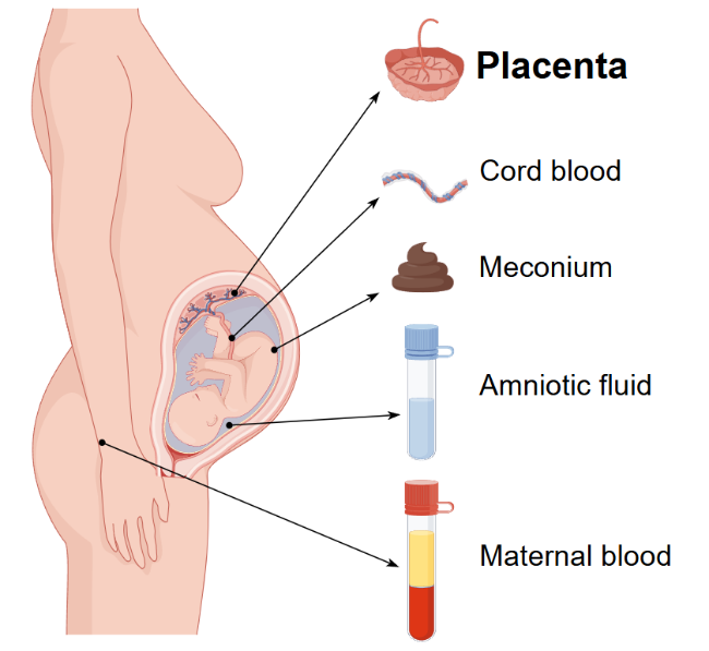
2 Exposure of placentas to PFAS

2.1 Exposure concentration and distribution of human placentas to PFAS

2.2 Factors influence PFAS retention and transport in the Placenta

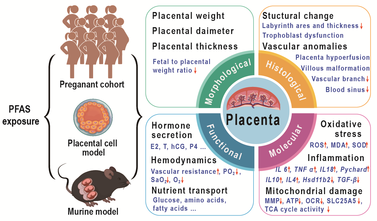
3 The effect of PFAS on placental structure and function

3.1 Effect on placental morphological

3.2 Effect on placental histological structure

3.3 Effect on placental vascular

3.4 Effect on nutrient metabolism and transport

3.5 Effect on placental endocrine

3.6 Molecular mechanisms of placental dysfunction

4 Conclusion and outlook

Cite this article

Feng Quan , Chuanzi Gao , Wenhui Qiu , Yi Zheng . The Impact of Prenatal Perfluorinated and Polyfluoroalkyl Substances Exposure on Placental Structure and Function[J]. Progress in Chemistry, 2025 , 37(11) : 1719 -1730 . DOI: 10.7536/PC20250610

1 Introduction

Per- and polyfluoroalkyl substances (PFAS), as persistent organic pollutants, have attracted global attention due to their widespread applications and environmental persistence. Among them, perfluorooctane sulfonate (PFOS), perfluorooctanoic acid (PFOA), perfluorohexane sulfonate (PFHxS), and related substances have been listed under the Stockholm Convention. PFAS are widely present in various environmental media and can enter the human body through dietary intake[1],daily drinking water[2],skin contact[3],and inhalation[4],where they accumulate in the body, disrupt the endocrine system, and thereby affect human health. The U.S. Agency for Toxic Substances and Disease Registry (ATSDR)[5]has released a report on the toxicological hazards of PFAS, indicating that PFAS exposure is associated with adverse health outcomes such as liver damage, thyroid disease, reduced fertility, obesity, asthma, hormonal suppression, endocrine disruption, and testicular and kidney cancers.
As understanding of the health impacts of PFAS deepens, their potential hazards to maternal and infant health are receiving increasing attention. During pregnancy, the mother’s hormonal signaling pathways are highly active to support normal fetal development and ensure a smooth pregnancy. In addition, to meet the nutritional needs of both the pregnant woman and the fetus, pregnant women tend to prefer high-protein foods such as eggs, fish, milk, shrimp, and chicken, which have relatively high PFAS levels[6],potentially increasing the level of exposure for pregnant women. Once PFAS enter the maternal body, they can interfere with normal hormone secretion through various complex mechanisms. Studies have found that PFAS exposure may affect the levels of key hormones such as estrogen and progesterone[7-9],thereby disrupting pregnancy-related physiological processes. These disruptive effects not only may increase the risk of pregnancy complications such as gestational diabetes, hypertension, and preterm birth, but also may pose a threat to fetal growth and development, including low birth weight and developmental delays[10-12].
As a key organ at the maternal-fetal interface, the placenta not only performs vital functions in substance exchange and endocrine regulation but also serves as a natural barrier protecting the fetus from harmful external substances. However, PFAS can cross the placental barrier, accumulate in placental tissues, and potentially disrupt normal placental physiological functions, thereby affecting fetal growth and development[13-15]. Studies have shown that PFAS exposure is associated with abnormal transport of certain key nutrients in the placenta[16-18], disrupts endocrine system homeostasis[19,20], and may increase the risk of adverse birth outcomes such as miscarriage, preterm birth, and low birth weight through multiple mechanisms, including inducing placental oxidative stress[21], inflammatory responses[22], and mitochondrial damage[23].
Given the severity and complexity of the potential harms posed by PFAS exposure during pregnancy to placental development and maternal–infant health, this review aims to systematically synthesize the literature on epidemiological studies, placental cell models, and animal exposure models to comprehensively assess the impact of PFAS exposure on the placenta. This study summarizes global PFAS exposure levels in the placenta, examines their effects on placental morphology, structure, and function, explores the possible mechanisms underlying these effects, and outlines future research directions, with the goal of providing a scientific basis and reference for mitigating the adverse health impacts of PFAS on mothers and infants.

2 PFAS Exposure in the Placenta

2.1 Exposure levels in the placenta of the general population

In recent years, the issue of prenatal PFAS exposure has garnered increasing attention. A large number of studies have focused on intrauterine PFAS exposure, with the primary biological matrices studied including maternal blood[24,25],umbilical cord blood[26,27],placenta[14,15,28-41],amniotic fluid[42,43],and meconium[40](Figure 1). As an important organ for substance exchange between the mother and fetus, and as a barrier to environmental pollutants, the placenta has become a key research subject for studying PFAS exposure levels and the mechanisms of transfer between mothers and infants.
图1 PFAS宫内暴露的生物基质

Fig.1 The biological matrices for PFAS exposure in utero

By conducting a systematic analysis of existing literature, this study summarizes the levels of PFAS exposure in placental tissues from populations worldwide, as shown in Table 1.Existing research data indicate that the five PFAS with the highest detection rates in the placenta are PFOA, PFOS, perfluorononanoic acid (PFNA), PFHxS, and perfluorodecanoic acid (PFDA). Among these, PFOA and PFOS have the highest detection rates, and their exposure concentrations dominate. The samples primarily originate from three regions: China, the United States, and Europe, with China exhibiting relatively high exposure levels. The sample sizes reported in these studies are relatively limited (ranging from 20 to 302), and the PFAS exposure concentrations vary between 0.3 and 33.8 ng/g (median or mean). Notably, the PFAS concentration in placentas from Fuxin, China (33.8 ng/g) is significantly higher than in other regions, primarily because the placental samples in this study were collected from individuals exposed near local fluorine chemical plants[35],suggesting that differences in concentration among samples from different regions are related to local industrial activities and environmental exposure levels.
表1 各国的人群胎盘中的PFAS暴露浓度 (ng/g)

Table 1 The concentration of PFAS (ng/g) in human placentas across different countries

Country Region Year Sample size Statistic PFOA PFOS PFNA PFHxS PFDA Ref
China Tianjing 2010 29 Median 1.41 7.32 0.96 0.36 0.67 15
China Fuxin 2020 50 Median 21.00 2.50 8.40 0.06 1.80 33
China Guangzhou 2021 302 Median 4.57 1.81 0.14 <MDL 0.17 36
China Wuhan 2015-2016 32 Median 0.46 2.42 / 0.21 / 28
China Henan 2016 54 Median 0.18 0.35 0.06 0.08 <MDL 32
China Henan 2019-2020 48 Median 0.57 0.77 0.09 <MDL 0.07 40
China Hebei 2013-2014 180 Median 1.33 1.21 0.23 0.05 0.21 39
Unite States North Carolina 2010-2011 120 Median 0.27 0.95 0.11 <MDL 0.06 34
Unite States North Carolina 2015-2018 122 Median 0.11 0.88 0.08 0.06 0.05 31
Unite States Arkansas 2010-2014 151 Median 0.07 0.43 0.05 0.05 0.04 41
Denmark Denmark / 34 Mean 0.23 1.30 0.14 <MDL 0.10 29
Sweden Sweden / 38 Mean 0.41 1.40 0.18 <MDL 0.21 30
Austria Austria 2017-2019 135 Median 0.22 0.34 0.06 0.03 0.03 37
Spain Granada / 20 Median 0.14 0.6 0.06 0.07 0.03 44
Israel Be'er Sheva 2018-2021 50 Median 0.08 0.26 0.04 0.06 0.02 45
Italy Reggio Emilia 2021-2022 20 Median 0.08 0.2 / 0.03 / 38

MDL is defined as method detection limit

Zhang et al.[15]were the first to investigate PFAS exposure concentrations in placental samples in 2013. Since then, numerous research teams worldwide have systematically examined the characteristics of PFAS exposure in the placenta from different regions, populations, and research methodologies. Mamsen et al.[29,30]analyzed placental, fetal organ, and blood samples obtained from legal abortions in Denmark and found that PFAS concentrations in the placenta exhibit a clear time-dependent cumulative effect. The highest concentration was observed for PFOS (1.3 ng/g), followed by PFOA, PFNA, PFDA, and perfluoroundecanoic acid (PFUnDA), with all PFAS concentrations increasing with gestational age. This finding is corroborated by subsequent studies; Li et al.[46]found that the efficiency of PFAS transport across the placenta in women with preterm delivery was significantly lower than in women with full-term delivery, possibly due to two factors: first, the longer duration of full-term pregnancy leads to greater PFAS accumulation on the fetal side; second, the barrier function of the placenta gradually weakens in late pregnancy. A study conducted in central North Carolina, USA, showed that the detection rates of PFNA, PFOS, PFOA, and PFDA in the placenta ranged from 96% to 100%, and that the concentrations of PFOA, PFNA, and PFDA in the placentas of primiparous women were significantly higher than those in multiparous women[34]. Notably, another study conducted in this region between 2015 and 2018 found that the concentrations of PFOS, PFHxS, and PFUnDA in the placenta showed a year-on-year decreasing trend, but only the reduction in PFHxS was statistically significant[31]. This change may reflect the effectiveness of local environmental controls, but it is also necessary to consider the impact of sample variability and population differences. In contrast, two studies conducted in Henan Province, China[32,40]indicated that the PFAS concentrations in placentas collected in 2019–2020 were higher than those collected in 2016. The PFAS concentrations in placentas collected in 2019–2020 were higher than those collected in 2016.
Existing research indicates that the distribution of PFAS in the placenta may be influenced by hemodynamic characteristics (including blood flow, flow velocity, and perfusion pressure) and the physicochemical properties of PFAS. In terms of hemodynamics, the unique structural features of the placenta significantly affect the distribution patterns of PFAS. Two studies[36,38]have found through comparative analysis that there are significant differences in the distribution of different PFAS monomers between the decidual tissue (maternal side) and the villous tissue (fetal side) of the placenta. Specifically, long-chain PFAS (such as PFOA, PFOS, PFUnDA, and 6∶2 Cl-PFESA) exhibit significantly higher concentrations on the fetal side than on the maternal side, whereas short-chain PFAS (including PFHxA, PFHxS, PFBS, and 6∶2 FTS) display the opposite distribution pattern[36,38]. This differential distribution may be attributed to the spongy villous tree structure on the fetal side, which has a larger surface area, combined with low-flow, low-pressure blood flow characteristics that facilitate the absorption and accumulation of PFAS in the placenta. In terms of physicochemical properties, the pK aand LogPvalues of PFAS are also closely related to their placental distribution characteristics. PFAS with higher pK avalues (PFOS, PFOA, PFUnDA) and higher LogPvalues (PFOS, PFOA) tend to accumulate more readily in the villous tissue[38]. This suggests that different PFAS monomers exhibit differentiated distribution patterns at the materno-fetal interface due to their unique physicochemical properties.

2.2 Factors Affecting Placental Transport of PFAS

PFAS primarily crosses the placental barrier through two mechanisms: passive diffusion and active transport. The efficiency of this transport is closely linked to multiple factors, including the mother’s exposure levels, the physical properties of PFAS, and their protein-binding capacity.
The passive diffusion mechanism of PFAS primarily relies on the transmembrane transport of free-state molecules, a process that is significantly influenced by their binding properties to plasma proteins. The structure of PFAS is similar to that of fatty acids, and approximately 90% of PFAS exist in a bound state with human serum albumin (HSA)[47]. This binding behavior directly determines the concentration levels of free PFAS. Molecular docking results indicate that the binding affinity between PFAS and HSA exhibits a clear dependence on carbon chain length: with the exception of PFUnDA, the binding affinities of perfluorocarboxylic acids (PFCAs), perfluorosulfonic acids (PFSAs), F-53B, and fluorotelomer sulfonates (FTSAs) with HSA all increase as the carbon chain length grows, with perfluorododecanoic acid (PFDoDA) serving as the turning point for the binding affinity between PFCAs and HSA[32]. When the carbon chain length of PFAS exceeds a certain critical length, the molecule may need to bend in order to fit into the binding site on the HSA protein; however, this bending leads to increased torsional energy and enhanced electrostatic repulsion[32]. Secondly, structural differences among PFAS also affect their binding characteristics with HSA: linear PFOA and PFOS exhibit stronger binding affinities with HSA than their branched isomers, which helps explain why branched PFAS are more readily transported across the placenta[48]. Finally, different PFAS monomers may display distinct binding patterns. The binding affinity of 6∶2 Cl-PFESA with HSA is higher than that of PFOS; the former preferentially binds to the Sudlow I site via its chlorine and oxygen atoms, whereas the optimal binding site for the latter is the Sudlow II site[49]. This specificity in binding differences may be an important molecular basis for the differing efficiencies of placental transport between the two compounds.
During active transport, placenta-specific transport proteins play a crucial role. Among them, organic anion transporter 4 (OAT4), as a key transporter, exhibits a significant negative correlation between its expression level and the efficiency of PFAS placental transfer[28,50]. The transport capacity of OAT4 for PFAS is also related to carbon chain length. The binding affinity of PFAS to OAT4 increases with the chain length of PFCAs, Cl-PFESAs, and FTSAs, while the binding affinity of PFSAs (except PFHpS and PFNS) to OAT4 also increases as the chain length increases[32]. The study by Chen et al.[14]further confirms that 8∶2 Cl-PFESA is more readily transported across the placenta than 6∶2 Cl-PFESA, a phenomenon associated with its higher hydrophobicity and lower plasma protein binding capacity. In addition to OAT4, molecular docking studies have identified MRP1, OATP2B1, ASCT1, and P-gp as key transport proteins involved in the placental transfer of PFAS[40]. Li et al.[46]conducted a systematic analysis of the relationship between the gene expression of nine transport proteins located on the maternal side of the placenta and placental transport efficiency, and found that the expression level of the MDR1 gene is significantly positively correlated with the transport efficiency of several long-chain PFAS (including PFNA, PFDA, PFUnDA, etc.); MRP2 shows a significant positive correlation with PFNA, PFDA, and various PFOS isomers; and BCRP primarily regulates the transplacental transport of PFNA and PFTrDA. These findings collectively reveal the complex regulatory mechanisms underlying the active transport of PFAS, providing a molecular-level explanation for understanding the differences in placental transfer among various PFAS monomers.

3 The Impact of PFAS on Placental Structure and Function

3.1 Impact on Placental Morphology

Placental morphological parameters are important indicators for assessing placental function, including placental weight, placental diameter, placental thickness, and the fetal-placental weight ratio (FPR). Clinical studies have shown that placental macrosomia (excessive placental growth) is associated with adverse outcomes such as low Apgar scores and respiratory distress, while placental hypoplasia is linked to an increased risk of fetal malformations[5]. Regarding the impact of PFAS exposure on placental morphology, existing epidemiological studies show significant heterogeneity in their findings. Table 2summarizes current epidemiological study results on the association between PFAS exposure and placental weight. A large Danish birth cohort study found no association between PFOA or PFOS and placental weight[51]. In contrast, a birth cohort study in Ma'anshan, China, observed a positive correlation between br-PFHxS and 6∶2 Cl-PFESA and placental weight, and found that ∑PFAS (particularly in mid- and late pregnancy) can significantly increase placental weight[52]. Conversely, the UPSIDE cohort study in the United States showed a significant negative correlation between PFNA and placental weight, and positive correlations between PFNA and PFDA with FPR, suggesting that PFAS exposure may reduce placental efficiency[53]. Although the French SEPAGES cohort did not find any association between individual PFAS compounds and placental weight or FPR, cluster analysis revealed that pregnant women in the medium- and high-concentration PFAS exposure groups had an average placental weight 30 g lower than that of the control group[54]. These differences may stem from variations in study population characteristics, methods for assessing exposure levels, and strategies for controlling confounding factors, highlighting the need for standardized, large-scale research.
表2 关于PFAS与胎盘重量关联的流行病学研究

Table 2 Epidemiologic studies on the associations between PFAS and placental weight

Author, Year Country Sample size Results of placenta weight Ref
Fei et al., 2008 Denmark
1996-2002
1400 Maternal PFOA and PFOS levels did not have any statistically significant effect on placental weight. 51
Gan et al., 2024 China
2013-2014
712 Positive associations found between br-PFHxS, 6∶2 Cl-PFESA, and placental weight.
Mixed PFAS exposure (especially in 2nd/3rd trimesters) was positively linked to placental weight and scaling exponent (indicating reduced placental efficiency).
52
Chowdhury et al., 2024 USA
2015-2019
158 PFNA was inversely associated with placental weight (β=-0.17, 95% CI: -0.33, -0.019).
PFDA showed a positive association with FPR.
PFOA was negatively linked to placental arterial vascularization (mean distance from arterial end point to perimeter), but no direct effect on birth weight was observed.
53
Khan et al., 2025 France
2014-2017
340 Moderate-to-higher PFAS exposure cluster was associated with lower placental weight (-30 g, 95% CI: -56, -4.3), but no individual PFAS was directly associated with placental weight or FPR. 54
Animal experimental studies have provided important evidence for understanding the impact of PFAS exposure on placental morphology, but the results also exhibit significant heterogeneity. Table 3summarizes the results related to placental weight from exposure experiments in rodent models. Suh et al.[55]found that PFOA exposure during pregnancy significantly reduced placental weight, whereas Blake et al.[56]observed the opposite result, possibly due to differences in dose and exposure window, which lead to different mechanisms by which PFOA affects placental weight. PFOS exposure, in contrast, consistently reduced placental weight and led to a decrease in placental diameter[57,58]. Exposure to PFHxS[59]resulted in a significant reduction in placental weight, but no significant changes in placental diameter were observed. In addition, as traditional PFAS are increasingly subject to regulatory control, the use of novel PFAS is becoming more widespread, and their impact on placental weight has attracted considerable attention. Adams et al.[18]found through comparison that exposure to fluorotelomer ethoxylates (FTEOs) has a greater impact on FPR than PFOA. Moreover, hexafluoropropylene oxide dimer acids (GenX)[56], PFDMO2HpA, and PFDMO2OA[60], among other novel PFAS, can cause placental enlargement. These differences may stem from the diverse molecular structures and mechanisms of action of different PFAS monomers, and they also reflect the complexity of the placenta's response to PFAS exposure.
表3 关于PFAS与胎盘重量关联的啮齿类动物模型研究

Table 3 Rodent models on the associations between PFAS and placental weight

Author, Year Animal species Exposure doses Duration of Exposure Results of placenta weight Ref
Suh et al., 2011 CD-1 Mice PFOA:0, 2, 10 and 25 mg/kg/day GD 11 to 16 Significantly reduced with increasing PFOA dose. 55
Lee et al., 2015 CD-1 Mice PFOS:0, 0.5, 2 and 8 mg/kg/day GD 11 to 16 Mean palcenntal weight was significantly reduced in all the treated groups and this reduction was seen in proportion to the PFOS dosage 58
Li et al., 2016 Sprague-Dawley (SD) rats PFOS: 0, 5 and 20 mg/kg/day GD 12 to 18 Significant reduction for the group treated with 20 mg PFOS 57
Blake et al., 2020 CD-1 Mice PFOA: 1 or 5 mg/kg/day; GenX: 2 or
10 mg/kg/day
GD 1.5 to 11.5 or 17.5 GD11.5: No significant difference.
GD17.5: placental weight and fetus-placenta weight ratioswas significantly higher in 5 mg/kg/d PFOA or
10 mg/kg/d GenX treated groups.
56
Zhang et al., 2023 ICR mice PFHxS: 0, 0.03, and 0.3 µg/kg/day GD 7 to 17 Placental weight significanty lower in high dose group 59
Adams et al., 2024 CD-1 PFOA: 0, 5, 100 ng/L; FTEOs: 0, 5,
100 ng/L (drinking water)
GD 0.5 to 17.5 With a notable increase in placental weights in the 100 ng/L FTEOs group (13% for females, 15% for males). FTEOs exposure causing a larger decrease in the fetus-placental weight ratio compared to PFOA exposure. 18
Zhu et al., 2024 CD-1 PFDMO2HpA :0.01, 0.04, 0.16, 0.63, 2.5 mg/kg/day; PFDMO2OA 0.04, 0.16, 0.63, 2.5, 10 mg/kg/day GD 2 to 12 or 18 Gestational exposure to PFDMO2HpA/PFDMO2OA induced placenta enlargement, decreased fetus weight and placental efficiency 60

3.2 Impact on Placental Tissue Structure

Animal studies have revealed that PFAS exposure has a significant impact on placental tissue structure, with effects that are clearly dose-dependent and compound-specific. Different PFAS monomers can induce characteristic placental pathological changes, such as destruction of trophoblast structures, tissue edema, and abnormal cellular composition[56,58,59,61]. At the cellular level, PFAS exposure leads to a significant reduction in the number of glycogen trophoblast cells (GlyT) in the junctional zone and sinusoidal trophoblast giant cells (S-TGC) in the labyrinth zone, accompanied by morphological abnormalities such as nuclear shrinkage[55,58]. A study by Blake et al.[56]found that in the PFOA (5 mg/kg/d) and GenX (10 mg/kg/d) exposure groups, the incidence of placental abnormalities was 2–3 times higher than in the control group, with distinct damage patterns for each compound. PFOA primarily causes vasodilation and red blood cell stasis in the placental labyrinth, disrupting maternal-placental-fetal exchange through microcirculatory impairment, whereas GenX leads to trophoblast cell atrophy and early fibrin clot formation, compromising placental structural integrity through cytotoxicity and procoagulant effects[56]. At the molecular level, PFOA can induce placental cell apoptosis by upregulating the expression of the pro-apoptotic proteins Bax and cleaved-caspase 3, leading to rupture of the placental nuclear membrane, karyopyknosis, and chromatin condensation[62]. PFOS, on the other hand, causes abnormal deposition of matrix collagen in the placental labyrinth layer[61]. F-53B exposure results in marked inflammatory cell infiltration in the placenta, an increase in syncytiotrophoblast nodules, and loosening of the trophoblast basement membrane[22]. GenX exposure leads to dose-dependent neutrophil infiltration and cellular degeneration in the decidual zone of the placenta, while the labyrinth zone exhibits marked congestion[63].

3.3 Impact on Placental Vessels and Function

The placental vascular system is the structural foundation for placental development and functional maintenance, serving as the critical conduit for inter-fetal substance exchange. Originating from the fetal vessels within the umbilical cord, the placental vascular system undergoes fine differentiation to form a villous tree-like capillary network, providing the fetus with an adequate supply of oxygen and nutrients[64].Normal development of placental vessels is crucial for maintaining placental function; any structural or functional abnormalities can directly affect the fetus's oxygen supply and nutrient uptake, thereby leading to adverse pregnancy outcomes.
Existing epidemiological studies have revealed a close association between PFAS exposure and abnormalities in placental structure and function. Chowdhury et al.[53]used human 2D placental images combined with shape-matching algorithms to quantify placental vascular features. They found that prenatal PFOA exposure was negatively correlated with the average distance from the arterial termini to the periphery of the placenta, suggesting a possible reduction in the degree of placental surface vascularization. PFNA was negatively correlated with total arterial arc length, and PFDA was negatively correlated with maximum arterial tortuosity. These findings indicate that PFAS exposure can disrupt the structural organization of the placental vascular network, thereby reducing the efficiency of fetal–maternal substance exchange. A French SEPAGES cohort study found that perfluorohexyl phosphonic acid (PFHxPA) and perfluoroheptanoic acid (PFHpA) were associated with a reduction in chorionic villous space, while exposure to PFHxPA and PFTrDA was associated with an increased percentage of syncytial knots in placental villi. 6∶2 diPAP was found to reduce villous capillary density by 17%, indicating that PFAS exposure impairs maternal–fetal blood flow exchange, leading to insufficient placental perfusion and fetal hypoxia[54]. At the molecular level, PFAS exposure leads to a significant upregulation of placental vascular endothelial growth factor (VEGF), angiopoietin-2 (ANG2), and their receptors (VEGFR2, TIE2). PFNA exposure increases VEGFlevels by 32.5%[36]. Notably, placental vascular biomarkers may mediate the association between PFAS exposure and adverse birth outcomes. Studies have shown that placental VEGFR2and ANG2mediate the effects of PFOS and 8∶2 Cl-PFESA on low birth weight, accounting for 9.5%–32.5% of the total effect[36]. In addition, research indicates that the effects of PFAS on the placenta differ by sex. In the Ma’anshan cohort, female fetuses were found to be more sensitive to PFAS-induced vascular damage, possibly due to sex-specific differences in the expression of X-chromosome–linked genes in the placenta[52].
Epidemiological studies have confirmed that PFAS exposure during pregnancy disrupts placental hemodynamic balance, primarily manifesting as increased vascular resistance and impaired oxygen exchange function. In the Spanish BiSC cohort, exposure to long-chain PFAS such as PFUnDA, PFTrDA, and PFDoDA was significantly associated with elevated pulsatility indices in the uterine and umbilical arteries, indicating that increased placental vascular resistance may lead to insufficient fetal blood perfusion[65]. Furthermore, PFBS exposure was associated with an increased cerebroplacental ratio, a phenomenon that reflects the fetus' activation of compensatory mechanisms in response to abnormal placental blood flow: by reducing peripheral vascular resistance, the fetus prioritizes cerebral blood supply. However, this compensatory regulation may come at the expense of nutrient supply to other organs[65]. Ji et al.[36]further found in a Chinese population study that exposure to PFOA, PFOS, and 6∶2 Cl-PFESA was significantly associated with reduced partial pressure of oxygen (PO2) and oxygen saturation (SaO2) in umbilical cord blood; for example, PFOA exposure was associated with a 6.49% decrease in SaO2, indicating that PFAS exposure can impair placental gas exchange function and potentially trigger fetal hypoxia. Collectively, these findings suggest that by disrupting placental hemodynamic parameters and gas exchange efficiency, PFAS may exert profound effects on fetal growth and development.
In vitro experimental studies have provided crucial evidence for elucidating the molecular mechanisms by which PFAS disrupt placental angiogenesis. Multiple studies using different placental cell models have demonstrated that PFAS can interfere with the expression and function of key angiogenic factors through multiple pathways. Pham et al.[66]using the human choriocarcinoma cell line (BeWo placental cell model) found that PFOS exposure significantly reduced the gene expression of placental growth factor (PlGF). As a key factor in placental angiogenesis, the downregulation of PlGFexpression may directly lead to insufficient angiogenesis, thereby triggering placental vascular abnormalities. Further studies using the human placental trophoblast cell line (HTR-8/Svneo cell model) and the human choriocarcinoma cell line (JEG-3 cell model) revealed that PFOS inhibits the PPARγ signaling pathway, thereby downregulating the gene expression of angiogenic factors VEGFAand ANGPTL4, as well as matrix metalloproteinases MMP-2and MMP-9. This, in turn, suppresses the migratory and angiogenic capabilities of placental trophoblast cells, resulting in inadequate placental vascularization[67]. Zhao et al.[61]further confirmed in JEG-3 cells that PFOS exposure impairs cell migration, invasion, and angiogenic capacity. In addition, Forsthuber et al.[68]used a 3D co-culture angiogenesis model involving human umbilical vein endothelial cells (HUVECs) and normal colonic fibroblasts (NCFs) and found that PFOS exposure led to a dose-dependent reduction in the number of vessel tips formed, branch density, tip extension length, and total area of the vascular network. Moreover, VEGFR2signaling was significantly inhibited, resulting in decreased endothelial cell metabolic activity and abnormal vascular morphogenesis.
Multiple mouse exposure models have confirmed that PFOS exposure can cause significant structural abnormalities in the labyrinthine layer of the mouse placenta, characterized by a marked reduction in vascular branching and a decrease in sinusoidal area[59,62,69]. PFOA, on the other hand, disrupts placental development by affecting the number of uterine natural killer (uNK) cells; studies show that its exposure can significantly reduce the number of uNK cells in the decidual region, where these cells play a crucial role in placental vascular remodeling and trophoblast invasion[62]. Subchronic GenX exposure, meanwhile, leads to vascular disorganization in the placenta, with discontinuous signaling between maternal and fetal vascular networks, a loose structure of the trophoblastic layer, and swelling and vacuolization of mitochondria in fetal endothelial cells. At the same time, placental expression of the VEGFA and ANGPT1genes is upregulated, while expression of the endothelial transmembrane receptor TIE2is downregulated[70].

3.4 Impact on the metabolic and transport functions of nutrients

The placenta is the primary conduit through which the fetus obtains oxygen and nutrients, primarily utilizing two major mechanisms—passive diffusion and active transport—to transfer essential nutrients such as oxygen, glucose, and amino acids from the maternal circulation to the fetus[71].In this process, passive diffusion is mainly responsible for the transmembrane transport of oxygen and small-molecule nutrients, while active transport is involved in the transport of specific nutrients against their concentration gradient. In addition, auxiliary mechanisms such as facilitated diffusion and endocytosis also play a role in regulating the placental transport of substances. PFAS exposure can disrupt placental metabolism and transport of nutrients through multiple mechanisms, and the mechanisms may differ among various PFAS monomers.
In vitro experimental studies have revealed the molecular mechanisms by which PFAS disrupt nutrient metabolism and transport in placental cells. In the JEG-3 cell model, PFAS exposure interferes with the synthesis and metabolism of cell membrane lipids, thereby affecting lipid homeostasis in placental cells and significantly altering their lipid composition, leading to a marked increase in intracellular phosphatidylcholine, ether phosphatidylcholine, and lysophosphatidylcholine[72,73]. This alteration in lipid composition may directly affect the structural integrity and functional properties of the cell membrane. In the human placental trophoblast cell line (CTB cell model), PFOA exposure disrupts lipid metabolism balance through a dual regulatory mechanism: on the one hand, it upregulates genes involved in fatty acid metabolism, promoting mitochondrial fatty acid oxidation; on the other hand, it downregulates genes involved in phospholipid metabolism, compromising membrane structural integrity[74]. This metabolic disruption not only leads to an imbalance in intracellular lipid homeostasis but may also reduce transmembrane transport efficiency by affecting membrane fluidity, thereby interfering with the placenta's normal substance transport function.
In a pregnant rat model, PFOS exposure significantly reduced the placental transport efficiency of glucose analogs (MeG) and amino acid analogs (MeAIB), accompanied by a marked decrease in the protein expression level of the amino acid transporter SNAT4[17]. Human-relevant doses of PFHxS exposure lead to a significant reduction in the levels of 29 amino acids and 44 amides in the placenta by downregulating amino acid transporter gene expression and inducing alternative splicing that results in abnormal protein structures, thereby causing fetal growth restriction[59]. Another study by this team focused on the effects of PFHxS exposure on lipid metabolism, revealing that PFHxS exposure increases total placental lipid content, causes abnormalities in phospholipid and triglyceride metabolic pathways, and reduces cholesterol ester levels. It may also disrupt transmembrane transport of fatty acids in the placenta by interfering with the functions of fatty acid-binding protein 2 (FABP2) and transporter CD36, thereby affecting the fetal lipid supply[16]. Adams et al.[18]found that FTEOs have a more pronounced impact on placental nutrient transport than PFOA and elucidated the specific mechanisms of action of these two PFAS. PFOA exposure primarily affects placental nutrient transport indirectly by influencing gluconeogenesis and amino acid metabolism, resulting in a significant increase in the relative concentrations of glucose and threonine in the placenta and a decrease in creatine concentration; in contrast, FTEOs exposure mainly disrupts nutrient transport by interfering with biotin and amino acid metabolism, leading to a decrease in the relative concentrations of asparagine and lysine and an increase in creatine concentration.

3.5 Impact on Placental Endocrine Function

The placenta can secrete various hormones that regulate maternal metabolism, thereby influencing maternal nutrient supply and regulating the course of pregnancy. The main hormones secreted by the placenta include human chorionic gonadotropin (hCG), estrogen (E2), progesterone (P4), and human placental lactogen (hPL), among others[71].These hormones play crucial roles in establishing and maintaining pregnancy, such as regulating endometrial decidualization, placental development, angiogenesis, endometrial receptivity, embryo implantation, immune tolerance, and fetal development[75,76].In addition, placental hormones also participate in regulating maternal metabolic activities to meet the demands of pregnancy and promote fetal growth and development. Steroid hormones are key endocrine factors that play an important role in maintaining normal fetal development and promoting fetal growth. Figure 2summarizes the main research findings on the effects of PFAS exposure on steroid hormones and their synthesizing enzymes in the placenta.
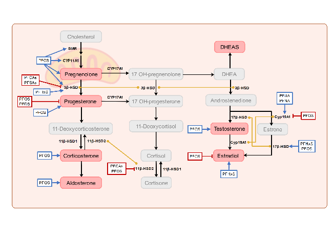
图2 关于PFAS暴露对类固醇激素及其合成酶影响的主要研究结果总结,带箭头的蓝色方框表示相关激素水平上升或上调合成酶,带短横线的红色方框表示下降或下调,黄色连接线表示调控同一基因,黑色的文字代表已有研究验证的潜在机制,灰色字体代表尚未研究的潜在机制

Fig.2 Summary of major findings on the effects of PFAS exposure on steroid hormones and steroidogenic enzymes. Blue boxes with arrows indicate increased levels of the corresponding hormone or enzyme, red boxes with lines indicate decreased levels, yellow connecting lines indicate the regulation of the same gene. Black text represents mechanisms that have been validated in previous studies, while gray text indicates potential mechanisms that have not yet been investigated

Epidemiological studies indicate that PFAS exposure affects the endocrine function of the placenta. Yao et al.[77]found, by measuring placental steroidogenic enzymes (including cytochrome P450 aromatase (CYP19A1), 3β-hydroxysteroid dehydrogenase 1 (3β-HSD1), and 17β-hydroxysteroid dehydrogenase 1 (17β-HSD1)) that PFUA and PFNA are positively correlated with the concentration levels of CYP19A1 in the placenta, and PFHxS is positively correlated with 3β-HSD1 and 17β-HSD1. Mediation analysis further shows that 17β-HSD1 mediates the positive correlation between PFHxS and E2, with the indirect effect accounting for 25% of the total effect. This suggests that PFAS may influence hormone synthesis pathways by regulating the expression levels of placental steroidogenic enzymes. In addition, the concentration of PFAS exposure affects the secretion of human chorionic gonadotropin (hCG) and its subtypes in the placenta. Specifically, PFHxS is positively correlated with the free β-subunit (hCGb), PFDA is negatively correlated with hCG, and the sum of PFAS is negatively correlated with the free α-subunit (hCGa) and positively correlated with hCGb[78]. This indicates that PFAS exert differentiated regulatory effects on hCG subunits.
In vitro cell exposure studies have shown that PFAS significantly inhibit the secretory function of placental cells, and this inhibitory effect is closely related to the carbon chain length of PFAS. PFOS can significantly inhibit the secretion of hCG, P4, and E2 by syncytiotrophoblast cells, and this inhibition is concentration-dependent. It affects the secretory function of placental cells by reducing cell viability and inducing apoptosis[79]. A human placental microsomal protein model found that C9-C14 PFCAs significantly inhibit the production of 3β-HSD1, an enzyme in the placenta responsible for catalyzing the conversion of pregnenolone to progesterone[80]. Further findings indicate that PFOS and PFDS significantly inhibit progesterone synthesis in JEG-3 placental cells. Docking analyses reveal that PFAS interact with the steroid-binding site of 3β-HSD1, with their inhibitory activity exhibiting a V-shaped pattern centered on PFOS (C8)[81]. Similarly, Zhao et al.[82]found that PFAS inhibit the activity of 11β-hydroxysteroid dehydrogenase 2 (11β-HSD2) in BeWo cells, with inhibitory activity also showing a V-shaped pattern centered on PFDA (C10). Among them, PFDA and PFOS exhibit the strongest inhibitory effects. PFAS may bind to the steroid-binding site of 11β-HSD2by forming hydrogen bonds with the catalytic residue tyrosine 232 (Tyr232), thereby affecting its activity. These research findings suggest that PFAS can influence the synthesis and secretion of endocrine hormones in placental cells by interacting with the binding sites of key enzymes.
Animal studies have confirmed that PFAS exposure can disrupt placental endocrine homeostasis through multiple pathways. In pregnant rat exposure models, exposure to PFOA and PFOS dose-dependently suppresses the expression of genes encoding placental prolactin-family hormones (including 7 subtypes such as mPL-II, mPLP-Cα) [55,58]. In terms of steroid hormone regulation, PFOS exposure inhibits the activity of 11β-HSD2, leading to elevated cortisone levels in both the placenta and the fetus [17,57]. C10–C12 PFCAs, on the other hand, exhibit a "U-shaped" dose-response effect on 3β-HSD4activity (with PFUnDA serving as the inflection point), thereby inhibiting progesterone synthesis [80]. PFOS exerts bidirectional regulatory effects on placental endocrine function: by upregulating genes involved in placental steroid synthesis (such as Cyp11A1, 3β-HSD1, StAR, and 17β-HSD1/3), it increases levels of progesterone, aldosterone, and testosterone; at the same time, it suppresses the expression of the Cyp19A1gene, reducing estradiol synthesis [83]; furthermore, it also lowers hCG and prolactin levels, thereby affecting placental trophoblast function and the maintenance of pregnancy.

3.6 Molecular Mechanisms of Placental Dysfunction

Existing research primarily uses placental cell models and animal exposure models to reveal the mechanisms by which PFAS exposure impairs placental function. Experimental evidence indicates that PFAS mainly impair placental function through three key pathways: induction of inflammatory responses, oxidative stress, and mitochondrial dysfunction.
Exposure experiments indicate that PFAS may interfere with placental function through inflammatory pathways. PFOS exposure leads to decreased levels of IL-6in HTR-8/Svneo cells and increased secretion levels of TNF-α and IL-10 [84]. F-53B activates the NF-κB signaling pathway by phosphorylating IκB-α, triggering activation of the NLRP3 inflammasome and resulting in upregulation of pro-inflammatory genes (IL 1b, IL 18, IL 2, IL 6, TNF α, IFNG) and anti-inflammatory genes (IL 4and IL 10), as well as the NLRP3 inflammasome gene (NLRP3, caspase1)[22]. PFOS exposure leads to upregulation of the pro-inflammatory gene Pycardand downregulation of the anti-inflammatory gene Hsd11b2in placental tissue[21]. Placental proteomics shows that GenX exposure activates Rap1 signaling, leading to changes in the expression levels of inflammation-related proteins in this pathway. Zhu et al.[60], using a CD-1 mouse model, found that exposure to HFPO-TA analogs (PFDMO2HpA and PFDMO2OA) significantly upregulates IL 6and TNF αand downregulates the anti-inflammatory factor TGF-β.
PFAS can impair placental function by disrupting the redox balance. HTR-8/SVneo cell experiments have shown that PFOS[85]and PFOA[86]both induce abnormal accumulation of reactive oxygen species (ROS). Animal studies further confirm that PFOS exposure significantly increases placental oxidative stress, as evidenced by elevated levels of malondialdehyde (MDA), a lipid peroxidation product, and reduced activity of the antioxidant enzyme superoxide dismutase (SOD)[21]. Molecular mechanism studies indicate that PFOS impairs placental antioxidant defenses by inhibiting the Nrf2 signaling pathway and reducing the expression of antioxidant enzymes such as catalase (CAT)[21].
Mitochondria, as the central organelles of cellular energy metabolism, play a crucial role in the generation of reactive oxygen species (ROS), the regulation of intracellular calcium ion (Ca²⁺) concentrations, and cell signaling pathways associated with cell proliferation, differentiation, and apoptosis[87]. However, PFAS exposure can damage mitochondria in the placenta, triggering a series of adverse effects, including a decline in mitochondrial membrane potential (MMP) and reduced ATP production. A JEG-3 cell model found that 45% of PFAS (42 compounds) can induce changes in MMP, with PFHxS exhibiting the strongest mitochondrial toxicity[23]. Hofmann et al.[88]found that PFOS exposure reduces mitochondrial content in trophoblast cells, impairs mitochondrial respiratory function, decreases the oxygen consumption rate (OCR), and inhibits the activity of mitochondrial electron transport chain complexes I, II, and III, thereby reducing ATP production. In addition, PFDA exposure inhibits the mitochondrial β-oxidation process, leading to reduced tricarboxylic acid cycle activity, which in turn decreases the production of NADH and NADPH and increases intracellular oxidative stress[89]. Animal models have shown that PFOS exposure leads to downregulation of SLC25A5 expression in the placenta, a decrease in MMP, and reduced ATP synthesis[61]. These findings indicate that PFAS-induced damage to placental mitochondria not only affects their energy metabolism but may also interfere with normal cellular physiological processes through multiple mechanisms, potentially exerting adverse effects on placental health and function. Di Credico et al.[90]investigated the effects of combined exposure to bisphenols (BPA and BPS) and PFAS (PFOA and PFOS) on human placental membranes—mesenchymal stem cells (hFM-MSCs)—and found that combined exposures (BPA+PFOS, BPA+PFOA, BPS+PFOS, BPS+PFOA) significantly reduced MMP in hFM-MSCs. However, neither single exposures nor four-substance mixtures affected MMP, suggesting that different combinations of these substances may specifically impair mitochondrial function through additive, synergistic, or antagonistic interactions. In future assessments of the potential risks of PFAS to placental health, it will be essential not only to consider the toxicological effects of individual PFAS compounds but also to examine the interactions between PFAS and other chemicals, in order to more comprehensively evaluate the potential hazards posed by complex mixtures in real-world environmental exposure scenarios on placental development and function.

4 Conclusion and Outlook

As a class of emerging environmental pollutants characterized by persistence and bioaccumulation, PFAS have raised significant research interest in the field of environmental health due to their potential impact on placental structure and function. Existing studies have demonstrated that PFAS can interfere with placental development and function through multiple pathways, including altering placental morphological characteristics, impairing vascular system homeostasis, disrupting endocrine balance, and inducing cellular dysfunction. These pathological changes may ultimately lead to adverse pregnancy outcomes such as fetal growth restriction and preterm birth. However, current research still has several limitations, and further in-depth investigations are needed to enhance our understanding of the mechanisms by which PFAS affect placental structure and function, thereby effectively safeguarding maternal and infant health.
In population-based studies, existing epidemiological investigations have certain limitations. First, these studies generally suffer from small sample sizes, and the geographic scope of research is largely confined to regions such as China, the United States, and Europe. Second, most detection and analysis efforts focus on traditional PFAS (such as PFOA and PFOS), with insufficient attention paid to increasingly widely used novel alternatives (such as GenX and F-53B). Third, current research primarily centers on analyzing placental concentrations and examining the impact on placental vasculature, with relatively limited exploration of underlying molecular mechanisms. To comprehensively and deeply investigate the long-term and far-reaching effects of PFAS exposure in the placenta on pregnant women and fetuses, future research must conduct large-scale, prospective cohort studies in more regions worldwide. This will enable the collection of more representative epidemiological data, enhance the detection and assessment of novel PFAS, and integrate emerging omics technologies (such as transcriptomics, proteomics, and metabolomics) to dissect, at the molecular level, the mechanisms by which PFAS exposure affects placental function. Such efforts will provide a scientific basis for developing more precise risk assessment models.
In in vitro cell exposure model studies, the primary cell lines currently used include human placental choriocarcinoma cell lines (JEG-3, BeWo) and human placental trophoblast cell lines (HTR-8/Svneo, CTBs), which struggle to fully reflect the specific responses of different placental cell types. Future research could conduct comparative analyses using multiple cell lines to gain a more comprehensive understanding of the effects of PFAS on placental cells. In addition, by integrating co-culture models, three-dimensional organoid models, and other in vitro models, it will be possible to more comprehensively assess the impact of PFAS on various placental cells and to delve deeper into their molecular mechanisms of action.
Currently, animal exposure models primarily focus on rodent exposure, with studies largely employing single-compound, high-dose exposure scenarios—patterns that starkly contrast with the real-world environmental conditions of low-dose, multi-component mixed exposures. Future animal exposure experiments should consider incorporating a broader range of animal models to enhance the extrapolatability and generalizability of research findings. At the same time, experimental designs should more closely reflect real-world environmental conditions by using environmentally relevant doses and multi-component mixed exposure scenarios to assess the combined toxicity of different compounds. In addition, attention should be given to the differential exposure effects associated with various exposure windows and factors such as gender: different exposure stages (early, mid, and late pregnancy) may exert distinct impacts on the placenta and fetus, while gender differences may lead to varying sensitivities to PFAS exposure. Finally, multi-omics analytical techniques should be integrated to comprehensively evaluate the impact of PFAS exposure on the placenta, thereby providing a scientific basis for developing effective risk assessment and management strategies.
In summary, future research needs to advance in a coordinated manner across multiple fronts, including population cohorts, in vivo animal exposure studies, and in vitro exposure models. By employing multi-omics approaches, researchers can explore the mechanisms by which PFAS exposure affects the placenta. Through multidimensional and multilevel research strategies, existing research gaps can be addressed, enabling the development of targeted interventions that provide robust scientific support for comprehensive protection of maternal and infant health.
[1]
Wang Y X, Gao X Y, Liu J Y, Lyu B, Li J G, Zhao Y F, Wu Y N. China CDC Week., 2022, 4(9): 168.

[2]
Gao C Z, Drage D S, Abdallah M A, Quan F, Zhang K, Hu S Y, Zhao X, University H, Zheng Y, Harrad S, Qiu W H. ACS ES&T Water, 2024, 4(11): 4881.

[3]
Ragnarsdóttir O, Abou-Elwafa Abdallah M, Harrad S. Environ. Int., 2024, 188: 108772.

[4]
Makey C M, Webster T F, Martin J W, Shoeib M, Harner T, Dix-Cooper L, Webster G M. Environ. Sci. Technol., 2017, 51(13): 7667.

[5]
Toxicological profile for perfluoroalkyls: United States. Agency for Toxic Substances and Disease Registry. 2021. [2025-06-01]http://dx.doi.org/10.15620/cdc:59198.

[6]
Surma M, Sznajder-Katarzyńska K, Wiczkowski W, Piskuła M, Zieliński H. Environ. Toxicol. Chem., 2023, 42(12): 2589.

[7]
Liu H X, Pan Y T, Jin S N, Li Y Y, Zhao L Q, Sun X J, Cui Q Q, Zhang B, Zheng T Z, Xia W, Zhou A F, Campana A M, Dai J Y, Xu S Q. Environ. Int., 2020, 140: 105636.

[8]
Liu H X, Pan Y T, Jin S N, Sun X J, Jiang Y Q, Wang Y Y, Ghassabian A, Li Y Y, Xia W, Cui Q Q, Zhang B, Zhou A F, Dai J Y, Xu S Q. J. Hazard. Mater., 2021, 407: 124378.

[9]
Rivera-Núñez Z, Kinkade C W, Khoury L, Brunner J, Murphy H, Wang C, Kannan K, Miller R K, O’Connor T G, Barrett E S. Environ. Res., 2023, 220: 115233.

[10]
Fábelová L, Beneito A, Casas M, Colles A, Dalsager L, Den Hond E, Dereumeaux C, Ferguson K, Gilles L, Govarts E, Irizar A, Lopez Espinosa M J, Montazeri P, Morrens B, Patayová H, Rausová K, Richterová D, Rodriguez Martin L, Santa-Marina L, Schettgen T, Schoeters G, Haug L S, Uhl M, Villanger G D, Vrijheid M, Zaros C, Murínová P. Chemosphere, 2023, 313: 137530.

[11]
McAdam J, Bell E M. Environ. Health, 2023, 22(1): 41.

[12]
Xing W Y, Sun J N, Liu F H, Shan L S, Yin J L, Li Y Z, Xu H L, Wei Y F, Liu J X, Zheng W R, Zhang Y Y, Song X J, Liu K X, Liu J C, Wang J Y, Jia M Q, Chen X, Li X Y, Liu C, Gong T T, Wu Q J. J. Hazard. Mater., 2024, 472: 134556.

[13]
Perez C, Sehgal N, Eick S M, Barr D B, Panuwet P, Yakimavets V, Chen K, Shankar K, Pearson K J, Andres A, Everson T M. Environ. Res., 2025, 270: 120868.

[14]
Chen F F, Yin S S, Kelly B C, Liu W P. Environ. Sci. Technol., 2017, 51(11): 6387.

[15]
Zhang T, Sun H W, Lin Y, Qin X L, Zhang Y F, Geng X, Kannan K. Environ. Sci. Technol., 2013, 47(14): 7974.

[16]
Yao W C, Xu J J, Tang W T, Gao C, Tao L, Yu J, Lv J, Wang H, Fan Y J, Xu D X, Huang Y C. Environ. Int., 2023, 177: 108014.

[17]
Wan H T, Wong A Y, Feng S, Wong C K. Environ. Sci. Technol., 2020, 54(24): 16050.

[18]
Adams H, Hanrahan J, Kiefte S, O’Brien T, Mercer G V, Steeves K L, Schneider C M, Jobst K J, Cahill L S. Chemosphere, 2024, 356: 141923.

[19]
Wielsoe M, Molina-Molina J M, Rodrlguez-Carrillo A, Mustieles V, Olea N, Fernandez M F, Bonefeld-Jorgensen E C. Reprod. Toxicol., 2023, 120: 108444.

[20]
Yao Q, Gao Y, Zhang Y, Qin K L, Liew Z, Tian Y. Chemosphere, 2021, 285: 131521.

[21]
Han J Q, Lu Z K, Qi Y L, Liu T F, Li Y M, Han H H, Zhao C, Ma X Y. Birth Defects Res., 2024, 116(12): e2423.

[22]
Chu C, Ran H, Zhou Y, Zhao K, Zhang Y T, Fan Y Y, Wu L Y, Liang L X, Huang J W, Guo L H, Zhou J X, Lin L Z, Ma J H, Zhang C F, Yu Y J, Dong G H, Zhao X M. Ecotoxicol. Environ. Saf., 2024, 279: 116453.

[23]
Blake B E, Rickard B P, Fenton S E. Front. Toxicol., 2022, 4: 881347.

[24]
Inoue K, Okada F, Ito R, Kato S, Sasaki S, Nakajima S, Uno A, Saijo Y, Sata F, Yoshimura Y, Kishi R, Nakazawa H. Environ. Health Perspect., 2004, 112(11): 1204.

[25]
Fan X R, Tang S, Wang Y, Fan W H, Ben Y J, Naidu R, Dong Z M. Environ. Sci. Technol., 2022, 56(7): 4282-4294.

[26]
Gao K, Zhuang T F, Liu X, Fu J J, Zhang J X, Fu J, Wang L G, Zhang A Q, Liang Y, Song M Y, Jiang G B. Environ. Sci. Technol., 2019, 53(11): 6529.

[27]
Pan Y T, Zhu Y S, Zheng T Z, Cui Q Q, Buka S L, Zhang B, Guo Y, Xia W, Yeung L W Y, Li Y Y, Zhou A F, Qiu L, Liu H X, Jiang M M, Wu C S, Xu S Q, Dai J Y. Environ. Sci. Technol., 2017, 51(1): 634.

[28]
Chen F F, Yin S S, Kelly B C, Liu W P. Environ. Sci. Technol., 2017, 51(10): 5756.

[29]
Mamsen L S, Jönsson B A G, Lindh C H, Olesen R H, Larsen A, Ernst E, Kelsey T W, Andersen C Y. Sci. Total Environ., 2017, 596-597: 97.

[30]
Mamsen L S, Björvang R D, Mucs D, Vinnars M T, Papadogiannakis N, Lindh C H, Andersen C Y, Damdimopoulou P. Environ. Int., 2019, 124: 482.

[31]
Bangma J, Eaves L A, Oldenburg K, Reiner J L, Manuck T, Fry R C. Environ. Sci. Technol., 2020, 54(13): 8158.

[32]
Lu Y, Meng L Y, Ma D H, Cao H M, Liang Y, Liu H W, Wang Y W, Jiang G B. Environ. Pollut., 2021, 273: 116460.

[33]
Bao J, Shao L X, Liu Y, Cui S W, Wang X, Lu G L, Wang X, Jin Y H. Chemosphere, 2022, 307: 135731.

[34]
Hall S M, Zhang S, Hoffman K, Miranda M L, Stapleton H M. Chemosphere, 2022, 295: 133873.

[35]
Bao J, Qu W e, Shao L, Liu Y, Wang X, Wu Y. China Environmental Science, 2023, 43(7): 3730

(鲍佳, 渠文娥, 邵立新, 刘洋, 王新, 吴雨晴. 中国环境科学, 2023, 43(7): 3730).

[36]
Ji D, Pan Y T, Qiu X L, Gong J J, Li X J, Niu C, Yao J Z, Luo S L, Zhang Z Y, Wang Q, Dai J Y, Wei Y H. Environ. Sci. Technol., 2023, 57(14): 5782.

[37]
Kaiser A M, Forsthuber M, Widhalm R, Granitzer S, Weiss S, Zeisler H, Foessleitner P, Salzer H, Grasl-Kraupp B, Moshammer H, Hartmann C, Uhl M, Gundacker C. Ecotoxicol. Environ. Saf., 2023, 259: 115006.

[38]
Pascali J P, Piva E, Bonasoni M P, Migliavacca C, Seidenari A, Fais P. Environ. Res., 2023, 229: 115955.

[39]
Wang H X, Li W Y, Yang J Q, Wang Y P, Du H Y, Han M H, Xu L J, Liu S P, Yi J P, Chen Y, Jiang Q W, He G S. Sci. Total Environ., 2023, 858: 159747.

[40]
Li J, Ma D H, Qian C G, Guo B B, Guan R N, Liu C, Luo Y D, He A N, Li Z, Zhao C Y, Wang Y W, Jiang G B. Environ. Sci. Technol., 2024, 58(5): 2260.

[41]
Everson T M, Sehgal N, Campbell K, Barr D B, Panuwet P, Yakimavets V, Chen K, Perez C, Shankar K, Eick S M, Pearson K J, Andres A. Environ. Pollut., 2025, 368: 125737.

[42]
Jensen M S, Nørgaard-Pedersen B, Toft G, Hougaard D M, Bonde J P, Cohen A, Thulstrup A M, Ivell R, Anand-Ivell R, Lindh C H, Jönsson B A G. Environ. Health Perspect., 2012, 120(6): 897.

[43]
Stein C R, Wolff M S, Calafat A M, Kato K, Engel S M. Reprod. Toxicol., 2012, 34(3): 312.

[44]
Vela-Soria F, García-Villanova J, Mustieles V, de Haro T, Antignac J P, Fernandez M F. Talanta, 2021, 221: 121577.

[45]
Groisman L, Berman T, Quinn A, Pariente G, Rorman E, Karakis I, Gat R, Sarov B, Novack L. Ecotoxicol. Environ. Saf., 2023, 262: 115165.

[46]
Li J, Cai D, Chu C, Li Q Q, Zhou Y, Hu L W, Yang B Y, Dong G H, Zeng X W, Chen D. Environ. Sci. Technol., 2020, 54(8): 5062.

[47]
Tiburtini G A, Bertarini L, Bersani M, Dragani T A, Rolando B, Binello A, Barge A, Spyrakis F. Arch. Toxicol., 2024, 98(9): 3035.

[48]
Beesoon S, Martin J W. Environ. Sci. Technol., 2015, 49(9): 5722.

[49]
Sheng N, Wang J H, Guo Y, Wang J S, Dai J Y. Chem. Res. Toxicol., 2020, 33(6): 1478.

[50]
Kummu M, Sieppi E, Koponen J, Laatio L, Vähäkangas K, Kiviranta H, Rautio A, Myllynen P. Placenta, 2015, 36(10): 1185.

[51]
Fei C, McLaughlin J K, Tarone R E, Olsen J. Am. J. Epidemiol., 2008, 168(1): 66.

[52]
Gan H, Xing Y N, Tong J, Lu M J, Yan S Q, Huang K, Wu X Y, Tao S M, Gao H, Pan Y T, Dai J Y, Tao F B. Environ. Sci. Technol., 2024, 58(14): 6117.

[53]
Chowdhury S F, Prout N, Rivera-Núñez Z, Barrett E, Brunner J, Duberstein Z, Kannan K, Salafia C M, Shah R, Miller R K, O’Connor T G. Placenta, 2024, 149: 54.

[54]
Khan S, Ouidir M, Lemaitre N, Jovanovic N, Bayat S, Lyon-Caen S, Hoffmann P, Desseux M, Thomsen C, Couturier-Tarrade A, Småstuen Haug L, Valmary-Degano S, Siroux V, Slama R, Alfaidy N, Philippat C. Environ. Int., 2025, 197: 109308.

[55]
Suh C H, Cho N K, Lee C K, Lee C H, Kim D H, Kim J H, Son B C, Lee J T. Mol. Cell. Endocrinol., 2011, 337(1-2): 7.

[56]
Blake B E, Cope H A, Hall S M, Keys R D, Mahler B W, McCord J, Scott B, Stapleton H M, Strynar M J, Elmore S A, Fenton S E. Environ. Health Perspect., 2020, 128(2): 027006.

[57]
Li X H, Ye L P, Ge Y F, Yuan K M, Zhang Y F, Liang Y, Wei J, Zhao C, Lian Q Q, Zhu X Q, Ge R S. Placenta, 2016, 39: 125.

[58]
Lee C K, Kang S G, Lee J T, Lee S W, Kim J H, Kim D H, Son B C, Kim K H, Suh C H, Kim S Y, Park Y B. Mol. Cell. Endocrinol., 2015, 401: 165.

[59]
Zhang Y H, Lv J, Fan Y J, Tao L, Xu J J, Tang W T, Sun N, Zhao L L, Xu D X, Huang Y C. Environ. Health Perspect., 2023, 131(11): 117011.

[60]
Zhu B, Sheng N, Dai J Y. Sci. Total Environ., 2024, 912: 169151.

[61]
Zhao Y, Zhao H Q, Xu H F, An P, Ma B, Lu H Q, Zhou Q J, Li X T, Xiong Y. Ecotoxicol. Environ. Saf., 2022, 247: 114256.

[62]
Jiang W, Deng Y, Song Z, Xie Y, Gong L, Chen Y, Kuang H. Front. Physiol., 2020, 11: 51.

[63]
Lv D, Liu H Y, An Q, Lei C W, Wang Y X, Sun J, Li C H, Lin Y F, Dong Q, Yang Z G, Che K, Liu W D, Han W C. J. Hazard. Mater., 2024, 461: 132536.

[64]
Leonard A S, Lee J, Schubert D, Croen L A, Fallin M D, Newschaffer C J, Walker C K, Salafia C M, Morgan S P, Vvedensky D D. Phys. Rev. E, 2017, 96(4): 040401.

[65]
Knox B, Güil-Oumrait N, Basagaña X, Cserbik D, Dadvand P, Foraster M, Galmes T, Gascon M, Dolores Gómez-Roig M, Gómez-Herrera L, Småstuen Haug L, Llurba E, Márquez S, Rivas I, Sunyer J, Thomsen C, Julia Zanini M, Bustamante M, Vrijheid M. Environ. Int., 2024, 193: 109090.

[66]
Pham A, Zhang J, Feng L P. Reprod. Toxicol., 2020, 98: 269.

[67]
Li J, Quan X J, Lei S F, Huang Z Y, Wang Q, Xu P F. Biomedicines, 2021, 9(6): 677.

[68]
Forsthuber M, Widhalm R, Granitzer S, Kaiser A M, Moshammer H, Hengstschläger M, Dolznig H, Gundacker C. Environ. Pollut., 2022, 293: 118543.

[69]
Chen G, Xu Lin L, Huang Ye F, Wang Q, Wang Bing H, Yu Ze H, Shi Qiao M, Hong Jia W, Li J, Xu Li C. Biomed. Environ. Sci., 2018, 31(11): 843.

[70]
Dai Y H, He J L, He F, Chen Z X, Jiang Y, Geng Y Q, Geng J W, Zhou Y R, Chen X M, Li F F, Wang Y X, Mu X Y. Environ. Pollut., 2024, 363: 125294.

[71]
Murphy V E, Smith R, Giles W B, Clifton V L. Endocr. Rev., 2006, 27(2): 141.

[72]
Gorrochategui E, Pérez-Albaladejo E, Casas J, Lacorte S, Porte C. Toxicol. Appl. Pharmacol., 2014, 277(2): 124.

[73]
Gorrochategui E, Casas J, Porte C, Lacorte S, Tauler R. Anal. Chim. Acta, 2015, 854: 20.

[74]
Chen H, Kapidzic M, Gantar D, Aksel S, Levan J, Abrahamsson D P, Jigmeddagva U, Basrai S, San A L, Gaw S L, Woodruff T J, Fisher S J, Robinson J F. Toxicol. Sci., 2023, 196(2): 187.

[75]
Wang J J, Han T, Zhu X M. Chin. Med. J., 2024, 137(12): 1399.

[76]
Costa M A. Reprod. Biomed. Online, 2016, 32(1): 14.

[77]
Yao Q, Shi R, Wang C F, Han W C, Gao Y, Zhang Y, Zhou Y J, Ding G D, Tian Y. Environ. Int., 2019, 129: 573.

[78]
Liang H W, Koistinen H, Barrett E S, Xun X S, Yin Q, Kannan K, Moog N K, Ng C, O’Connor T G, Miller R, Adibi J J. Environ. Health Perspect., 2024, 132(4): 047008.

[79]
Zhang N, Wang W S, Li W J, Liu C, Wang Y, Sun K. Placenta, 2015, 36(5): 575.

[80]
Wang S W, Zhang B R, Zhai Y N, Tang Y B, Lou Y Z, Zhu Y, Wang Y Y, Ge R S, Li H T. Toxicology, 2022, 480: 153334.

[81]
Tang L M, Mao B P, Zhang B R, Li J J, Tang Y B, Li H T, Ge R S. Biomed. Environ. Sci., 2023, 36(11): 1015.

[82]
Zhao C C, Wang S W, Zhai Y N, Wang M Y, Tang Y B, Li H T, Im Y J, Ge R S. Toxicology, 2023, 488: 153484.

[83]
Dangudubiyyam S V, Mishra J S, Kumar S. Reprod. Toxicol., 2023, 118: 108390.

[84]
Zhu T R, Cao J, Hong J W, Li J. Chinese Journal of Industrial Hygiene and Occupational Diseases, 2020, 38(7): 481

(朱天如, 曹静, 洪加威, 李静. 中华劳动卫生职业病杂志, 2020, 38(07):481).

[85]
Sonkar R, Kay M K, Choudhury M. Chem. Res. Toxicol., 2019, 32(10): 2016.

[86]
Du Y T, Cai Z Z, Zhou G D, Liang W, Man Q H, Wang W Y. Ecotoxicol. Environ. Saf., 2022, 236: 113508.

[87]
Osellame L D, Blacker T S, Duchen M R. Best Pract. Res. Clin. Endocrinol. Metab., 2012, 26(6): 711.

[88]
Hofmann A, Mishra J S, Yadav P, Dangudubiyyam S V, Blesson C S, Kumar S. J. Environ. Sci. Public Health, 2023, 7(4): 164.

[89]
Widhalm R, Granitzer S, Natha B, Zoboli O, Derx J, Zeisler H, Salzer H, Weiss S, Schmitner N, Kimmel R A, Österreicher T, Oberle R, Hengstschläger M, Distel M, Gundacker C. Environ. Pollut., 2025, 367: 125595.

[90]
Di Credico A, Gaggi G, Bucci I, Ghinassi B, Di Baldassarre A. Int. J. Mol. Sci., 2023, 24(19): 15018.

Outlines

/