Home Journals Progress in Chemistry
Progress in Chemistry

Abbreviation (ISO4): Prog Chem      Editor in chief: Jincai ZHAO

About  /  Aim & scope  /  Editorial board  /  Indexed  /  Contact  / 
Review

Fluorescent Copper Nanoclusters: From Synthesis to Environmental Pollutants Sensing

  • Lingwei Hu 1 ,
  • Xiangqian Li 2 ,
  • Zhuohan Zhou 3 ,
  • Rumeng Zhao 3 ,
  • Lingling Sun 3, * ,
  • Jitao Li , 3, *
Expand
  • 1 College of Life Science and Agronomy, Zhoukou Nomal University, Zhoukou 466001, China
  • 2 College of Automotive and Mechanical Engineering, Zhoukou Vocational and Technical College, Zhoukou 466001, China
  • 3 School of Physics and Telecommunications Engineering, Zhoukou Normal University, Zhoukou 466001, China

Received date: 2025-08-21

  Revised date: 2025-09-18

  Online published: 2025-12-10

Supported by

Natural Science Foundation of Henan Province(252300423022)

Natural Science Foundation of Henan Province(252300423540)

Science and Technology Development Plan Project of Henan Province(252102210202)

Key Scientific Research Project of Colleges and Universities in Henan Province(26A140018)

Project of Innovation and Entrepreneurship Training for College Students in Henan Province(S202510478047)

Abstract

Copper nanoclusters (CuNCs) have gained prominence due to their remarkable color-tunable light emission and cost-effective, versatile solution-based synthesis. The use of various functional ligands in the synthesis of CuNCs enables the modulation of their emission wavelengths and enhances their environmental stability. These nanoclusters have found applications across diverse fields, including catalysis, sensing, bioimaging, and optoelectronics. This review offers a focused and up-to-date perspective by covering literature from the past decade (2015―2025) with an explicit emphasis on practical environmental matrices, including heavy metal ions, organic pollutants, pharmaceuticals, and other environmental contaminants. It systematically compares sensing mechanisms (e.g., fluorescence quenching, turn-on responses, ratiometric and inner-filter effects) and provides tabulated limits of detection for key heavy metals, organic pollutants, and pharmaceuticals to facilitate direct benchmarking. Finally, the review highlights translational gaps for in-field deployment, such as matrix interferences, long-term stability of ligand-stabilized CuNCs, sample pre-treatment needs, and the absence of standardized validation protocols and proposes targeted research directions to bridge laboratory advances with real-world environmental monitoring.

Contents

1 Introduction

2 Fundamental of CuNCs

2.1 Chemical composition and structural properties

2.2 Fluorescence properties

2.3 Sensing mechanisms

3 Synthetic approaches of CuNCs

3.1 Bottom-up method

3.2 Top-down method

3.3 Inter-cluster conversion method

3.4 Monolayer-protected method

3.5 Etching method

3.6 Electrochemical synthesis

3.7 Template method

4 Recent advances of CuNCs for environmental pollutants analysis

4.1 Ions

4.2 Organic pollutants

4.3 Pharmaceutical/Pesticides

4.4 H2O and H2O2

4.5 Biomacromolecules and small biomolecules

4.6 Enzyme activity detection

4.7 Others

5 Conclusions and perspectives

Cite this article

Lingwei Hu , Xiangqian Li , Zhuohan Zhou , Rumeng Zhao , Lingling Sun , Jitao Li . Fluorescent Copper Nanoclusters: From Synthesis to Environmental Pollutants Sensing[J]. Progress in Chemistry, 2025 , 37(12) : 1792 -1819 . DOI: 10.7536/PC20250812

1 Introduction

The excessive release of pollutants has emerged as a pressing global concern, as these substances, characterized by high solubility and mobility, can easily infiltrate air, soil, and water, thereby threatening both environmental health and human well-being [1]. Around 20% of incurable illnesses have been directly attributed to exposure to heavy metal ions and organic contaminants [2]. The accumulation of heavy metals can lead to severe health issues, including cancer, developmental malformations, neurodegeneration, and organ dysfunction. Furthermore, the overuse of pesticides and antibiotics contributes to the emergence of drug-resistant pathogens [3]. Since Goppelsroeder’s introduction of the first chemosensor for Al3+ in 1867 [4-5], numerous materials and technologies have rapidly evolved to detect pollutants, leading to various instrumental methods that have distanced these substances from our daily lives [6]. However, conventional detection methods, such as liquid chromatography and elemental analysis, are often costly and complex [7-8]. Moreover, the management of high-level radioactive waste, which frequently involves high concentrations of acid, poses compatibility issues for most sensing instruments due to their instability under such conditions[9-10]. Consequently, there is a critical need to develop novel pollutant sensors that exhibit high anti-interference capabilities and broad applicability.
Fluorescent sensing is a prominent optical detection technique recognized for its exceptional sensitivity, specificity, and accuracy, making it particularly effective for practical applications compared to other optical methods [11-13]. A range of fluorescent probes has been developed for fluorescence-based measurements, including organic dyes, fluorescent proteins, quantum dots (QDs), and up-conversion nanomaterials[14-16]. Organic dyes feature high quantum efficiency and small temperature dependence, but they tend to photobleach and have limited Stokes shifts, restricting their applicability in complex matrices. Fluorescent proteins are convenient but can denature, making them less suitable for many in vitro uses. Semiconductor QDs show size-tunable excitation and emission, yet their relatively narrow emission bands and excitation-dependent behavior create difficulties. Moreover, numerous QDs carry potential toxicity, reducing their suitability for biochemical assays. Up-conversion materials are especially beneficial for biological assays and imaging because they provide stable luminescent signals in complicated environments. Nonetheless, the toxicity of rare-earth-doped up-conversion nanoparticles and their byproducts may restrict their broader application. Consequently, there remains a critical need for the development of new fluorescent agents tailored for fluorescence sensors to enhance their effectiveness and applicability in various fields.
Metal nanoclusters (MNCs) have gained attention over the last decade as viable substitutes for organic fluorophores and QDs, owing to optical features that suit biological imaging and complex sample matrices[17-19]. Relative to QDs and organic dyes, MNCs combine excellent photostability, large Stokes shifts, and low toxicity [20]. Their precise atomic arrangement confers molecule-like properties and distinct electronic energy levels, setting them apart from larger nanoparticles[21]. At dimensions comparable to the electron Fermi wavelength, MNCs possess quantized electronic states that enable fluorescence emission [22]. Recent work has concentrated mainly on gold and silver nanoclusters; nonetheless, although copper is cheaper and more abundant, producing copper nanoclusters (CuNCs) is more difficult and they typically show lower photoluminescence quantum yields (PLQY)[23-25]. Nevertheless, CuNCs often outperform organic dyes in photostability. Unlike many Ⅱ-Ⅵ and Ⅳ-Ⅵ semiconductor quantum dots, which can exhibit high PLQY but raise toxicity issues, CuNCs tend to be less toxic and their smaller dimensions may support easier cellular internalization. Because of their favorable chemical, optical, and electronic attributes, CuNCs have expanded to explore uses in catalysis, chemical sensing, bioimaging, and electronic applications.
In this review, we focus on the use of CuNCs for environmental analysis. Our aim is to deliver a comprehensive survey of how CuNCs relate to environmental pollutants, which has received limited attention in earlier literature reviews. We first summarize common synthetic approaches and the fluorescence characteristics of CuNCs, then analyze recent synthetic advances, noting their strengths and drawbacks. We also analyze CuNC properties that are relevant for detecting environmental contaminants. The article closes with a discussion of future directions and potential developments for CuNC applications in this field. We hope this review deepens researchers’ awareness of recent progress and stimulates increased interest in employing CuNCs for environmental analytical purposes.

2 Fundamental of CuNCs

2.1 Chemical composition and structural properties

The physicochemical behavior of copper nanoclusters (CuNCs) is governed by their core metal atoms, the surrounding ligands, the cluster charge, and overall composition. MNCs are often described by the stoichiometry [Mn(SR)mq], where n is the number of metal atoms, m the count of thiolate-type ligands (a frequent motif), and q the net cluster charge. Tuning the metal identity, ligand shell, and charge at the atomic scale enables control over CuNC performance and physical-chemical traits. Their dimensions (on the order of ~2 nm) and internal architecture can be precisely adjusted, broadening potential applications. By analogy to proteins, CuNCs exhibit hierarchical structural organization: the metallic core corresponds to a primary structure; recurring local metal-ligand motifs serve as intermediate (secondary-like) linkages between core and ligands; and the spatial arrangement of surface ligands forms the outer or tertiary-like shell. These organic ligand motifs can be varied in length, bulk, and conformation, yielding a wide variety of CuNCs. Such as, Yan et al. reported the structural characterization of a newly isolated atomically precise CuNCs with an uncommon flattened oblate geometry with a multi-ligand strategy (trifluoroacetic acid, 4-fluorothiophenol, and triphenylphosphine) to prepare Cu62(4-F-PhS)30(CF3COO)8(PPh36H10, which features a flat metal core with an aspect ratio up to 2.6 (Fig. 1[26]. This work is presented as a model system linking shape control of CuNCs to optoelectronic and photothermal properties and as a stimulus for further structure-property studies and applications. In addition, resolving the crystal structure is essential because it reveals core atomicity, the nature of Cu-ligand bonding, cluster chirality, and how ligands are arranged around the metal center.
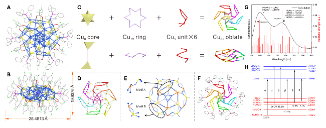
图1 (A) 透视和(B) 侧视图观察的Cu62簇的总体结构,颜色标签:浅蓝色为Cu;黄色为S;红色为O;亮绿色为F;粉色为P;灰色为C;(C) Cu62金属核心的解剖结构。所有原子均为Cu;(D) Cu48S30笼,原子的颜色编码:黄色球体为硫(S);多彩风车为Cu;(E) 表面硫醇配体的配位模式,颜色标签:浅蓝色为Cu;黄色和橙色为S;(F) 表面膦配体和羧酸配体的配位模式,颜色标签:多彩风车为Cu;玫瑰色为P;浅红色为O;海绿色为F;灰色为C;(G) Cu48S30的计算紫外-可见-近红外吸收光谱;(H) Kohn-Sham分子能级图(为清晰起见,所有H原子均已省略)[26]

Fig. 1 Total structure of the Cu62 cluster from the top (A) and side (B) views. Color codes for atoms: light blue, Cu; yellow, S; red, O; bright green, F; pink, P; gray, C. (C) Anatomy of the metal core of Cu62. All atoms are Cu. (D) Cu48S30 cage. Color codes for atoms: yellow spheres, S; chromatic windmill, Cu. (E) Coordination modes of thiolate ligands on the surface. Color labels: light blue, Cu; yellow and orange, S. (F) Coordination modes of phosphine ligands and carboxylic acid ligands on the surface. Color codes: chromatic windmill, Cu; rose, P; light red, O; sea green, F; gray, C. (G) Calculated UV-vis-NIR absorption spectra of Cu62. (H) Kohn-Sham molecular energy level diagram. All H atoms are omitted for clarity. Reprinted with permission from ref. 26. Copyright 2025 The Authors

2.2 Fluorescence properties

Unlike bulk copper particles, ultra-small CuNCs possess discrete electronic levels and exhibit molecule-like optical behavior. A variety of water-soluble CuNCs with emissions spanning blue to red have been reported [27]. Their fluorescence generally originates from electronic transitions involving occupied d-bands and states above the Fermi level, or between the highest occupied and lowest unoccupied molecular orbitals (HOMO and LUMO). Emission characteristics can be modulated by the choice of ligands and synthetic parameters. For instance, BSA-templated CuNCs can be prepared to emit either blue or red light depending on synthesis conditions[28-29]. dsDNA-templated CuNCs emit brightly near 570 nm, whereas ssDNA-templated clusters produce red emission around 615 nm when excited at 340 nm[30-31]. Certain CuNCs also demonstrate aggregation-induced emission (AIE)[32]. This AIE behavior is attributed to the restriction of intramolecular motion in the aggregated state, which promotes radiative decay while suppressing nonradiative pathways, leading to enhanced quantum yields. Wang et al. reported AIE properties in glutathione (GSH)-protected CuNCs[33]. The as-prepared clusters showed weak red fluorescence, but ethanol-driven aggregation enhanced the quantum yield and shifted the emission to shorter wavelengths. CuNCs can exhibit both one-photon and two-photon fluorescence, enabling deeper tissue imaging and lower autofluorescent backgrounds[34]. Compared with noble metals like gold and silver, copper precursors are more plentiful, less costly, and easier to obtain, making CuNCs attractive for many uses. However, CuNCs remain vulnerable to aggregation and oxidation, which quench their fluorescence; improving their stability will therefore be a key priority for future work.

2.3 Sensing mechanisms

Several CuNC-based probes have been designed to sense diverse analytes due to CuNCs can combine strong luminescence with low cost. Common photophysical mechanisms employed include photoinduced electron transfer (PET) and photoinduced charge transfer (PCT) (Fig. 2[35], fluorescence/Föster resonance energy transfer (FRET), aggregation-induced emission (AIE), and the inner-filter effect (IFE). The majority of CuNC fluorescence sensors function through a “turn-off” response, in which the target analyte diminishes the emitted signal. A major interference for PET/PCT-based sensors is the presence of other electron-accepting or electron-donating species that can also interact with the CuNCs, leading to non-specific quenching or enhancement of the signal. To distinguish between static and dynamic quenching, one can perform fluorescence lifetime measurements. In static quenching, the lifetime of the fluorophore remains constant because the quenching occurs upon formation of a non-fluorescent complex before excitation. In contrast, dynamic quenching involves collisional deactivation, which reduces the fluorescence lifetime of the fluorophore.
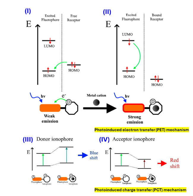
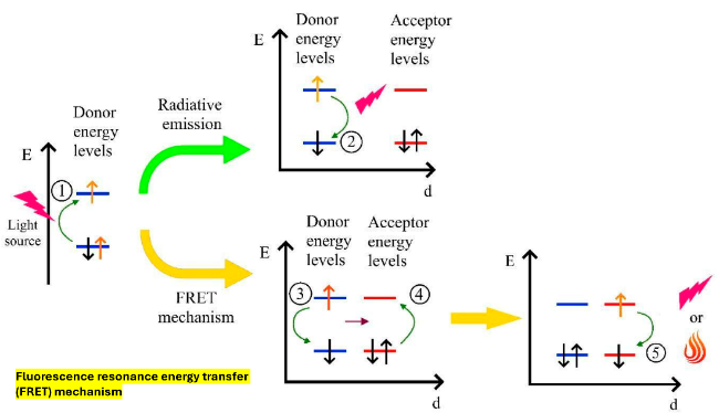
图2 PET机制(I-II)与PCT机制(III-IV):展示供体离子载体情况(III)和受体离子载体情况(IV)中与金属阳离子相互作用前后所涉及的能级跃迁过程[35]

Fig. 2 PET mechanism (I-II) and PCT mechanism (III-IV): energy levels involved in the transitions before and after the interaction with metal cations in the donor ionophore case (III) and in the acceptor ionophore case (IV). Reprinted from ref. 35 with permission from MDPI

A common strategy for constructing “turn-off” sensors is the inner filter effect (IFE), where an absorbing species reduces the fluorophore’s excitation or emission intensity by attenuating the relevant light (Fig. 3). For example, a label-free assay for nitrofurantoin employed adenosine-stabilized CuNCs: nitrofurantoin’s UV absorption (250~430 nm) overlaps the CuNC excitation/emission bands, so it shields both excitation and emitted light, lowering fluorescence[36]. The IFE is often non-specific and can be caused by any substance that absorbs light at the same wavelength as the CuNC’s excitation or emission. This can lead to significant interference from other colored or UV-absorbing compounds in the sample matrix. Distinguishing IFE from other quenching mechanisms is straightforward because the IFE affects the total light intensity but does not change the fluorescence lifetime of the fluorophore, unlike dynamic quenching. To confirm IFE, one can measure the absorbance of the sample at both the excitation and emission wavelengths of the CuNCs. A strong correlation between the absorbance and fluorescence quenching would suggest an IFE mechanism.
图3 基于内滤效应(IFE)传感器的构建策略[35]

Fig. 3 Strategies of inner-filter effect (IFE) based sensors. Reprinted from ref. 35 with permission from MDPI

Some probes quench fluorescence through electron-transfer processes. Cao et al. reported tannic acid (TA)-capped CuNCs for selective Fe3+ detection: Fe3+ forms a donor-acceptor complex with TA at the CuNC surface and accepts electrons, producing fluorescence quenching. Because the fluorescence lifetime is unchanged with Fe3+ concentration, the quenching is consistent with a static mechanism. The high selectivity arises from Fe3+’s electronic structure with five half-filled d orbitals; Fe3+ exhibits a stronger electron-accepting capability than many other ions. AIE quenching is another major quenching pathway used in metal-ion sensing. Such as, the GSH-CuNCs probe for Hg2+ detection relies on Hg2+-induced aggregation of NCs[37]. Hg2+ interacts with GSH ligands, linking CuNCs and quenching fluorescence. Zeta-potential measurements show a reduction in the negative surface charge of GSH-CuNCs after Hg2+ addition, indicating destabilization and aggregation. TEM reveals increased particle sizes and light-scattering signals grow stronger, supporting the aggregation-driven quenching mechanism.
The main design for “turn-on” sensors is an off-on sequence: fluorescence is suppressed initially and recovered when the target is introduced. Surfactant-free CuNCs have been employed as turn-on probes for vitamin C quantification [38]. Initially, fluorescence quenching occurs due to the formation of Fe-Cu NCs conjugates via a static quenching mechanism. Vitamin C’s strong reducing ability converts Fe3+ to Fe2+, inducing dissociation of the Fe-CuNCs conjugate and thereby restoring fluorescence intensity.
Growing interest in AIE has spawned many turn-on probes that exploit this effect. A “turn-on” AIE sensor can be designed where aggregation triggers fluorescence, as seen in some probes for fluoroquinolones (FQs). A major interference for AIE-based sensors is the presence of other ions or molecules that can also cause non-specific aggregation of the nanoclusters. This can be caused by changes in pH, ionic strength, or the presence of other coordinating species. To distinguish AIE from other mechanisms, one can use FRET. Liu and co-workers developed a ratiometric, visually readable sensor for fluoroquinolones (FQs) by leveraging Tb3+-activated AIE from DTE-CuNCs plus an antenna interaction between Tb3+ and FQs[39]. FQs coordinate to Tb3+ through carboxyl and carbonyl functionalities, acting as bridges that trigger Tb3+’s inherent fluorescence. Other approaches use the in situ formation of CuNCs as a selective turn-on method for Cu2+ detection[40]. Beyond AIE, confinement-induced enhanced emission (CIEE) is widely used to boost CuNC brightness by restricting molecular motion inside host matrices, thereby increasing quantum yield[41-42]. For instance, Yang et al.[43] reported a surface CIEE system in which GSH-CuNCs were incorporated into a Zn-HDS host to form GSH-CuNCs/Zn-HDS; this composite showed higher quantum yield, improved stability, and prolonged fluorescence lifetime, enabling sensitive enzyme biosensing.
In addition, ratiometric fluorescence sensors have garnered significant research attention due to their superior accuracy, sensitivity, and stability. This sensing method relies on the intensity ratio of two emission peaks, enabling self-calibration that effectively eliminates environmental and probe concentration interferences. Liu and colleagues [44] developed a CuNC-based ratiometric probe for real-time Ca2+ detection and imaging in neurons. They prepared a Ca2+-specific ligand containing two formaldehyde groups, then linked it to polyethylenimine (PEI) to form a tailored ligand for CuNC synthesis. The resulting nanoprobe showed a strong linear response to Ca2+ concentrations with a low detection limit of 220 ± 11 nM, along with excellent selectivity and stability. Benefiting from its low cytotoxicity and high biocompatibility, the nanoprobe revealed differential histamine-induced cytoplasmic Ca2+ elevations across various neuronal regions.
Aptamer-modified Cu@AuNCs (apt-Cu@AuNCs) were developed as a novel ratiometric fluorescent probe for detecting Hg2+ in Porphyra[45]. Typically, ratiometric sensing systems operate via Förster resonance energy transfer (FRET), where one emission peak is quenched and another enhanced upon analyte interaction (Fig. 4). In the absence of Hg2+, apt-Cu@AuNCs remain well dispersed; however, the addition of Hg2+ leads to the formation of thymidine-Hg-thymidine (T-Hg2+-T) complexes, resulting in NC aggregation and changes in fluorescence intensities due to FRET between AuNCs and CuNCs. The T-rich nucleic acid strands on the surface of the nanoclusters specifically capture Hg2+ ions, facilitating this aggregation and modulation of fluorescence. Additionally, a portable device combining a smartphone and microfluidic chip enables colorimetric detection of Hg2+ within 0.5 to 7.0 μM, demonstrating practicality and ease of use for on-site analysis. FRET requires the donor and acceptor to be in close proximity, which can be disrupted by changes in the sample matrix, leading to false signals. Other species that can act as acceptors or donors at the relevant wavelengths can also cause interference. Unlike collisional quenching, FRET results in a decrease in the fluorescence lifetime of the donor fluorophore and an increase in the fluorescence intensity of the acceptor. Time-resolved fluorescence spectroscopy is a powerful tool to confirm a FRET mechanism by observing the reduction in the donor's excited-state lifetime.
图4 FRET机制:供体与受体两种荧光团间能量转移过程的示意图[35]

Fig.4 FRET mechanism: schematic representation of processes involved between two fluorophores, a donor and an acceptor. Reprinted from ref. 35 with permission from MDPI

3 Synthetic approaches of CuNCs

A uniform, monodisperse catalyst with well-defined structure and composition is highly desirable for the development of efficient catalysts. Prior to 2010, reports on CuNCs were limited due to their susceptibility to oxidation and challenges in synthesis [46]. Since then, significant efforts have focused on the preparation and applications of CuNCs exhibiting diverse emission properties and good biocompatibility. Currently, multiple synthetic techniques enable the production of CuNCs with varied sizes, structures, and surface characteristics (Fig.5). Typical approaches include template-directed assembly, photoreduction, sonochemical methods, microemulsions,radiolysis, electrochemical routes, microwave-assisted synthesis, seed-mediated growth, monolayer protection, phase-transfer techniques, and chemical etching. For instance, in template-directed methods, Cu(II) ions coordinate to an appropriate template or stabilizer and are reduced on the template surface to form Cu atoms and subsequently NCs. Electrochemical preparation generates copper species via anodic dissolution of a copper electrode, whereas etching strategies start from larger, nonemissive copper nanoparticles (CuNPs) that are chemically eroded to yield small, luminescent CuNCs. The following section surveys recent advances in CuNC synthesis, organized by various strategies and then examines the fluorescence characteristics of the prepared clusters.
图5 铜纳米簇的主要合成方法

Fig.5 Main synthetic approaches of CuNCs

3.1 Bottom-up method

The bottom-up strategy builds MNCs from metal salts, ligands, and reductants. In this route, metal salts are reduced in solution by an appropriate reducing agent. First, a metal(I)-thiolate intermediate forms when the metal salt reacts with a thiolate ligand. Subsequent treatment with a reductant (for example, NaBH4 or ascorbic acid) reduces M(I) to M(0), producing M(0)@M(I)-type NCs, often termed a one-step synthesis. In some cases, the capping ligand itself serves as the reductant, obviating an added reducing agent; this approach is commonly called biomineralization. Besides conventional chemical reduction, photoreduction can generate luminescent MNCs by using light to drive the reduction. For example, Zhou et al.[47] applied photoreduction to prepare silane-stabilized AuNCs, which were then used to photodegrade the dye methylene blue.

3.2 Top-down method

The top-down route generates ultra-small MNCs by chemically etching larger metal nanoparticles or preformed clusters. In this approach, an excess of ligand or other etchant reacts with bigger metal particles to carve them down into smaller NCs; the reaction is typically carried out in homogeneous solution or at a liquid-liquid interface. Based on the nature of the etchant, etching strategies are commonly classified as ligand-driven or solvent-driven. A key benefit of etching is precise size control, enabling the production of monodisperse metal NCs and even alloyed clusters [48].

3.3 Inter-cluster conversion method

Inter-cluster conversion designs new NCs from existing clusters via seed-mediated growth, cluster-to-cluster transformation, metal exchange, ligand exchange, or motif exchange. Preformed nanoclusters act as precursors whose structures are tuned by changing kinetic or thermodynamic conditions. Ligand exchange reactions (LERs) are the most widely applied conversion technique[49]. LERs are particularly powerful for post-synthesis modification because of the flexible gold-sulfur chemistry; advantages include tuning cluster size and phase, endowing clusters with fluorescence for bio-labeling, increasing enantiomeric excess of chiral clusters, or inducing chirality in originally achiral species. As a result, LER expands the structural and functional diversity of MNCs [50]. Wang et al. [50] provide a thorough review of LERs on thiolate-protected AuNCs and the benefits they confer. This conversion pathway can also yield large quantities of high-quality crystalline MNCs, facilitating structural elucidation and deeper insights into their physicochemical behavior.

3.4 Monolayer-protected method

The monolayer-protected strategy is a straightforward, general approach for producing uniformly sized MNCs. First reported by Brust and co-workers in 1994 for thiol-capped metal particles, the method employs a two-phase extraction of Au(III) chloride and strict control of the thiol-to-Au(III) stoichiometry to directly form monolayer-protected AuNCs [51]. Commonly known as the Brust-Schiffrin or “direct synthesis” method, it has since become a standard route for synthesizing thiol-stabilized MNCs.

3.5 Etching method

The etching method represents a significant top-down strategy for synthesizing precise MNCs. This technique employs suitable etching agents to transform larger metal particles into nanoclusters of defined sizes. Etching strategies are typically divided into ligand-driven and solvent-driven etching, based on the nature of the etchant. Although etching has been widely exploited for Au and Ag cluster production, its application to CuNCs is still relatively underexplored [52]. Xie and coworkers [53] reported a solvent-etching protocol that tuned cluster hydrophilicity/hydrophobicity via electrostatic adsorption, enabling phase transfer from water to an organic solvent followed by mild etching to produce uniform clusters. This allowed for the transfer of clusters from the aqueous to the organic phase, followed by mild etching to achieve a uniform size distribution. Wang et al.[54] prepared oleylamine (OA)- stabilized Cu nanoparticles by dissolving Cu2+ in an OA/1-octadecene (ODE) medium; etching was achieved by adding an aqueous polyethyleneimine (PEI) solution to OA-Cu NPs in chloroform at 50 ℃. In another study, Cu2+ was reduced by ascorbic acid, which also acted as a capping agent; the resultant Cu NPs were then etched in glutathione (GSH) aqueous solution to yield CuNCs with a strong emission at ~600 nm[55]. The resulting CuNCs exhibited a strong emission peak at 600 nm. Additionally, other thiol ligands, such as cysteine and penicillamine, have likewise been used for etching.

3.6 Electrochemical synthesis

Electrochemical methods are widely used to generate metal nanoparticles and nanoclusters. Here, anodic dissolution of a copper electrode supplies Cu2+ ions that are reduced and stabilized at the cathode to form CuNCs. Adjusting current density provides an effective handle on final cluster size [56]. A major advantage is that CuNCs can be produced at low current densities without added surfactants or ligands; under optimized conditions, the PLQY of such ligand-free (“naked”) CuNCs can reach ~13%, comparable to many chemically synthesized counterparts[57]. Vilar-Vidal et al.[58] synthesized highly stable, ligand-free Cu13 clusters with blue emission (410 nm) using tetrabutylammonium nitrate as electrolyte, and showed that heating to 80 ℃ followed by redissolution in acetonitrile can tune core atom counts up to Cu20. Additionally, Smaller, green-emitting CuNCs (down to Cu5 cores) have been isolated by ethanol centrifugation during purification [59].

3.7 Template method

Template directed syntheses are among the most prevalent routes to CuNCs. In these protocols, templating ligands act both as reductants and stabilizers during cluster formation. Typical templates include peptides, proteins, polymers, dendrimers, DNA, and enzymes (Fig. 6). Cu2+ ions can effectively bind to these templates, subsequently being reduced to form clusters, with steric hindrance preventing aggregation. The choice of template can be tailored to specific applications, while factors such as pH, temperature, and the ratio of Cu2+ to templates influence the functional ligand groups and the metal core. Recent literature reviews highlight various protocols for synthesizing ligand-stabilized nanoclusters, detailing their properties and applications. The characteristics and structures of nanoclusters vary according to the stabilizing ligands used. For instance, in the synthesis of CuNCs using protein templates, increasing the pH promotes the cleavage of disulfide bonds, allowing thiol groups to stabilize the CuNCs within the protein framework. By optimizing template-assisted conditions, aqueous CuNCs with PLQYs exceeding 44% have been reported [60]. The following section provides a concise summary of the resulting CuNC types and their characteristics.
图6 铜纳米簇合成所用配体的示意图

Fig.6 Schematic representation of the ligands used for CuNCs synthesis

Proteins present a rich array of functional groups (notably amino and carboxyl moieties) that coordinate readily with Cu(II) and help anchor nascent Cu(0) atoms on the protein scaffold. Many proteins also contain thiol groups capable of reducing metal ions, enabling them to act simultaneously as templates and reductants for CuNC formation. Bovine serum albumin (BSA) is a widely used example: in an alkaline environment (with NaOH), BSA both stabilizes and helps reduce copper species to produce BSA-stabilized CuNCs. Bi et al. [61] successfully prepared CuNCs with outstanding optical properties using BSA as a template, then proposed BSA-stabilized CuNCs (BSA-CuNCs) for the detection of circulating tumor cells (CTCs) (Fig. 7A, B). These clusters emit red light near 640 nm; when CTCs are present, the BSA-CuNC red fluorescence is progressively quenched via the IFE, while BSA-sensitized green emission from CTCs increases, enabling a simple mix-and-read assay with a reported LOD of 12.01 nM (Figure 7C, D). Subsequently, Zhang et al.[29] showed that using hydrazine hydrate (N2H4·2H2O) as a mild reducer at room temperature yields BSA-CuNCs with improved fluorescence quantum yield (QY), emission centered at ~625 nm and a QY of 4.1% compared with clusters made without hydrazine. Beyond BSA, other proteins such as papain, lysozyme, trypsin, egg white proteins, and yeast extracts have been employed as templates for CuNC synthesis.
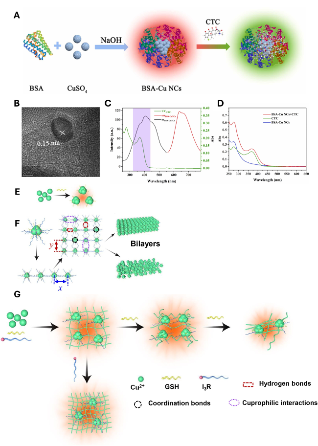
图7 (A) BSA-CuNCs的合成及CTC的视觉检测;(B) HRTEM图和(C) BSA-CuNCs的激发、发射光谱及紫外-可见吸收光谱;(D) BSA-CuNCs、CTC及BSA-CuNCs + CTC的紫外-可见吸收光谱[61];自组装I3R-GSH-CuNCs的示意图:(E) 具有弱荧光的GSH-CuNCs;(F) 小I3R-GSH-CuNCs的二次组装形成多层结构;(G) I3R-GSH-CuNCs分层自组装成纳米纤维、螺旋纳米纤维和短纳米纤维,具有强荧光特性[62]

Fig.7 (A) Synthesis of BSA-CuNCs and visual detection of CTC. (B) HRTEM and (C) the excitation, emission spectrum and the UV-vis absorption spectrum of BSA-CuNCs. (D) UV-vis absorption spectrum of BSA-CuNCs, CTC, and BSA-CuNCs + CTC. Reprinted from ref. 61 with permission from Elsevier, 2023. Scheme of the self-assembly I3R-GSH-CuNCs. (E) GSH-CuNCs with weak fluorescence. (F) The secondary assembly of small I3R-GSH-CuNCs into multilayered architectures; (G) I3R-GSH-CuNCs hierarchical self-assembly into nanofibers, helical nanofibers and short nanofibers, with strong fluorescence. Reprinted from ref. 62 with permission from Elsevier, 2020

Peptides, constructed from natural amino acids, offer tunable functionality, good biocompatibility, and the ability to form self-assembled architectures that can carry drugs, nucleic acids, or metal ions. Their sequences can be engineered to serve as templates for fluorescent nanomaterials. For instance, Yang et al.[62] reported a simple one-pot route to orange-red emitting CuNCs using an ultrashort peptide (I3R) together with GSH. The coordination between the copper core and capping ligands promotes ligand-to-metal charge transfer (LMCT), producing I3R-GSH-CuNCs with intensified emission and prolonged lifetimes (Fig.7E~G). Circular dichroism of samples with helical nanofibers showed a negative band around 300~320 nm, indicating the clusters are chiral, which induced by the chiral ligands GSH and I3R. Zhang and coworkers [63] designed a copper-loaded peptide nanoparticle (MHRC@Cu) to induce cuproptosis by reprogramming metabolism in glycolysis-dependent tumor cells: MHRC shifted cells toward greater mitochondrial respiration and heightened copper sensitivity, yielding up to 96% tumor growth suppression. Despite their performance, peptide-templated CuNCs face scalability challenges because peptide synthesis can be expensive.
DNA is an attractive scaffold for nanomaterial construction due to its nanoscale architecture and abundant functional sites. In 2010, Mokhir et al.[30] showed that double-stranded DNA (dsDNA) can stabilize CuNCs in the presence of Cu2+ and ascorbate, whereas single-stranded DNA (ssDNA) did not support cluster formation. Template length influences both fluorescence intensity and cluster size: DNA-CuNCs are aggregates of Cu atoms protected by DNA ligands, and varying the template sequence or length tunes optical properties. Practical DNA-CuNC sensing protocols, for example, Hg2+ assays often require multiple steps (pre-incubation of Hg2+ with DNA, then addition of Cu2+ and reductant), which increases assay time and potential error sources; moreover, oxidation of copper by dissolved oxygen can produce radicals that limit fluorescence stability to roughly 30 min [64]. To overcome this, Kim et al.[65] incorporated fructose into the synthesis, producing Fru@DNA-CuNCs with dramatically enhanced stability: the operational lifetime increased from about 30 min to 108 days (a >5000-fold improvement). The stabilization likely arises from reduced oxygen diffusion and solubility in high-fructose, higher-viscosity media. Building on this, Li et al. [66] developed Fru@DNA-CuNCs as robust Hg2+ sensors: Hg2+ interaction rapidly quenches fluorescence via metallophilic interactions that facilitate electron transfer between Hg and Cu, altering the cluster electronic structure (Fig. 8A). Additionally, poly(thymine)-templated CuNCs demonstrate that template length can tune cluster size and quantum yield [67], highlighting how sequence design controls DNA-CuNC properties and widening their application scope.
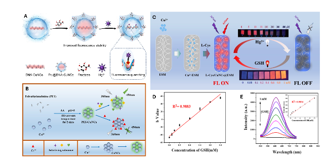
图8 (A) Hg2+引起的Fru@DNA-铜纳米簇荧光猝灭机制示意图[66];(B) PEI-铜纳米簇的制备、光学特性及其在Cr(VI)检测中的应用示意图[68];(C) 基于蛋壳膜和半胱氨酸的固相铜纳米簇荧光膜制备示意图;(D) Lab-b值与0.05~1 mM浓度范围内GSH的线性关系;(E) 不同浓度GSH(0.05~1 mM)对Hg2+猝灭的L-半胱氨酸/CuNCs@ESM荧光光谱的影响[69]

Fig. 8 (A) Scheme of the fluorescence quenching of Fru@DNA-CuNCs caused by Hg2+. Reprinted from ref. 66 with permission from Elsevier, 2024. (B) The preparation, optical properties, and applications of PEI-CuNCs for Cr(VI) detection. Reprinted from ref.68 with permission from Wiley, 2024. (C) Scheme of solid-phase CuNCs fluorescence membranes with the ESM and cysteine. (D) Linear relationship between Lab-b value and GSH concentration between 0.05 and 1 mM. (E) Fluorescence spectra of Hg2+ quenched l-Cys/CuNCs@ESM with different GSH from 0.05 to 1 mM. Inset: Linear relationship between the fluorescence intensity of L-Cys/CuNCs@ESM and GSH concentration. Reprinted from ref. 69 with permission from the American Chemical Society, 2023

Polymers bearing appropriate functional groups provide multiple coordination sites for Cu(II), facilitating nanocluster formation. In 2013, Fernández-Ujados and co-workers [70] reported a route to water-soluble CuNCs using dendrimers as templates and stabilizers, where direct metal reduction was carried out under reflux in the presence of a PEGylated ligand. The resulting clusters showed long-term stability under varied conditions and uniform size distribution. Polyethyleneimine (PEI), rich in amino groups, has likewise been applied as a protective matrix to prepare fluorescent CuNCs using reducers such as hydrazine hydrate or ascorbic acid [71-72]. Because of its abundant amine functionalities and strong metal affinity, PEI can act both as a template and a capping agent. For example, PEI with a molecular weight of ~10 000 was used together with ascorbic acid to yield water-soluble, reasonably stable PEI-protected CuNCs (PEI-CuNCs). More recently, Gan et al. [68] developed a one-pot, mild synthesis of PEI-CuNCs that selectively respond to Cr(VI). The PEI-CuNC fluorescence is quenched proportionally to Cr(VI) concentration, affording a low detection limit of 8.9 nM and eliminating the need for extra reductants; the products display high quantum yield and robust photostability against competing metal ions (Fig. 8B).
Small molecules are also effective as stabilizers or reductants in CuNC synthesis; many are thiol- or carboxyl-containing compounds that reduce metal salts and bind strongly to metal surfaces[73]. Zhang et al.[69] prepared an eggshell membrane (ESM)-based fluorescent sensor (L-Cys/CuNCs@ESM) by in situ formation of red-emitting CuNCs using L-cysteine as both reducer and protector for visual detection of Hg2+ and GSH (Fig. 8C). This fluorescent composite exhibited excellent stability and portability, a large Stokes shift (~250 nm), and a long lifetime (~7.3 μs). L-Cys/CuNCs@ESM showed selective fluorescence quenching by Hg2+, and fluorescence could be restored by GSH, enabling an “ON-OFF-ON” sensing cycle suitable for smartphone-assisted readout. Liang et al.[11] similarly exploited ESM and smartphone imaging to build an “ON-OFF-ON” platform for visual dual detection of H2O2 and GSH: red GSH-CuNCs@ESM were produced in situ with GSH acting as both protector and reducer, and H2O2 specifically quenched their fluorescence. Self-assembly strategies also permit tuning of CuNC arrangement within assemblies, producing polymorphic clusters with varied emissions.
Considering the above-mentioned methods for the preparation of CuNCs, we summarize the pros and cons of the synthetic strategies discussed in this Section. Each method is listed with key advantages and limitations was also compared, as seen in Table 1.
表1 本文对CuNCs合成方法的系统比较

Table 1 Comprehensive comparison of CuNCs synthetic methods in this work

Method PLQY potential Typical stability Scalability Cost Typical yield Pro. Cons.
Bottom-up chemical/photo-reduction Low-Moderate (variable; often up to
~10%~20%)
Moderate (depends on ligand/passivation) Moderate (bench-scale; some scale-up issues) Low-Moderate Moderate Simple, tunable by reagents/conditions, versatile ligand compatibility Oxidation sensitivity, reproducibility issues, and residual reductant impurities
Top-down/chemical etching Moderate (good size control can improve PL) Moderate (depends on passivation after etching) Low-Moderate (difficult to scale uniformly) Moderate Low-Moderate (loss during etching) Precise size/phase control and tight size distributions Material loss, complex control, potential residues from etchants
Inter-cluster conversion Moderate-High (can produce desirable optical states) Variable (depends on final ligands/structure) Low (often lab-scale, stepwise) Moderate-High Low-Moderate Post-synthesis tunability, access to metastable structures Complex pathways, purification challenges, variable yields
Monolayer-protected (Brust-Schiffrin style) Moderate (good when thiol shells optimized) Moderate-Good (thiol passivation effective but Cu issues) Low-Moderate (two-phase processes limit scale) Moderate Moderate Produces monodisperse, thiol-protected clusters; established protocols Cu-thiol reactivity/oxidation issues, solvent and scale limitations
Electrochemical synthesis Low-Moderate (ligand-free variants often lower PLQY) Variable (ligand-free less stable; can passivate post-synthesis) Moderate-High (flow/electrochemical reactors possible) Moderate (instrumentation cost) Moderate Precise temporal control, cleaner products (no chemical reductants) Requires electrochemical setup, electrode/electrolyte impurities, possibly low PLQY
Template-directed (general: proteins/peptides/DNA/polymers) Moderate-High (can reach high PLQYs with optimized templates) Moderate-Good (templates often protect against oxidation) Low-Moderate (depends on template: polymers scale better than DNA/peptides) Moderate-High (DNA/peptides costly; polymers less so) Moderate Mild/biocompatible conditions, built-in functionality, sequence/size control Cost (DNA/peptides), heterogeneity (proteins), purification complexity
Protein-templated
(e.g., BSA)
Moderate (commonly red-emitting CuNCs, PLQY variable) Moderate (proteins afford protection but can denature) Low-Moderate (biological reagents limit scale) Low-Moderate Moderate Biocompatible, simple one-pot syntheses, useful for sensing/bioimaging Heterogeneity, batch variability, susceptibility to denaturation
Peptide-templated clusters Moderate-High (programmable environment; can be high) Moderate (depends on peptide stability) Low (peptide cost and synthesis limit scale) High (peptide synthesis cost) Low-Moderate Sequence programmability, chiral assemblies, controlled environment High cost, limited scalability, potential stability issues
DNA-templated CuNCs Moderate-High (sequence can yield strong emitters) Moderate (oxidation is a concern; can be mitigated) Low (DNA cost and handling limit bulk use) High Low-Moderate Precise sequence control of emission, good for sensing/bioconj. High cost, environmental sensitivity, and nuclease susceptibility
Polymer/dendrimer templates (PEI, PEGylated, etc.) Moderate (can be tuned; good colloidal PL) Good (steric stabilization) Moderate-High (polymers scale well) Moderate Moderate-High Water solubility, steric stabilization, and easier scalability Polymer polydispersity affects uniformity; some polymers may be toxic
Small-molecule stabilizers (thiols, carboxylates, amino acids) Low-Moderate (can be optimized) Low-Moderate (ligand exchange/oxidation risk) High (low cost, simple reagents) Low High Low cost, fast kinetics, easy functionalization for sensors Lower long-term stability, less steric protection, ligand-induced quenching risk

4 Recent advances of CuNCs for environmental pollutants analysis

As industrial activities expand, pollutants may increasingly accumulate in the environment, threatening ecosystems and human health; thus, real-time monitoring is essential for environmental protection and disease prevention. The following section summarizes advances in employing CuNCs for environmental analysis, organized by pollutant class.

4.1 Ions

Heavy metals pose persistent, toxic, and bioaccumulative hazards. Fluorescent CuNCs have been widely used as ion sensors. Mercury, in particular, is a pervasive pollutant with serious ecological and human health impacts, yet many existing Hg2+ biosensors lack adequate sensitivity and selectivity. A high signal-to-noise analytical probe was engineered by templating CuNCs on a reticular poly(T) DNA scaffold, which markedly enhanced fluorescence and resisted enzymatic degradation [74]. This DNA-templated sensor achieved excellent sensitivity and selectivity for Hg2+ across a linear range from 50 pM to 500 μM and a very low LOD of 16 pM. Because Hg2+ interacts with GSH, the probe can also be applied to monitor GSH inhibition by Hg2+ in biological samples. Jain et al. [75] designed a dual colorimetric-fluorimetric Hg2+ assay (Fig. 9A, B) combining blue fluorescent CuNCs-doped Zr-MOF (CuNCs@Zr-MOF) and red N-methyl mesoporphyrin IX (NMM) together with a peroxidase-mimicking G-quadruplex DNAzyme (PMDNAzyme). Hg2+ mediates T-Hg2+-T base pairing between a dabcyl-tagged complementary DNA and the CuNCs@Zr-MOF@G4 construct, quenching CuNC fluorescence at 463 nm through a FRET mechanism. Additionally, the G-quadruplex structure enhances NMM fluorescence emission (610 nm). This probe quantitatively detects Hg2+ with a LOD of 0.59 nM (fluorimetry), demonstrating resilience against interference from other metal ions in real aqueous samples, thus highlighting its potential for monitoring Hg2+ concentrations in aquatic products.
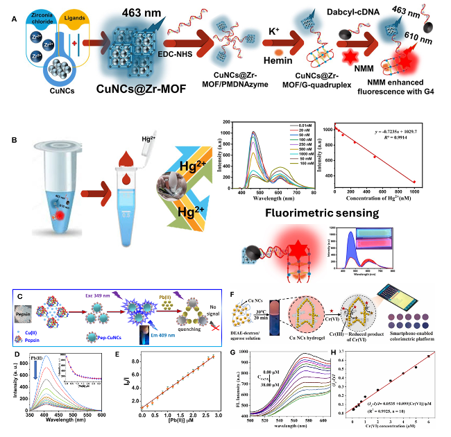
图9 (A) PMDNAzyme传感器的制备流程;(B) Hg2+存在下的荧光检测机制示意图[75];(C) Pep-铜纳米簇及其Pb(Ⅱ)离子检测示意图;(D) 不同浓度Pb(Ⅱ)离子(0.1~6 mM)存在下Pep-铜纳米簇的荧光发射光谱;(E) 荧光强度比与Pb(Ⅱ)离子浓度的Stern-Volmer关系图[76];(F) 基于铜纳米簇水凝胶的合成流程及扩增荧光检测策略示意图;(G) 添加不同浓度Cr(Ⅵ)(0~38 μM)后铜纳米簇水凝胶的荧光发射光谱;(H) 荧光强度比与Cr(Ⅵ)浓度(0.07~6.00 μM)的关系曲线[77]

Fig.9 (A) Fabrication of the PMDNAzyme sensor. (B) Fluorimetric-detection mechanism in the presence of Hg2+ ion. Reprinted from ref. 75 with permission from the authors, 2024. Available under the CC-BY-NC-ND 4.0 license. (C) Scheme of Pep-CuNCs and their Pb(Ⅱ) ions sensing. (D) FL emission of Pep-CuNCs in the presence of Pb(Ⅱ) ions (0.1 to 6 mM); (E) Stern-Volmer plot of the FL intensity ratio to the Pb(Ⅱ) ions. Reprinted from ref. 76 with permission from MDPI 2022. (F) Synthetic scheme and amplified fluorescence sensing strategy based on CuNCs hydrogel. (G) Fluorescence emission spectra of CuNCs hydrogel after adding various concentrations of Cr(Ⅵ) from 0 to 38 μM. (H) Plots of intensity ratio vs. the concentration of Cr(Ⅵ) (0.07-6.00 μM). Reprinted from ref. 77 with permission from Elsevier, 2024

Similarly, numerous probes have been developed to detect lead ions (Pb2+) in environmental and drinking water systems due to lead’s high toxicity, particularly to children. Even minimal lead exposure can lead to severe health issues, including cardiovascular, neurological, and reproductive disorders. Han et al.[78] prepared GSH-protected CuNCs (CuNCs@GSH) for Pb2+ detection via AIE. Initially exhibiting low fluorescence, these nanoclusters emitted bright orange fluorescence upon Pb2+ addition, demonstrating a linear response from 200 to 700 μM, with a LOD of 106 μM (S/N = 3). This method is rapid, straightforward, and highly selective, allowing for visual detection of Pb2+ under UV light. Goswami et al. [79] introduced BSA-capped Cu quantum clusters (CuQC@BSA) as a sensitive probe for Pb2+, utilizing fluorescence quenching in the presence of low concentrations of H2O2. Additionally, Saleh et al.[76] developed high-luminescent blue-emitting pepsin CuNCs (Pep-CuNCs), synthesized using an eco-friendly microwave-assisted method, which showed remarkable selectivity for Pb(Ⅱ) ions through fluorescence quenching, achieving an LOD of 11.54 nM (Fig. 9C~E).
Chromium (Cr(Ⅵ)), a common environmental contaminant from industrial processes, has prompted the development of various analytical methods for detection. Bai et al. presented a ratiometric fluorescent probe for Cr2O72- or Cd2+ by integrating GSH-based carbon dots (CDs) with CuNCs, resulting in two distinct emission peaks at 450 and 750 nm [80]. The nanohybrid demonstrated excellent sensitivity and selectivity, with successful fluorescence test strips showing color changes from pink to purple under UV light and recovery rates between 102% and 109% in real water samples. Another study by Cao et al. [77] developed a smartphone-enabled colorimetric platform combining agarose hydrogel with CuNCs, enhancing optical performance and target recognition (Fig. 9F~H). This platform achieved a fluorescence quantum yield three times higher than that of CuNCs alone, enabling sensitive Cr(Ⅵ) detection from 0.07 to 38.00 μM. Notably, excellent analytical performance for Cr(Ⅵ) detection is preserved when the CuNCs hydrogel is directly immobilized on paper, highlighting the potential of this approach for advancing hydrogel applications. Furthermore, this method has been effectively utilized to detect Cr(Ⅵ) in environmental samples, indicating its suitability for portable, instrument-free devices for real-time, on-site environmental monitoring.
Sulfide ions represent a significant category of environmental pollutants and are by-products of extensive industrial activities. Their presence in various forms (e.g., H2S, HS- and S2-) poses a latent threat to human health. CuNCs have been widely employed for the detection of sulfide ions. A sensing platform was created by modifying CuNCs with 3-mercaptopropionic acid, resulting in Cu2+@MPA-CuNCs, which displayed AIE phenomena [81]. This luminescent probe was easy to prepare, with a linear detection range for S2- from 0 to 600 μM and an LOD of 26.3 nM, showing high selectivity and accuracy in simulated environments (Fig. 10A~E). Additionally, a novel "turn-on" detection strategy for S2- was developed using silk fibroin-templated CuNCs (SF@CuNCs) [82]. This method exhibited increased fluorescence intensity upon S2- addition due to aggregation-induced emission enhancement (AIEE) effect, with a linear detection range of 5.0 to 110.0 μM and an LOD of 0.286 μM, achieving recovery rates between 95.6% and 101.6% in real sample tests (Fig. 10F~H).
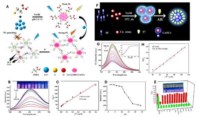
图10 (A) Cu2+@MPA-CuNCs的制备流程;(B) 缓冲溶液中S2-存在时Cu2+@MPA-CuNCs的荧光发射光谱;(C) 荧光峰强度与S2-浓度的拟合曲线;(D) Cu2+@MPA-CuNCs 24 h内荧光强度变化曲线;(E) 不同阴离子对Cu2+@MPA-CuNCs荧光强度的影响(红色柱),以及不同阴离子与S2-共存时对传感器的影响(绿色柱)[81]; (F) SF@CuNCs的合成流程;(G) 添加不同浓度S2-后SF@CuNCs的荧光响应;(H) 荧光强度与S2-浓度(5~110 μM)的Stern-Volmer关系图[82]

Fig.10 (A) Preparation of Cu2+@MPA-CuNCs. (B) FL emission spectra of Cu2+@MPA-CuNCs in the presence of S2- in the buffer solution. (C) Fitting curve between peak intensity and concentration of S2-. (D) FL intensity curve of Cu2+@MPA-CuNCs within 24 h. (E) Effect of different anions on the FL intensity of Cu2+@MPA-CuNCs (red pillars), and the influence on the sensor in the presence of different anions coexisting with S2- ions (green pillars). Reprinted from ref. 81 with permission from the American Chemical Society, 2020. (F) Synthesis of SF@CuNCs. (G) Fluorescence response of SF@CuNCs upon addition of different S2- concentrations, (H) Stern-Volmer equation of FL intensity on S2- (5~110 μM). Reprinted from ref. 82 with permission from Elsevier, 2019

4.2 Organic pollutants

Industrialization driven by globalization has led to a significant increase in harmful pollutants to meet global demands, resulting in alarming environmental contamination. Organic pollutants, particularly azo dyes from the textile industry, and inorganic pollutants, especially oxidized heavy metals, are prevalent in industrial effluents. These inadequately treated carcinogenic effluents are discharged into soil and water, causing irreversible harm to humans and aquatic ecosystems. Effective action plans are essential to safely eliminate these contaminants without generating stable secondary pollutants. Common dyes found in industrial effluents include methyl orange (MO), rhodamine B (RB), malachite green (MG), and methylene blue (MB). Reactive oxygen species (ROS), such as superoxide radicals (O2-·) and hydroxyl radicals (OH), play a crucial role as primary agents in degrading these toxic substances [83-84]. Addressing this pollution crisis requires thorough examination and innovative solutions to mitigate the impact on health and the environment. Yang et al. [85] developed a Fenton-like nanocatalyst by synthesizing ultrasmall CuNCs on hydroxyapatite (HAp) through doping and modification, resulting in a composite catalyst termed Cu NCs/HAp. Utilizing H2O2, Cu NCs/HAp demonstrates exceptional catalytic performance, effectively degrading organic dyes like MB under mild neutral conditions, achieving over 83% removal within 30 min. This performance highlights its catalytic universality and stability.
In addition to this research, numerous studies have focused on the use of functionalized CuNCs for organic pollutants [84]. Su et al. [86] developed a new fluorometric method for the identification and determination of kojic acid (KA), utilizing KA-induced static fluorescence quenching of BSA-CuNCs. This method is highly selective due to the unique interaction between KA and copper ions and has been successfully applied to real samples (Fig. 11A~C). Similarly, Xu et al. [84] introduced the first fluorescent sensor array based on CuNCs to detect 12 metal ions (Pb2+, Fe3+, Cu2+, Cd2+, Cr3+, Co2+, Ni2+, Zn2+, Ag+, Fe2+, Hg2+, and Al3+) and dissolved organic matter (DOM), including humic substances, lipids, fatty acids, amino acids, and lignans. CuNCs synthesized with PEI, histidine (His), and GSH demonstrated varied binding affinities. Principal component analysis (PCA) and linear discriminant analysis (LDA) were utilized for identification, achieving classification at a limit concentration of 1.5 μM and quantification down to 0.83 μM (Zn2+), proving effective for environmental monitoring in tap and seawater samples. Moreover, tetracycline (TC), a widely used antibiotic, is poorly absorbed in the human gastrointestinal tract, with about 75% excreted into wastewater. Zheng et al.[87] enhanced the fluorescence properties of CuNCs by employing cetyltrimethylammonium bromide (CTAB) to improve sensitivity and selectivity. They developed fluorescence-enhanced CuNCs (TA-CuNCs@CTAB) using tannic acid as a template. The electrostatic interaction between CTAB and TA-CuNCs promotes aggregation, leading to enhanced fluorescence through the AIE effect. This probe shows linear detection for TC in two ranges: 5~50 μM and 50~130 μM, with a LOD of 65 nM. The detection mechanism involves IFE induced by TC, resulting in significant fluorescence quenching, making it a convenient and accurate method for detecting TC in real water samples.
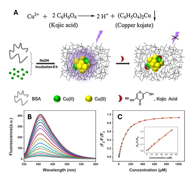
图11 (A) BSA封端的CuNCs用于KA检测的示意图;(B) 不同KA浓度(0~1000 μM)下CuNCs的荧光发射光谱;(C) 407 nm处荧光猝灭率与KA浓度的关系曲线[86]

Fig. 11 (A) Scheme of BSA-capped CuNCs for KA sensing. (B) FL emission spectra of CuNCs under KA concentrations (0-1000 μM). (C) Plots of FL quenching rate at 407 nm as a function of KA concentration. Reprinted from ref. 86 with permission from Elsevier, 2014

Hosseini et al. presented a rapid, cost-effective, and sensitive fluorescence turn-on sensing system for the trace detection of ciprofloxacin (CIP) and ofloxacin (OFL) in aqueous samples[19]. Using a one-pot method, they synthesized CuNCs under 15 nm and functionalized them with sodium gluconate (Gl@CuNCs). The maximum fluorescence emission of Gl@CuNCs at 414 nm, excited at 322 nm, shifted to longer wavelengths upon adding CIP and OFL. The incorporation of Gl@CuNCs with the target drugs activated fluorescence through a chelation-enhanced fluorescence mechanism. A linear correlation was established between fluorescence intensity differences with and without the analytes, covering a concentration range of 15 to 900 nM, with limits of detection of 9 nM for CIP and 8 nM for OFL. This detection system demonstrated high selectivity, even in the presence of a 100-fold excess of interfering substances, and was successfully applied to quantify CIP and OFL in drinking water, cow milk, human urine, and serum samples, yielding satisfactory recovery rates.
Thiol compounds are significant environmental pollutants due to their characteristic odor and biological toxicity. Zhang et al. [88] introduced a novel triplet-induced fluorescence “turn-off” strategy for detecting thiol pollutants using a GSH-stabilized CuNC (GSH-CuNC) probe. The synthesized GSH-CuNCs exhibited small size, excellent water solubility, and strong red fluorescence emission at 630 nm, which could be quantitatively quenched as the concentration of thiol pollutants increased. This probe effectively detected thioglycolic acid (TGA), 3-mercaptopropionic acid (MPA), 2-mercaptoethanol (ME), and 2-(diethylamino)ethanethiol (2-AT) across a broad linear range of 1~100 μM, with LOD of 0.73, 0.43, 0.37, and 0.69 μM, respectively. The method successfully detected these thiol pollutants in lake water, yielding satisfactory recovery rates. Furthermore, the application of GSH-CuNCs was extended to the development of luminous test strips, utilizing their outstanding fluorescence properties for rapid, real-time detection of thiol pollutants.

4.3 Pharmaceutical/Pesticides

Pesticide residues are prevalent in the environment due to the extensive use of pesticides in agricultural practices. Even trace amounts of these highly toxic substances can pose significant risks to ecosystems and human health. This section reviews detection systems based on CuNCs for various common pesticides, including paraoxon, dinotefuran (DNF), o-phenylenediamine (OPD), dithiocarbamates (DTCs), thiram, paraquat, fluazinam, and nitrofurantoin (NFT).
CuNCs have been successfully utilized to develop an enzyme-free electrochemical biosensor specifically for paraoxon, a representative organophosphate (OP)[89]. The biosensor is based on a biocompatible CuNCs@BSA-SWCNT, where CuNCs@BSA serves as the sensing probe and SWCNT enhances its electrocatalytic performance. The detection range for paraoxon spans 0.5~35 µM, with a LOD of 12.8 nM. Furthermore, the composite sensor effectively detects paraoxon in real water samples, achieving recovery rates between 93% and 104%. For monitoring DNF residues, which are commonly used in agriculture, advanced sensing platforms based on fluorescent CuNCs have been developed. Yang et al. [90] designed a dual-emission ratiometric fluorescent probe was designed by combining sulfur-doped carbon quantum dots (S-CQDs) with CuNCs, yielding excellent sensitivity and selectivity for DNF within a linear range of 10~500 µM. The IFE caused a reduction in S-CQDs fluorescence upon the addition of CuNCs; however, in the presence of DNF, the IFE diminished due to CuNC aggregation, restoring S-CQDs fluorescence. When tested with honey as a real sample, the ratiometric sensor demonstrated strong analytical performance for DNF detection.
CuNCs also exhibit several advantageous characteristics, including cost-effectiveness, biocompatibility, and appealing emission properties, making them suitable candidates for the detection and quantification of various pharmaceuticals in biological fluids. Recently, a high-performance chemiluminescence method was developed for the sensitive quantification of tramadol, achieving a linear concentration range of 0.003~2.5 μM and a LOD of 0.8 nM[91]. This method utilized a nanoporous flake-like Cu-based MOF encapsulated with BSA-CuNCs as the support. To enhance selectivity for tramadol, a pre-extraction step was implemented using MIP-Fe3O4@SiO2 in a dispersive solid-phase extraction process. Additionally, fluorescent bimetallic NCs composed of Cu and other metals, including Pd, Au, Ag, and Mo, have been prepared [92]. A fluorescence sensor based on Cu/Mo bimetallic NCs, with PLQY of 26%, has been utilized for methotrexate detection within a linear range of 50 nM to 100 μM and a LOD of 13.7 nM. Moreover, CTAB-coated CuNCs were employed to detect carbamazepine in exhaled breath condensate, achieving a linear range of 0.2 to 20 μg·mL-1 and a LOD of 0.08 μg·mL-1 [93].
Wang et al.[36] introduced a label-free, “turn-off” analytical method utilizing water-soluble adenosine-stabilized CuNCs. This fluorescent sensing probe demonstrated a rapid, sensitive, and selective response to NFT across a broad linear range of 0.05~4.0 μM, with a LOD of 30 nM. The emission intensity of the adenosine-stabilized CuNCs decreased, accompanied by a gradual shift towards longer wavelengths at elevated NFT concentrations. Minimal changes in the PL spectra of CuNCs in the presence of various other substances, including phenylalanine, proline, isoleucine, and others at 1.0 μM, confirmed the probe's high selectivity for NFT. The proposed quenching mechanism is attributed to the IFE between the adenosine-stabilized CuNCs and NFT, due to the overlap of NFT's absorption band (250~430 nm) with the excitation and emission spectra of CuNCs. Additionally, a ratiometric sensor based on GSH-stabilized CuNCs was developed for detecting o-phenylenediamine (OPD), an intermediate in pharmaceutical production, exhibiting a linear detection range of 0.15 to 110 μg·L-1 and a LOD of 93 ng·L-1[94].
Orange-fluorescent CuNCs emitting at 560 nm, with a PLQY of 5.8%, were synthesized using ovalbumin for the detection of doxycycline [95]. The CuNCs@OVA probe demonstrated a linear detection range from 1 to 1000 µM and a LOD of 270 nm. It was effectively utilized to detect doxycycline in urine samples, achieving recoveries exceeding 90%. The presence of doxycycline resulted in significant PL enhancement, attributed to the interaction between doxycycline and ovalbumin, leading to a more compact structure of the NCs. Upon UV excitation, the solution color shifted from orange to yellow with varying doxycycline concentrations. Furthermore, CuNCs@OVA exhibited high selectivity for doxycycline compared to other antibiotics. It also demonstrated good stability in hyper-saline conditions (at 40 ℃ and under photobleaching) and in the presence of various organic solvents and metal ions.

4.4 H2O and H2O2

The detection and quantification of humidity are vital in various fields, including environmental monitoring and quality control within biotechnology, food processing, and beverage industries. Therefore, developing a straightforward and cost-efficient analytical method for humidity detection is imperative. Recent research has focused on electronic, acoustic, and optical sensing mechanisms to meet this need. Among these, fluorescence sensors are particularly notable due to their simplicity and potential for rapid handling. Cheng et al. [96] developed a nanoswitch using CuNCs stabilized by dual ligands: 2-amino-5-mercapto-1,3,4-thiadiazol (AMTD) and acetate (Ac-) for the sensitive detection of trace water in the organic phase. This nanoswitch operates reversibly between two states: “fluorescence off” in solid form and “fluorescence on” in the presence of water. The strong hydrogen bonding between water and AMTD/Ac promotes cluster aggregation. The sensor exhibited high sensitivity, excellent reversibility, and consistent responses, with maximum fluorescence intensities for water content of 50.0%, 35.48%, 35.48%, and 39.39% in ethanol, THF, ACT, and MeCN, respectively. Time-resolved luminescence decay curves indicated a long lifetime of 3.14 ns, suggesting phosphorescence behavior. Importantly, aqueous solutions of these CuNCs displayed long-term stability over several months. Wen et al. [97] developed a dual-emitting film utilizing GSH-CuNCs and CDs for atmospheric humidity detection within the 40%~80% range (Fig. 12A~E). The sensor was fabricated by infiltrating a CD solution into filter paper, followed by dipping it into a CuNC solution and drying it under vacuum. This visual sensor exhibits bright fluorescence and has a diameter of approximately 8 cm with a uniform surface. An increase in air humidity caused the film’s color to shift from red to blue, enabling simultaneous detection of ethanol and water. The detection mechanism is based on the fluorescence intensity variation of CuNCs in different environments, demonstrating a linear relationship between the fluorescence intensity ratio (red/blue) and humidity, indicating its potential for straightforward atmospheric humidity measurements.
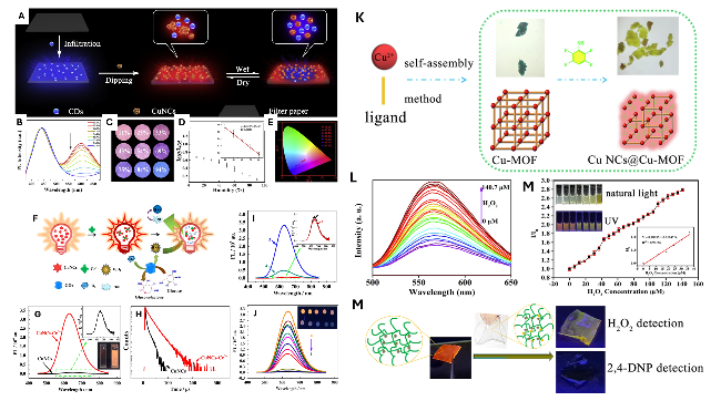
图12 (A) CuNCs-CDs复合材料的合成示意图;(B) 不同湿度下CuNCs-CDs的光致发光光谱;(C) 紫外灯下不同湿度CuNCs-CDs薄膜的荧光图像;(D) 比率荧光强度与湿度的关系曲线;(E) 随湿度变化的CuNCs-CDs色度坐标(xy)的CIE色度图[97]。(F) CuNCs-Ce3+荧光探针用于H2O2检测的示意图;(G) 350 nm激发波长下的荧光光谱和(H) CuNCs与CuNCs-Ce3+的时间分辨荧光衰减曲线;(I) 制备样品在350 nm激发波长下的荧光光谱;(J) HAc-NaAc缓冲液中加入200 μL不同浓度H2O2后CuNCs-Ce3+的荧光光谱[98]。(K) CuNCs@Cu-MOF的合成示意图;(L) 以0.5 mL OPD为过氧化物酶底物时,CuNCs@Cu-MOF暴露于不同浓度H2O2的荧光光谱;(M) CuNCs@Cu-MOF响应值与H2O2浓度(0~140.7 μM)的关系曲线;(N) 用于H2O2和2,4-DNP检测的便携式水凝胶传感器制备示意图[99]

Fig.12 (A) Synthesis of CuNCs-CDs. (B) PL spectra of CuNCs-CDs in different humidity. (C) Images of CuNCs-CDs film in different humidity under the UV light. (D) The relationship between ratiometric fluorescence intensity and humidity. (E) CIE chromaticity diagram of the (xy) color coordinates of the CuNCs-CDs varying with humidity. Reprinted from ref. 97 with permission from Elsevier, 2019. (F) Scheme of CuNCs-Ce3+ fluoroprobe for H2O2 detection. (G) Fluorescence spectra at an excitation wavelength of 350 nm and (H) time-resolved fluorescent decay curves of CuNCs and CuNCs-Ce3+. (I) Fluorescence spectra of the prepared samples at the excitation wavelength of 350 nm. (J) Fluorescence spectra of CuNCs-Ce3+ with 200 μL of different H2O2 concentrations in HAc-NaAc buffer. Reprinted from ref. 98 with permission from Springer Nature, 2021. (K) Synthetic scheme of CuNCs@Cu-MOF. (L) FL spectra of CuNCs@Cu-MOF exposed to different concentrations of H2O2 with 0.5 mL of OPD as the peroxidase substrate; (M) the relation of response values of CuNCs@Cu-MOF with concentrations of H2O2 (0~140.7 μM). (N) The prepared hydrogel portable sensor for H2O2 and 2,4-DNP detection. Reprinted from ref. 99 with permission from Elsevier, 2024

Hydrogen peroxide (H2O2) is a vital signaling molecule that regulates key biological processes. However, elevated levels of H2O2 can cause irreversible oxidative damage to lipids, proteins, and DNA, leading to various diseases, such as neurodegeneration, diabetes, Alzheimer’s disease, and cancer. Du et al.[100] developed a rapid, direct colorimetric probe for detecting H2O2 in aqueous solutions, with the capability to measure concentrations ranging from 0.001 to 1000 mM, without the need for chromogenic reagents or expensive instruments. This probe employs water-soluble mercaptosuccinic acid-stabilized CuNCs (MSA-CuNCs). The color of a diluted CuNC solution changes from claret-red at low H2O2 concentrations (~0.001 mM) to saffron yellow at higher concentrations (around 1000 mM). As H2O2 concentration increases from 0 to 1 mM, the absorption peak at 520 nm decreases due to the aggregation and growth of Cu NPs, which occurs as a result of Cu—S bond cleavage and the detachment of MSA from the Cu NPs surface. Further increases in H2O2 concentration from 1 mM to 1000 mM led to an increase in the absorption peak at 375 nm, attributed to the formation of Cu2O. Hence, MSA-CuNCs serve effectively as a colorimetric sensor for H2O2 across a wide concentration range. Mei et al.[98] synthesized GSH-capped CuNCs via a simple one-pot chemical reduction method, using GSH as both a capping and reducing agent (Fig. 12F~J). The AIE of CuNCs was activated by Ce(Ⅲ), resulting in the formation of the CuNCs-Ce3+ fluoroprobe through electrostatic and coordination interactions. The fluorescence intensity of CuNCs-Ce3+ increased by approximately 40-fold compared to CuNCs, accompanied by a 20-nm blue shift in maximum emission and a 3.45-fold increase in average fluorescent lifetime. The fluorescence of CuNCs-Ce3+ was selectively quenched at 650 nm by H2O2 through a redox reaction, enabling sensitive detection of H2O2 within a linear range of 14~140 μM. The high selectivity of this fluorescent assay highlights its potential for sensitive H2O2 detection in various applications, including real-world care products and human serum samples.
Li et al. [99] synthesized a novel CuNCs@Cu-MOF that exhibits remarkable catalytic properties and intense red emission at 610 nm, utilizing Cu-MOF as a template and 2,3,5,6-tetrafluorobenzenethiol (TFTP) as a reducing agent (Fig. 12K~N). This in situ approach enabled the final product to retain the peroxidase-like activity of Cu-MOF, facilitating the efficient catalysis of H2O2 to produce hydroxyl radicals. These radicals subsequently oxidize o-phenylenediamine (OPD) to yield its oxidation product (OxOPD), resulting in yellow emission at 560 nm. Furthermore, the strong red fluorescence of CuNCs@Cu-MOF was quantitatively quenched as the concentration of 2,4-dinitrophenol (2,4-DNP) increased, due to the IFE. Using 560 and 610 nm as response signals, this composite serves as a sensor for H2O2 and 2,4-DNP, with LOD of 72.5 and 67 nM, respectively. Additionally, a portable sensor was developed using PVA hydrogel for efficient microparticle sampling from various substrates for on-site detection.

4.5 Biomacromolecules and small biomolecules

Electrochemiluminescence (ECL) of CuNCs has widespread applications in sensing and biosensing. However, the ECL performance of protein-templated CuNCs is often hindered by the IFE, which occurs due to the close packing of CuNCs, restricting the activation of internal emitters. Therefore, optimizing molecular arrangements through controlled spatial distribution of CuNCs is crucial, as DNA-templated CuNCs typically exhibit enhanced characteristics compared to protein-based versions. Zhou et al.[101] developed a DNA nanocrane structure on the GCE using DNA hybridization and binding. The tetrahedral configuration of the nanocrane, combined with the manipulator’s sequence, allowed for both lateral and longitudinal separation of CuNCs. A strand displacement reaction, aided by Mn2+ DNAzyme for target recycling, facilitated autonomous DNA movement, generating AT-rich double-stranded DNA sequences on the structure. This method enabled ECL detection of miRNA155 on GCE in the presence of S2O82-, achieving a LOD of 36 attomolar. In another study, Borghei et al.[102] employed DNA templating to detect miRNA155 using two distinct sequences, resulting in an increase in fluorescence of the DNA-CuNC complex from 50 pM to 10 nM, with an LOD of 11 pM.
Monkeypox (MPXV), an Orthopox zoonotic viral infection, is a significant global health concern. The virus can be transmitted easily between humans through both direct and indirect sexual contact, making accurate and early detection essential for reducing mortality rates. Dhanasekaran et al.[103] developed red-fluorescent CuNCs measuring under 10 nm, exhibiting strong fluorescence properties (Fig. 13A). These CuNCs were employed to detect the MPXV antigen (A29P) through antigen-antibody conjugation, utilizing fluorescence and smartphone colorimetric sensing methods. The interaction mechanisms between the antigen (A29P) and antibody (Ab A29) were investigated (Fig. 13B, C). Fluorescence sensing in PBS yielded detection limits of 0.096 nM. For practical applications, the immunosensor array successfully detected A29P in spiked serum samples. Additionally, a smartphone-integrated sensor array facilitated point-of-care (POC) analysis by measuring RGB color changes (Fig. 13D). The results indicate that the synthesized CuNCs could detect A29P through fluorescence and smartphone colorimetric techniques.
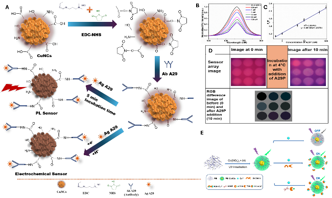
图13 (A) 通过EDC-NHS偶联法修饰的铜纳米簇进行A29P的荧光与电化学检测示意图;(B) 加标血清样本中A29P的光致发光光谱(500 pM~100 nM);(C) 相应的线性拟合图;(D) 不同浓度A29P作用下铜纳米簇的RGB差异图像(紫外灯下通过颜色传感器阵列分析)[103]。(E) 基于Cu2+猝灭的荧光开启式检测方法用于生物硫醇和乙酰胆碱酯酶(AChE)检测的示意图[105]

Fig.13 (A) Fluorescence and electrochemical detection of A29P using Ab A29-modified CuNCs via EDC-NHS coupling. (B) PL spectrum of A29P in the spiked serum sample (500 pM~100 nM). (C) corresponding linear fit diagram, and (D) RGB difference image of CuNCs with different concentrations of A29P, analysis by color sensor array under UV light. Reprinted from ref. 103 with permission from the American Chemical Society, 2024. (E) Scheme of Cu2+-quenched fluorescence turn-on assay for the detection of biothiols and AChE. Reprinted from ref. 105 with permission from Elsevier, 2018

CuNCs have also been utilized for protein detection. Cao et al.[104] developed a method for identifying a small molecule and its interacting protein, using streptavidin and biotin as a model system. The detection strategy comprised two components: protein molecules immobilized on magnetic NPs (MNPs) and ssDNA oligonucleotides with a small molecule linked to their 5′-end. By monitoring the fluorescent intensity of both components, dual signals were generated for the solid detection of either the protein or the small molecule. These signals originated from the formation of CuNCs synthesized through a polythymine templating method. The location of the generated signals, whether in the supernatant or precipitate, indicated the presence of interacting proteins. This biosensor successfully detected streptavidin and biotin within linear ranges of 1~200 nM and 10~1000 nM, achieving LOD of 0.47 and 3.1 nM, respectively, demonstrating potential for broader biomedical applications.

4.6 Enzyme activity detection

Monitoring enzyme activity in living cells is crucial, and MNCs, such as CuNCs, are extensively used for enzyme activity biosensing. Two primary strategies are employed, including the in situ synthesis of CuNCs, which generates fluorescence signals, particularly for nucleic acid-related enzymes. Acetylcholinesterase (AChE), an essential enzyme linked to Alzheimer’s disease, has emerged as a significant target for innovative sensing probes using CuNCs. Due to the mechanism of AChE inhibition by organophosphates, these methods can also be adapted for detecting pesticides.
Yang et al. [105] presented an effective fluorescence sensing method for the detection of multiple analytes using UV-light induction. The PEI-protected CuNCs (PEI-CuNCs) demonstrated exceptional stability and fluorescence intensity, enabling the development of a label-free assay for the sensitive detection of Cu2+Fig. 13E). The interaction between Cu2+ and the —SH functional groups in biothiols led to the formation of the RSH-Cu2+ complex, which triggered the fluorescence recovery of the PEI-CuNCs. GSH and cysteine (Cy), both biothiols, were detected within linear ranges of 1~25 μM and 0.5~25 μM, respectively. For AChE activity, thiocholine (TCh), produced from the hydrolysis of acetylthiocholine by AChE, interacts with Cu2+ to form the TCh-Cu2+ complex, leading to fluorescence recovery in the PEI-CuNCs. The response to AChE was linear within the range of 3~200 UL-1, with a limit of detection of 1.38 UL-1. This sensing platform was also used to detect tacrine, a well-known AChE inhibitor with an IC50 value of 53.4 nM. Additionally, a novel ratiometric fluorescence probe was created to evaluate AChE activity by integrating a single-atom nanozyme (SAzyme) with polyvinylpyrrolidone-protected CuNCs (PVP-CuNCs)[106]. The SAzyme catalyzed the oxidation of OPD to produce 2,3-diaminophenazine (DAP) in the presence of H2O2. DAP emitted fluorescence at 566 nm, while PVP-CuNCs exhibited a peak at 438 nm. As DAP concentration increased, the fluorescence intensity of PVP-CuNCs decreased, confirming FRET between PVP-CuNCs and DAP through fluorescence lifetime measurements. This dual-emission signal system demonstrated high sensitivity for AChE activity, emphasizing the potential of SAzymes in AChE analysis, with further exploration needed for evaluating inhibitors, particularly pesticide residues.
Thiolated CuNCs exhibiting AIE characteristics are emerging as innovative luminescent materials. However, maintaining their intense luminescence in neutral solutions remains challenging. Such as, Zhao et al.[107] synthesized stable CuNCs with improved AIE characteristics by employing a hydrophobic capping agent, 4-methylthiophenol, during the synthesis process. Initially, the CuNCs exhibited weak emission due to the presence of hydrophobic ligands. However, following further processing, they self-assembled into highly red-emissive particles with uniform rod-like morphologies, measuring in the hundreds of nanometers, and displayed robust luminescence that remained stable in neutral and alkaline environments. The introduction of a hydrophobic electron acceptor, 4-nitrophenol, led to approximately 80% quenching of the CuNCs' emission. This quenching mechanism, combined with 4-nitrophenyl-β-D-galactopyranoside (NPGal) as a substrate for β-galactosidase, enabled quantification of β-galactosidase activity in serum, achieving a LOD of 0.9 U·L-1 within a linear range of 2.5~212.0 U·L-1. This work successfully demonstrated the preparation of CuNCs under neutral conditions and highlighted their potential in enzyme activity monitoring. Additionally, Qing et al. [108] utilized micrococcal nuclease (MNase), an enzyme that degrades nucleic acids, to detect Staphylococcus aureus. They found that in the absence of MNase, double-stranded DNA (dsDNA), with sodium ascorbate, served as a template for CuNC formation, exhibiting excitation/emission peaks at 340/570 nm. The fluorescence turn-on strategy based on DNA-templated CuNC synthesis was also applied to detect the activity of enzymes such as T4 polynucleotide kinase phosphatase, DNA polymerase, and uracil-DNA glycosylase (UDG), with both dsDNA and ssDNA serving as templates for CuNC synthesis.

4.7 Others

Quantitative detection of explosive compounds, such as nitroaromatics (e.g., trinitrophenol, dinitrobenzene, TNT) and heterocyclic nitramines (e.g., trinitrotriazine, RDX), has been accomplished using CuNCs as fluorescent turn-on/turn-off probes. Cysteine-stabilized CuNCs were employed for the selective detection of dinitrobenzene and picric acid via a turn-on fluorescence mechanism, demonstrating a linear detection range from 99 nM to 1.3 μM with a LOD of 0.13 μM[109]. For RDX detection, BSA-stabilized CuNCs achieved a LOD of 1.62 nM within a linear concentration range of 0 to 0.238 μM, utilizing a turn-off/on fluorescence mechanism modulated by Zn2+ ions. The presence of RDX promoted Lewis acid-base interactions between Zn2+ and trinitrotriazine, resulting in rapid fluorescence quenching of the CuNCs [110].
TNT, a commonly used military explosive, poses significant environmental risks as a water contaminant. The ability to detect and differentiate 2,4,6-trinitrotoluene (TNT) from its analogs, particularly 2,4,6-trinitrophenol (TNP), is critical for global security and presents substantial challenges in trace detection. Wu et al.[111] successfully synthesized PEI-capped CuNCs with a high density of ―NH2 groups. By carefully controlling the size and structure of these PEI-CuNCs, the FRET mechanism occurred between the PEI-CuNCs and the Meisenheimer complex during TNT detection (Fig. 14A). The calculated affinity energy between PEI-CuNCs and TNP was -9.09 kcal·mol-1, indicating effective electron transfer due to energy level alignment and strong interactions between TNP’s ―OH and the ―NH2 groups on PEI-CuNCs (Fig. 14B). Consequently, TNT and TNP were detectable among 26 types of analogs and cations, achieving LODs of 26.57 and 12.82 nM, respectively. Building on the promising performance of PEI-CuNCs, a CuNC-paper sensing chip was developed by integrating it into commercial filter paper for on-site visual detection of TNT and TNP in simulated environments (Fig. 14C~E).
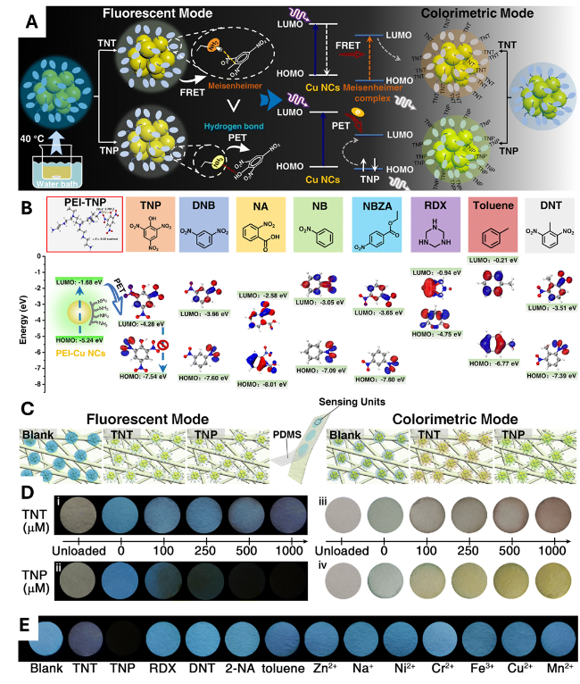
图14 (A) PEI-CuNCs用于TNT和TNP检测的示意图;(B) TNP荧光猝灭中的PET过程及PEI-CuNCs的能级示意图;(C) 用于TNT和TNP检测的CuNCs纸基传感芯片;(D) 传感芯片在荧光和比色模式下检测TNT和TNP的图像;(E) 所用传感芯片的荧光图像[111]

Fig.14 (A) Scheme of PEI-CuNCs for TNT and TNP determination. (B) PET process in fluorescence quenching of TNP and energy levels of PEI-CuNCs. (C) CuNC-paper sensing chip for TNT and TNP determination. (D) Images of the sensing chip for detecting TNT and TNP in fluorescent and colorimetric modes. (E) Fluorescent images of the sensing chip are used. Reprinted from ref. 111 with permission from the Royal Society of Chemistry, 2022

Accurate pH measurement at both environmental and cellular levels is essential for numerous applications in environmental science, biomedicine, and bioprocessing. The initial use of fluorescent CuNCs as pH probes was demonstrated by Zhang’s group[29] in 2014. The prepared BSA-CuNCs exhibited red fluorescence at 620 nm, with a quantum yield of 4.1%. Notably, the fluorescence intensity of BSA-CuNCs significantly increased when the pH decreased from 12 to 6. To broaden the pH response range, Huang et al.[112] developed a pH sensor utilizing trypsin-stabilized fluorescent CuNCs, which demonstrated a linear and reversible decrease in fluorescence as pH increased from 2.02 to 12.14. Similarly, Li et al. [113] created an ultra-sensitive pH sensor based on BSA-capped CuNCs, showing a linear fluorescence response across a pH range of 2 to 14, with a 20-fold increase at elevated pH levels. In addition, Wang et al. [54] prepared CuNCs through interfacial etching of PEI-protected CuNCs, which changed color from colorless to blue as pH varied from 2.0 to 13.2, functioning as an effective colorimetric pH indicator.
Artificial food coloring is vital in the food manufacturing sector, enhancing product appeal through vibrant hues. Synthetic dyes have largely supplanted natural colorants due to their cost-effectiveness, consistent color quality, and stability across a range of temperatures and pH levels. Common food items such as chocolates, baked goods, soft drinks, and dairy products often contain artificial organic colors like Allura Red, Amaranth, Safranin, Brilliant Blue, Quinoline Yellow, Indigo Carmine, Sunset Yellow (SY), and Tartrazine (Tz). The regulation of these colorants is crucial due to potential health risks. For instance, in soft drinks with added juices or flavors, the concentration of SY and Tz should not exceed 100 μg·mL-1. Identifying these colorants is essential, as Tz, a water-soluble synthetic dye, can trigger allergies, asthma, and childhood hyperactivity due to its toxic azo groups and nitrogen-containing aromatic rings (―N $\stackrel{\mathrm{ }\mathrm{ }\mathrm{ }\mathrm{ }}{=}$N―) that release harmful substances such as aromatic amines. Zhang et al.[114] synthesized blue-emissive CuNCs through a straightforward chemical reduction method, using copper sulfate, folic acid, and ascorbic acid as the metal source, capping agent, and reducing agent, respectively. The resulting CuNC sensors exhibited excellent performance and selectivity, utilizing the interfacial fluorescence enhancement and static quenching mechanisms.

5 Conclusions and perspectives

Copper nanoclusters (CuNCs) are emerging as promising candidates for environmental sensing due to their low cost, aqueous dispersibility, tunable luminescence, and relatively low toxicity compared to many semiconductor QDs. When compared to organic dyes and various QDs, CuNCs exhibit greater photostability, larger Stokes shifts, and lower toxicity, although they typically demonstrate lower PLQY than Au/Ag NCs and high-quality QDs. The incorporation of proteins, peptides, DNA, and small-molecule ligands allows for the modulation of emission wavelength, stability, and selectivity, with the choice of ligand and surface chemistry playing a pivotal role in sensor performance. CuNCs have shown high sensitivity in laboratory settings for detecting heavy metal ions, pesticides, pharmaceuticals, and other environmental contaminants. However, key limitations persist, including broad size distributions resulting from current synthesis methods, excitation-dependent emission characteristics, modest PLQY, variable stability in complex matrices, and challenges related to reproducibility and scalability for large-scale production (Fig. 15).
图15 用于提升纳米铜团簇传感器检测环境污染物的策略

Fig. 15 Strategies to improve the CuNC-based sensors towards environmental pollutants sensing

Current sensor designs often rely on straightforward “turn-off” quenching mechanisms, which are susceptible to matrix effects and false positives. To enhance reliability, more robust sensor formats, such as ratiometric, FRET, and “turn-on” approaches, are needed, along with multiplexing strategies. Practical deployment of CuNCs will require their integration with microfluidics, portable devices, and wearable platforms, in addition to establishing standardized protocols for sample handling and validation in real-world conditions. Furthermore, the development of greener synthesis routes, lifecycle assessments, and scale-up strategies is essential for the broader adoption of CuNCs in environmental monitoring.
In addition, the trade-offs between cost and performance must be carefully quantified, as this work emphasizes the low cost of Cu while underestimating the hidden expenses associated with stabilizing ligands, purification processes, and quality control measures necessary to achieve reliable PLQY and stability. Translational efforts should incorporate techno-economic analyses that compare the total cost per test, including materials, processing, and quality control, against existing standard methods.
Moreover, reproducibility and standardization are urgent priorities in CuNC studies. The literature reveals a variety of synthetic recipes that yield disparate size distributions and photophysical properties. Without community standards, such as the reporting of quantum yields measured under standardized conditions, size-distribution metrics, and long-term stability tests, cross-study comparisons and regulatory acceptance will be challenging. Additionally, mechanistic gaps hinder rational design, as the fundamental understanding of how ligand structure, cluster nuclearity, and surface oxidation state influence emission wavelength, PLQY, and susceptibility to quenchers remains incomplete. Targeted mechanistic studies using time-resolved spectroscopy, atomistic modeling, and in situ characterization under realistic matrices are essential for transitioning from trial-and-error approaches to predictive engineering.
Selectivity is still a challenge in complex environmental matrices, such as wastewater, soils, and biological fluids, which present further obstacles. These samples often contain multiple interferents and exhibit variable pH and ionic strength. Many CuNC-based sensors have only been validated in buffered or spiked matrices; therefore, systematic interference studies, competitor ion panels, and blind field tests are necessary to establish real-world selectivity and limits of detection. Then, the trade-off between stability and reactivity must be addressed, as increasing stability through thicker ligand shells or crosslinking may reduce sensor reactivity and sensitivity. Design strategies should explicitly balance robustness with responsiveness, potentially employing modular shells that can adjust permeability or utilizing sacrificial protective groups that are removed under controlled assay conditions.
Despite being presented as low-toxicity alternatives, the environmental fate, persistence, and potential transformation products of CuNCs, such as the release of copper ions and ligand degradation, require thorough ecotoxicological assessments before widespread environmental deployment. Scale-up of CuNC production introduces new challenges, as lab-scale syntheses that maintain tight control over reducing conditions and ligand stoichiometry may not easily translate to industrial batch or continuous-flow production. Therefore, attention to scalable chemistries, including flow synthesis, the use of benign solvents, and minimal purification processes, should be prioritized from the outset.
Opportunities for computational and data-driven design are also noteworthy. This review mentions the potential of machine learning; specifically, combining high-throughput synthesis and characterization with machine learning could expedite the discovery of ligand-cluster combinations that optimize PLQY, stability, and target specificity. However, success relies on the availability of standardized, high-quality datasets. Furthermore, the validation of multifunctional systems for dual roles, such as combining sensing and remediation (e.g., detection coupled with catalytic degradation or sequestration), is essential. While this dual-function approach is appealing, it often results in compromises on both fronts. Rigorous demonstrations of simultaneous, durable sensing and remediation performance are required, ideally with metrics that allow comparisons to standalone remediation technologies.
Finally, the pathways to regulatory approval and user acceptance are complex. For CuNC-based methods to effect change in environmental monitoring practices, they must meet regulatory validation criteria, including repeatability, robustness, and inter-laboratory reproducibility. Additionally, these methods must be designed for ease of use by non-experts. Early engagement with regulators, end-users, and field practitioners will facilitate adoption and uncover unmet practical constraints.
[1]
Zuo Z S, Zhang H, Gao S S, Wang C Q, Chen W T, Hu G Z. J. Environ. Chem. Eng., 2024, 12(5): 113263.

[2]
Lustig W P, Mukherjee S, Rudd N D, Desai A V, Li J, Ghosh S K. Chem. Soc. Rev., 2017, 46(11): 3242.

[3]
Liang M P, Liu Y J, Lu S, Wang Y, Gao C R, Fan K, Liu H Y. Trac Trends Anal. Chem., 2024, 177: 117800.

[4]
Nguyen M P, Kelly S P, Wydallis J B, Henry C S. Anal. Chim. Acta, 2020, 1100: 156.

[5]
Goppelsröder F. J. Prakt. Chem., 1867, 101(1): 408.

[6]
Mako T L, Racicot J M, Levine M. Chem. Rev., 2019, 119(1): 322.

[7]
Huang J L, Qin Y H, Tang S, Kong D Z, Liu C. Prog. Chem., 2024, 36(1): 120

(黄家麟, 秦垚华, 唐盛, 孔德昭, 刘畅. 化学进展, 2024, 36(1): 120)

[8]
Zhou C Y, Huang J, Wang Q, Tang H, Hu Y C, Wang W L. Prog. Chem., 2024, 36(6): 893

(周存银, 黄娟, 王琼, 唐浩, 胡云楚, 王文磊. 化学进展, 2024, 36(6): 893)

[9]
Peng Q, Zhang J J, Fang X Y, Ni J, Song C Y. Prog. Chem., 2022, 34(12): 2573

(彭倩, 张晶晶, 房新月, 倪杰, 宋春元. 化学进展, 2022, 34(12): 2573)

[10]
Zhang H Z, Xu W L, Meng F S, Q. Zhao, Y. Qiao Y Y, Tian Y Y. Prog. Chem., 2025, 37(2):226

(张浩哲, 许文龙, 孟繁升, 赵强, 乔英云, 田原宇. 化学进展, 2025, 37(2): 226)

[11]
Liang M N, Yuan L L, Shao C Y, Zheng X Q, Song Q C, Gu Z R, Lu S. IEEE Sens. J., 2023, 23(16): 18142.

[12]
Yuan L L, Shao C Y, Zhang Q, Webb E, Zhao X H, Lu S. Environ. Res., 2024, 251: 118610.

[13]
Zhang Q, Wang X Q, Yuan L L, Yu L N, Shao C Y, Jia H X, Lu S. Anal. Methods, 2024, 16(14): 2063.

[14]
Du P, Zhang J, Ma J Y, Chu Z K, Cao F, Liu J. Crit. Rev. Anal. Chem., 2024, 54(6): 1416.

[15]
Kumar J V, Rhim J W. J. Environ. Chem. Eng., 2024, 12(2): 111999.

[16]
Liu S D, Zhang L W, Kim H, Sun J Y, Yoon J. Coord. Chem. Rev., 2024, 501: 215575.

[17]
Aparna R S, Anjali Devi J S, Sachidanandan P, George S. Sens. Actuat. B Chem., 2018, 254: 811.

[18]
Cheng Z H, Gu L Q, Zhao Y Y, Yang L L, Chen L, Wang T, Luo M, Wei J C, Li P. J. Hazard. Mater., 2022, 424: 127555.

[19]
Mohammad Hosseini S, Sadeghi S. Microchem. J., 2024, 202: 110751.

[20]
Liang M N, Shao C Y, Zhang Q, Zhang C, Wang Y X, Zheng X Q, Lu S. IEEE Sens. J., 2023, 23(3): 2076.

[21]
Zhang B H, Chen J S, Cao Y T, Chai O J H, Xie J P. Small, 2021, 17(27): 2004381.

[22]
Lu S. Precis. Chem., 2025, 3(10):570.

[23]
Matus M F, Häkkinen H. Nat. Rev. Mater., 2023, 8(6): 372.

[24]
Yao Q F, Zhu M, Yang Z C, Song X R, Yuan X, Zhang Z P, Hu W P, Xie J P. Nat. Rev. Mater., 2025, 10(2): 89.

[25]
Pichugina D A, Kuz’menko N E, Shestakov A F. Russ. Chem. Rev., 2015, 84(11): 1114.

[26]
Yan B Z, University I M, University I M, Sun J, University I M, Liu J H, Li L, University X, Deng H W, University X, Xu Q H, University I M, Wang S, Shen H, University I M. JACS Au, 2025, 5(4): 1884.

[27]
Basu K, Gayen K, Mitra T, Baral A, Roy S S, Banerjee A. ChemNanoMat, 2017, 3(11): 808.

[28]
Serag A, Abduljabbar M H, Althobaiti Y S, Alnemari R M, Almutairi F M, Aldhafeeri M M, Almalki A H. Microchem. J., 2025, 208:112523.

[29]
Wang C, Wang C X, Xu L, Cheng H, Lin Q, Zhang C. Nanoscale, 2014, 6(3): 1775.

[30]
Rotaru A, Dutta S, Jentzsch E, Gothelf K, Mokhir A. Angew. Chem. Int. Ed., 2010, 49(33): 5665.

[31]
Qing Z, He X, He D, Wang K, Xu F, Qing T, Yang X. Angew. Chem. Int. Ed., 2013, 52.

[32]
Shi Y E, Ma J Z, Feng A R, Wang Z G, Rogach A L. Aggregate, 2021, 2(6): e112.

[33]
Wang Z G, Chen B K, Susha A S, Wang W H, Reckmeier C J, Chen R, Zhong H Z, Rogach A L. Adv. Sci., 2016, 3(11): 1600182.

[34]
Wang Y L, Cui Y Y, Liu R, Wei Y T, Jiang X L, Zhu H R, Gao L, Zhao Y L, Chai Z F, Gao X Y. Chem. Commun., 2013, 49(91): 10724.

[35]
Burratti L, Ciotta E, De Matteis F, Prosposito P. Nanomaterials, 2021, 11(2): 276.

[36]
Wang Y, Chen T X, Zhuang Q F, Ni Y N. Talanta, 2018, 179: 409.

[37]
Hu X, Wang W, Huang Y M. Talanta, 2016, 154: 409.

[38]
Sahu D K, Singha D, Sahu K. Sens. Bio Sens. Res., 2019, 22: 100250.

[39]
Liu W Y, Wang K, Liu P, Jiang W Q, Feng Y Y, Hu Y Y, Zheng M M, Zhou Y B, Xiao Y Q, Liu Y N. Anal. Chim. Acta, 2024, 1316: 342842.

[40]
Wang Y, Chen T X, Zhang Z T, Ni Y N. RSC Adv., 2018, 8(17): 9057.

[41]
Hu X, Liu X D, Zhang X D, Chai H X, Huang Y M. Biosens. Bioelectron., 2018, 105: 65.

[42]
Yang J L, Song N Z, Jia Q. Nanoscale, 2019, 11(45): 21927.

[43]
Yang J L, Li Z, Jia Q. Chem. Commun., 2020, 56(20): 3081.

[44]
Liu Z C, Jing X, Zhang S J, Tian Y. Anal. Chem., 2019, 91(3): 2488.

[45]
Shi Y Q, Li W T, Feng X P, Lin L, Nie P C, Shi J Y, Zou X B, He Y. Food Chem., 2021, 344: 128694.

[46]
Biswas S, Miller J T, Li Y H, Nandakumar K, Kumar C S S R. Small, 2012, 8: 687.

[47]
Zhou S C, Duan Y Y, Wang F, Wang C Y. Nanoscale, 2017, 9(15): 4981.

[48]
Li Y, Zaluzhna O, Xu B L, Gao Y, Modest J M, Tong Y J. J. Am. Chem. Soc., 2011, 133(7): 2092.

[49]
Meng X M, Xu Q, Wang S X, Zhu M Z. Nanoscale, 2012, 4(14): 4161.

[50]
Wang Y N, Bürgi T. Nanoscale Adv., 2021, 3(10): 2710.

[51]
Brust M, Walker M, Bethell D, Schiffrin D J, Whyman R. J. Chem. Soc., Chem. Commun., 1994, (7): 801.

[52]
Maity S, Bain D, Patra A. J. Phys. Chem. C, 2019, 123(4): 2506.

[53]
Yuan X, Luo Z T, Zhang Q B, Zhang X H, Zheng Y G, Lee J Y, Xie J P. ACS Nano, 2011, 5(11): 8800.

[54]
Wang C, Yao Y G, Song Q J. Colloids Surf. B Biointerfaces, 2016, 140: 373.

[55]
Jia X F, Li J, Wang E K. Small, 2013, 9(22): 3873.

[56]
Kawasaki H, Kosaka Y, Myoujin Y, Narushima T, Yonezawa T, Arakawa R. Chem. Commun., 2011, 47(27): 7740.

[57]
Reetz M T, Helbig W. J. Am. Chem. Soc., 1994, 116(16): 7401.

[58]
Vilar-Vidal N, Blanco M C, López-Quintela M A, Rivas J, Serra C. J. Phys. Chem. C, 2010, 114(38): 15924.

[59]
Vilar-Vidal N, Rey J R, López Quintela M A. Small, 2014, 10(18): 3632.

[60]
Li Y L, Feng L Y, Yan W, Hussain I, Su L, Tan B E. Nanoscale, 2019, 11(3): 1286.

[61]
Bi N, Xi Y H, Hu M H, Xu J, Gou J, Li Y X, Zhang L N, Jia L. Colloids Surf. B Biointerfaces, 2023, 228: 113404.

[62]
Yang S Q, Li G H, Song C K, Liu M Y, Yuan Z W. Colloids Surf. A Physicochem. Eng. Aspects, 2020, 606: 125514.

[63]
Zhang W, Chen Z L, Xiong C, Yuan L Z, Hu J J, Dai J, Xia F, Lou X D. ACS Nano, 2024, 18(50): 34244.

[64]
Qing Z H, Bai A L, Xing S H, Zou Z, He X X, Wang K M, Yang R H. Biosens. Bioelectron., 2019, 137: 96.

[65]
Kim S, Lee E S, Cha B S, Park K S. Nano Lett., 2022, 22(15): 6121.

[66]
Li X, Xie S Y, Qin C, Liu S R, Su F L, Zhang X, Chen Y, Li B Z. Microchem. J., 2024, 197: 109859.

[67]
Xu F Z, Qiao Z Z, Luo L, He X X, Lei Y L, Tang J L, Shi H, Wang K M. Talanta, 2022, 243: 123323.

[68]
Gan M Y, Wang Y Q, Wang F, Tan J, Pei Y H, Wang J H, Choi M M F, Bian W. Luminescence, 2024, 39(2): e4689.

[69]
Zhang C, Liang M N, Shao C Y, Li Z W, Cao X, Wang Y X, Wu Y N, Lu S. ACS Appl. Bio Mater., 2023, 6(3): 1283.

[70]
Fernández-Ujados M, Trapiella-Alfonso L, Costa-Fernández J M, Pereiro R, Sanz-Medel A. Nanotechnology, 2013, 24(49): 495601.

[71]
Gan M Y, Wang Y Q, Wang F, Tan J, Pei Y H, Wang J H, Choi M M F, Bian W. Luminescence, 2024, 39(2): e4689.

[72]
Mu G M, Zou J, Ma M, Yan P, Li M T, Xia H, Xu F J. Microchem. J., 2025, 213: 113806.

[73]
Deb A K, Biswas B, Goswami N, Hilder E F, Naidu R, Rahman M M. Appl. Surf. Sci., 2021, 546: 149122.

[74]
Li J Y, Fu W X, Bao J C, Wang Z Y, Dai Z H. ACS Appl. Mater. Interfaces, 2018, 10(8): 6965.

[75]
Jain S, Nehra M, Dilbaghi N, Chaudhary G R, Kumar S. BME Front., 2024, 5: 78.

[76]
Saleh S M, El‐Sayed W A, El-Manawaty M A, Gassoumi M, Ali R. Biosensors, 2022, 12: 197.

[77]
Cao H Y, Zhang X D, Tang M J, Dong W F, Shi W B. Sens. Actuat. B Chem., 2023, 392: 134066.

[78]
Han B Y, Hou X F, Xiang R C, Yu M B, Li Y, Peng T T, He G H. Chin. J. Anal. Chem., 2017, 45(1): 23.

[79]
Goswami N, Giri A, Bootharaju M S, Xavier P L, Pradeep T, Pal S K. Anal. Chem., 2011, 83(24): 9676.

[80]
Ding M, Tang L, Ma X S, Song C X, Wang S X. Commun. Chem., 2022, 5: 172.

[81]
Wang D W, Wang Z Q, Wang X B, Zhuang X M, Tian C Y, Luan F, Fu X L. J. Agric. Food Chem., 2020, 68(40): 11301.

[82]
Zhang G M, Wang R R, Shi L H, Zhang C H, Zhang Y, Zhou Y, Dong C, Li G, Shuang S M. Sens. Actuat. B Chem., 2019, 279: 361.

[83]
Wei D L, Li M W, Wang Y, Zhu N F, Hu X L, Zhao B Y, Zhang Z, Yin D Q. J. Hazard. Mater., 2023, 441: 129890.

[84]
Xu J M, Zhou H M, Zhang Y X, Zhao Y, Yuan H, He X X, Wu Y, Zhang S J. J. Hazard. Mater., 2022, 428: 128158.

[85]
Yang Y, Zhang Y H, Jiang S T, Shan X H, Guo X, Liu H Q, Shen L R, Chang W G. Langmuir, 2024, 40(1): 827.

[86]
Gao Z, Su R X, Qi W, Wang L B, He Z M. Sens. Actuat. B Chem., 2014, 195: 359.

[87]
Zheng X, Chen Q M, Zhang Z X, Wang Z L, Gong Z J. Microchem. J., 2022, 180: 107570.

[88]
Zhang J H, Zhang Z T, Ou Y J, Zhang F, Meng J, Wang G, Fang Z L, Li Y. Nanoscale, 2020, 12(37): 19429.

[89]
Bagheri H, Afkhami A, Khoshsafar H, Hajian A L, Shahriyari A. Biosens. Bioelectron., 2017, 89: 829.

[90]
Yang Y, Wei Q Y, Zou T, Kong Y L, Su L F, Ma D, Wang Y D. Sens. Actuat. B Chem., 2020, 321: 128534.

[91]
Yousefzadeh A, Hassanzadeh J, Mousavi S M J, Yousefzadeh M. Sens. Actuat. B Chem., 2019, 286: 154.

[92]
Nerthigan Y, Sharma A K, Pandey S, Wu H F. Microchim. Acta, 2019, 186(3): 130.

[93]
Hatefi A, Rahimpour E, Khoubnasabjafari M, Edalat M, Jouyban-Gharamaleki V, Alvani-Alamdari S, Nokhodchi A, Pournaghi-Azar M H, Jouyban A. Microchim. Acta, 2019, 186(3): 194.

[94]
Sivasankaran U, Radecki J, Radecka H, Girish Kumar K. Luminescence, 2019, 34(2): 243.

[95]
Yang K C, Wang Y Y, Lu C S, Yang X M. J. Lumin., 2018, 196: 181.

[96]
Cheng Y, Sun F F, Zhou Y H. J. Lumin., 2018, 197: 376.

[97]
Wen Z Q, Song S L, Wang C X, Qu F D, Thomas T, Hu T T, Wang P, Yang M H. Sens. Actuat. B Chem., 2019, 282: 9.

[98]
Mei H, Ma Y G, Wu H M, Wang X D. Anal. Bioanal. Chem., 2021, 413(8): 2135.

[99]
Li H J, Wu Y Y, Xu Z Q, Wang Y. Chem. Eng. J., 2024, 479: 147508.

[100]
Du Y B, Fang J, Wang H L, Yang Y. ACS Appl. Mater. Interfaces, 2017, 9(12): 11035.

[101]
Zhou Y, Wang H J, Zhang H, Chai Y Q, Yuan R. Anal. Chem., 2018, 90(5): 3543.

[102]
Borghei Y S, Hosseini M, Ganjali M R. Microchim. Acta, 2017, 184(8): 2671.

[103]
Dhanasekaran B, Chandran M, Chellasamy G, Veerapandian M, Govindaraju S, Yun K. ACS Appl. Bio Mater., 2024, 7(9): 6065.

[104]
Cao J P, Wang W, Bo B, Mao X X, Wang K M, Zhu X L. Biosens. Bioelectron., 2017, 90: 534.

[105]
Yang J L, Song N Z, Lv X J, Jia Q. Sens. Actuat. B Chem., 2018, 259: 226.

[106]
Wang M K, Liu L, Xie X L, Zhou X B, Lin Z H, Su X G. Sens. Actuat. B Chem., 2020, 313: 128023.

[107]
Zhao M Z, Qian Z S, Zhong M T, Chen Z T, Ao H, Feng H. ACS Appl. Mater. Interfaces, 2017, 9(38): 32887.

[108]
Qing T P, Long C C, Wang X, Zhang K W, Zhang P, Feng B. Microchim. Acta, 2019, 186(4): 248.

[109]
Hambarde G, Bothra S, Upadhyay Y, Bera R K, Sahoo S K. Microchem. J., 2019, 147: 899.

[110]
Aparna R S, Anjali Devi J S, Anjana R R, Nebu J, George S. Sens. Actuat. B Chem., 2019, 291: 298.

[111]
Wu H T, Wang G F, Cai Z Z, Li D Z, Xiao F F, Lei D, Dai Z H, Dou X C. Anal. Methods, 2022, 14(44): 4485.

[112]
Wang W, Leng F, Zhan L, Chang Y, Yang X X, Lan J, Huang C Z. Anal., 2014, 139(12): 2990.

[113]
Liao X Q, Li R Y, Long X H, Li Z J. RSC Adv., 2015, 5(60): 48835.

[114]
Zhang S, Zhu M L, Zhang W T, Guo Y Y. Microchem. J., 2024, 207: 112146.

Outlines

/