Home Journals Progress in Chemistry
Progress in Chemistry

Abbreviation (ISO4): Prog Chem      Editor in chief: Jincai ZHAO

About  /  Aim & scope  /  Editorial board  /  Indexed  /  Contact  / 
Review

The Single-Cell RNA Sequencing Technology Based on Microfluidic Chips

  • Luxi Shu ,
  • Yan Zhang , *
Expand
  • Faculty of Science, Kunming University of Science and Technology, Kunming 650500, China

Received date: 2025-07-04

  Revised date: 2025-08-05

  Online published: 2025-08-29

Supported by

National Natural Science Foundation of China(32460624)

Kunming University of Science and Technology “Double First-Class” Science and Technology Project(202201BE070001-028)

Yunnan Provincial Education Science Planning Project(BC23002)

Technology 19th “Challenge Cup” National College Student Extracurricular Academic and Technological Works Competition Incubation Project(2024ZK128)

Abstract

Cell heterogeneity is key to understanding life processes such as embryonic development and disease evolution,while traditional bulk cell RNA sequencing cannot resolve gene expression differences at the single-cell level. Although single-cell RNA sequencing (scRNA-seq) technology can construct transcriptomic maps at single-cell resolution,it faces challenges such as low efficiency in single-cell isolation and capture,and large deviations in trace RNA manipulation. Microfluidic chip technology,through a microscale fluid manipulation system,integrates processes such as single-cell isolation,lysis,reverse transcription,amplification,and sequencing library construction,achieving high-throughput,low sample loss,and automated operations,which significantly improve the efficiency and data reliability of scRNA-seq. This paper outlines the sequencing process of scRNA-seq,including steps such as single-cell isolation and capture,RNA extraction,reverse transcription and amplification,and single-cell sequencing. It analyzes the core advantages of microfluidic chips in adapting to single cells,precisely controlling reaction volumes,and realizing process automation,and briefly describes the technical principles and characteristics of representative platforms such as Fluidigm C1,10X Genomics Chromium,and BD Rhapsody. Microfluidic chip technology provides an efficient and precise technical platform for scRNA-seq. In the future,with the continuous optimization of chip design and the improvement of multi-omics integrated analysis capabilities,we expect it to play a more profound role in resolving complex biological systems,revealing disease mechanisms,and even promoting precision medicine.

Contents

1 Introduction

2 Single-cell RNA sequencing workflow

2.1 Isolation and capture of single cells

2.2 RNA extraction,reverse transcription and amplification

2.3 Single-cell sequencing

3 Single-cell RNA sequencing technology based on microfluidic chips

3.1 Development history of scRNA-seq based on microfluidic chips

3.2 Core advantages of microfluidic chips in scRNA-seq

4 Representative microfluidic single-cell RNA sequencing platforms

4.1 Fluidigm C1 platform

4.2 10X Genomics chromium platform

4.3 BD rhapsody platform

5 Summary and prospects
Cite this article

Luxi Shu , Yan Zhang . The Single-Cell RNA Sequencing Technology Based on Microfluidic Chips[J]. Progress in Chemistry, 2026 , 38(2) : 283 -297 . DOI: 10.7536/PC20250706

1 Introduction

As the basic unit of life activities, cells do not exist with homogeneous functional characteristics. Even within the same tissue or cell population, individual cells exhibit significant differences in gene expression patterns, metabolic activity, and functional states. This cellular heterogeneity has a decisive impact on embryonic development[1], disease evolution[2], immune response[3], and other key biological processes. However, traditional bulk cell RNA sequencing (Bulk RNA-seq) mixes millions of cells for detection, essentially performing a statistical average of gene expression across heterogeneous cell populations, thereby masking cellular heterogeneity[4]. In 2009, Tang et al.[5]achieved mammalian single-cell whole-transcriptome sequencing for the first time, marking the beginning of the single-cell omics era. The breakthrough development of single-cell RNA sequencing (scRNA-seq) technology enabled, for the first time, the construction of genome-wide expression maps at single-cell resolution. It not only precisely identifies cell subpopulations and their molecular features but also dynamically tracks cell state transition trajectories, providing an unprecedented perspective for deeply understanding life processes[6].
scRNA-seq technology enables precise measurement of the expression levels of thousands of genes in each cell by performing reverse transcription, amplification, and high-throughput sequencing on the RNA of single cells[7]. Utilizing this technology, it is possible to achieve identification of new cell types and subpopulations, map cell differentiation and developmental trajectories, and analyze dynamic changes in cell states during disease progression. In the field of developmental biology, scRNA-seq aids in studying the differentiation pathways and molecular regulatory mechanisms from embryonic stem cells to various terminally differentiated cells[8]; in cancer research, this technology helps reveal tumor cell heterogeneity, discover cancer stem cells and drug-resistant subpopulations, and provides a theoretical basis for precision cancer therapy[9]; in immunological research, scRNA-seq enables in-depth analysis of the diversity and functional states of immune cells, explaining the molecular mechanisms of immune responses[10].
Although scRNA-seq technology holds great potential, it still faces numerous challenges in practical applications. Among these, the isolation and capture of single cells are critical steps in scRNA-seq experiments; traditional single-cell isolation methods such as flow cytometry[11], micromanipulation[12]suffer from issues such as low throughput, high cost, and significant cell damage, making them difficult to meet the demands of large-scale single-cell analysis. Furthermore, the RNA content in single cells is extremely low, and biases and noise are easily introduced during RNA capture, reverse transcription, and amplification processes, affecting the quality and accuracy of sequencing data[13].
The emergence of microfluidic chip technology provides an effective solution to the challenges faced by scRNA-seq[14]. Microfluidic chips are miniaturized devices that integrate microstructures such as microchannels, micropumps, and microvalves onto a chip of a few square centimeters, enabling precise manipulation of trace fluids at the micrometer scale[15]. They offer advantages such as small size, high integration, high throughput, low reagent consumption, and fast analysis speed; therefore, using microfluidic chip devices for cell biology research, especially single-cell analysis, has inherent advantages. Combining microfluidic chip technology with scRNA-seq enables efficient separation, capture, and processing of single cells, reducing sample loss and contamination during experimental operations, and improving the quality and reliability of sequencing data[16]. Meanwhile, the multi-functional integration characteristics of microfluidic chips allow multiple experimental steps such as single-cell sorting, lysis, reverse transcription, amplification, and library construction to be completed continuously on the chip, achieving automation and integration of scRNA-seq experiments, greatly improving detection efficiency and reducing analysis costs[17].
In summary, microfluidic chip-based single-cell RNA sequencing technology holds significant importance in life science research, providing a powerful tool for deeply analyzing cellular heterogeneity and revealing mechanisms of cell function and fate regulation. This article first introduces the workflow of single-cell RNA sequencing, including steps such as single-cell isolation and capture, RNA extraction, reverse transcription and amplification, and single-cell sequencing. It also summarizes the methods and core advantages of using microfluidic chips for scRNA-seq and discusses representative microfluidic single-cell RNA sequencing platforms. Finally, it offers forward-looking perspectives on the application of this technology in areas such as spatial multi-omics integration and clinical point-of-care testing, aiming to promote the development of precision medicine and personalized therapy.

2 Single-cell RNA sequencing workflow

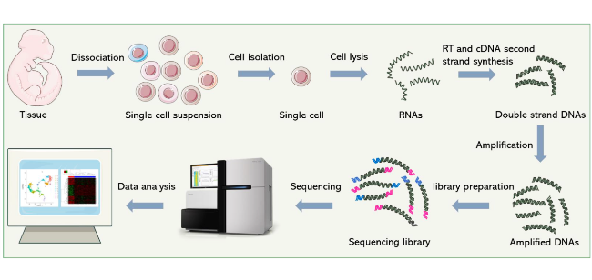
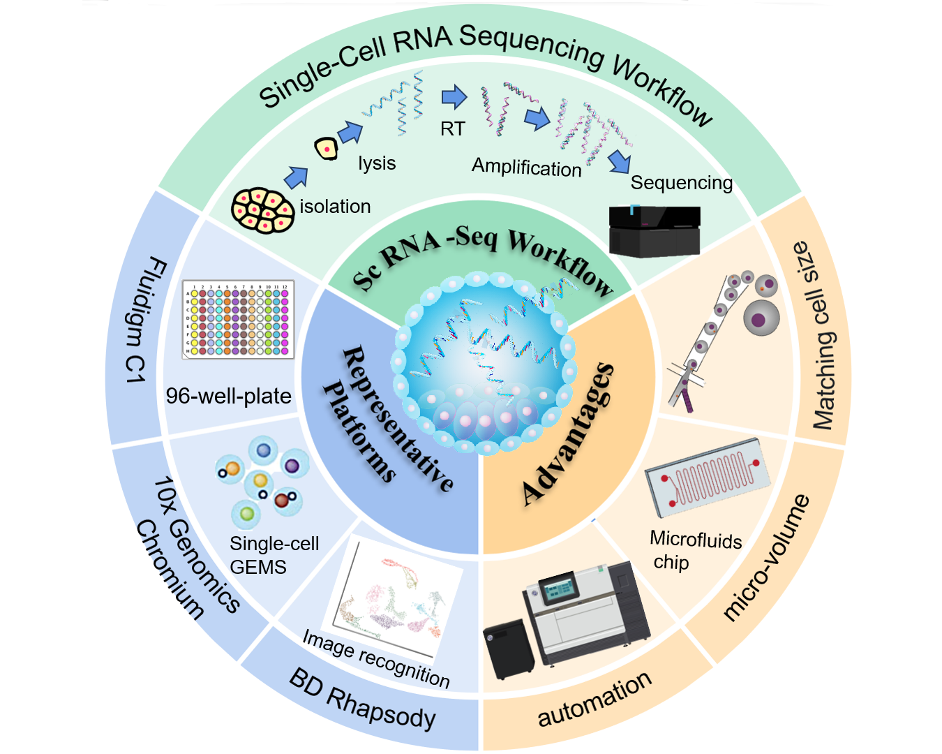
scRNA-seq can precisely resolve the gene expression status of each cell within a cell population, revealing heterogeneity among cells. It can be used to discover new cell types and states, gain deep insights into developmental processes and disease progression mechanisms, and has broad applications and significant importance in biological and medical research fields. Its sequencing workflow mainly includes several steps such as single-cell isolation and capture, RNA extraction, reverse transcription and amplification, and single-cell sequencing (Figure 1)[18].
图1 单细胞RNA测序流程[18]

Fig.1 The workflow of single-cell RNA sequencing[18]

2.1 Isolation and Capture of Single Cells

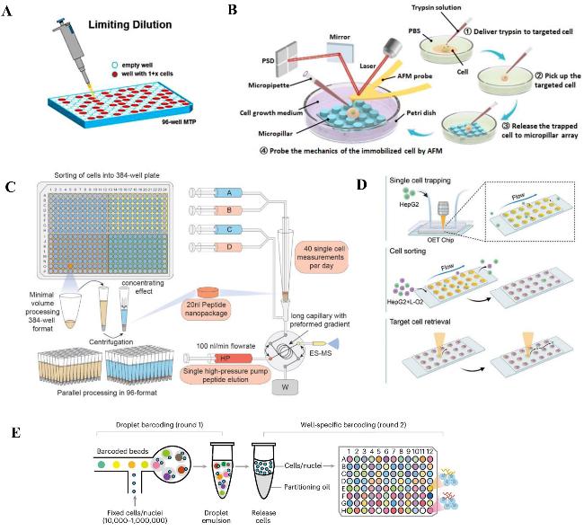
Isolation and capture of single cells are the primary critical steps in scRNA-seq[19], and their isolation efficiency directly affects the quality and accuracy of subsequent sequencing data. Currently, commonly used methods for single-cell isolation and capture mainly include limiting dilution[20], micromanipulation[21], fluorescence-activated cell sorting[22](FACS), microwell technology[23], and droplet microfluidics[24]and other techniques; each method has its unique principles, advantages, and limitations.Table 1provides a comparison of these methods.
表1 常见的单细胞分离方法对比

Table 1 Comparison of common single-cell isolation methods

Method Principle Throughput Advantages Limitations Ref
Limited dilution Based on Poisson distribution,single cells are randomly distributed in the well plate through gradient dilution (Fig.2A Low
(≤ 384 well plates)
Simple equipment,low cost,suitable for small-scale research High empty well rate,low throughput,dependent on operational experience 20
Micromanipulation Manually manipulating a micropipette under a microscope to directly aspirate single cells (Fig.2B Extremely low Precisely select cells of specific morphology or position without labeling interference Extremely low throughput,time-consuming operation,dependent on highly skilled personnel 21
Fluorescence-activated cell sorting (FACS) After fluorescent labeling,target single cells are sorted by droplet electric field (Fig.2C High
(> 104 cells/hour)
High throughput,multi-parameter sorting (supporting combined labeling of multiple markers) Expensive equipment,fluorescent labeling may change cell status,requiring high-quality single-cell suspensions 22
Micro-well technology The size matches single cells,and cells randomly fall into the micro-wells (Fig.2D High
(about
200 000 micro-wells)
High throughput,low damage,compatible with various cell types The empty well rate is about 30 %,and the size of the micro-wells needs to strictly match the cell type 23
Droplet microfluidics Microfluidics generates independent droplets containing single cells + barcoded microbeads (Fig.2E Ultra-high
(> 104 cells/time)
Ultra-high throughput,high capture efficiency,high degree of automation High equipment and consumable costs,strict requirements for droplet stability,requiring complex bioinformatics for deduplication 24

2.2 RNA Extraction, Reverse Transcription, and Amplification

Extracting RNA from single cells, reverse transcribing it into cDNA, and subsequently amplifying it are critical steps in the scRNA-seq workflow, directly determining whether high-quality sequencing data can be obtained. Due to the extremely low RNA content in single cells, typically ranging from a few picograms to tens of picograms, these processes impose exceptionally high requirements on RNA extraction, reverse transcription, and amplification technologies.
图2 常见的单细胞分离方法:(A) 有限稀释示意图[20];(B) 显微操作法分离单细胞[21];(C) FACS流程示意图[22];(D) 微孔列阵捕获单细胞[23];(E) 液滴微流控工作流程图[24]

Fig.2 The most-used single-cell isolation methods. (A) Isolation of single cells by micromanipulation method[20];(B) schematic diagram of micromanipulation[21];(C) FACS flow diagram[22];(D) microporous arrays capture single cells[23];(E) diagram of droplet microfluidics workflow[24]

2.2.1 RNA extraction

RNA extraction is the first step in obtaining single-cell RNA information. After separation and capture, cells are transported through a preset microchannel network within a microfluidic chip to the RNA extraction functional zone under precise fluid control. Subsequently, lysis buffer is delivered through the same channel system to achieve cell lysis and release RNA. Currently, common single-cell RNA extraction methods mainly include chemical lysis[25]and automated extraction based on microfluidic chips[26]. The chemical lysis method utilizes a lysis buffer containing components such as surfactants and proteases to rapidly lyse single cells, releasing the RNA inside. A commonly used lysis buffer is Trizol[27]. As a total RNA extraction reagent, Trizol contains substances such as guanidine isothiocyanate. Its principle is that guanidine isothiocyanate can rapidly break down cells while denaturing proteins in nucleoprotein complexes to release nucleic acids; this process requires the addition of RNase inhibitors to prevent RNA degradation. Subsequently, impurities such as proteins and DNA are removed via layered centrifugation to obtain pure RNA. This method is relatively simple to operate but is susceptible to RNase contamination, which can lead to RNA degradation and affect extraction efficiency. Furthermore, Trizol is expensive and contains toxic substances such as phenol and isothiocyanates; proteinase K, which offers biosafety, provides a solution to these issues[28]. The automated extraction method based on microfluidic chips involves capturing single cells into micro-reaction units on the chip. By utilizing integrated structures such as micropumps and microvalves on the chip, the flow of lysis buffer, wash buffer, and elution buffer is precisely controlled to achieve automated single-cell RNA extraction. Zhang et al.[29]designed a microfluidic chip named Paired-seq based on the principle of differential flow resistance, achieving efficient separation and pairing of single cells and magnetic beads with a pairing efficiency as high as 95%. Through the integration of valves and pumps, this chip effectively removes free RNA, enabling efficient cell lysis and mRNA capture. This method reduces human operational errors and lowers the risk of RNase contamination, thereby improving the efficiency and quality of RNA extraction. Meanwhile, the miniaturization and integration characteristics of microfluidic chips[30]allow for the processing of single cells within extremely small volumes, reducing reagent consumption and sample loss. However, this method requires specialized microfluidic chip equipment, resulting in higher costs.

2.2.2 Reverse transcription

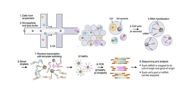
Reverse transcription is the process of converting extracted RNA molecules into complementary DNA (cDNA), laying the foundation for subsequent PCR amplification and sequencing analysis. In scRNA-seq, oligo(dT) primers are typically used to bind to the poly(A) tail of mRNA, synthesizing cDNA from an RNA template under the action of reverse transcriptase.[31]. To improve the efficiency and accuracy of reverse transcription, Macosko et al.[32]introduced special molecular tags called UMIs (Unique Molecular Identifiers) into the reverse transcription primers, synthesized oligonucleotide structures on magnetic beads, and utilized these beads to deliver a large number of primers with unique barcodes into individual droplets. By employing droplet microfluidic devices, cells and beads were co-encapsulated, asFigure 3shows. After cell lysis, mRNA is released and binds to primer-carrying beads for reverse transcription, forming STAMPs (Single-cell Transcriptomes Attached to Microparticles). Reading the sequences on the STAMPs allows inference of the cellular origin of each transcript. In this process, the UMI, as a short random nucleotide sequence, assigns a unique tag to each mRNA molecule, effectively correcting biases generated during PCR amplification. Sun et al.[33]co-encapsulated human and mouse cells with barcodes incorporating homotrimer UMIs, significantly reducing transcript count bias introduced by PCR amplification and improving the accuracy of gene expression quantification. Furthermore, conditions of the reverse transcription reaction, such as temperature[34], time[35], primers[36], and reverse transcriptase[37], among others, have a significant impact on the efficiency of reverse transcription and the quality of cDNA. Reverse transcription reaction conditions need to be optimized according to different experimental requirements and sample characteristics to obtain high-quality cDNA.
图3 微珠共封装细胞的微流控装置[32]

Fig.3 Microfluidics device for co-encapsulating cells with beads[32]

2.2.3 Amplification

Since the initial amount of RNA in a single cell is extremely small, the amount of cDNA obtained through reverse transcription is insufficient to meet the requirements of high-throughput sequencing; therefore, the cDNA needs to be amplified. Currently, there are two main methods commonly used for single-cell cDNA amplification: PCR-based amplification methods[38]and in vitro transcription (IVT)-based amplification methods[39]. PCR-based amplification methods rely on PCR technology, using the cDNA obtained from reverse transcription as a template. Through the specific binding of primers and the action of DNA polymerase, the cDNA is exponentially amplified. This method is simple to operate and has high amplification efficiency, enabling the acquisition of large amounts of cDNA in a short time. However, PCR amplification processes are prone to introducing amplification bias; the amplification efficiency of cDNA fragments of different lengths and sequences may vary, resulting in an amplified cDNA library that does not accurately reflect the expression levels of the original RNA[40]. To reduce PCR amplification bias, one can employ touchdown PCR[41]amplification, or add an appropriate amount of amplification correction reagents during the amplification process[42]and other methods. The in vitro transcription-based amplification method was first introduced by Eberwine[43]. In this method, a first round of reverse transcription is performed first, using T7-oligo(dT) primers to reverse transcribe mRNA into double-stranded cDNA containing a T7 promoter. Subsequently, the in vitro transcription enzyme T7 RNA polymerase recognizes the T7 promoter and uses the double-stranded cDNA as a template to linearly synthesize antisense RNA (aRNA). These aRNAs are then reverse transcribed back into cDNA, thereby achieving cDNA amplification. The advantage of this method is that it can avoid PCR amplification bias, making the amplified cDNA library more accurately reflect the expression levels of the original RNA[44]. However, the in vitro transcription amplification method is relatively complex to operate, requiring multiple enzymatic reaction steps. After multiple rounds of amplification, long transcripts may be lost. Additionally, the experimental cost is higher, the amplification efficiency is relatively lower, and the amplification time is longer[45].

2.3 Single-cell sequencing

High-throughput sequencing technology is one of the core technologies for single-cell RNA sequencing. It can rapidly and accurately determine the sequence information of cDNA in single cells, providing a massive data foundation for subsequent data analysis. The principle of high-throughput sequencing is mainly based on Sequencing by Synthesis (SBS) technology[46]. Taking the Illumina sequencing platform as an example, it is currently one of the most widely used sequencing platforms in scRNA-seq[47]. During the sequencing process, the amplified cDNA library is first fragmented, and specific adapter sequences are ligated to both ends of the fragments. These cDNA fragments with adapters are immobilized on the surface of a sequencing flow cell to form single-stranded DNA templates[48]. Then, a sequencing reaction solution containing DNA polymerase, dNTPs, and fluorescently labeled reversible terminators is added. Under the action of DNA polymerase, dNTPs bind to the template strand according to the principle of complementary base pairing, adding one dNTP at a time. Since the dNTPs carry fluorescent labels and reversible terminating groups, once a dNTP is added to the growing strand, the reversible terminating group prevents the addition of the next dNTP. At this point, laser scanning can detect the fluorescent signal, thereby determining the base type at that position[48-49]. Subsequently, the reversible terminating groups are removed to continue the next round of base addition and detection. This cycle repeats to achieve base-by-base sequencing of the cDNA fragments. Through parallel sequencing of a large number of cDNA fragments, the Illumina sequencing platform can generate billions of sequencing reads in a single sequencing reaction, covering most of the transcriptome information in a single cell[50].
In addition to the Illumina sequencing platform, the PacBio single-molecule real-time sequencing platform and the Oxford Nanopore nanopore sequencing platform also play key roles. The PacBio platform[51]utilizes single-molecule real-time sequencing technology to achieve direct sequencing of individual DNA molecules without pre-amplification steps. Leveraging the advantage of long-read sequencing data, it excels in resolving full-length transcript structures. The Oxford Nanopore platform[52]is based on the principle of nanopore sensing; when a DNA molecule passes through a nanopore, it triggers changes in electrical current. By precisely detecting these current signals, the DNA base sequence can be deciphered. It features real-time sequencing, long reads, and portable equipment. Different high-throughput sequencing platforms vary in sequencing principles, read length, throughput, accuracy, and cost; therefore, the appropriate sequencing platform must be selected based on specific research objectives and requirements.

3 Single-cell RNA sequencing technology based on microfluidic chips

3.1 Development history of scRNA-seq based on microfluidic chips

Single-cell RNA sequencing is a core technology for resolving cellular heterogeneity, providing an unprecedented perspective for understanding the complexity of life. In 2009, Tang et al.[5]developed a mouse oocyte sequencing technology based on Smart-seq[53], marking the beginning of a new era in single-cell analysis. For the first time, full-length cDNA sequencing of mammalian single cells was achieved, but at this stage, single-cell isolation still relied on low-throughput micromanipulation. The rise of microfluidic chip technology completely revolutionized this situation[54]. In 2011, Fluidigm launched the C1 integrated single-cell microfluidic chip[55], which separated individual cells into independent reaction chambers via microchannels, achieving parallel lysis, reverse transcription, and pre-amplification of 96 cells for the first time, initially demonstrating the integration advantages of microfluidic chips in single-cell processing. However, specific models of C1 chips could only capture cells of certain sizes, resulting in low capture efficiency and high costs. Subsequently, breakthroughs in droplet microfluidics technology[56] spurred the emergence of inDrop[57], Drop-seq[32,58], and 10X Genomics' Chromium[59]and other epoch-making platforms. The emergence of this technology greatly improved the throughput of scRNA-seq, pushing the throughput of a single experiment to thousands of cells through the combination of water-in-oil droplets and barcoded magnetic beads, while significantly reducing costs. After 2016, the popularization of high-throughput scRNA-seq promoted research developments in cancer heterogeneity and the immune microenvironment[60]. Since then, single-cell sequencing platforms based on microfluidic chips and microdroplet technology have continuously emerged and been optimized, with multimodal integration becoming a trend. Competitive platforms such as BD Rhapsody[61] have expanded technological diversity through microwell plate and magnetic bead combination schemes. Microfluidic chips have begun to integrate proteomics and epigenetic detection, optimizing capture efficiency and clinical translation capabilities. With the maturation of commercialized microfluidic chip-based scRNA-seq, microfluidic chips have been applied to single-cell spatial transcriptomics research[14], resolving the spatial distribution of gene expression at single-cell resolution and enabling automated, portable clinical point-of-care testing[62]. Recently, deep learning methods have further empowered cell annotation and drug prediction[63], accelerating the penetration of this technology from a research tool into clinical application scenarios such as early tumor screening[64], personalized medicine, and more. In the future, it may reshape the boundaries of precision medicine through million-level throughput, live-cell dynamic tracking, and standardized diagnostic protocols.

3.2 Core advantages of microfluidic chips in scRNA-seq

The rapid development of microfluidic chips has opened new avenues for scRNA-seq[65]. Through ingeniously designed micrometer-scale channels and reaction chambers, it integrates processes such as single-cell capture and amplification onto the chip, demonstrating unique advantages in the field of scRNA-seq: First, microfluidic chips exhibit high flexibility in structural and functional design, capable of precisely adapting to diverse single-cell analysis requirements[66]; Second, microfluidic channels have dimensions ranging from tens to hundreds of micrometers, suitable for solution volumes from picoliters to nanoliters, significantly reducing sample loss and enhancing detection sensitivity[67]; Third, the high integration of multifunctional units with microfluidic chips achieves process automation, largely avoiding errors introduced by manual operations[68].

3.2.1 Adapted for single-cell

The bottlenecks of early single-cell isolation techniques have severely constrained research progress at the single-cell level. Due to their small volume, single-cell isolation remains a major challenge in scRNA-seq. Microfluidic devices typically feature internal dimensions below 100 μm and reaction volumes in the picoliter to nanoliter range, making them highly suitable for single-cell isolation.
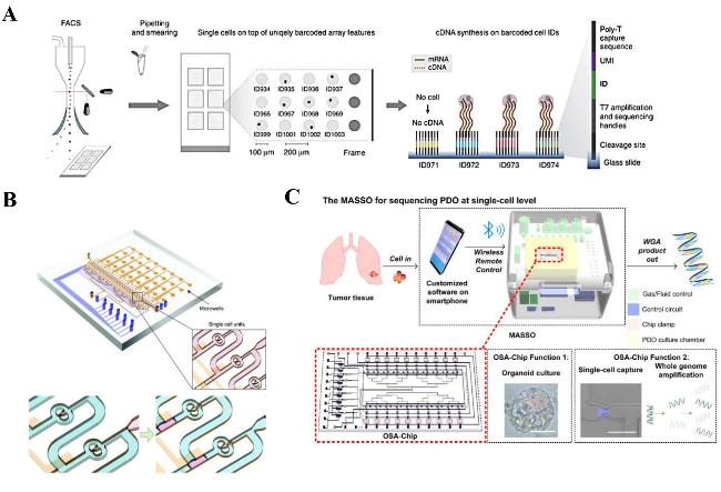
Currently, microfluidics-based cell separation and capture have become the mainstream methods for scRNA-seq, primarily including two types of microfluidic chip technologies: microwell-based and droplet-based. For microwell array-based microfluidic chips, the microwell dimensions on the chip are precisely designed to accommodate only a single cell (Figure 4A); cells randomly fall into the microwells under the action of gravity or hydrodynamic forces, achieving precise capture[69]. For example, the microwell chip of the BD Rhapsody platform contains approximately 200,000 microwells, enabling rapid capture of a large number of single cells. Shi et al.[70]utilized this platform to efficiently capture T cells on a large scale and reveal their transcriptomic dynamic changes, with throughput far exceeding traditional methods. Droplet microfluidics based on microdroplets is a technology that has developed rapidly and been widely applied in recent years; it encapsulates single cells together with gel beads or magnetic beads carrying specific barcodes within tiny droplets, where each microdroplet forms an independent reaction unit. This design not only enables efficient labeling and capture of thousands of single cells[71], but also effectively reduces cell cross-contamination. Zheng et al.[59]successfully achieved rapid encapsulation and capture of 293T and 3T3 cells using the droplet-based 10X Genomics Chromium platform, with an encapsulation efficiency of 80%.
图4 (A) FACS机器将单细胞分选到带条形码的微阵列上,当单细胞落在条形码oligo-dTVN引物(ID)顶部时,才会转录cDNA[69];(B) 通过气动控制进行单细胞分离和皮升液滴生成的微流控芯片[75];(C) 基于微流控芯片的单细胞类器官自动测序系统示意图[84]

Fig.4 (A) The FACS machine sorted single cells onto a barcoded microarray,and the cDNA was transcribed only when the single cells landed on top of the barcoded ogo-dTVN primer (ID)[69]. (B) Microfluidic chip for single cell isolation and picolitre droplet generation by pneumatic control[75]. (C) Schematic diagram of a Microfluidic chip-based Automatic System for Sequencing Organoids at the single-cell level[84]

Microfluidic chips can adapt to the capture and analysis requirements of different cell types by adjusting channel geometry, dimensions, and reaction chamber layouts. For larger tumor cells, wide-diameter channels can be designed to avoid cell damage.[72]; for rare circulating tumor cells, specific capture can be achieved by modifying the channel inner walls with immunomagnetic beads.[73]. Through hydrodynamic capture mechanisms, microfluidic chips can efficiently capture single cells at fixed positions, with capture efficiency reaching nearly 100%, significantly reducing cell loss rates.[74].

3.2.2 Precise control of reaction volume

Microfluidic technology minimizes reaction volumes to the picoliter or nanoliter scale[32], achieving precise fluid control via microchannels, which significantly reduces sample loss and enhances analytical sensitivity. The capability to handle trace samples makes microfluidic systems particularly suitable for processing limited quantities of precious cell samples, such as rare tumor cells or stem cells. Zhang et al.[75]integrated hydrodynamic trapping and phase switching techniques to encapsulate single cells in picoliter-scale hydrogel droplets, asFigure 4Bshows, achieving nearly 100% single-cell capture efficiency.
Precise fluid control in microfluidic chips makes the cell lysis and nucleic acid extraction processes more efficient, reducing the risk of RNA degradation and preserving more transcriptome information. Li et al.[76]By controlling the ratios of enzymes, primers, and templates in the reaction system, multiple displacement amplification (MDA) of tumor cells was achieved on a microfluidic chip, aiding research into anticancer drug resistance mechanisms. Furthermore, the organic combination of microfluidic chips and digital PCR technology enables absolute quantification of low-abundance RNA in single cells.[77], providing more accurate data for subsequent sequencing analysis.
In the single-cell lysis step, the chemical lysis method ensures full contact between the lysis buffer and cells by precisely controlling the flow rate and mixing ratio of fluids in the microfluidic chip, thereby improving lysis efficiency.[78]. Meanwhile, the microscale environment of the microfluidic chip reduces the amount of lysis buffer required, lowering experimental costs. Physical lysis methods utilize specific microstructures fabricated within the microchip to generate shear forces, frictional forces, or compressive forces that rupture cell membranes.[79-80]. Cheng et al.[81]developed a Pump-on-a-chip microfluidic platform capable of efficiently lysing 50 μL of cell samples within 36 s. This physical lysis method is simple to operate, fast, and avoids potential damage to RNA caused by chemical reagents.

3.2.3 Process Automation

For complex processes requiring multi-step handling, microfluidic chips can integrate multiple functional modules to achieve integrated operations, realizing full-process automation from cell capture to nucleic acid analysis. This integrated design significantly reduces human operational errors and improves the accuracy and reproducibility of single-cell analysis. Ruan et al.[82]integrated a high-precision temperature control module on the chip to regulate nucleic acid amplification temperatures, constructing a rapid and stable thermal cycling system. This compressed the time for a series of processes including single-cell automatic separation, lysis, and amplification to within 2 hours, significantly improving scRNA-seq efficiency and automation levels. The chip can be designed with multi-channel arrays to process thousands of single cells in a single run, and through standardized process control, experimental reproducibility is significantly enhanced. Based on the development of optical sensors, microfluidic chips integrated with optical detection modules can monitor single-cell capture status and reaction progress in real time, and further optimize experimental results by feedback-regulating fluid parameters.[83]. Wu et al.[84]integrated all steps of scRNA-seq on a microfluidic chip (Figure 4C), performing automated sequencing of organoid single-cell lung cancer at the single-cell level for the first time, providing a brand-new technical platform for precise modeling of lung cancer organoids and research on tumor heterogeneity.
The design characteristics of microfluidic chips make them adaptable to diverse analytical needs. In the library preparation stage, microfluidic chips integrate multiple operations such as nucleic acid fragmentation, end repair, and adapter ligation into a single system, reducing human error and sample loss through automation. During the fragmentation process, the microfluidics-based ultrasonic shearing module can achieve uniform control of nucleic acid fragment sizes by precisely regulating acoustic energy parameters.[85]; in the end repair and adapter ligation steps, Maguire et al.[86]optimized multi-enzyme reaction conditions by setting up a temperature gradient control unit within the chip, reducing sequence bias caused by differences in enzyme reaction kinetics in traditional methods by more than 40%. To improve the quality of library preparation, Zhang et al.[87]introduced magnetic field control on the chip to manipulate magnetic bead motion for DNA purification, achieving a cDNA purification rate of 92%. Its enclosed hydrophobic environment reduces the risks of sample evaporation, cross-contamination, and exogenous RNase contamination, providing technical assurance for the efficient utilization of rare single-cell samples.

4 Representative microfluidic single-cell RNA sequencing platform

The following section will briefly describe representative single-cell RNA sequencing platforms based on microfluidic chip technology, including Fluidigm C1, 10X Genomics, and BD Rhapsody, summarizing their technical principles, performance characteristics, application scenarios, as well as their respective advantages and limitations (Table 2)[59,88-89]. Through a comprehensive comparison and analysis of these platforms, this provides researchers with a comprehensive and accurate reference basis for selecting single-cell RNA sequencing platforms.
表2 代表性的微流控单细胞RNA测序平台

Table 2 Representative microfluidic single-cell RNA sequencing platforms

Platform Core Technology Throughput Core Advantages Limitations Applicable Samples Ref
Fluidigm
C1
Microwell Array Low Stable automated workflow Low throughput,high cost Immune cells,neurons
cell lines
88
10X Genomics Droplet Microfluidics High Standardized analysis,compatible with multiple species Omission of expressed genes Blood cells,tumor cells,organoids 59
BD Rhapsody Magnetic Bead Encoding High Multi-omics integration Require cell viability
> 90%
Fresh immune cells,stem cells,early-stage embryos 89

4.1 Fluidigm C1 Platform

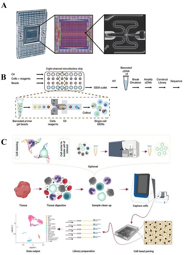
Fluidigm C1[90]As an automated preparation system applied early in single-cell genomics research, its core advantage lies in the precise design of microfluidic chips with 96 single-cell capture sites (Figure 5A), enabling efficient capture and manipulation of cells with a diameter ≤25 μm and regular shapes. Furthermore, captured cells can be directly observed under a microscope to confirm single-cell capture status and cell viability. The system integrates key steps such as cell capture, lysis, reverse transcription, and pre-amplification. Through precise control of microfluidic flow, it achieves independent reactions for single cells, significantly reducing cross-contamination while minimizing manual operation errors, thereby enhancing experimental accuracy and reproducibility.[91]. Its high sensitivity makes it particularly suitable for detecting low-abundance transcripts.[92], Xin et al.[93]utilized C1 to identify all islet cell types in mice, successfully detecting gene expression in Gcg-Ppy cells that were difficult to discover using traditional sequencing methods. Additionally, the experiment had a short cycle, offering advantages in time cost.
The Fluidigm C1 platform demonstrates significant advantages in spatial omics integration studies due to its powerful full-length transcript analysis capabilities and high-depth sequencing characteristics, particularly excelling in research on microenvironmental dynamic changes such as keratinocyte differentiation. Siriwach et al.[94]utilized this platform to first identify a migratory keratinocyte subpopulation expressing THBS1 (Thrombospondin-1) during epidermal wound healing, and elucidated the dynamic process of this subpopulation from basal differentiation and polarized migration to terminal differentiation, laying a theoretical foundation for revealing skin re-epithelialization mechanisms and developing new targets for chronic wound treatment. Furthermore, as a technical platform providing an end-to-end solution from single-cell isolation to full-length mRNA analysis, its data quality has surpassed similar microfluidic technologies in multiple benchmark tests.[95]. However, this platform also has obvious limitations; fluid flow or pressure within the C1 capture circuit may cause cell damage or intercellular fusion, thereby leading to alterations in single-cell gene expression patterns.

4.2 10X Genomics Chromium Platform

The 10X Genomics Chromium platform is a widely applied and highly influential scRNA-seq technology[59]. Based on microdroplet technology and utilizing an eight-channel microfluidic chip, it generates approximately 100,000 barcoded gel beads (GEMs) every 6 minutes, precisely encapsulating single cells into independent reaction units, thereby minimizing UV-induced cellular damage (Figure 5B). With an encapsulation rate of 80% and a cell capture efficiency of 50%, it is suitable for rare sample analysis. Furthermore, the UMI technology employed by this platform effectively corrects PCR amplification bias[96]. In regions with low GC (guanine-cytosine base pair) content, the platform demonstrates significant advantages in controlling GC content bias, reducing gene expression quantification errors to below 5% and improving the accuracy of gene quantification.
图5 (A) Fluidigm C1的芯片设计[108]。(B) 10X Genomics Chromium平台的工作原理[59]。(C) 用于获取高质量的scRNA-seq数据的BD Rhapsody工作流程[89]

Fig.5 (A) The chip design of Fluidigm C1[108]. (B) The working principle of the 10X Genomics Chromium platform[59]. (C) BD Rhapsody workflow for high-quality scRNA-seq data[89]

The 10X Genomics platform is the preferred platform for cancer research[97]. Its revolutionary single-cell resolution and spatial omics technologies can systematically resolve core cancer challenges that are difficult to capture with traditional techniques: precisely revealing malignant cell subpopulations, rare drug-resistant cells, and genomic heterogeneity within tumors through scRNA-seq[98-99], and combining Visium spatial transcriptomics technology to intuitively locate the spatial distribution and interactions of components such as immune cells, stroma, and blood vessels in the tumor microenvironment[100], comprehensively driving the translation process from basic cancer biology to clinical precision diagnosis and treatment. However, this platform has stringent requirements for total cell count and viability, exhibits a low gene detection rate, tends to miss lowly expressed genes, and incurs high R&D costs for personalized experimental design and analysis.

4.3 BD Rhapsody Platform

BD Rhapsody[89]Based on the classic microwell plate principle, single cells are isolated and captured via natural sedimentation. The platform utilizes a microwell array containing 220,000 wells, achieving a cell capture rate of up to 80%, which is higher than that of 10X Genomics Chromium, enabling efficient single-cell isolation and capture.[101]. This natural sedimentation method causes no damage to cells, offers excellent sample compatibility, and maximizes the preservation of cell viability and integrity. Meanwhile, by combining oligonucleotide magnetic beads, the platform enables dual labeling with cell barcodes and UMIs, supporting joint analysis of transcriptomes, surface proteins, and immune repertoires.[102], providing possibilities for multi-dimensional analysis of cellular functional characteristics (Figure 5C). Leveraging real-time monitoring via an image recognition system, the platform can precisely capture the single-cell trapping process, ensuring that only one single cell is captured per microwell, thereby significantly improving experimental accuracy and reliability.
The BD Rhapsody system performs excellently in processing cells with low mRNA content[103], an advantage that has enabled its rapid rise in the field of immune cell research[104]; Scheiber et al.[105] effectively recovered immune cells with low mRNA content using the BD Rhapsody platform, further validating the utility of this system in rare cell research. As the integration of spatial omics and scRNA-seq becomes a new trend, Sennikov et al.[106] obtained effective anti-tumor T cells and their T cell receptors through single-cell multi-omics BD Rhapsody data analysis, and observed that dominantly clonotype-transduced TCR T cells exhibited potent cytotoxicity against MAGE-A3-positive tumors, providing a new perspective for revealing cellular heterogeneity and personalized therapy. Although this platform occupies an important position in single-cell research due to its high-precision gene capture capability and multi-omics compatibility, the randomness of cell sedimentation leads to phenomena such as empty wells or multiple cells per well, necessitating screening and validation of capture results, which increases experimental complexity and time costs[107].

5 Conclusion and Outlook

Microfluidic chip technology integrates processes such as single-cell isolation, lysis, reverse transcription, amplification, and sequencing library construction through a micrometer-scale fluid manipulation system, providing an efficient and precise technical platform for scRNA-seq and significantly enhancing its efficiency and data reliability. Its core advantages lie in the microscale design adapted for single cells, precise control of reaction volumes at the pico- to nanoliter scale, and full-process automation from cell capture to nucleic acid analysis, effectively addressing challenges in traditional techniques such as low single-cell isolation and capture efficiency and significant deviations in trace RNA operations.
In terms of technical processes, scRNA-seq covers four core steps: single-cell isolation and capture, RNA extraction, reverse transcription and amplification, and single-cell sequencing. Among these, microfluidic chips demonstrate unique advantages in the single-cell isolation stage: chips based on microwell arrays achieve single-cell capture through precisely designed microwell dimensions, while droplet microfluidics-based technologies encapsulate single cells with barcoded magnetic beads within individual droplets, significantly increasing throughput to thousands of cells per run. In RNA extraction and amplification, microfluidic chips control reaction volumes at the picoliter scale; combined with UMI labeling technology to correct PCR biases, this ensures accurate detection of low-abundance transcripts. The integration of high-throughput sequencing platforms such as Illumina with microfluidic technology has further advanced the in-depth analysis of single-cell transcriptome data.
Representative platforms such as Fluidigm C1, 10X Genomics Chromium, and BD Rhapsody each possess distinct technical features and application advantages, playing a significant role in life science research fields including the analysis of cellular heterogeneity and the elucidation of disease mechanisms. The Fluidigm C1 performs best in full-length transcript analysis and minimizing gene length bias, making it suitable for small-scale sample studies requiring high-quality, deep sequencing. The 10X Genomics Chromium platform excels in controlling GC content bias and achieving high cell throughput, making it particularly ideal for large-scale cell population studies such as those involving tumor cells. BD Rhapsody demonstrates unique advantages in handling cells with low mRNA content, providing a reliable tool for immune cell research. Given the differences in technical performance and application scenarios among these platforms, researchers must comprehensively consider factors such as research objectives, sample types, required cell numbers, and budget when selecting an appropriate platform.
Although microfluidic chip-based single-cell RNA sequencing technology has advanced single-cell gene expression analysis, bottlenecks still exist. In the cell capture stage, limited by the Poisson distribution, pairing efficiency is only about 50%, and cell heterogeneity along with sample preprocessing losses lead to capture bias, making fragile or large cells prone to damage. During molecular operations, the extremely low RNA content in single cells faces issues of degradation and amplification bias, while library preparation also carries risks of cross-contamination. Furthermore, high equipment and consumable costs, coupled with the complexity of clinical sample processing, limit the widespread adoption and translation of this technology. These bottlenecks involve capture efficiency, molecular stability, data standardization, and cost control, requiring breakthroughs across the entire chain to promote its application in fields such as precision medicine.
Future microfluidic chip scRNA-seq technology will achieve breakthroughs in multiple aspects: In terms of technical performance optimization, through continuous iteration of chip design, it is expected to achieve million-level throughput, and combined with deep learning to optimize droplet generation and cell capture, reducing the cost and threshold for rare cell detection. In the field of multi-omics integration applications, integrating multi-omics data such as proteomics and epigenomics, and combining them with spatial transcriptomics technology, will deeply analyze the spatial distribution of gene expression and microenvironment interaction mechanisms at the single-cell level, providing a new perspective for organ development and tumor research. In clinical translation, developing portable automated systems for single-cell sequencing early screening of circulating tumor cells, combined with live cell dynamic tracking to monitor drug responses, will strongly promote the development of precision medicine. Furthermore, establishing standardized operation and analysis systems to resolve data compatibility issues will accelerate the widespread application of this technology in basic research and clinical diagnosis, bringing more profound impacts to the fields of life sciences and medicine.
[1]
Fiorentino J, Torres-Padilla M E, Scialdone A. Annu. Rev. Genet., 2020, 54: 167.

[2]
Lee H O, Park W Y. Single Cell Genomics for Tumor Heterogeneity. Eds.: Noh D Y, Han W, Toi M. Singapore: Springer, 2021.

[3]
Minnema-Luiting J, Vroman H, Aerts J, Cornelissen R. Int. J. Mol. Sci., 2018, 19(4): 1041.

[4]
Li X M, Wang C Y. Int. J. Oral Sci., 2021, 13: 36.

[5]
Tang F C, Barbacioru C, Wang Y Z, Nordman E, Lee C, Xu N L, Wang X H, Bodeau J, Tuch B B, Siddiqui A, Lao K Q, Surani M A. Nat. Meth., 2009, 6(5): 377.

[6]
Gupta R K, Kuznicki J. Cells, 2020, 9(8): 1751.

[7]
Simmons S K, Lithwick-Yanai G, Adiconis X, Oberstrass F, Iremadze N, Geiger-Schuller K, Thakore P I, Frangieh C J, Barad O, Almogy G, Rozenblatt-Rosen O, Regev A, Lipson D, Levin J Z. Nat. Biotechnol., 2023, 41(2): 204.

[8]
McCracken I R, Taylor R S, De La Cuesta F, Dobie R, Henderson B E, Mountford J C, Caudrillier A, Henderson N C, Ponting C P, Baker A H. Eur. Heart J., 2019, 41(9): 1024.

[9]
Zhang Y, Wang D, Peng M, Tang L, Ouyang J, Xiong F, Guo C, Tang Y, Zhou Y, Liao Q, Wu X, Wang H, Yu J, Li Y, Li X, Li G, Zeng Z, Tan Y, Xiong W. J. Exp. Clin. Cancer Res., 2021, 40(1): 81.

[10]
Papalexi E, Satija R. Nat. Rev. Immunol., 2018, 18(1): 35.

[11]
Drescher H, Weiskirchen S, Weiskirchen R. Biomedicines, 2021, 9(11): 1613.

[12]
Emmert-Buck M R, Bonner R F, Smith P D, Chuaqui R F, Zhuang Z P, Goldstein S R, Weiss R A, Liotta L A. Science, 1996, 274(5289): 998.

[13]
Wu Y, Zhang K. Nat. Rev. Nephrol., 2020, 16(7): 408.

[14]
Cao J, Chen X, Huang S Q, Shi W X, Fan Q, Gong Y L, Peng Y, Wu L L, Yang C Y. Trac Trends Anal. Chem., 2023, 158: 116868.

[15]
Pattanayak P, Singh S K, Gulati M, Vishwas S, Kapoor B, Chellappan D K, Anand K, Gupta G, Jha N K, Gupta P K, Prasher P, Dua K, Dureja H, Kumar D, Kumar V. Microfluid. Nanofluid., 2021, 25(12): 99.

[16]
Streets A M, Zhang X N, Cao C, Pang Y H, Wu X L, Xiong L, Yang L, Fu Y S, Zhao L, Tang F C, Huang Y Y. Proc. Natl. Acad. Sci. U. S. A., 2014, 111(19): 7048.

[17]
Chen Y W, Wang X Q, Na X, Zhang Y K, Cai L F, Song J, Yang C Y. Anal. Chem., 2024, 96(31): 12916.

[18]
Pan Y T, Cao W J, Mu Y, Zhu Q Y. Biosensors, 2022, 12(7): 450.

[19]
Pensold D, Zimmer-Bensch G. Methods for Single-Cell Isolation and Preparation. Eds.: Yu B, Zhang J, Zeng Y, Li L, Wang X. Singapore: Springer, 2020.

[20]
Gross A, Schoendube J, Zimmermann S, Steeb M, Zengerle R, Koltay P. Int. J. Mol. Sci., 2015, 16(8): 16897.

[21]
Feng Y Q, Li M. Nanoscale, 2023, 15(32): 13346.

[22]
Brunner A D, Thielert M, Vasilopoulou C, Ammar C, Coscia F, Mund A, Hoerning O B, Bache N, Apalategui A, Lubeck M, Richter S, Fischer D S, Raether O, Park M A, Meier F, Theis F J, Mann M. Mol. Syst. Biol., 2022, 18(3): e10798.

[23]
Gan C Y, Zhang J Y, Chen B, Wang A, Xiong H Y, Zhao J W, Wang C T, Liang S Z, Feng L. Small, 2024, 20(23): 2307329.

[24]
Li Y, Huang Z, Xu L B, Fan Y L, Ping J, Li G C, Chen Y J, Yu C W, Wang Q F, Song T R, Lin T, Liu M M, Xu Y Q, Ai N, Meng X N, Qiao Q, Ji H B, Qin Z, Jin S, Jiang N, Wang M X, Shu S K, Zhang F, Zhang W Q, Liu G H, Chen L M, Jiang L. Nat. Meth., 2025, 22(6): 1199.

[25]
Roszkowski M, Mansuy I M. Front. Cell Dev. Biol., 2021, 9 : 648274.

[26]
Subramanian Parimalam S, Oguchi Y, Abdelmoez M N, Tsuchida A, Ozaki Y, Yokokawa R, Kotera H, Shintaku H. Anal. Chem., 2018, 90(21): 12512.

[27]
Schactler S A, Scheuerman S J, Lius A, Altemeier W A, An D, Matula T J, Mikula M, Kulecka M, Denisenko O, Mar D, Bomsztyk K. BMC Genom., 2023, 24: 446.

[28]
Lizardi P M, Engelberg A. Anal. Biochem., 1979, 98(1): 116.

[29]
Zhang M X, Zou Y, Xu X, Zhang X B, Gao M X, Song J, Huang P F, Chen Q, Zhu Z, Lin W, Zare R N, Yang C Y. Nat. Commun., 2020, 11: 2118.

[30]
Kutluk H, Viefhues M, Constantinou I. Small Sci., 2024, 4(4): 2300206.

[31]
He P, Williams B A, Trout D, Marinov G K, Amrhein H, Berghella L, Goh S T, Plajzer-Frick I, Afzal V, Pennacchio L A, Dickel D E, Visel A, Ren B, Hardison R C, Zhang Y, Wold B J. Nature, 2020, 583(7818): 760.

[32]
Macosko E Z, Basu A, Satija R, Nemesh J, Shekhar K, Goldman M, Tirosh I, Bialas A R, Kamitaki N, Martersteck E M, Trombetta J J, Weitz D A, Sanes J R, Shalek A K, Regev A, McCarroll S A. Cell, 2015, 161(5): 1202.

[33]
Sun J F, Philpott M, Loi D, Li S, Monteagudo-Mesas P, Hoffman G, Robson J, Mehta N, Gamble V, Brown T Jr, Brown T Sr, Canzar S, Oppermann U, Cribbs A P. Nat. Meth., 2024, 21(3): 401.

[34]
Becskei A, Rahaman S. Comput. Struct. Biotechnol. J., 2022, 20: 4325.

[35]
Abdulmalik O, Lombardi A A, Russell J E. PLOS One, 2012, 7(7): e40827.

[36]
Micheel J, Safrastyan A, Aron F, Wollny D. Nat. Commun., 2024, 15: 5858.

[37]
Ellefson J W, Gollihar J, Shroff R, Shivram H, Iyer V R, Ellington A D. Science, 2016, 352(6293): 1590.

[38]
Li S Q, Sun J, Allesøe R, Datta K, Bao Y, Oliveira G, Forman J, Jin R, Olsen L R, Keskin D B, Shukla S A, Wu C J, Livak K J. Nat. Protoc., 2019, 14(8): 2571.

[39]
Hashimshony T, Wagner F, Sher N, Yanai I. Cell Rep., 2012, 2(3): 666.

[40]
Aird D, Ross M G, Chen W-S, Danielsson M, Fennell T, Russ C, Jaffe D B, Nusbaum C, Gnirke A. Genome Biol., 2011, 12(2): R18.

[41]
Korbie D J, Mattick J S. Nat. Protoc., 2008, 3(9): 1452.

[42]
Zhou Y, Jia E T, Sheng Y Q, Qiao Y, Wang Y, Shi H J, Liu Z Y, Pan M, Tu J, Bai Y F, Zhao X W, Ge Q Y, Lu Z H. ACS Appl. Mater. Interfaces, 2022, 14(17): 19154.

[43]
Morris J, Singh J M, Eberwine J H. J. Vis. Exp., 2011, 50: e2634.

[44]
Sasagawa Y, Hayashi T, Nikaido I. Strategies for Converting RNA to Amplifiable cDNA for Single-Cell RNA Sequencing Methods. Eds.: Suzuki Y. Singapore: Springer, 2019.

[45]
Popova P G, Lagace M A, Tang G, Blakney A K. Eur. J. Pharm. Biopharm., 2024, 198: 114247.

[46]
Uhlen M, Quake S R. Trends Biotechnol., 2023, 41(12): 1565.

[47]
Pease J, Sooknanan R. Nat. Meth., 2012, 9(3): i.

[48]
Marie R, Pødenphant M, Koprowska K, Bærlocher L, Vulders R C M, Wilding J, Ashley N, McGowan S J, van Strijp D, van Hemert F, Olesen T, Agersnap N, Bilenberg B, Sabatel C, Schira J, Kristensen A, Bodmer W, van der Zaag P J, Mir K U. Lab Chip, 2018, 18(13): 1891.

[49]
Chaitankar V, Karakülah G, Ratnapriya R, Giuste F O, Brooks M J, Swaroop A. Prog. Retin. Eye Res., 2016, 55: 1.

[50]
Tan G, Opitz L, Schlapbach R, Rehrauer H. Sci. Rep., 2019, 9(1): 2856.

[51]
Rhoads A, Au K F. Genom. Proteom. Bioinform., 2015, 13(5): 278.

[52]
Jain M, Olsen H E, Paten B, Akeson M. Genome Biol., 2016, 17(1): 239.

[53]
Goetz J J, Trimarchi J M. Nat. Biotechnol., 2012, 30(8): 763.

[54]
Mehling M, Tay S. Curr. Opin. Biotechnol., 2014, 25: 95.

[55]
Durruthy-Durruthy R, Ray M. Using Fluidigm C1 to Generate Single-Cell Full-Length cDNA Libraries for mRNA Sequencing. Eds.: DiStefano J K. New York:Springer, 2018.

[56]
Kaminski T S, Garstecki P. Chem. Soc. Rev., 2017, 46(20): 6210.

[57]
Klein A M, Mazutis L, Akartuna I, Tallapragada N, Veres A, Li V, Peshkin L, Weitz D A, Kirschner M W. Cell, 2015, 161(5): 1187.

[58]
Bageritz J, Raddi G. Single-Cell RNA Sequencing with Drop-Seq. Eds.: Proserpio V. New York: Springer, 2019.

[59]
Zheng G X Y, Terry J M, Belgrader P, Ryvkin P, Bent Z W, Wilson R, Ziraldo S B, Wheeler T D, McDermott G P, Zhu J J, Gregory M T, Shuga J, Montesclaros L, Underwood J G, Masquelier D A, Nishimura S Y, Schnall-Levin M, Wyatt P W, Hindson C M, Bharadwaj R, Wong A, Ness K D, Beppu L W, Deeg H J, McFarland C, Loeb K R, Valente W J, Ericson N G, Stevens E A, Radich J P, Mikkelsen T S, Hindson B J, Bielas J H. Nat. Commun., 2017, 8(1): 14049.

[60]
Juzenas S, Goda K, Kiseliovas V, Zvirblyte J, Quintinal-Villalonga A, Siurkus J, Nainys J, Mazutis L. Nucleic Acids Res., 2025, 53(2): gkae1312.

[61]
Shum E Y, Walczak E M, Chang C, Christina Fan H. Quantitation of mRNA Transcripts and Proteins Using the BD Rhapsody™ Single-Cell Analysis System. Eds.: Suzuki Y. Singapore: Springer, 2019.

[62]
Maïno N, Hauling T, Cappi G, Madaboosi N, Dupouy D G, Nilsson M. Sci. Rep., 2019, 9: 3542.

[63]
Chen J, Wang X, Ma A, Wang Q E, Liu B, Li L, Xu D, Ma Q. Nat. Commun., 2022, 13(1): 6494.

[64]
Bhagwat N, Dulmage K, Pletcher C H Jr, Wang L, DeMuth W, Sen M E, Balli D, Yee S S, Sa S L, Tong F, Yu L P, Moore J S, Stanger B Z, Dixon E P, Carpenter E L. Sci. Rep., 2018, 8: 5035.

[65]
Liu Y X, Fan Z H, Qiao L, Liu B H. Trac. Trends Anal. Chem., 2022, 157: 116822.

[66]
Zhu J C, Wang Y C, Chen P, Su H Y, Du W, Liu B F. Sens. Actuat. B Chem., 2019, 283: 685.

[67]
Denyer T, Timmermans M C P. Trends Plant Sci., 2022, 27(1): 104.

[68]
Sarma M, Lee J, Ma S, Li S, Lu C. Lab Chip, 2019, 19(7): 1247.

[69]
Vickovic S, Ståhl P L, Salmén F, Giatrellis S, Westholm J O, Mollbrink A, Navarro J F, Custodio J, Bienko M, Sutton L A, Rosenquist R, Frisén J, Lundeberg J. Nat. Commun., 2016, 7: 13182.

[70]
Shi S, Baracho G V, Song H W, Acob R A, Ayer A, Widmann S J, Tyznik A J. J. Immunol., 2023, 210(Supplement 1): 251.09.

[71]
Wen N, Zhao Z, Fan B Y, Chen D Y, Men D, Wang J B, Chen J. Molecules, 2016, 21(7): 881.

[72]
Li G L, Ji Y, Wu Y H, Liu Y S, Li H, Wang Y M, Chi M B, Sun H Y, Zhu H Q. Biosens. Bioelectron., 2023, 237: 115451.

[73]
Zhuo Y, Wang Y R, Gao R K, Zhan C B, Liu H C, Liu Y Y, Wang F F, Chen X Z, Feng Y J, Yu L D. Microchem. J., 2025, 212: 113330.

[74]
Sauzade M, Brouzes E. Lab Chip, 2017, 17(13): 2186.

[75]
Zhang B Y, Xu H, Huang Y Q, Shu W L, Feng H T, Cai J, Zhong J F, Chen Y. Analyst, 2019, 144(24): 7185.

[76]
Li R, Zhou M X, Yue C Y, Zhang W K, Ma Y, Peng H L, Hu Z Y, Wei Z W. Lab Chip, 2018, 18(5): 723.

[77]
Fang W B, Liu X D, Maiga M, Cao W J, Mu Y, Yan Q, Zhu Q Y. Biosensors, 2024, 14(2): 64.

[78]
Kim S C, Clark I C, Shahi P, Abate A R. Anal. Chem., 2018, 90(2): 1273.

[79]
Li Z G, Liu A Q, Klaseboer E, Zhang J B, Ohl C D. Lab Chip, 2013, 13(6): 1144.

[80]
Kim J, Hong J W, Kim D P, Shin J H, Park I. Lab Chip, 2012, 12(16): 2914.

[81]
Cheng Y N, Wang Y, Wang Z Y, Huang L, Bi M Z, Xu W X, Wang W H, Ye X Y. Biomicrofluidics, 2017, 11(2): 024112.

[82]
Ruan Q Y, Shi C R, Lin X Y, Ruan W D, Xu M H, Yang C Y, Ren Y K. Chem. Eng. J., 2025, 511: 162099.

[83]
Liao Z R, Zhang Y, Li Y R, Miao Y F, Gao S M, Lin F K, Deng Y L, Geng L N. Biosens. Bioelectron., 2019, 126: 697.

[84]
Wu X, Li B W, Wang Y D, Xue J C, Zhao H T, Huang Z C, Zheng Z B, Liang N X, Wei Z W. Anal. Chem., 2024, 96(42): 17027.

[85]
Sun L, Lehnert T, Gijs M A M, Li S J. Lab Chip, 2022, 22(21): 4224.

[86]
Maguire S, Lohman G J S, Guan S X. Nucleic Acids Res., 2020, 48(14): e80.

[87]
Zhang Q Q, Xu X, Lin L, Yang J, Na X, Chen X, Wu L L, Song J, Yang C Y. Lab Chip, 2022, 22(10): 1971.

[88]
DeLaughter D M. Curr. Protoc. Mol. Biol., 2018, 122: e55.

[89]
Li W Y, Bazaz S R, Mayoh C, Salomon R. Curr. Protoc., 2024, 4(2): e963.

[90]
Pollen A A, Nowakowski T J, Shuga J, Wang X H, Leyrat A A, Lui J H, Li N Z, Szpankowski L, Fowler B, Chen P L, Ramalingam N, Sun G, Thu M, Norris M, Lebofsky R, Toppani D, Kemp D W II, Wong M, Clerkson B, Jones B N, Wu S Q, Knutsson L, Alvarado B, Wang J, Weaver L S, May A P, Jones R C, Unger M A, Kriegstein A R, West J A A. Nat. Biotechnol., 2014, 32(10): 1053.

[91]
Gong H B, Do D, Ramakrishnan R. Single-Cell mRNA-Seq Using the Fluidigm C1 System and Integrated Fluidics Circuits. Eds.: Raghavachari N, Garcia-Reyero N. New York: Springer, 2018.

[92]
Wu A R, Neff N F, Kalisky T, Dalerba P, Treutlein B, Rothenberg M E, Mburu F M, Mantalas G L, Sim S, Clarke M F, Quake S R. Nat. Meth., 2014, 11(1): 41.

[93]
Xin Y R, Kim J, Ni M, Wei Y, Okamoto H, Lee J, Adler C, Cavino K, Murphy A J, Yancopoulos G D, Lin H C, Gromada J. Proc. Natl. Acad. Sci. U. S. A., 2016, 113(12): 3293.

[94]
Siriwach R, Ngo A Q, Higuchi M, ARIMA K, Sakamoto S, Watanabe A, Narumiya S, Thumkeo D. iScience, 2022, 25(4): 104130.

[95]
Arya A, Tripathi P, Dubey N, Aier I, Kumar Varadwaj P. Genom. Inform., 2025, 23: 13.

[96]
Islam S, Zeisel A, Joost S, La Manno G, Zajac P, Kasper M, Lönnerberg P, Linnarsson S. Nat. Meth., 2014, 11(2): 163.

[97]
Prakash P G, Kumar N, Gurumurthy R K, Chumduri C. Sci. Data, 2024, 11: 1238.

[98]
Li Y Q, Wang W J, Wang D Y, Zhang L C, Wang X Z, He J, Cao L, Li K, Xie H Y. Genes, 2022, 13(12): 2276.

[99]
Xin S Y, Liu X, Li Z Y, Sun X C, Wang R, Zhang Z H, Feng X W, Jin L, Li W Y, Tang C Z, Mei W L, Cao Q, Wang H J, Zhang J G, Feng L J, Ye L. Exp. Hematol. Oncol., 2023, 12(1): 49.

[100]
Wang C, Lu M Y, Chen C M, Chen J J, Cai Y S, Wang H, Tao L L, Yin W H, Chen J K. Int. Immunopharmacol., 2025, 145: 113752.

[101]
Ulbrich J, Lopez-Salmeron V, Gerrard I. BD Rhapsody™ Single-Cell Analysis System Workflow: From Sample to Multimodal Single-Cell Sequencing Data. Eds.: Calogero R A, Benes V. New York: Springer, 2023.

[102]
Timms J A, Winter S, Hargreaves S, McLornan D P, Platzbecker U, Kordasti S. Blood, 2019, 134(Supplement 1): 2980.

[103]
Nguyen R, Vaidyanathan S, Hlathu Z, Zhao X Y, Moskwa J, Acob R A, Gordon C, Wang L, Ayer A. Cancer Res., 2025, 85(8_Supplement 1): 4078.

[104]
Ji X H, Miranda M, Suresh S, Bader C S, Zoubeidi A, Meyer E H, Maecker H T. J. Immunol., 2023, 210(Supplement 1): 249.03.

[105]
Scheiber A, Trebo M, Pittl A, Heidegger I, Hautz T, Oberhuber R, Trajanoski Z, Augustin F, Sopper S, Wolf D, Pircher A, Salcher S. STAR Protoc., 2024, 5(4): 103475.

[106]
Sennikov S, Volynets M, Alrhmoun S, Perik-Zavodskii R, Perik-Zavodskaia O, Fisher M, Lopatnikova J, Shevchenko J, Nazarov K, Philippova J, Alsalloum A, Kurilin V, Silkov A. Front. Immunol., 2024, 15 :1470130.

[107]
Hautz T, Salcher S, Fodor M, Sturm G, Ebner S, Mair A, Trebo M, Untergasser G, Sopper S, Cardini B, Martowicz A, Hofmann J, Daum S, Kalb M, Resch T, Krendl F, Weissenbacher A, Otarashvili G, Obrist P, Zelger B, Öfner D, Trajanoski Z, Troppmair J, Oberhuber R, Pircher A, Wolf D, Schneeberger S. Nat. Commun., 2023, 14: 2285.

[108]
Szulwach K E, Chen P L, Wang X H, Wang J, Weaver L S, Gonzales M L, Sun G, Unger M A, Ramakrishnan R. PLOS One, 2015, 10(8): e0135007.

Outlines

/