Home Journals Progress in Chemistry
Progress in Chemistry

Abbreviation (ISO4): Prog Chem      Editor in chief: Jincai ZHAO

About  /  Aim & scope  /  Editorial board  /  Indexed  /  Contact  / 
Review

Carbon Dots as Fluorescent Probes in Environmental Monitoring

  • Congyin Zhang 1 ,
  • Kuiyu Yi , 1, * ,
  • Jia Feng 1 ,
  • Hongwei Shi 2
Expand
  • 1 School of Petrochemical Engineering, Shenyang University of Technology, Liaoyang 111003, China
  • 2 School of Materials Science and Engineering, Shenyang University of Technology, Shenyang 110870, China

Received date: 2025-06-23

  Revised date: 2025-10-19

  Online published: 2026-02-05

Supported by

National Natural Science Foundation of China(52171089)

Liaoning Provincial Key Research and Development Program(2024JH2/102500080)

Fundamental Research Project of Liaoning Provincial Department of Education(LJKMZ20220514)

Postgraduate Education Reform Project of Shenyang University of Technology(SYJG20222036)

Abstract

With the acceleration of industrialization and urbanization,the types of environmental pollutants have multiplied,and sample matrices have become increasingly complex,placing higher demands on the selectivity and anti-interference capability of detection technologies. Conventional methods such as ICP-MS and GC-MS are limited by cumbersome sample pretreatment and high operational costs,making them unsuitable for rapid and on-site monitoring. Carbon dots (CDs),as a promising class of zero-dimensional carbon-based nanomaterials,offer a green and sensitive alternative for constructing fluorescent probes due to their exceptional optical properties,low toxicity,and tunable surface functionalities. This review systematically summarizes recent advances in CDs-based fluorescent probes for environmental monitoring,covering synthesis strategies,luminescence mechanisms,characterization techniques,and their applications in detecting metal ions,inorganic anions,and organic pollutants. Special emphasis is placed on the design principles and response mechanisms of three types of probes:fluorescence quenching,fluorescence enhancement,and ratiometric fluorescence sensing. Notably,ratiometric probes utilize built-in reference signals to achieve self-calibration in complex matrices,significantly improving accuracy and anti-interference performance. Furthermore,this review highlights the integration of CDs-based probes with smartphone-based sensing platforms,demonstrating their great potential for on-site,rapid,and visual detection of pollutants. These advances provide a clear pathway toward making environmental monitoring more convenient and intelligent. Finally,current challenges and future prospects in material design,mechanism studies,application expansion,and intelligent platform development are discussed,offering theoretical and technical support for further innovations in the field.

Contents

1 Introduction

2 Carbon dots

2.1 Synthesis of carbon dots

2.2 Carbon dots fluorescence detection mechanism

2.3 Characterization of carbon dots

3 The application of carbon dots in environmental detection

3.1 Environmental detection applications of fluorescence quenched carbon dots

3.2 Environmental detection applications of fluorescence enhanced carbon dots

3.3 Environmental detection applications of ratiometric fluorescent carbon dots

4 Conclusion and outlook
Cite this article

Congyin Zhang , Kuiyu Yi , Jia Feng , Hongwei Shi . Carbon Dots as Fluorescent Probes in Environmental Monitoring[J]. Progress in Chemistry, 2026 , 38(2) : 298 -318 . DOI: 10.7536/PC20250625

1 Introduction

With the rapid growth of the economy and the accelerated processes of industrialization and urbanization, environmental pollution has become increasingly severe, emerging as a critical factor constraining sustainable development[1-2]. The types of environmental pollutants are continuously increasing, with complex sources, posing serious threats to human health, ecosystems, and the global climate[3-4]. Therefore, developing efficient, sensitive, and environmentally friendly pollutant detection technologies is of great significance for accurately assessing environmental quality, providing timely warnings of potential risks, and formulating effective governance strategies.
Traditional methods for detecting environmental pollutants include Inductively Coupled Plasma Mass Spectrometry (ICP-MS)[5-6], Atomic Absorption Spectroscopy (AAS)[7-8], Gas Chromatography (GC)[9-10], High Performance Liquid Chromatography (HPLC)[11-12]as well as Gas Chromatography-Mass Spectrometry (GC-MS)[13-14]and High Performance Liquid Chromatography-Mass Spectrometry (HPLC-MS)[15-16]. These methods are highly regarded for their high sensitivity and accuracy; however, the sample pretreatment process is tedious, the operation is relatively complex, which increases time costs, and the daily maintenance costs of the required instruments are high. Currently, among numerous detection technologies, fluorescence sensing technology has attracted significant attention due to its advantages such as high sensitivity, good selectivity, and ease of operation[17-19]. Carbon dots (CDs), as an emerging class of zero-dimensional carbon-based nanomaterials, have shown broad application prospects in fields such as bioimaging, sensing detection, and photocatalysis since their discovery, owing to their unique optical properties, good biocompatibility, and low toxicity[20-22]. Particularly in the field of environmental detection, carbon dots as fluorescent probes can not only effectively identify and respond to various environmental pollutants but also achieve specific detection of different pollutants through flexible regulation of their surface chemical properties.
This article aims to comprehensively review the research progress of carbon dots as fluorescent probes in the field of environmental detection. It focuses on introducing synthesis strategies, fluorescence detection mechanisms, characterization techniques, and application cases of carbon dots in detecting typical environmental pollutants such as metal ions, inorganic anions, and organic pollutants. In particular, this paper will deeply explore the construction schemes and response mechanisms of three types of carbon dot probes: fluorescence quenching, fluorescence enhancement, and ratiometric types, analyzing their anti-interference capabilities and self-calibration characteristics in complex environmental matrices. Furthermore, it looks ahead to their practical application value, including the integrated innovation of ratiometric probes in intelligent sensing platforms, as well as detection strategies and applicability of carbon dot probes for complex matrices such as soil and industrial wastewater. By comprehensively elaborating on the theoretical basis and practical applications of carbon dot fluorescent probes in environmental detection, this article aims to provide a scientific basis and practical guidance for the innovation of environmental detection methods, promoting the green, convenient, and intelligent development of environmental detection technologies, and contributing to environmental protection and sustainable development.

2 Carbon dots

2.1 Synthesis of Carbon Dots

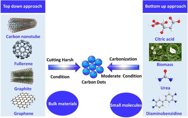
The synthesis strategies of carbon dots can be mainly divided into two categories: "top-down" and "bottom-up". The common synthetic precursors and basic routes for both methods are asFigure 1shown[23].
图1 CDs的合成方法[23]

Fig.1 Methods for synthesizing CDs[23]

2.1.1 Top-down approach

This method primarily involves cutting and decomposing large-sized carbon materials such as carbon nanotubes, fullerenes, graphite, and graphene into small-sized carbon dots through physical or chemical means. Common methods include laser ablation, arc discharge, and electrochemical oxidation.[24-26]Specifically, the fundamental principle of carbon dot synthesis via laser ablation in liquid involves focusing high-energy pulsed lasers onto solid carbon targets, such as graphite, submerged in liquid media like ethanol or water. The instantaneous high temperature and high-pressure plasma induced by the laser vaporizes, exfoliates, and fragments the carbon target. During this process, the carbon source reacts with the surrounding liquid or added dopants, such as nitrogen-containing precursors, within the cavitation bubble micro-reactors, ultimately self-assembling into carbon dots with uniform size and good crystallinity, which are stably dispersed in the solution. This method requires no harmful chemicals such as strong acids, features a simple one-step process, and allows for the regulation of carbon dot size, structure, and surface chemistry by adjusting laser parameters and precursors.[27]The fundamental principle of synthesizing carbon nanomaterials via the arc discharge method utilizes the high-temperature plasma generated by a direct current arc between graphite electrodes to evaporate the anode graphite, forming high-density carbon atoms, ions, and clusters. These carbon species undergo transport, nucleation, and growth within specific temperature gradients and spatial domains, eventually depositing on the cathode or reaction chamber walls to form the target products. By precisely controlling key parameters such as buffer gas composition and pressure, catalysts, current and voltage, and external magnetic fields, effective control over carbon nanomaterials like carbon dots and their structural characteristics is achieved. This method is renowned for preparing carbon nanomaterials with high crystallinity and few defects, making it suitable for the large-scale synthesis of high-quality nanomaterials.[28]The electrochemical oxidation method uses macroscopic carbon materials, such as graphite rods, as electrodes. By applying a current in a specific electrolyte solution, large-sized carbon sources are efficiently exfoliated and functionalized through electrochemically driven oxidative etching, thereby producing carbon dots with high purity and uniform size. Although this process allows for good regulation of carbon dot properties at the laboratory level, it involves complex electrolysis systems and post-treatment steps with high operational requirements; therefore, its application potential for large-scale green synthesis remains to be further explored.[29]However, these methods typically involve intense physical or chemical processes, requiring large amounts of strong acids and harsh synthesis conditions. These stringent conditions lead to high energy consumption and relatively low yields. Furthermore, the resulting carbon dots possess a limited variety of surface functional groups, restricting the flexibility of subsequent functionalization modifications and limiting their large-scale application.

2.1.2 Bottom-up approach

The bottom-up approach uses small molecule substances such as citric acid, biomass, urea, and o-phenylenediamine as raw materials to prepare carbon dots through carbonization processes like hydrothermal treatment, microwave irradiation, and pyrolysis. These methods include the hydrothermal method, pyrolysis method, microwave-assisted method, and solvothermal method.[30-33]. The basic principle of the hydrothermal method involves placing biomass or organic molecular carbon precursors with solvents such as water in a sealed high-pressure autoclave. Under high temperature and pressure conditions, this promotes a series of reactions including dehydration, carbonization, and surface functionalization of the precursors, ultimately forming carbon dots with small sizes and good water solubility. This method is currently receiving significant attention in the field of synthesizing carbon dots using green precursors such as fruit juices, plant extracts, and waste materials.[34]. The pyrolysis method involves subjecting organic or biomass precursors to high-temperature heat treatment in an inert atmosphere, causing them to undergo a series of complex reactions such as dehydration, degradation, and carbonization, thereby forming carbon dots with sizes smaller than 10 nm. This process is simple to operate, low-cost, rapid, and solvent-free, thus offering good scalability. However, the main challenge faced by this method is that it easily leads to the aggregation of carbon dots during the carbonization process. Nevertheless, by precisely controlling parameters such as pyrolysis temperature, duration, and the pH value of the reaction system, the properties of the resulting carbon dots can be effectively adjusted, demonstrating application potential in fields such as sensing, bioimaging, and energy storage.[35]. As an efficient and green method for synthesizing carbon dots, the microwave-assisted method core utilizes the dielectric heating effect of microwaves, causing reactant molecules to generate heat rapidly through friction in an alternating electric field, thereby achieving rapid carbonization and functionalization of the precursors. This method typically involves mixing biomass raw materials with a reaction medium and exposing them to microwave radiation in a sealed system for tens of seconds to several minutes, completing the synthesis and surface passivation of carbon dots in a single step. Its advantages include fast reaction rates, concentrated energy, uniform heating, and usually no need for complex post-treatment.[36]. Among these, the hydrothermal method is widely applied due to its advantages of simple operation, low cost, and ease of control. Notably, in this method, precise combination of precursors and regulation of reaction parameters such as temperature, pH value, and time allow for effective control over the size, degree of graphitization, and the types and quantities of surface functional groups of the carbon dots, thereby enabling on-demand design of their fluorescence properties.
Overall, the bottom-up approach has become the mainstream strategy for synthesizing functionalized carbon dots due to its mild reaction conditions, abundant precursor sources, excellent tunability, and higher yield.

2.2 Mechanism of Carbon Dot Fluorescence Detection

As a novel type of fluorescent nanomaterial, carbon dots demonstrate immense potential in the field of environmental detection. Their fluorescence detection mechanisms are diverse; currently, the most extensively studied primarily include Fluorescence Resonance Energy Transfer (FRET), Aggregation-Caused Quenching (ACQ), Aggregation-Induced Emission (AIE), Photoinduced Electron Transfer (PET), and the Inner Filter Effect (IFE).

2.2.1 Fluorescence Resonance Energy Transfer

As a non-radiative energy transition process, the theory of Fluorescence Resonance Energy Transfer was first proposed in 1948 by the German scientist Förster. Fluorescence Resonance Energy Transfer refers to the phenomenon where, when the fluorescence spectrum of one fluorescent molecule (donor) overlaps with the excitation spectrum of another fluorescent molecule (acceptor), the excitation of the donor fluorescent molecule is directly transferred to the nearby acceptor molecule through non-radiative dipole-dipole coupling, while the fluorescence intensity of the donor fluorescent molecule itself attenuates. Therefore, in an effective FRET system, one can observe the attenuation of the donor fluorescence intensity and the enhancement of the acceptor fluorescence intensity. In carbon dot fluorescence detection, carbon dots can serve as donor or acceptor molecules; by connecting carbon dots with specific recognition units, the presence of an analyte causes changes in the distance between the donor and acceptor or the degree of spectral overlap, thereby altering the FRET efficiency. Energy transfer is achieved through interaction with other fluorescent substances. The extent of this energy transfer is closely related to the spatial distance between the donor and acceptor molecules, generally occurring when the distance is 1~10 nm. By monitoring changes in the fluorescence intensity of the donor or acceptor, the presence or concentration of the target analyte can be indirectly detected[37-38].

2.2.2 Aggregation-induced quenching effect

Aggregation-caused quenching (ACQ) effect refers to the phenomenon where, in the aggregated state, most carbon dots undergo non-radiative transitions such as energy transfer, surface electron transitions, and π-π interactions between carbon cores due to interparticle interactions, leading to fluorescence quenching. Among these, π-π interactions between carbon cores are the primary cause of fluorescence quenching, providing a channel for non-radiative decay of excited-state energy. This effect limits the application of carbon dots in solid-state luminescence. For instance, when preparing high-concentration test strips or light-emitting devices, the ACQ effect can cause a significant reduction or even complete quenching of the signal. However, through rational design and regulation, such as introducing functional groups onto the surface of carbon dots or increasing surface charge, solid-state luminescent carbon dots capable of effectively suppressing π-π stacking and thus exhibiting anti-ACQ effects can be prepared, thereby broadening their application scope in environmental detection.[39-40].

2.2.3 Aggregation-Induced Emission Enhancement Effect

Contrary to the ACQ effect, the aggregation-induced emission (AIE) effect refers to the phenomenon where certain fluorescent substances exhibit enhanced fluorescence intensity in the aggregated state. Its core mechanism can be attributed to the restriction of intramolecular motion (RIM). In the dispersed state, motions such as chemical bond rotation or molecular vibration within AIE molecules consume excited-state energy, resulting in weak fluorescence; whereas in the aggregated state, these motions are physically restricted, forcing energy to be released via radiative transition pathways, thereby significantly enhancing fluorescence. In the field of carbon dots, carbon dots possessing the AIE effect can maintain or enhance fluorescence emission in the aggregated state, giving them unique advantages in environmental detection. They are particularly suitable for preparing highly sensitive solid-state or high-concentration detection probes, as they can effectively overcome the signal attenuation caused by the ACQ effect in traditional probes under solid-state or high-concentration conditions, significantly improving detection stability. Such carbon dots typically possess abundant surface functional groups and special molecular structures, enabling them to form a microenvironment favorable for fluorescence emission in the aggregated state.[41-42].

2.2.4 Photoinduced Electron Transfer

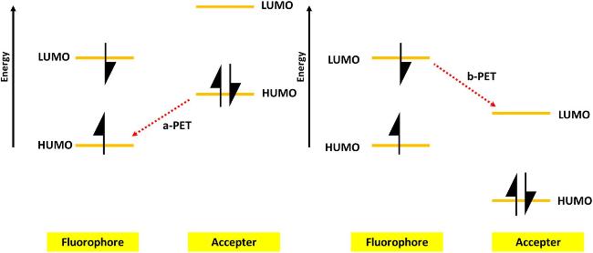
Photo-induced electron transfer refers to the process where electrons transition from the ground state to an excited state under photoexcitation, followed by electron transfer within or between molecules. In carbon dot fluorescence detection, carbon dots can act as electron donors or acceptors, achieving electron transfer through interactions with other substances. Asshown in Figure 2, based on the relative energy level positions between the carbon dots and the interacting substances, PET can be specifically divided into two modes: If the HOMO energy level of the electron acceptor is higher than that of the carbon dots, photoexcited electrons will spontaneously transfer from the acceptor to the HOMO vacant orbital of the carbon dots. This non-radiative electron transfer process competes with the radiative recombination process of fluorescence emission, consuming excited-state energy and leading to fluorescence quenching of the carbon dots; this phenomenon is termed a-PET. Conversely, in the b-PET process, when the LUMO energy level of the carbon dots is higher than that of the acceptor, excited-state electrons transfer from the carbon dots to the acceptor, similarly consuming excited-state energy and causing fluorescence quenching via electron transfer. In practical probe design, the acceptor is coupled with the carbon dots; the acceptor initiates PET to keep the probe fluorescence off. The binding of the target analyte to the acceptor inhibits PET, restoring the carbon dot fluorescence and achieving fluorescence enhancement detection. By monitoring changes in fluorescence intensity, the presence or concentration of the target analyte can be indirectly detected.[43-44].
图2 PET从CD到受体猝灭剂和目标分析物的示意图[23]

Fig.2 A scheme showing PET from the CDs to the quencher and target analyte[23]

2.2.5 Fluorescence inner filter effect

The inner filter effect is an apparent fluorescence quenching phenomenon in fluorescence analysis caused by light-absorbing substances. IFE refers to the phenomenon where fluorescence is weakened due to the absorption of emitted light, excitation light, or both by the fluorophore or absorber when the fluorophore concentration is high or coexists with other absorbers. Its essence is not a molecular interaction such as electron transfer or energy transfer between the fluorophore and the analyte, but rather a physical optical process. Given the significant overlap between the fluorescence spectrum of carbon dots and the absorption spectrum of the analyte, and the fact that the absorption peak and fluorescence lifetime of the carbon dots remain unchanged after adding the analyte, it is concluded that the inner filter effect exists during the quenching process.[45-46].
In summary, the fluorescence detection mechanisms of carbon dots are diverse, each with its unique advantages and applicable scope. In practical applications, it is necessary to select an appropriate fluorescence detection mechanism based on the characteristics of the target analyte and detection requirements to achieve efficient and accurate environmental monitoring.

2.3 Characterization of Carbon Dots

To comprehensively understand the properties of carbon dots, multiple characterization methods need to be employed for their analysis[47]. The following are several commonly used characterization methods.

2.3.1 Microscopy

Scanning Electron Microscopy (SEM) and Transmission Electron Microscopy (TEM) are common microscopy techniques that can provide information on the size, size distribution, and morphology of carbon dots. Through SEM and TEM images, the morphology and dispersion of carbon dots can be directly observed.

2.3.2 Spectral analysis

Photoluminescence (PL) spectroscopy and ultraviolet-visible (UV-Vis) absorption spectra are commonly used spectral analysis methods for studying the optical properties of carbon dots. PL spectroscopy provides information on the fluorescence emission wavelength and intensity of carbon dots, while UV-Vis absorption spectra offer insights into their absorption characteristics.

2.3.3 Structural Component Analysis

Methods such as X-ray diffraction (XRD), Raman spectroscopy (RS), X-ray photoelectron spectroscopy (XPS), and Fourier transform infrared spectroscopy (FTIR) are used to analyze the structure and chemical composition of carbon dots. These methods can provide information on the crystal structure, chemical bonding states, and surface functional groups of carbon dots.

2.3.4 Other analysis methods

Methods such as mass spectrometry (MS), nuclear magnetic resonance spectroscopy (NMR), and thermogravimetric analysis (TGA) can also be used for the characterization of carbon dots. These methods can provide information on the molecular mass distribution, atomic composition, and thermal stability of carbon dots.

3 Application of Carbon Dots in Environmental Detection

Probes constructed based on carbon dots demonstrate significant application potential in the field of environmental monitoring due to their notable advantages, including high sensitivity, rapid response, and ease of operation. These probes can efficiently detect metal ions, inorganic anions, and organic pollutants in the environment, providing strong support for the precise assessment of environmental quality. Starting from three types—fluorescence quenching carbon dots, fluorescence enhancement carbon dots, and ratiometric fluorescent carbon dots—we will elaborate in detail on the extensive applications of carbon dots in environmental monitoring.

3.1 Environmental detection applications of fluorescence quenching carbon dots

Fluorescence quenching carbon dots (Q-CDs), when interacting with specific analytes, exhibit a significant reduction in fluorescence intensity, even to the point of complete quenching. As a negative detection strategy, fluorescence quenching signals hold a place in the field of analytical detection due to their broad applicability and high selectivity. Particularly in sensing research targeting metal cations, Q-CDs probes have demonstrated excellent selectivity and practicality in recognizing ions such as Fe3+, Cu2+, Al3+, and Hg2+and other highly toxic metal ions, among which the detection of Fe3+is particularly noteworthy.
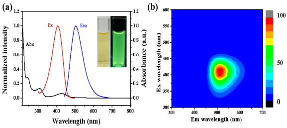
Fe3+is widely sourced from environmental emissions such as steel production, ore refining, and the use of fertilizers and pesticides. Abnormal levels in the human body may pose health risks, such as triggering Parkinson's disease. Therefore, developing efficient Fe3+detection technologies is crucial. In recent years, research on CDs-based Fe3+probes has emerged endlessly, with significant progress made particularly in the selection of CDs precursors. Ma et al.[48]synthesized nitrogen-doped carbon dots (N-CDs) fluorescent probes using citric acid (CA) and melamine as precursors via a facile one-step hydrothermal method. Their FRET effect caused quenching of blue fluorescence at 430 nm, with a detection range of 20~80 μmol/L and a limit of detection of 3.18 μmol/L, demonstrating good accuracy and repeatability in actual water samples (recovery rates of 97.04%~100.58%). Ye et al.[49]synthesized green fluorescent carbon dot (FNCDs) probes using 4,5-difluoro-o-phenylenediamine and ethylenediamine as precursors via a hydrothermal method. As shown inFigure 3, FNCDs possess excellent optical properties and can achieve Fe3+detection via the inner filter effect (fluorescence quenching at 512 nm). The detection range is 0.2~300 μmol/L with a limit of detection of 0.08 μmol/L; spiked recovery rates in laboratory/dormitory water samples were ≤107.5% with RSD < 1.1%, indicating their applicability for multi-source water Fe3+detection.
图3 FNCDs荧光探针的光学性质:(a) 吸收、激发与发射光谱(插图为日光与紫外光下的照片);(b) 三维荧光光谱[49]

Fig.3 Optical properties of the FNCDs probe. (a) Abs,Ex,and Em spectra (inset:visible and UV light images);(b) 3D fluorescence spectrum[49]

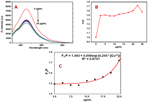
Cu2+is a metal pollutant widely present in water and the atmosphere. In these environments, Cu2+can accumulate in the human body through various pathways. Abnormal levels of Cu2+in the human body may trigger anemia, copper poisoning, cardiovascular diseases, and neurological disorders. Therefore, using carbon dots as fluorescent probes to detect Cu2+in the environment is particularly important. Sanni et al.[50]selected pine cone biomass as a raw material and successfully synthesized pine cone carbon dot (PC-CDs) fluorescent probes via microwave pyrolysis. The interaction between Cu2+and specific functional groups on the surface of PC-CDs leads to fluorescence quenching of the PC-CDs. The PC-CDs achieved a detection range for Cu2+of 2.5~22.5 μg/mL, with a limit of detection as low as 0.005 μg/mL; these performances are demonstrated inFigure 4. On the other hand, Zhang et al.[51]first utilized FeCl3to prepare Fe3O4, and then used it together with riboflavin as precursors, combined with a hydrothermal method, to prepare a novel magnetic carbon dot (M/CDs) fluorescent probe. This probe also possesses high selectivity for detecting Cu2+. In the presence of Cu2+, the photo-induced electron transfer process leads to fluorescence quenching of the CDs, enabling the probe to achieve a detection range for Cu2+of 0.2~50 mg/L, with a limit of detection of 0.096 mg/L.
图4 PC-CDs对Cu2+的荧光检测:(a) 添加不同浓度Cu2+时PC-CDs的荧光发射光谱;(b) PC-CDs荧光强度与Cu2+浓度关系;(c) F0/F与Cu2+浓度的校准曲线[50]

Fig.4 Fluorescence detection of Cu2+ using PC-CDs.(a) Fluorescence emission spectra of PC-CDs with the addition of different concentrations of Cu2+;(b) relationship between the fluorescence intensity of PC-CDs and the concentration of Cu2+;(c) calibration curve of F0/F versus the concentration of Cu2+ [50]

Al3+Mainly originates from fossil fuel combustion, aluminum processing wastewater, and pesticides, migrating into soil and water environments and accumulating in the human body. Excessive Al3+can cause damage to the nervous, digestive, skeletal, and renal systems, necessitating carbon dot fluorescent probes for detection. Jian et al.[52]prepared yellow fluorescent carbon dot (Y-CDs) fluorescent probes via a microwave method using chitosan ando-phenylenediamine (OPD) as raw materials for the detection of Al3+in water. The interaction between Al3+and functional groups on the surface of Y-CDs triggers static quenching, enabling the probe to achieve a detection range for Al3+of 10~100 μmol/L, with a limit of detection of 0.2 μmol/L.
Hg2+Originating from natural geological activities and emissions from fuel combustion and waste incineration, it pollutes the environment through atmospheric deposition. Its toxicity to multiple systems, including neurological, renal, and reproductive systems, highlights the necessity of detection. Song et al.[53]Using glutathione (GSH), diethylenetriamine (DETA), and ammonium ferric citrate (AFC) as raw materials, blue-emitting carbon dot (M-CDs) fluorescent probes were prepared via a one-step solvent method for Hg2+detection. Fluorescence quenching occurs through static quenching and the inner filter effect, with a detection range of 0.008~0.08 μmol/L and a limit of detection of 5.40 nmol/L.
Furthermore, the detection of Pb2+and Cr6+and other metal ions is equally significant for environmental remediation. The Ren team[54]employed1H-benzotriazole and choline chloride as precursors to synthesize nitrogen-rich carbon quantum dots (N-CQDs) fluorescent probes via solvothermal methods, which exhibited excellent fluorescence detection performance for Co2+. Co2+can coordinate with surface functional groups of N-CQDs, causing significant quenching of their blue fluorescence at 325 nm. The probe showed good linearity in the range of 5~250 μmol/L, with a detection limit as low as 1.2269 μmol/L. Notably, the research team successfully applied N-CQDs to the analysis of real industrial high-salinity wastewater, demonstrating the probe's excellent adaptability to complex matrices: in high-salinity wastewater, N-CQDs exhibited strong tolerance to Cl⁻ but were relatively sensitive to SO42-, thus requiring pretreatment via barium salt precipitation to remove sulfur before detection. This demonstrates the great potential of N-CQDs for achieving high-precision and high-reliability detection in industrial wastewater systems.
Idrees et al.[55]used fenugreek seeds as raw materials to synthesize carbon dot (CDs) fluorescent probes via one-step hydrothermal synthesis for Pb2+detection. The —NH₂ and —OH groups on the CDs coordinate with Pb2+, causing fluorescence quenching through non-radiative electron transfer. The detection range is 39.5–395 μmol/L, with a limit of detection of 9.345 μmol/L. This environmentally friendly fluorescent probe shows great promise for detecting Pb2+in natural water bodies. As for Cr6+, although its Cr3+form is an essential trace element for humans, Cr6+has become a carcinogen, posing a serious threat to human health. Tall et al.[56]prepared 515 nm green-light nitrogen and phosphorus co-doped carbon dots (N,P-CDs) fluorescent probes using PA and L-arginine as raw materials via a one-pot hydrothermal method. Upon addition of Cr6+, the fluorescence of N,P-CDs is quenched due to FRET, PET, and IFE effects. The detection range for Cr6+is 0.05–3 μmol/L, with a limit of detection of 26 nmol/L. Bardhan et al.[57]selected citric acid and ethylenediamine as starting materials to synthesize nitrogen-containing carbon dots (CDs) via hydrothermal methods, which were then incorporated into the natural mineral kyanite to construct KYCDs fluorescent probes for detecting Cr6+in the environment. In the presence of Cr6+, the induced PET process significantly quenches the fluorescence of the carbon dots at 460 nm. The limit of detection is as low as 0.11 μmol/L, with a linear range of 0–34 μmol/L. Good detection performance is maintained across a wide pH range and in various real water samples such as tap water, pond water, and tannery wastewater. Even in complex tannery wastewater, KYCDs effectively resist interference without pretreatment and achieve rapid, significant fluorescence quenching responses to Cr(VI) concentrations as high as 19.8 mg/L, demonstrating excellent anti-interference capability and selectivity.
With the continuous in-depth development of fluorescence quenching-based carbon dots in the field of sensing technology, their application scope has gradually expanded to the detection of inorganic anions. Although these inorganic anions differ significantly from metal ions in terms of structure and charge characteristics, they can form stable complexes with specific functional groups on the surface of carbon dots or participate in electron transfer processes, thereby effectively modulating fluorescence signals and achieving precise detection of inorganic anions.
Sulfide ions (S2⁻) mainly originate from the decomposition of sulfides during ore processing, the discharge of sulfur-containing wastewater in the pulping processes of the paper and textile industries, and the release of hydrogen sulfide in chemical production. Their absorption by crops and enrichment through the food chain can cause liver and kidney damage in humans, necessitating precise detection. Zeng et al.[58]ingeniously used melamine and 2,4-difluorobenzoic acid as precursors to hydrothermally synthesize a novel nitrogen and fluorine co-doped carbon dot (N,F-CDs) fluorescent probe. This probe achieves S2⁻ detection via static quenching through the formation of a ground-state complex, with a wide detection range of 0–30 μmol/L and a low limit of detection of 168 nmol/L. Furthermore, based on the principle that Cd2+binds with S2⁻ in the complex to form CdS, it enables linked detection of Cd2+, with a detection range of 0–30 μmol/L and a limit of detection of 340 nmol/L. Notably, the spike recovery rates of this fluorescent probe in tap water samples ranged from 96.2% to 103.5%, with an RSD < 6.13%.
Hypochlorite ions (ClO⁻) are reactive oxygen species with strong oxidizing and bleaching capabilities; appropriate amounts of ClO⁻ can also reduce inflammation and kill bacteria. However, excessive accumulation of ClO⁻ from industrial and domestic sewage discharge in water bodies can trigger severe health risks such as cardiovascular diseases and cancer due to its strong oxidizing nature, necessitating precise detection via carbon dot fluorescent probes. Wang et al.[59]synthesized aminophenylboronic acid-functionalized nitrogen-doped carbon dots (GAAP-CDs) via a hydrothermal method for specific detection of ClO⁻. This probe exhibits a significant response at 375 nm and generates a new absorption peak at 490 nm through a mechanism involving ClO⁻-induced C—B bond cleavage and fluorescence quenching. The detection range is 0.1~1000 μmol/L with a limit of detection of 0.50 μmol/L. Recovery rates in tap water and drinking water tests reached 99.2%~100.8%, demonstrating its potential for detecting environmental water samples.
Fluorescence quenching-type carbon dots not only demonstrate unique value in the detection of metal cations and inorganic anion pollutants but have also successfully broadened their application scope, particularly in the identification of organic pollutants. They are especially prominent in applications involving tetracycline antibiotics, aromatic compounds, and volatile organic compounds, highlighting their extensive versatility.
Tetracycline antibiotics (Tetracyclines, TCs), as broad-spectrum antibacterial agents widely used in the medical field, exhibit potent antibacterial activity against various cocci and bacilli. However, their environmental residues, due to non-degradability and highly toxic metabolites, threaten water and soil safety, highlighting the need for precise detection. He et al.[60]synthesized graphitic carbon nitride quantum dots (g-CNQDs) fluorescent probes using citric acid and urea as precursors via a one-step microwave-assisted method. Upon the addition of TC, the fluorescence intensity of the probe significantly decreased due to static quenching and the inner filter effect (IFE). The detection concentration range of this probe for TC was 7–100 μmol/L, with a limit of detection as low as 0.48 μmol/L. In the detection of TC in actual water samples, the recovery rates of the probe ranged from 97.77% to 103.4%, with relative standard deviations between 1.05% and 5.87%, demonstrating excellent performance.
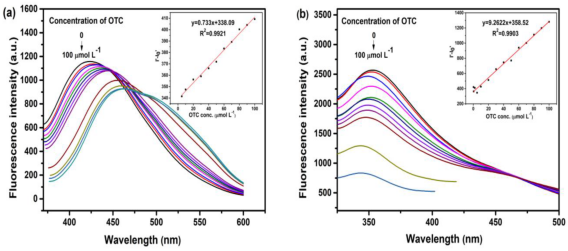
Oxytetracycline (OTC), as a member of the tetracycline antibiotic family, is widely used in animal husbandry, aquaculture, and human medicine. However, its residual presence in the environment and potential risks to humans have become a global focus. Therefore, accurate detection of OTC levels in the environment is crucial. Gao et al.[61]Using orange peels and watermelon rinds as raw materials, nitrogen-doped carbon dots (ON-CQDs and WN-CQDs) were synthesized via a one-step hydrothermal method, emitting bright fluorescence under excitation at 320 nm and 280 nm, respectively. Based on the fluorescence quenching effect, these two types of N-CQDs were developed as highly efficient fluorescent probes for the high-sensitivity detection of oxytetracycline (OTC) in environmental samples. OTC interacts with the amino groups of the N-CQDs via FRET, leading to a significant reduction in fluorescence intensity.Figure 5demonstrates the excellent detection performance of the two N-CQDs for OTC. The detection concentration ranges for ON-CQDs and WN-CQDs are 2–100 μmol/L and 0.25–100 μmol/L, respectively, with limits of detection as low as 0.973 μmol/L and 0.077 μmol/L. In the detection of actual samples such as soil, tap water, and lake water, this method exhibited superior accuracy and anti-interference capabilities. Specifically, for complex soil matrices, matrix interference was effectively eliminated through a pretreatment process involving acetonitrile extraction, centrifugation, and filtration. This resulted in OTC spike recovery rates ranging from 91.7% to 103.2% with RSD < 5.35% for all samples, validating the practical potential of N-CQDs probes in complex environments. This study provides a green and efficient solution for the rapid detection of antibiotic residues in the environment.
图5 N-CQDs探针对OTC的检测性能:(a) ON-CQDs与(b) WN-CQDs在加入不同浓度OTC后的荧光发射光谱,插图:荧光强度与OTC浓度的校准曲线[61]

Fig.5 Detection performance of the N-CQDs probes for OTC. Fluorescence emission spectra of (a) ON-CQDs and (b) WN-CQDs with the addition of different concentrations of OTC. Insets:the corresponding calibration curves of fluorescence intensity versus OTC concentration[61]

4-Nitrophenol (abbreviated as 4-NP), as a core aromatic compound, occupies an indispensable position in the production of numerous industrial fields such as agrochemicals, explosives, textiles, leather, and pharmaceuticals. Its high toxicity, resistance to degradation, and water solubility urgently demand efficient detection technologies. Venugopalan et al.[62]ingeniously utilized the dried rhizomes of Swertia flagellaris as starting materials to synthesize Swertia flagellaris-derived carbon dots (SFCDs) via microwave-assisted technology. As a novel and cost-effective fluorescent probe, these carbon dots demonstrate significant potential in monitoring 4-NP content in water bodies. Their fluorescence quenching, induced by the formation of Meisenheimer complexes causing static quenching and a slight inner filter effect (IFE), enables the detection of 4-NP within a linear range of 0–14.28 μmol/L, with a limit of detection as low as 0.207 μmol/L. Ashokan et al.[63]selected o-phenylenediamine and oleic acid as precursors and successfully prepared carbon dots exhibiting yellow fluorescence at a wavelength of 555 nm using a one-pot synthesis strategy. The fluorescent probe constructed based on these carbon dots achieves selective detection of 2-nitrophenol (2-NP) and 4-NP in water bodies, respectively, relying on static quenching caused by the interaction of —NO₂ and —OH groups with the surface functional groups of the carbon dots. The detection concentration spans 0.74–134 μmol/L, with limits of detection refined to 6.4 and 4.8 nmol/L, respectively.
Formaldehyde (FA), as a volatile organic pollutant released in large quantities during industrialization and urbanization, has widespread sources. For humans, environmental formaldehyde can impair respiratory, neurological, and immune system functions, and even increase cancer risk. Regarding environmentally friendly detection technologies for formaldehyde, Li et al.[64]creatively synthesized nitrogen-doped tannin/urea carbon dots (TA-CDs) fluorescent probes using the natural polyphenolic compound tannic acid (TA) and urea as raw materials via a hydrothermal method. This probe demonstrates highly specific and sensitive detection capabilities for formaldehyde based on fluorescence quenching. The interaction between the probe and formaldehyde induces structural changes in the fluorescent probe, thereby affecting its luminescent properties. The detection range of this fluorescent probe for formaldehyde is broad, spanning from 0.00015 to 0.035 mmol/L, with a limit of detection as low as 0.296 μmol/L, providing a practical solution for indoor air quality monitoring.
The practical value of fluorescence quenching carbon dot probes is further reflected in their detection capability for complex real-world environmental samples. Subsequent research will increasingly focus on systematic analysis of more challenging matrices such as soil and industrial wastewater, supplemented by targeted pretreatment strategies, fully demonstrating the immense application potential of carbon dot probes in complex systems.
Du et al.[65]Using p-phenylenediamine as a precursor, red fluorescent nitrogen-doped carbon quantum dots (N-CQDs) were synthesized via a microwave-assisted hydrothermal method to serve as a high-performance fluorescent probe for detecting alachlor. Aromatic π-π stacking and hydrogen bonding interactions between alachlor and the surface of N-CQDs caused significant fluorescence quenching of the N-CQDs at 601 nm, with a wide linear range of 0.005–150 μmol/L and a detection limit of 0.2 nmol/L. Furthermore, the probe demonstrated strong potential in soil extracts; the research team directly applied it to soil extracts pre-treated by homogenization, drying, sieving, ethanol ultrasonic extraction, centrifugation, and membrane filtration, achieving satisfactory recoveries of 86.6%–114.3%, which highlights the excellent matrix compatibility and practical prospects of the N-CQDs.
Mao et al.[66]Ingeniously selected dragon fruit seeds as natural precursors and successfully synthesized blue fluorescent carbon dots (P-CDs) using an ultrasound-assisted hydrothermal method. As a high-performance fluorescent probe, these carbon dots demonstrated highly specific detection capabilities for humic acid. Based on FRET between humic acid and P-CDs, the fluorescence of P-CDs at 463 nm was significantly quenched by humic acid, with a linear range of 3.0–120.0 mg/L. Regarding practical applications, the research team validated the probe's reliability in real samples, including lake water, tap water, and soil samples from campus flower beds and medicinal plant gardens following alkali extraction and centrifugation pretreatment. The spike recovery rates for all samples ranged from 96.4% to 104.5%, with RSD values less than 1.2%. These results were consistent with those obtained via high-performance liquid chromatography, confirming the excellent accuracy and anti-interference capability of P-CDs in complex soil matrices.
Keerthana et al.[67]Nitrogen-doped carbon quantum dots (N-CQDs) were synthesized via microwave irradiation using L-serine and citric acid as precursors, followed by boric acid functionalization to successfully prepare boron/nitrogen co-doped carbon quantum dots (B/N-CQDs) as a fluorescent probe for the precise detection of picric acid (PA) in industrial wastewater. In the presence of PA, the fluorescence of B/N-CQDs at 427 nm is quenched based on the FRET mechanism. The probe exhibits excellent analytical performance with a detection limit as low as 1.8 nmol/L and a linear range of 37 nmol/L to 30 μmol/L. It demonstrates remarkable applicability in sewage and complex industrial wastewater from fireworks and dye industries. In practical applications, wastewater samples require only simple membrane filtration before direct detection. Under this simplified protocol, the recovery rate for picric acid remains as high as 98%–101%, proving the probe's strong anti-interference capability and suitability for rapid on-site screening of industrial wastewater without complex pretreatment. In summary, fluorescence-quenching carbon dots have been widely applied in detecting various environmental targets; a summary comparison of their representative detection performance parameters is presented inTable 1.
表1 荧光猝灭型碳点的检测性能对比

Table 1 Comparison of detection performance in fluorescence quenching carbon dots

Analyte Precursors Synthesis Method Linear Range/
(μmol/L)
LOD/
(μmol/L)
Response Time Real Samples Ref
Fe3+ Citric acid (CA) and melamine Hydrothermal method 20~80 3.18 15 min Tap water,
Sand lake water
48
Fe3+ 4,5-Difluoro-1,2-phenylenediamine and ethylenediamine Hydrothermal method 0.2~300 0.08 <5 min Laboratory water,
Dormitory water
49
Cu2+ Pine-cone biomass Microwave pyrolysis 39.4~354.3 0.079 10 min Wastewater effluent 50
Cu2+ Riboflavin (C₁₇H₂₀N₄O₆) and Fe₃O₄ Hydrothermal method 3.15~787.4 1.51 30 min Tap water,River water 51
Al3+ Chitosan,and o-Phenylenediamine (OPD) Microwave method 10~100 0.2 - Tap water 52
Hg2+ Glutathione (GSH),ammonium ferric citrate (AFC),and diethylenetriamine (DETA) solvothermal method 0.008~0.08 0.0054 3 min Tap water,Yellow sea water 53
Co2+ BTA and choline chloride Solvothermal method 5~250 1.2269 1 min Industrial astewater 54
Pb2+ Fenugreek seeds Hydrothermal method 39.5~395 9.345 2 min - 55
Cr6+ Phytic acid (PA) and L-arginine (Arg) Hydrothermal method 0.05~3 0.026 10 min Water,Soil 56
Cr6+ Citric acid and ethylenediamine Hydrothermal method 0~34 0.11 - Tannery wastewater,Living cells 57
S2 Melamine,and 2,4-difluorobenzoic acid Hydrothermal method 0~30 0.168 1.5 min Tap water 58
ClO⁻ 3-Aminophenylboronic acid (3-APBA) and glutaric acid Hydrothermal method 0.1~1000 0.5 2 h Tap water,
Drinking water
59
TC Citric acid and Urea microwave-assisted method 7~100 0.48 23 min Tap water,River water,Lake water 60
OTC ON-CQDs:Orange peel
WN-CQDs:Watermelon peel
Hydrothermal method ON-CQDs:2~100
WN-CQDs:0.25~100
ON-CQDs:0.973
WN-CQDs:0.077
20 min Tap water,
Lake water,
Soil
61
4-NP Sweet flagdried rhizomes Microwave irradiation 0~14.28 0.207 1 min Tap water,
River water
62
2-NP/4-NP o-phenylenediamine and oleic acid carbonization 0.74~134 2-NP:0.0064
4-NP:0.0048
- Industrial water,
Drinking water,
River water
63
FA Tannic acid (TA) and urea Hydrothermal method 0.15~35 0.296 - - 64
Alachlor p-Phenylenediamine Microwave-assisted hydrothermal method 0.005~150 2×10⁻⁴ - Soil 65
Humic acid Pitaya seeds Ultrasound-assisted hydrothermal method 8.4~336.8 - 8 min Tap water,
Lake water,
Flower bed soil,Botanical garden soil
66
Picric acid(PA) L-serine and citric acid Microwave-assisted 0.0374~30 0.0018 2 min Firework industrial effluents,Dye industrial effluents 67

3.2 Environmental Detection Applications of Fluorescence-Enhanced Carbon Dots

In stark contrast to quenching-type carbon dots, fluorescence-enhancing carbon dots (E-CDs) exhibit a significant surge in fluorescence intensity when encountering specific environmental factors or analytes. As a positive detection strategy, fluorescence enhancement signals stand out in the field of detection due to their high sensitivity and low background noise interference. Although fluorescence-enhancing probes are widely used in anion detection, they have also demonstrated considerable potential in metal ion analysis; these metal ions promote an increase in fluorescence intensity through coordination with the surface of the carbon dots or by inducing their aggregation, laying a solid foundation for the application of fluorescence-enhancing probes in metal ion detection.
Peng team[68]ingeniously utilized triethoxysilane (APTES) and citric acid (CA) as precursors to synthesize silicon-doped carbon dots (Si/CDs) via a solvothermal method, and combined them with zeolitic imidazolate framework-90 (ZIF-90) to create a novel fluorescent probe—CDs@ZIF-90. This probe exhibits highly selective detection capabilities for Al3+and Hg2+, wherein Al3+significantly enhances its fluorescence at 453 nm, while Hg2+causes fluorescence quenching. The detection ranges of CDs@ZIF-90 cover Al3+ (1~200 μmol/L) and Hg2+ (0.05~240 μmol/L), with limits of detection as low as 0.81 μmol/L (Al3+) and 19.6 nmol/L (Hg2+).
Yan team[69]Taking a different approach, using citric acid and sodium sulfide nonahydrate as starting materials, sulfur-containing carbon dots CDs(s) were synthesized hydrothermally, and then combined with UiO-66-NH2(Eu) to innovatively develop the dual-emission fluorescent probe UiO-66-NH2(Eu)-CDs(s). Hg2+chelates with the amino and sulfur-containing functional groups on the probe surface, causing a significant increase in the fluorescence intensity of both emission peaks at 436 and 712 nm. The detection range is as wide as 0~100 μmol/L, and the detection limit is precisely 76.67 nmol/L. Even more remarkably, the detection of Pb2+by this probe is also based on a fluorescence enhancement mechanism, but it only enhances the emission peak at 436 nm. Based on this characteristic, the Yan team constructed a 3D analysis model, successfully achieving simultaneous detection of Hg2+and Pb2+, and achieved significant results in actual water sample analysis, providing guidance for synchronous detection of multiple metal ions.
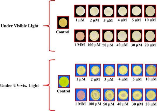
Kaur team[70]employed gallic acid (GA) andN,N-dimethylformamide (DMF) as precursors to prepare nitrogen-doped carbon dots (GA-DMFCDs) fluorescent probes via a one-pot hydrothermal method. Based on the chelation between surface functional groups and Pb2+, aggregation-induced emission enhancement (AIEE) was triggered, leading to fluorescence enhancement for the detection of Pb2+. The detection range of this probe covers 30~120 μmol/L of Pb2+, with a limit of detection of 715 nmol/L. Owing to their excellent selectivity, sensitivity, and convenience, GA-DMFCDs fluorescent probes are expected to become an effective means for Pb2+detection in the environmental field. To expand their practical application, the team further immobilized GA-DMFCDs on filter paper to construct a portable detection platform; asshown in Figure 6, upon addition of Pb2+, a distinct colorimetric response from pale yellow to light pink was observed in the detection zone under daylight, and corresponding fluorescence changes were also observed under UV irradiation. The research team integrated this test strip into a plastic box equipped with built-in sample wells and microfluidic channels, allowing samples to automatically flow to the detection zone and display color changes, confirming its potential for rapid on-site detection.
图6 使用GA-DMF CDs纸基传感器对Pb2+的双模式可视化检测:(a,b) 日光下的比色响应;(c,d) 紫外光下的荧光响应[70]

Fig.6 Dual-mode visual detection of Pb2+ using the GA-DMF CDs-based paper sensor.(a,b) Colorimetric response under daylight;(c,d) fluorimetric response under UV light[70]

Compared to other pollutants, fluorescence-enhanced carbon dots demonstrate broader application potential in the field of inorganic anion detection. The detected anions can significantly enhance the fluorescence effect by undergoing redox reactions or forming complexes with the carbon dots.
Ge et al.[71]Creatively selected hexamethylenetetramine and ammonium citrate as precursors to hydrothermally synthesize green-emitting nitrogen-doped carbon dots (N-CDs) fluorescent probes, which detect Cu based on an "on-off-on" mechanism2+and S2⁻. Cu2+coordinates with amino groups on the surface of N-CDs, causing fluorescence quenching; subsequently, S2-combines with Cu2+to form CuS, restoring fluorescence. The detection concentration ranges of this probe for Cu2+and S2-are 0.05~5 μmol/L and 0.05~10 μmol/L, respectively, with limits of detection as low as 25 and 32 nmol/L. This probe also performs well in practical detection applications for Cu2+and S2-in lake water, showing promising prospects for applications in analytical chemistry and environmental remediation.
Fluoride ions (F-) mainly originate from industrial emissions such as aluminum smelting and agricultural activities. Excessive fluoride intake can cause dental fluorosis, skeletal fluorosis, kidney damage, and parathyroid hyperplasia in humans. Therefore, developing green, highly sensitive, and simple F⁻ fluorescent probes is crucial for safeguarding health and environmental safety. Chen et al.[72]synthesized nitrogen-doped carbon dots (NCDs) fluorescent probes using citric acid and urea as precursors via a hydrothermal method. This probe detects F3+-mediated fluorescence "off-on" mode for F-in water bodies. Al3+reacts with oxygen-containing functional groups on the surface of NCDs, leading to fluorescence quenching; upon addition of F-, it coordinates with Al3+, causing the desorption of Al3+and restoring fluorescence. The detection concentration range of this probe for F⁻ is 20~300 μmol/L, with a limit of detection reaching 0.65 μmol/L.
Zhang et al.[73]synthesized selenium and nitrogen-doped carbon dots (Se, N-CDs) via a hydrothermal method using 2,3-diaminopyridine and selenourea as precursors, and combined them with curcumin to construct a fluorescent probe for F-detection. The detection mechanism is based on the quenching of the yellow fluorescence of Se, N-CDs by curcumin through IFE; upon addition of F-, a curcumin/F-complex is formed, causing a blue shift in the absorption peak and weakening the IFE, thereby restoring fluorescence. The system has a detection concentration range for F-of 2~60 μmol/L, with a limit of detection as low as 0.39 μmol/L. The probe has been successfully applied to the quantitative analysis of F-in tap water and milk, achieving recoveries of 92.6%~109.5% and RSD < 7.9%, demonstrating its potential for environmental and food testing.
Pang et al.[74]synthesized nitrogen-doped carbon dots (N-CDs) fluorescent probes via a one-step hydrothermal method using the nitrogen-rich materials adenine (AD) and ethylenediamine. Based on the fluorescence enhancement effect, this probe detects hypochlorite ions (ClO-); ClO-oxidizes surface groups of N-CDs, triggering AIEE-induced absorption redshift and significantly enhancing green fluorescence at 535 nm. The detection concentration range of this probe for ClO-is 0.01~1 mmol/L, with a detection limit of 0.82 μmol/L. In tap water, pool water, and river water samples, the recovery rate of this probe ranges from 90.0% to 125.0%, with RSD ≤ 5.0%. More surprisingly, the fluorescence enhancement effect of this probe can be regenerated through reduction by NaBH4. This fluorescent probe is the first ClO-probe based on fluorescence-enhanced N-CDs, providing a new strategy for monitoring disinfectants in environmental water bodies.
PO43-As a key component of phosphate fertilizers, it is indispensable for plant growth and development. However, excessive application of phosphate fertilizers leads to rising PO levels in water bodies.43-concentrations. In view of this, developing an environmentally friendly, rapid, and highly sensitive fluorescent detector to accurately monitor phosphate content in water bodies is particularly important. Han et al.[75]utilized excess sludge from paper mills (PMS) as a precursor to hydrothermally synthesize paper mill sludge carbon dots (PMS-CDs), serving as a green "turn-on" carbon dot fluorescent probe for detecting PO43-. PO43-reacts with the amino groups on the surface of PMS-CDs and enhances the fluorescence signal through electrostatic interactions. This fluorescent probe for PO43-has a wide detection range, covering a concentration interval of 9.96~500 μg/L, with a detection limit as low as 3.28 μg/L.
Zhu et al.[76]took a different approach, synthesizing blue fluorescent carbon dots from urea and sodium citrate via a microwave method. They further combined these with zinc nitrate and 2-methylimidazole to prepare a novel fluorescent probe, CDs@ZIF-8, using a one-pot method. This probe ingeniously utilizes a sequential "on"-"off"-"on" mode to achieve the sequential detection of Fe3+and PO43-. In the presence of Fe3+, CDs@ZIF-8 interacts with it to form static quenching; upon the addition of PO43-, it spontaneously reacts with Fe3+to form a complex, restoring fluorescence. The probe exhibits a wide detection range covering the concentration interval of 0~1 mmol/L, with a limit of detection reaching 5.2 μmol/L.
In addition to inorganic anions, certain organic small molecules with specific recognition sites can also induce a fluorescence enhancement effect in carbon dots, and this application has shown initial success in the detection of antibiotics and volatile organic compounds.
Li et al.[77]selected o-phenylenediamine (OPD) and urea as precursors to successfully synthesize nitrogen-doped carbon dots (N-CDs) fluorescent probes via a microwave-assisted method. This probe can perform "turn-off" fluorescence detection for Fe3+, and can also achieve "turn-on" fluorescence detection for tetracycline (TC). Fe3+significantly reduced the fluorescence intensity of N-CDs at 524 nm through dynamic quenching, while the detection of TC involved adding triethanolamine (TEA) to the N-CDs to mask Fe3+; subsequently, TC adsorbed onto the surface of the N-CDs via hydrogen bonding, resulting in fluorescence enhancement. The detection ranges of this probe for Fe3+ and TC were 1~70 μmol/L and 0.1~10 μmol/L, respectively, with limits of detection as low as 0.1011 and 0.0555 μmol/L, respectively.
Li et al.[78]also used neutral red and thiourea as precursors to hydrothermally synthesize red fluorescent carbon dot (R-CDs) fluorescent probes. Based on AIEE, this probe achieves dual-functional "turn-on" detection for tetracycline antibiotics (TCs) and pH. OTC induces the aggregation of R-CDs through π-π stacking and electrostatic interactions, thereby enhancing their fluorescence; on the other hand, the negative charges on the surface of R-CDs interact with H+neutralization, which also triggers AIEE. Among these, R-CDs exhibit a broad detection range for OTC, covering a concentration interval of 2~50 μmol/L, with a limit of detection as low as 23 nmol/L. This represents the first report of a red-light carbon dot TCs fluorescent probe based on AIEE with a detection limit reaching the nmol/L level, demonstrating promising prospects for antibiotic residue monitoring.
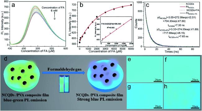
Wang et al.[79]pioneered the hydrothermal synthesis of a novel nitrogen-doped carbon dot (NCQD) fluorescent probe using alkali lignin (AL) extracted from spruce and m-phenylenediamine (MPD) as precursors for detecting formaldehyde (FA) in water. AsFigure 7shows, the characterization of these carbon dots demonstrates the fluorescent response of NCQDs to formaldehyde: the reaction between NCQDs and FA generates Schiff bases, disrupting the hydrogen bonding between NCQDs and water, which significantly enhances fluorescence at 490 nm; furthermore, the probe exhibits excellent quantitative capability and lifetime response (Figure 7a~c). The probe offers a wide detection range for FA, covering a concentration interval of 0.05~2 mmol/L, with a limit of detection reaching 4.64 μmol/L. Notably, the team also extended this work by constructing an NCQD/polyethylene composite film probe capable of detecting FA gas. Upon exposure to formaldehyde gas, the film's fluorescence intensifies and shifts from blue-green to blue, asFigure 7d~hshows. Fluorescence-enhanced carbon dots demonstrate excellent detection performance in various environmental analysis systems; a summary comparison of their representative detection performance parameters is presented inTable 2.
图7 NCQDs 对甲醛的荧光增强响应与双模式检测:(a) NCQDs 溶液荧光光谱随甲醛浓度的变化;(b) 荧光强度与浓度的定量关系;(c) 反应前后荧光寿命衰减曲线;(d) NCQDs/PVA薄膜气体传感示意图;(e~g) 薄膜在空气、水汽和甲醛气体中的荧光显微图像;(h) 从甲醛气体切换回空气环境时的荧光可逆性[79]

Fig.7 Fluorescence enhancement response and dual-mode detection of FA by NCQDs. (a) Fluorescence emission spectra of NCQDs solution with increasing FA concentrations;(b) the quantitative relationship between fluorescence intensity and FA concentration;(c) fluorescence lifetime decay curves of NCQDs before and after reaction with FA;(d) schematic diagram of the NCQDs/PVA composite film for FA gas sensing;(e~g) fluorescence microscope images of the film under ambient air,water vapor,and FA gas;(h)fluorescence reversibility upon switching from FA gas back to air environmen[79]

表2 荧光增强型碳点的检测性能对比

Table 2 Comparison of detection performance in fluorescence enhancement carbon dot

Analyte Precursors Synthesis Method Linear Range/
(μmol/L)
LOD/
(μmol/L)
Response Time/min Real Sample Ref
Al3+/Hg2+ (3-aminopropyl) triethoxysilane (APTES) and citric acid Solvothermal method Al3+:1~200
Hg2+:0.05~240
Al3+:0.81
Hg2+:0.0196
5 Tap water,
Yellow river water
68
Hg2+ Citric acid and sodium sulfide nonahydrate (Na₂S·9H₂O) Hydrothermal method 0~100 0.07667 2 Tap water,
Groundwater
Sewage plant effluent,
Lead-containing seepage water
69
Pb2+ Gallic acid (GA) and NN-Dimethylformamide (DMF) Hydrothermal method 30~120 0.715 - Tap water,RO water,
distilled water
70
Cu2+/S2 Hexamethylene-tetramine and ammonium citrate Hydrothermal method Cu2+:0.05~5
S2⁻:0.05~10
Cu2+:0.025
S2⁻:0.032
10 Tap water,
Lake water
71
F⁻ Citric acid and urea Hydrothermal method 20~300 0.65 1 Tap water,
Taihu lake water
72
F⁻ 2,3-diaminopyridine,and selenourea Hydrothermal method 2~60 0.39 - Tap water,
Milk
73
ClO⁻ Adenine (AD) and ethylenediamine (EDA) Hydrothermal method 10~1000 0.82 - Tap water,
pool water,
River water
74
PO43 Paper mill sludge (PMS) Hydrothermal method 0.103~5.16 0.034 30 Tap water,
Natural water
75
PO43 Sodium citrate and urea Microwave method 0~1000 5.2 - - 76
TC/Fe3+ o-phenylenediamine and urea Microwave-assisted method TC:0.1~10
Fe3+:1~70
TC:0.0555
Fe3+:0.1011
<1 Tap water,
Milk
77
OTC Neutral red and thiourea Hydrothermal method 2~50 0.023 2 Milk products 78
FA Alkali lignin (AL) and m-phenylenediamine (MPDA) Hydrothermal method 50~2000 4.64 1 Gaseous FA 79

3.3 Environmental Detection Applications of Ratiometric Fluorescent Carbon Dots

Ratiometric fluorescent probes, as a rising star in the field of fluorescence detection, demonstrate significant advantages in analyzing complex matrices such as wastewater and soil, owing to their strong anti-interference capability, wide dynamic detection range, and excellent sensitivity and selectivity. Their unique dual-signal output mode not only provides an intrinsic self-calibration mechanism for precise detection in complex environmental matrices but also makes changes in fluorescence color or intensity ratios easy to capture and analyze by portable devices (such as smartphones and portable spectrometers), thereby laying a solid foundation for building intelligent, on-site rapid sensing platforms. The core of ratiometric fluorescence sensing lies in possessing two non-overlapping emission peaks targeted at the analyte, meaning it has dual fluorescence signals: one is a response/indicator signal that fluctuates sensitively with changes in the concentration of the measured substance; the other is a reference signal that remains constant under the same conditions, together constituting a system that ensures measurement stability through self-calibration. By introducing a reference signal, ratiometric fluorescent probes effectively reduce errors caused by external interference while maintaining the high selectivity and high sensitivity of fluorescence detection. By dividing the fluorescence intensity of the indicator signal by that of the reference signal, which exhibits a distinct difference, the probe can eliminate interference from factors such as concentration variations and light source fluctuations, achieving internal calibration and thus significantly enhancing the accuracy and reliability of measurements.[80-81].
Currently, numerous researchers have successfully developed ratiometric fluorescent carbon dot detection systems with environmental response characteristics. These nanoprobes, which perfectly integrate signal self-calibration with target recognition functions, provide a new perspective for the precise detection of various pollutants in complex matrices. In recent years, significant achievements have been made in three major areas: the detection of heavy metal cations, the tracking of toxic anions, and the screening of organic pollutants using carbon dot ratiometric fluorescent probes.
Pang et al.[82]successfully prepared carbon dot (GN-CDs) fluorescent probes with dual emission peaks at 470 and 570 nm via a facile method of directly carbonizing gallic acid and o-phenylenediamine in aqueous solution. This probe exhibits extremely high selectivity and sensitivity towards Fe3+in acidic environments; the addition of Fe3+causes fluorescence quenching at 570 nm while the fluorescence intensity at 470 nm remains unchanged, thereby achieving precise detection of Fe3+. Its detection range is as wide as 0~50 μmol/L, with a limit of detection as low as 0.8 μmol/L.
Zhang et al.[83]used microbial fermentation products of Pu'er tea as precursors to prepare carbon dots rich in surface functional groups (T-CDs). They innovatively mixed T-CDs with o-phenylenediamine to construct a T-CDs-OPD ratiometric-colorimetric sensing platform for Cu2+detection. The detection mechanism of this platform lies in the fact that Cu2+quenches the 470 nm fluorescence peak of T-CDs through coordination interactions, while simultaneously catalyzing the oxidation of OPD to generate an oxidized product (oxOPD), thereby introducing a new fluorescence peak at 573 nm. This platform offers a wide detection range for Cu2+(0~170 μmol/L), with a limit of detection as low as 51 nmol/L.
Wang et al.[84]used p-phenylenediamine and toluene as precursors to synthesize red-emitting carbon dots (RCDs) via a solvothermal method, and ingeniously combined them with quercetin (QCT) to construct an RCDs-QCT fluorescent probe. This probe exhibits excellent visual and ratiometric fluorescence detection performance for Al3+. The detection mechanism lies in the fact that Al3+ has no effect on the red fluorescence peak of the RCDs-QCT probe at 360 nm, but with the formation of the QCT-Al3+ complex, the fluorescence intensity of the green fluorescence peak at 506 nm is significantly enhanced. The system has a wide detection range for Al3+, ranging from 0 to 35 μmol/L, with a limit of detection as low as 90 nmol/L. Furthermore, RCDs-QCT has achieved quantitative analysis of Al3+ in ultrapure water and tap water, demonstrating significant application potential.
Yin et al.[85]Using citric acid and glutathione as raw materials, N,S-co-doped carbon dots (NS-CDs) assembled with silver nanoclusters (AgNCs) were synthesized via microwave heating to construct a NS-CDs/AgNCs ratiometric fluorescent probe. This probe undergoes a redox reaction with Hg2+, forming an NS-CDs/Agn-2NCsHg complex, where the fluorescence of AgNCs is quenched while that of NS-CDs is enhanced. The detection range of this fluorescent probe for Hg2+reaches 0.1~90 μmol/L, with a limit of detection as low as 0.041 μmol/L. Notably, the probe demonstrates excellent recovery rates in samples (92.90%~103.64%), showing great application potential in water environment analysis.
Wang et al.[86]Using sodium citrate, polyacrylamide, and p-phenylenediamine as precursors, blue fluorescent carbon dots (BCDs) and red fluorescent carbon dots (RCDs) were synthesized via hydrothermal and solvothermal methods, respectively. A dual-emission ratiometric fluorescent probe was constructed by simply mixing them. This probe utilizes Pb2+-induced selective quenching of BCD fluorescence (based on the inner filter effect), while the fluorescence of RCDs serves as a stable internal reference signal, achieving highly sensitive and selective detection of Pb2+. As the concentration of Pb2+increases, the fluorescence color of the system changes significantly from blue to red. The detection range of this probe for Pb2+is 0–200 nmol/L, with a limit of detection as low as 2.89 nmol/L. The research team further fabricated fluorescent test strips from the probe solution and deeply integrated them with a smartphone platform, constructing an intelligent detection system comprising test strips, a portable UV lamp, and a color recognition app. The detection process is clear and convenient: after immersing the test strip in the sample, a fluorescence image is captured under a UV lamp using a mobile phone; the app automatically extracts RGB values and calculates the B/R ratio, ultimately enabling on-site semi-quantitative detection of Pb²⁺. The entire process can be completed within 5 minutes, providing a powerful tool for rapid on-site screening of heavy metal ions in water environments.
Zhang et al.[87]rapidly synthesized nitrogen and sulfur co-doped carbon dots (NSCDs) using ascorbic acid and thiourea as precursors via a one-step microwave method, and simply mixed them with gold nanoclusters (AuNCs) synthesized by the bovine serum albumin template method to construct an NSCDs/AuNCs ratiometric fluorescent probe for the detection of Hg in the environment.2+. The probe utilizes the strong metallophilic interaction between Hg2+ and Au+ to specifically quench the red fluorescence of AuNCs at 690 nm, while the blue fluorescence of NSCDs at 406 nm remains almost unaffected, thereby causing a distinct change in the system's fluorescence color from pinkish-purple to blue. The probe has a detection range for Hg2+ of 0.05~7.0 μmol/L, with a limit of detection of 18 nmol/L. The research team also developed a smartphone-assisted sensing platform that captures fluorescence images of the probe solution at different Hg2+ concentrations in a dark box using a mobile phone, reads RGB values via an app, and calculates the B/R ratio to achieve visual quantitative detection of Hg2+. Its reliability was verified in actual water samples, demonstrating the great application potential of this method in fields such as environmental monitoring.
Although the hazards of heavy metal cations to the environment have attracted widespread attention, the risks posed by anionic pollutants are equally worthy of high vigilance. Carbon dot-based ratiometric fluorescent probes, leveraging their unique dual-signal self-calibration function, demonstrate significant advantages in addressing the challenges of anion detection in complex matrices, bringing innovative solutions for the precise tracking of such pollutants.
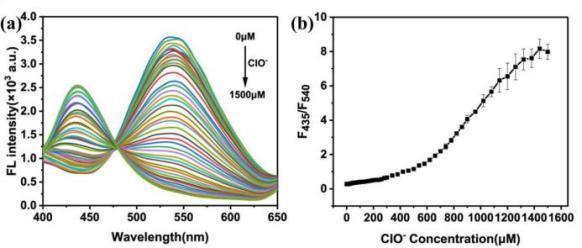
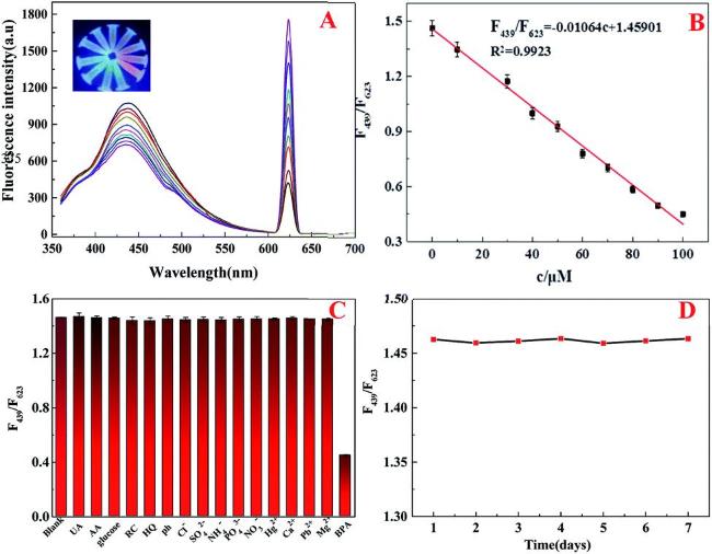
Shao et al.[88]utilized glutathione as a precursor to hydrothermally synthesize blue-emitting carbon dots (CDs) and combined them with curcumin (CCM) to assemble a CDs-CCM ratiometric fluorescent probe for efficient detection of ClO⁻. CCM quenches the blue fluorescence of CDs at 435 nm via the inner filter effect (IFE) and induces a green fluorescence peak at 540 nm. The oxidation of CCM by ClO⁻ weakens the IFE, resulting in the recovery of fluorescence at 435 nm and the reduction of fluorescence at 540 nm, as confirmed by the changes in their fluorescence spectra (Figure 8a) and the linear calibration curve established thereby (Figure 8b). This system achieves a detection range for ClO-of 1–1500 μmol/L with a limit of detection of 36.54 nmol/L. Superior recoveries (90%–101%) were verified in tap water, swimming pool water, and milk samples, providing a new strategy for the rapid and sensitive detection of ClO⁻ in complex matrices and demonstrating broad practical prospects.
图8 (a) CDs和CCM(20 μmol/L)与不同浓度的ClO-(0~1500 μmol/L)混合的荧光光谱;(b) ClO-浓度和荧光强度之间的线性关系(F435/F540[88]

Fig.8 (a) Fluorescence spectra of CDs and CCM (20 μmol/L) mixed with various concentrations of ClO- (0~1500 μmol/L);(b) linear relationship between the ClO- concentration and fluorescence intensity (F435/F540[88]

Zhang et al.[89]synthesized green-emitting carbon dots (GCDs) using perylene tetracarboxylic dianhydride (PTCDA) and triethylamine (TEA) as precursors, and red-emitting carbon dots (RCDs) using phenylenediamine (p-PDA) as a precursor. They mixed them at a fluorescence intensity ratio of 6:1 and added Eu3+ to construct a novel GCDs-RCDs ratiometric fluorescent probe for detecting inorganic phosphate (Pi). The sensing mechanism lies in the fact that Eu3+ induces aggregation-caused quenching (ACQ) of GCDs, quenching the green fluorescence at 475 nm, while Pi binds with Eu3+, disrupting the ACQ and restoring the green fluorescence, while the red fluorescence of RCDs at 620 nm remains stable. The GCDs-RCDs ratiometric fluorescent probe has a detection range for Pi of 0–55 μmol/L, with a limit of detection as low as 0.09 μmol/L. In terms of actual sample testing, the probe achieved accurate detection in tap water, lake water, and soil. Particularly for soil samples, after pretreatment with sodium bicarbonate extraction, the phosphate recovery rate reached 94.00%–106.40%, a result that strongly verifies the probe's excellent accuracy and anti-interference capability in complex environmental matrices. Furthermore, fluorescent test strips based on this probe enabled on-site visual detection of Pi, showing a color change from red to green under UV light, providing a new method for environmental phosphate monitoring.
Wang et al.[90]ingeniously mixed blue-emitting carbon dots (CDs) with red-emitting bovine serum albumin-capped gold nanoclusters (BSA-AuNCs) to construct a CDs/AuNCs ratiometric fluorescent probe for cyanide (CN⁻) detection. CN⁻ forms a stable [Au(CN)₂]⁻ complex with BSA-AuNCs, leading to quenching of the red fluorescence at 612 nm, while the blue fluorescence at 438 nm remains stable. The CN⁻ detection range of this system is 8 nmol/L to 75 μmol/L, with a detection limit as low as 8 nmol/L. This carbon dot fluorescent probe successfully achieved sensitive detection of complex organic pollutants, paving a new direction for the transformation of environmental monitoring technologies towards greener and smarter solutions.
Nejad et al.[91]made pioneering progress in the field of visual sulfide detection by successfully developing a CDs/AuNCs dual-emission ratiometric fluorescent probe for sulfide detection. This probe ingeniously combines blue fluorescent CDs with orange fluorescent AuNCs; sulfides form Au₂S with the AuNCs, causing fluorescence quenching of the AuNCs while the CD fluorescence remains stable, resulting in a color change from pink to blue. The probe has a sulfide detection range of 1.0–50.0 μmol/L, a limit of detection as low as 0.35 μmol/L, and a visual detection limit of 4.0 μmol/L, which is far below the maximum allowable concentration of sulfides in drinking water (10.0 μmol/L). Notably, the research team combined smartphones with UV lamps to achieve portable detection in river water and wastewater samples, demonstrating recovery rates of 94.3%–108.8%, RSD < 5.2%, and response times < 2 min. This ratiometric fluorescent probe demonstrates significant application value in the field of environmental monitoring.
Hao et al.[92]used 1,2-phenylenediamine and citric acid as precursors to synthesize nitrogen-doped carbon dots (N-CDs) via hydrothermal carbonization. These were combined with a morin-Al3+complex (morin-Al3+) based on the inner filter effect (IFE) to construct a ratiometric fluorescent probe with dual emission wavelengths, N-CDs/morin-Al3+, for detecting F⁻. F⁻ competes for coordination with Al3+to form a more stable Al3+-F⁻ complex, disrupting the IFE. This causes quenching of the green fluorescence of morin-Al3+at 492 nm and recovery of the blue fluorescence of N-CDs at 432 nm, achieving a fluorescence transition from green to blue. The detection range of this probe for F⁻ is 0.5–150 μmol/L, with a limit of detection (LOD) of 55.8 nmol/L. Furthermore, a smartphone RGB analysis-assisted visual sensing system based on this probe achieved a detection range of 2.5–150 μmol/L for F⁻ with an LOD of 2.09 μmol/L. The probe has been successfully applied to detect F⁻ in tap water and surface water from the Fen River. Additionally, Yan et al.[93]synthesized dual-emission carbon dots (CDs) using alizarin red and citric acid as precursors via a hydrothermal method. They introduced Al3+to construct a CDs@Al3+ratiometric fluorescent probe for detecting F⁻ in the environment. In the presence of F⁻, based on the strong coordination between F⁻ and Al3+, the red light of the probe weakens while the green light recovers as F⁻ competitively binds to Al3+, leading to the disruption of the aggregated structure of CDs@Al3+. This results in weakened red light and recovered green light, enabling visual detection with a range of 150–1200 µmol/L and an LOD of 7.998 µmol/L. The research team designed and built an AI-integrated handheld smartphone detection platform, comprising test strips, a 3D-printed dark box, and an app equipped with the YOLO v3 algorithm. By simply placing the test strip in the dark box to capture a fluorescence image, the app automatically identifies the image location, extracts R/G values, and directly outputs the F⁻ concentration based on a built-in standard curve. This achieves intelligent, portable, real-time quantitative detection without human intervention, with recovery rates of 99.24%–101.66% in tap water, toothpaste, and milk. This study provides an innovative solution for rapid on-site detection of F⁻ in fields such as environmental monitoring and food safety, demonstrating the immense potential of deep integration between artificial intelligence and carbon dot detection technologies.
Carbon dot fluorescent probes, by combining the high anti-interference capability of ratiometric fluorescence analysis with molecular recognition functions, have also successfully achieved sensitive detection of complex organic pollutants, paving a new path for the green and intelligent development of environmental monitoring technologies.
Zhang et al.[94]selected bovine serum albumin (BSA) as the raw material to prepare bovine serum albumin-derived carbon dots (BCDs) via a one-step hydrothermal method, and combined them with Eu3+to develop a Eu-BCDs ratiometric fluorescent probe. This probe can specifically detect TC; TC quenches the blue fluorescence at 448 nm through the IFE mechanism, while simultaneously enhancing the Eu3+red fluorescence at 618 nm based on the antenna effect. The Eu-BCDs exhibit a wide detection range for TC, covering 0~80 μmol/L, with a detection limit as low as 3 nmol/L. Experiments further verified the probe's quantitative detection capability for TC in samples such as river water, achieving recovery rates of 96.8%~103.6%. The ratiometric fluorescent probe features simple preparation, high sensitivity, and strong anti-interference capabilities.
Fu et al.[95]developed a carbon dot (CDs)-based fluorescent probe for oxytetracycline (OTC) utilizing FRET. The binding of OTC to the CDs triggers FRET, resulting in the quenching of blue fluorescence at 405 nm and the enhancement of green fluorescence at 505 nm. The probe exhibits a wide linear detection range for OTC covering 0–40 μmol/L, with a limit of detection as low as 0.41 μmol/L and a response time of only 30 s. Furthermore, the probe enables high-precision detection of OTC in complex matrices such as river water, tap water, milk, and pork, achieving recovery rates between 93% and 102% with relative standard deviations below 6%. This probe provides an intuitive and reliable sensing platform for detecting oxytetracycline residues in food and environmental samples.
Qu et al.[96]UsingN-(phosphonomethyl)iminodiacetic acid (PMIDA) and branched polyethyleneimine (BPEI) as precursors, N,P co-doped carbon dots (BPEI-CDs) ratiometric fluorescent probes were synthesized via a hydrothermal method. Based on AIEE induced by Schiff base reactions and enhanced π-conjugation, detection of formaldehyde (FA) was achieved. The addition of FA resulted in fluorescence enhancement at 501 nm and simultaneous fluorescence quenching at 408 nm, with the fluorescence color changing from colorless to light yellow. BPEI-CDs exhibited a wide detection range for FA covering 0–40 μmol/L, with a limit of detection of 0.47 μmol/L. In contaminated food samples, recoveries ranged from 96.9% to 101.8%, with RSD values between 2.23% and 3.21%, providing a simple and reliable solution for FA detection in complex environments.
Lu team[97]ingeniously used blue-emitting carbon dots (CDs) and gold nanoparticles (AuNPs) to construct a highly sensitive fluorescence sensor based on the inner filter effect (IFE) for detecting the carbamate pesticide carbaryl, asshown in Figure 9. AuNPs quench the fluorescence of CDs via the inner filter effect; thiocholine generated from the acetylcholinesterase (AChE)-catalyzed reaction induces AuNP aggregation, leading to fluorescence recovery; whereas carbaryl inhibits AChE activity, preventing fluorescence recovery, thereby achieving sensitive detection of the target. The probe exhibits a good response to carbaryl within a linear range of 5×10⁻⁴~1 μmol/L, with a detection limit as low as 2.5×10⁻⁴ μmol/L. It achieved satisfactory recoveries of 96.8%~103.6% with RSD < 5% in actual strawberry samples, demonstrating its excellent potential for future detection of the pesticide carbaryl.
图9 基于CDs和AuNP的荧光传感器的示意图,用于通过IFE检测甲萘威[97]

Fig.9 Schematic illustration of the fluorescent sensor based on CDs and AuNPs for carbaryl detection through the IFE[97]

Clermont-Paquette et al.[98]synthesized dual-emission carbon dots (CDs) via a microwave-assisted method using L-glutathione and formamide as precursors, constructing a fluorescent probe for ratiometric detection of glyphosate. The interaction between glyphosate and the surface functional groups of the CDs leads to quenching of the red fluorescence at 600–800 nm, while the blue fluorescence at 415–600 nm remains stable as an internal reference signal, asFigure 10shows. This probe enables glyphosate detection across a broad pH range of 3–10, with a limit of detection as low as 0.03 ppm in the low concentration range of 0–10 ppm (1 ppm = 1×10⁻⁶) and excellent detection performance in the high concentration range of 0–500 ppm.
图10 基于比率检测的低浓度草甘膦传感[98]

Fig.10 Low concentration of glyphosate sensing based on ratiometric measurements[98]

Li et al.[99]utilized carbon dots (CDs) containing abundant hydroxyl and carbonyl groups as carriers to coordinate and polymerize Eu3+with 5'-adenosine monophosphate (AMP) on their surface, successfully constructing a novel CDs@Eu-AMP ratiometric fluorescent probe for the detection of bisphenol A (BPA). The strong coordination between BPA and Eu3+quenches the blue fluorescence of CDs at 439 nm, while the red fluorescence of Eu3+at 623 nm is enhanced, thereby achieving a ratiometric fluorescent response.Figure 11fully demonstrates that the probe has a detection range for BPA of 0.1~100 µmol/L, with a limit of detection as low as 20 nmol/L. Furthermore, the probe exhibits good recovery rates (91.80%~102.7%) in actual water samples such as river water, lake water, and tap water. Additionally, coffee-ring test strips prepared based on CDs@Eu-AMP enable naked-eye visual detection of BPA under UV light, asshown in Figure 12, where the fluorescent color of the test strip changes distinctly from blue to red with increasing BPA concentration.
图11 CDs@Eu-AMP比率荧光探针的分析性能表征:(a) CDs@Eu-AMP荧光光谱对BPA浓度的响应;(b) F439/F623与BPA的线性关系[99]

Fig.11 Analytical performance of the CDs@Eu-AMP ratiometric fluorescence probe:(a) fluorescence spectral response of CDs@Eu-AMP to BPA concentration;(b) linear relationship between F439/F623 and BPA concentration[99]

图12 基于(a) Eu-AMP,(b) CDs@Eu-AMP与(c) CDs的咖啡环试纸在不同BPA浓度下的对比[99]

Fig.12 Comparison of coffee-ring test strips based on(a) Eu-AMP,(b) CDs@Eu-AMP,and(c) CDs at different BPA concentrations[99]

Guo et al.[100]Using melamine and o-phenylenediamine as precursors, dual-excitation nitrogen-doped carbon dots (N-CDs) were successfully synthesized via microwave irradiation. The N-CDs serve as a ratiometric fluorescent probe for detecting 4-NP based on the IFE mechanism. The addition of 4-NP causes fluorescence quenching at 450 nm and enhancement at 550 nm, shifting the fluorescence color from blue to green. This probe exhibits a wide detection range for 4-NP, covering 0.083–80 μmol/L, with a detection limit of 83 nmol/L. Notably, by combining 3D printing technology, they constructed a self-calibrating sensing platform that enables visual detection of 4-NP in real samples; this method is simple, rapid, and reliable.
Chu et al.[101]Using citric acid and polyethyleneimine as precursors, blue fluorescent carbon dots (bCDs) were synthesized via a solvothermal method. Innovatively, the bCDs were covalently linked to SiO2 NPs embedding red-emitting CdTe quantum dots (rQDs) to construct a novel dual-emission ratiometric fluorescent probe, rQDs@SiO2@CDs. This probe leverages the specific interaction between gold nanoparticles (AuNPs) and thiram to achieve highly sensitive and selective detection of thiram. The detection mechanism is based on an "on-off-on" fluorescence response: the specific interaction between thiram and AuNPs disrupts fluorescence resonance energy transfer (FRET), restoring the blue fluorescence of bCDs, while the red fluorescence of rQDs remains stable as an internal reference signal. The probe exhibits a detection range for thiram of 0–10 µmol/L, with a limit of detection as low as 59 nmol/L. Looking ahead, this technology can be deeply integrated with smartphone platforms. The research team has constructed an intelligent detection system comprising test strips, a portable dark box, and a mobile app, asFigure 13shows. The detection process is clear and convenient: after adding the sample solution to the test strip, the strip is placed in the dark box, a fluorescence image is captured with a smartphone, and finally, the app analyzes the R/B value to achieve quantitative detection of thiram. This lays a solid foundation for developing portable intelligent equipment oriented toward rapid on-site detection.
图13 福美双的可视化定量检测:(a~c) 3D打印智能手机传感平台的构建与工作原理;(d,e) 荧光响应图像及定量校准曲线[101]

Fig.13 Visual and quantitative detection of Thiram:(a~c) fabrication and working mechanism of the 3D-printed smartphone sensing platform;(d,e) fluorescence response images and quantitative calibration curves[101]

Xiao et al.[102]synthesized blue fluorescent carbon dots (CDs) using citric acid and ethylenediamine as precursors via a hydrothermal method, and mixed them with polystyrene-encapsulated red fluorescent quantum dots (QDs) to construct a CDs/QDs ratiometric fluorescent probe. Based on the acetylcholinesterase (AChE)/choline oxidase (ChOx) dual enzyme-mediated Fe2+/Fe3+conversion cascade catalytic reaction, the probe achieves detection of chlorpyrifos with a detection range of 5~60 nmol/L and a limit of detection of 1.45 nmol/L. Regarding implementation, the research team further developed a smartphone-based fluorescence spectrometer (SBS) and a supporting app as shown inFigure 14, which can rapidly acquire fluorescence spectra within 380~760 nm, enabling spectral reconstruction, peak extraction, and quantitative analysis. The system is low-cost (approximately $156 per set), easy to operate, and suitable for on-site detection. In actual sample tests on apples and cabbage, recovery rates ranged from 91.6% to 104.2%, demonstrating its accuracy and reliability. This study provides a scalable, low-cost, and easy-to-operate solution for rapid on-site screening of pesticide residues, particularly suitable for food safety and environmental monitoring in resource-limited regions. Overall, ratiometric fluorescent carbon dots effectively improve the reliability of detection results through internal reference signal correction; a summary comparison of representative environmental detection performance parameters is presented inTable 3.
图14 智能手机生物传感器(SBS)的便携式设备与光学设计:(a) SBS的设计图与实物照片;(b) 定制SBS-App的用户界面与操作流程;SBS的组件分解图(c)与内部光路示意图(d)[102]

Fig. 14 Smartphone-based biosensor (SBS):portable device and optical design.(a) Design diagram and photograph of the SBS;(b) user interface and operational flowchart of the custom SBS-App;parts assembly drawing of the SBS(c) and internal light path schematic(d)[102]

表3 比率型荧光碳点的检测性能对比

Table 3 Comparison of detection performance for ratiometric fluorescent carbon dots

Analyte Precursors Synthesis Method Linear Range/
(μmol/L)
LOD/
(μmol/L)
Response Time Real Sample Ref
Fe3+ Gallic acid and o-henylenediamine Hydrothermal method 0~50 0.8 5 min Tap water,Spring water 82
Cu2+ Pu-erh tea Brewing with hot water 0~170 0.051 1 h --- 83
Al3+ p-phenylenediamine and toluene Solvothermal method 0~35 0.09 5 min Tap water,Ultrapure water 84
Hg2+ Citric acid and glutathione Microwave-assisted method 0.1~90 0.041 5 min Tap water,River water,
Lake water
85
Hg2+ Ascorbic acid and thiourea Microwave-assisted method 0.05~7.0 0.018 5 min Tap water,River water 87
Pb2+ BCDs:Sodium citrate and polyacrylamide
RCDs:p-Phenylenediamine and ethanol
Hydrothermal method 0~0.2 2.89×10⁻³ 5 min Tap water,Lake water 86
ClO⁻ Glutathione Hydrothermal method 1~1500 0.03654 30 s Tap water,Swimming pool water,Milk 88
PO43 GCDs:Perylene-3,4,9,10-tetracarboxylic (PTCDA) and triethylamine (TEA)
RCDs:p-phenylenediamine (p-PDA)
Hydrothermal method 0~55 0.09 2 min Tap water,Lake water,
Soil extracts
89
CN⁻ Citric acid and ethylenediamine anhydrous Hydrothermal method 0.008~75 0.008 10 min Tap water,Cassava roots,Sprouted potatoes,Liquor 90
S2 Citric acid and urea Hydrothermal method 1~50 0.35 2 min River water,wastewater 91
F⁻ o-Phenylenediamine and citric acid Hydrothermal method 0.5~150 0.0558 1 min Tap water,Surface water 92
F⁻ Alizarin carmine and citric acid Hydrothermal method 150~1200 7.998 1 min Tap water,Toothpaste,Milk 93
TC Bovine Serum Albumin (BSA) and Eu(NO33 Hydrothermal method 0~80 0.003 2 min River water,Milk,Honey,Serum 94
OTC Spinach Solvothermal method 0~40 0.41 30 s River water,Tap water,Mineral water,Milk,Pork 95
FA N-(phosphonomethyl)iminodiacetic acid (PMIDA) and branched polyethyleneimine(BPEI) Hydrothermal method 0~40 0.47 100 min Bean sprout 96
Carbaryl Citric acid and ethylenediamine Hydrothermal method 5×10⁻⁴~1 2.5×10⁻⁴ 30 min Strawberry 97
Glyphosate L-glutathione and formamide Microwave-assisted Low range:
0~59;High range:0~2957
0.177 - - 98
BPA Sodium citrate and ethylenediamine Hydrothermal method 0.1~100 0.02 3 min Tap water,River water,
Lake water
99
4-NP Melamine and o-phenylenediamine Microwave radiation 0.083~80 0.083 2 min Tap water,Sewage,
Yongjiang river water
100
Thiram Citric acid and polyethylenimine Solvothermal method 0~10 0.059 4 min Tap water,Apple peel,
Milk
101
Chlorpyrifos Citric acid and ethylenediamine Hydrothermal method 0.005~0.060 0.00145 60 min Apple,Cabbage 102

4 Conclusion and Outlook

In recent years, as environmental pollution issues have intensified, there is an increasingly urgent demand for efficient and agile technologies to detect environmental pollutants. As a unique class of zero-dimensional carbon-based nanomaterials, carbon dots (CDs) have emerged in the field of environmental monitoring, demonstrating broad application prospects. This study comprehensively reviews the latest research achievements of carbon dot fluorescent probes in environmental monitoring by meticulously examining their synthesis pathways, fluorescence detection principles, and characterization methods. Of particular note are three types of carbon dot probes—fluorescence quenching, fluorescence enhancement, and ratiometric—which, owing to their innovative design concepts and response modes, exhibit excellent performance in detecting various environmental pollutants such as metal ions, inorganic anions, and organic contaminants. Especially, ratiometric fluorescent carbon dot probes stand out in environmental monitoring applications due to their superior anti-interference capabilities and self-calibration functions in complex environmental backgrounds. Furthermore, the successful integration of carbon dot probes with intelligent sensing platforms like smartphones has opened new technical pathways for rapid, on-site, and visual detection of pollutants; meanwhile, systematic detection studies targeting complex environmental matrices such as soil and industrial wastewater fully validate the immense potential of carbon dot probes transitioning from laboratory research to practical applications. These research findings not only deepen our understanding of the theoretical framework of carbon dot fluorescent probes but also provide solid support for advancing environmental detection technologies toward greener, more convenient, and intelligent directions.
Although carbon dot fluorescent probes have made significant progress in the field of environmental detection, they still face numerous challenges and opportunities. Future research directions can focus on the following aspects.
First, innovation and optimization of synthesis methods: Further explore environmentally friendly, efficient, and low-cost carbon dot synthesis methods to improve the yield and fluorescence performance of carbon dots, reduce production costs, and promote their widespread application in practical scenarios.
Second, in-depth exploration of fluorescence mechanisms: Strengthen research on the fluorescence mechanisms of carbon dots, particularly the regulation mechanisms of key parameters such as surface states and energy level structures, with the aim of designing fluorescent probes with higher sensitivity and selectivity.
Third, improvement of probe performance: by improving probe design, enhance its anti-interference capability, stability, and long-term monitoring ability to meet the requirements for accurate detection of pollutants in complex environmental matrices.
Fourth, diversification of application expansion: applying carbon dot fluorescent probes to the detection of more types of environmental pollutants, such as emerging organic pollutants and microplastics, while exploring their application potential in fields like food safety and biomedicine.
Fifth, intelligent and integrated development: By integrating advanced technologies such as the Internet of Things and big data, promote the intelligent and integrated development of carbon dot fluorescent probes to achieve real-time monitoring and early warning of environmental pollutants, providing more precise and efficient technological support for environmental protection.
Sixth, promote industrialization and overcome application bottlenecks: To achieve the leap from the laboratory to practical application, it is necessary to focus on overcoming three key bottlenecks: the reproducibility and stability of batch synthesis, which urgently requires the development of standardized synthesis processes to ensure inter-batch consistency; the long-term storage life of probe materials, which can be enhanced through modification strategies such as surface encapsulation and matrix immobilization to improve their durability; and anti-interference performance and reliability in complex environments, which necessitates establishing a systematic performance evaluation standard system to cope with extreme or complex matrices. Solving these problems is key to the market-oriented application of carbon dot probes.
Carbon dots have broad application prospects as fluorescent probes in the field of environmental detection. Future research will be dedicated to addressing existing challenges, promoting continuous innovation and upgrading of carbon dot fluorescent probe technology, and contributing to the construction of a green and sustainable ecological environment.
[1]
Zhong Q M, Wen H W, Lee C C. Environ. Impact Assess. Rev., 2022, 95: 106799.

[2]
Chen X F, Li Z J, Chao L Q, Hao Y T, Wang Y M, Liang R F, Li K F, Pu X C. Water Res., 2024, 252: 121237.

[3]
Chen B, Wang M, Duan M X, Ma X T, Hong J L, Xie F, Zhang R R, Li X Z. J. Clean. Prod., 2019, 228: 101.

[4]
Shetty S S, Deepthi D, Harshitha S, Sonkusare S, Naik P B, Kumari N S, Madhyastha H. Heliyon, 2023, 9(9): e19496.

[5]
Zhou X, Gui L Y, Lu Z Y, Chen B B, Wu Z K, Zhou Z, Liang Y, He M, Hu B. Anal. Chim. Acta, 2024, 1324: 343003.

[6]
Elseblani R, Cobo-Golpe M, Godin S, Jimenez-Lamana J, Fakhri M, Rodríguez I, Szpunar J. Sci. Total Environ., 2023, 887: 164111.

[7]
Rains T C, Watters R L, Epstein M S. Environ. Int., 1984, 10(2): 163.

[8]
Tüzen M. Microchem. J., 2003, 74(3): 289.

[9]
You D W, Seon Y S, Jang Y, Bang J, Oh J S, Jung K W. J. Chromatogr. A, 2020, 1625: 461267.

[10]
Santos F J, Galceran M T. Trac Trends Anal. Chem., 2002, 21(9/10): 672.

[11]
Boukhobza I, Crans D C. Arab. J. Chem., 2020, 13(1): 1198.

[12]
Hameedat F, Hawamdeh S, Alnabulsi S, Zayed A. Molecules, 2022, 27(6): 1807.

[13]
Fang J, Zhao H Z, Zhang Y H, Lu M H, Cai Z W. Trends Environ. Anal. Chem., 2020, 25: e00076.

[14]
Xu Y H, Ou Q, Wang X T, Hou F, Li P, van der Hoek J P, Liu G. Environ. Sci. Technol., 2023, 57(8): 3114.

[15]
Picó Y, Barceló D. Trac Trends Anal. Chem., 2020, 130: 115964.

[16]
Okoffo E D, Thomas K V. J. Hazard. Mater., 2024, 464: 133013.

[17]
Yi K Y, Zhang L. Food Chem., 2021, 354: 129584.

[18]
Yi K Y, Li H, Zhang X T, Zhang L. Inorg. Chem., 2021, 60(5): 3172.

[19]
Yi K Y, Zhao Q F, Kang X Q. Bull. Chem. Soc. Jpn., 2025, 98(2): uoaf004.

[20]
Li H X, Yan X, Kong D S, Jin R, Sun C Y, Du D, Lin Y H, Lu G Y. Nanoscale Horiz., 2020, 5(2): 218.

[21]
Long C C, Jiang Z X, Shangguan J F, Qing T P, Zhang P, Feng B. Chem. Eng. J., 2021, 406: 126848.

[22]
Yao Y, Zhang H Y, Hu K S, Nie G, Yang Y Y, Wang Y X, Duan X G, Wang S B. J. Environ. Chem. Eng., 2022, 10(2): 107336.

[23]
Kayani K F, Ghafoor D, Mohammed S J, Shatery O B A. Nanoscale Adv., 2025, 7(1): 42.

[24]
Cui L, Ren X, Wang J, Sun M. Mater. Today Nano, 2020, 12: 100091.

[25]
Su Y J, Zhang Y F. Carbon, 2015, 83: 90.

[26]
Li R L, Zhao Z B, Leng C Y, Li Y, Ai L S, Sun Y, Wang X Z, Qiu J S. New Carbon Mater., 2023, 38(2): 347.

[27]
Kang S, Jeong Y K, Ryu J H, Son Y, Kim W R, Lee B, Jung K H, Kim K M. Appl. Surf. Sci., 2020, 506: 144998.

[28]
Alas M O, Alkas F B, Aktas Sukuroglu A, Genc Alturk R, Battal D. J. Mater. Sci., 2020, 55(31): 15074.

[29]
Kurian M, Paul A. Carbon Trends, 2021, 3: 100032.

[30]
Gao S Y, Wang X, Xu N, Lian H L, Xu L, Zhang W Q, Xu C Y. Cellulose, 2021, 28(3): 1647.

[31]
Luo H, Lari L, Kim H, Hérou S, Tanase L C, Lazarov V K, Titirici M M. Nanoscale, 2022, 14(3): 910.

[32]
Zhu J, Wu C X, Cui Y M, Li D X, Zhang Y J, Xu J, Li C F, Iqbal S, Cao M W. Colloids Surf. A Physicochem. Eng. Aspects, 2021, 623: 126673.

[33]
Zhou Y, Duan H L, Tan K J, Dong L. Nanoscale, 2024, 16(24): 11642.

[34]
Liu H X, Zhong X, Pan Q, Zhang Y, Deng W T, Zou G Q, Hou H S, Ji X B. Coord. Chem. Rev., 2024, 498: 215468.

[35]
Etefa H F, Tessema A A, Dejene F B. J. Carbon Res., 2024, 10(3): 60.

[36]
George H S, Selvaraj H, Ilangovan A, Chandrasekaran K, Kannan V R, Parthipan P, Almutairi B O, Balu R. Inorg. Chem. Commun., 2023, 157: 111348.

[37]
Liu H Y, Li C Y, Li J, Cheng Y Q, Zhao J F, Chen J N, Sun M T. Appl. Mater. Today, 2023, 30: 101731.

[38]
Liao S, Ding Z, Wang S, Tan F Y, Ge Y, Cui Y Q, Tan N, Wang H Q. Spectrochim. Acta Part A Mol. Biomol. Spectrosc., 2021, 259: 119897.

[39]
Zhang S T, Li J H, Zhou J Y, Xu P Y, Li Y, Zhang Y M, Wu S P. Talanta, 2025, 282: 126983.

[40]
Guo T X, Yang G X, Li Y, Liu C, Yang F L, Hou D F, Sun H, Zheng Y W, Lin X, Liu L X. Nanoscale Adv., 2024, 6(8): 1997.

[41]
Zhou Y W, Liu F P, Shi Q, Sun Z H, Yang B, Zhao L, Cui F Z, Nie Z G. J. Lumin., 2025, 286: 121335.

[42]
Hun Seo Y, Elizabeth Aguilar Estrada D, Jang D, Baik S, Lee J, Ha Kim D, Kim S. Spectrochim. Acta Part A Mol. Biomol. Spectrosc., 2024, 310: 123925.

[43]
Huang J Y, Long C C, Zhang L, Qing T P, Zhang P, Qing Z H, Feng B. J. Environ. Chem. Eng., 2022, 10(6): 108951.

[44]
Zhang L Q, Wei S Q, Zhang L M, Yin X H, Yang R. Spectrochim. Acta Part A Mol. Biomol. Spectrosc., 2024, 304: 123276.

[45]
Zhang J Y, Zhou R H, Tang D D, Hou X D, Wu P. Trac Trends Anal. Chem., 2019, 110: 183.

[46]
Mu X W, Wu M X, Zhang B, Liu X, Xu S M, Huang Y B, Wang X H, Song D Q, Ma P Y, Sun Y. Talanta, 2021, 221: 121463.

[47]
Hu Q, Gong X J, Liu L Z, Choi M M F. J. Nanomater., 2017, 2017: 1804178.

[48]
Ma Y L, Mao L H, Cui C C, Hu Y, Chen Z X, Zhan Y, Zhang Y H. Spectrochim. Acta Part A Mol. Biomol. Spectrosc., 2024, 316: 124347.

[49]
Ye S, Zhang M M, Guo J Q, Song J, Zeng P J, Qu J L, Chen Y, Li H. Nanomaterials, 2022, 12(9): 1487.

[50]
Sanni S O, Moundzounga T H G, Oseghe E O, Haneklaus N H, Viljoen E L, Brink H G. Nanomaterials, 2022, 12(6): 958.

[51]
Zhang Y, Li G Z, Lu M R, Li J X, Zhou Q Q, Zou X L, Yang W R, Yang M, Wang H B. Microchem. J., 2024, 206: 111424.

[52]
Jian K, Fu L M, Zhang Y J, Zhang H M, Guo X J, Zhao X H. Int. J. Biol. Macromol., 2024, 260: 129413.

[53]
Song Y, Xie R Y, Tian M M, Mao B D, Chai F. J. Hazard. Mater., 2023, 457: 131683.

[54]
Ren H W, Li M Y, Liu Y Z, Zhao T D, Zhang R Y, Duan E H. Sci. Total Environ., 2022, 811: 152389.

[55]
Idrees M, Saqib N, Zaman G. Spectrochim. Acta Part A Mol. Biomol. Spectrosc., 2025, 326: 125303.

[56]
Tall A, Antônio Cunha F, Kaboré B, d’Angeles do E S Barbosa C, Rocha U, Sales T O, Fonseca Goulart M O, Tapsoba I, Carinhanha Caldas Santos J. Microchem. J., 2021, 166: 106219.

[57]
Bardhan S, Roy S, Das S, Saha I, Mondal D, Roy J, Kr Chanda D, Das S, Karmakar P, Das S. Spectrochim. Acta Part A Mol. Biomol. Spectrosc., 2022, 273: 121061.

[58]
Zeng Y T, Xu Z B, Liu A K, Cai H J, Zhang M M, Song J, Zeng P J, Qu J L, Guo J Q, Li H. Dyes Pigm., 2022, 203: 110379.

[59]
Wang L L, Jana J, Chung J S, Hur S H. Spectrochim. Acta Part A Mol. Biomol. Spectrosc., 2021, 260: 119895.

[60]
He Y Z, Li X P, Yao G X, Fang S J, Yu H, Zou T R, Tan W, Wang H B. Chemosphere, 2024, 362: 142863.

[61]
Gao R, Wu Z B, Wang L, Liu J, Deng Y J, Xiao Z H, Fang J, Liang Y S. Nanomaterials, 2020, 10(8): 1561.

[62]
Venugopalan P, Vidya N. J. Photochem. Photobiol. A Chem., 2023, 439: 114625.

[63]
Ashokan I, Bhunia S K. Inorg. Chim. Acta, 2024, 573: 122312.

[64]
Li Y N, Chi C X, Zhao Y, Jiang G Q, Wu J H, Song J X. Chem. Eng. Sci., 2025, 304: 120914.

[65]
Du F Y, Cheng Z F, Lai Z, Ruan G H, Zhao C X. New J. Chem., 2019, 43(47): 18695.

[66]
Mao C L, Zhao H Q, Ye H, Zhao L S. Spectrochim. Acta Part A Mol. Biomol. Spectrosc., 2023, 286: 121950.

[67]
Keerthana P, Cherian A R, Sirimahachai U, Thadathil D A, Varghese A, Hegde G. J. Environ. Chem. Eng., 2022, 10(2): 107209.

[68]
Peng L P, Guo H, Wu N, Wang M Y, Hao Y R, Ren B R, Hui Y F, Ren H L, Yang W. Anal. Chim. Acta, 2024, 1288: 342171.

[69]
Yan D F, Yuan F, Chen Z, Zhang J B, Song S B. Chem. Eng. J., 2024, 499: 156102.

[70]
Kaur H, Bhattu M, Chakroborty S, Aulakh M K, Mutreja V, Verma M, Tiwari K, Chakraborty C, Darwish I A. ACS Omega, 2025, 10(2): 2354.

[71]
Ge J, Shen Y M, Wang W X, Li Y Q, Yang Y. J. Environ. Chem. Eng., 2021, 9(2): 105081.

[72]
Chen B N, Wang E Y, Yang Z S, Fu H Y. Environ. Chem., 2024, 43(3): 875.

(陈倍宁, 王恩语, 杨正爽, 付翯云. 环境化学, 2024, 43(3): 875.)

[73]
Zhang X, Tan X Y, Hu Y P. J. Hazard. Mater., 2021, 411: 125184.

[74]
Pang X P, Li L, Wang P Y, Zhang Y, Dong W F, Mei Q. Microchem. J., 2021, 168: 106400.

[75]
Han Y T, Zheng J J, Wei L C, Zhu L. J. Water Process. Eng., 2024, 61: 105269.

[76]
Zhu C C, Yang J, Ni W, Zeng W Y, Xu J, Zhang K D, Zhan X Q, Ma N, Tsai F C. J. Environ. Chem. Eng., 2023, 11(6): 111414.

[77]
Li C Y, Liu L, Zhang D H. Spectrochim. Acta Part A Mol. Biomol. Spectrosc., 2024, 305: 123504.

[78]
Li L, Shi L H, Jia J, Eltayeb O, Lu W J, Tang Y H, Dong C, Shuang S M. Sens. Actuat. B Chem., 2021, 332: 129513.

[79]
Wang Y, Liu Y S, Zhou J, Yue J Q, Xu M C, An B, Ma C H, Li W, Liu S X. RSC Adv., 2021, 11(47): 29178.

[80]
Wang Q, Li X C, Yang K, Zhao S J, Zhu S H, Wang B H, Yi J N, Zhang Y, Song X Z, Lan M H. Ind. Eng. Chem. Res., 2022, 61(17): 5825.

[81]
Yang K R, Jia P, Hou J J, Bu T, Sun X Y, Liu Y N, Wang L. ACS Sustainable Chem. Eng., 2020, 8(46): 17185.

[82]
Pang S, Liu S Y. Anal. Chim. Acta, 2020, 1105: 155.

[83]
Zhang W J, Li N, Chang Q, Chen Z F, Hu S L. Colloids Surf. A Physicochem. Eng. Aspects, 2020, 586: 124233.

[84]
Wang B G, Liu X, Duan W M, Dai S S, Lu H S. Microchem. J., 2020, 156: 104807.

[85]
Yin P Y, Li S Q, He Y Z, Yao G X, Yu H, Yang W R, Wang H B, Tan W. Process. Saf. Environ. Prot., 2024, 189: 267.

[86]
Wang H Q, Yang L, Chu S Y, Liu B H, Zhang Q K, Zou L M, Yu S M, Jiang C L. Anal. Chem., 2019, 91(14): 9292.

[87]
Zhang J Y, Wang T, Du Y X, Wu Y, Jiang X H, Zou H C, Zhang Z W. Microchem. J., 2024, 199: 110201.

[88]
Shao K Y, Guo L J, Zhong Y P, Zhang L L, Lu Z T, Wang D. ACS Appl. Nano Mater., 2024, 7(8): 8645.

[89]
Zhang Z, Tao H H, Cao Q, Li L F, Xu S H, Li Y C, Liu Y Y. Anal. Bioanal. Chem., 2022, 414(11): 3507.

[90]
Wang J, Qiu Y, Li D Q, Liu X Y, Jiang C X, Huang L, Wen H M, Hu J. Microchim. Acta, 2019, 186(12): 809.

[91]
Nejad M A F, Bigdeli A, Hormozi-Nezhad M R. Microchem. J., 2020, 157: 104960.

[92]
Hao Y M, Dong W J, Liu Y, Wen X L, Shuang S M, Hu Q, Dong C, Gong X J. J. Hazard. Mater., 2022, 439: 129596.

[93]
Yan L R, Zhang B X, Zong Z W, Zhou W, Shuang S M, Shi L H. J. Colloid Interface Sci., 2023, 651: 59.

[94]
Zhang T T, Chen Z H, Shi G Y, Zhang M. J. Anal. Test., 2022, 6(4): 365.

[95]
Fu Y Z, Huang L, Zhao S J, Xing X J, Lan M H, Song X Z. Spectrochim. Acta Part A Mol. Biomol. Spectrosc., 2021, 246: 118947.

[96]
Qu J, Zhang X, Liu Y F, Xie Y C, Cai J W, Zha G J, Jing S. Microchim. Acta, 2020, 187(6): 355.

[97]
Lu Y, Bao C Q, Yang M H. Biosensors, 2025, 15(10): 691.

[98]
Clermont-Paquette A, Mendoza D A, Sadeghi A, Piekny A, Naccache R. Sensors, 2023, 23(11): 5200.

[99]
Li Y X, Min Q, Wang Y F, Zhuang X M, Hao X W, Tian C Y, Fu X L, Luan F. RSC Adv., 2022, 12(12): 7306.

[100]
Guo G Q, Zheng Q, Li T T, Zhang T, Tang S Y, Li L T, Gao S N, Tang Y, Chen D. Chem. Eng. Sci., 2023, 281: 119201.

[101]
Chu S Y, Wang H Q, Ling X, Yu S M, Yang L, Jiang C L. ACS Appl. Mater. Interfaces, 2020, 12(11): 12962.

[102]
Xiao M, Xu N X, He A T, Yu Z P, Chen B, Jin B H, Jiang L L, Yi C Q. iScience, 2023, 26(4): 106553.

Outlines

/