Home Journals Progress in Chemistry
Progress in Chemistry

Abbreviation (ISO4): Prog Chem      Editor in chief: Jincai ZHAO

About  /  Aim & scope  /  Editorial board  /  Indexed  /  Contact  / 
Review

Recent Advances in the Application of Element Doped Carbon Dots in Multimodal Biological Imaging

  • Zhihong Liu ,
  • Faqi Liang ,
  • Qitong Huang , * ,
  • Shuisheng Hu , * ,
  • Xiaofeng Lin , * ,
  • Weijia Zeng , *
Expand
  • Key Laboratory of Biomedical Biosensors of Ganzhou, School of Medical and Information Engineering, School of Pharmacy, Gannan Medical University, Ganzhou 341000, China
* (Qitong Huang);
(Shuisheng Hu);
(Xiaofeng Lin);
(Weijia Zeng)

Received date: 2025-12-25

  Revised date: 2026-03-13

  Online published: 2026-03-21

Supported by

National Natural Science Foundation of China(82360647)

National Natural Science Foundation of China(82060599)

Natural Science Foundation of Jiangxi Province(20232BAB216101)

Natural Science Foundation of Jiangxi Province(20242BAB22006)

Abstract

Carbon dots (CDs), as an emerging class of zero-dimensional carbon nanomaterials, have demonstrated significant potential in the field of biomedical imaging due to their unique photoluminescence properties, excellent biocompatibility, and low toxicity. This review systematically summarizes the recent progress in the application of CDs as dual-modal or multimodal probes in computed tomography (CT), magnetic resonance imaging (MRI), and fluorescence imaging (FL). It particularly focuses on the synergistic effects of metal ion and heteroatom doping on the physicochemical properties of CDs, with an emphasis on their optical, magnetic, and X-ray attenuation characteristics. The findings reveal that element doping and surface functionalization can significantly enhance the performance of multimodal imaging. For instance, doping with metal ions or heteroatoms can effectively improve the relaxivity in MRI/FL dual-modal imaging and optimize the X-ray attenuation properties in CT/FL dual-modal imaging. Furthermore, some CD-based nanomaterials have successfully achieved MRI/CT/FL trimodal imaging, providing innovative solutions for precision medicine. Despite the progress made, CDs-based multimodal probes still face several challenges, including the imbalance in multimodal performance and the lack of comprehensive long-term biosafety assessments. For future clinical translation, further optimization of material design and the implementation of standardized toxicological evaluations will be essential. These efforts will significantly advance the diagnosis and treatment of diseases.

Contents

1 Introduction

2 Classification and synthesis of doped CDs

2.1 Solvothermal method

2.2 Microwave method

2.3 Pyrolysis method

2.4 Other methods

3 Properties of doped CDs

3.1 Optical properties

3.2 Biocompatibility

3.3 Magnetic properties

3.4 X-ray attenuation properties

4 Advances in multimodal imaging applications

4.1 Doped CDs for CT/FL imaging

4.2 Doped CDs for MRI/FL imaging

4.3 Doped CDs for MRI/CT/FL imaging

4.4 The potential of multimodal imaging for clinical applications

5 Conclusion
Cite this article

Zhihong Liu , Faqi Liang , Qitong Huang , Shuisheng Hu , Xiaofeng Lin , Weijia Zeng . Recent Advances in the Application of Element Doped Carbon Dots in Multimodal Biological Imaging[J]. Progress in Chemistry, 2026 , 38(3) : 577 -600 . DOI: 10.7536/PC20251218

1 Introduction

The history of carbon dots (CDs) as novel zero-dimensional carbon nanomaterials can be traced back to the accidental discovery of fluorescent carbon nanoparticles in 2004 during the purification of single-walled carbon nanotubes (SWCNTs) produced by the arc discharge method. In 2006, Sun’s research team[1] successfully prepared stable photoluminescent carbon-based nanoparticles sized about 5 nm via the laser-mediated ablation of carbon targets. Their work marked the formal discovery of CDs as a novel type of nanomaterial. Owing to their unique photoluminescent and nanoscale properties, CDs have shown potential across a wide range of interdisciplinary applications, including biomedical imaging, molecular sensing, and targeted drug delivery[2]. They have garnered significant attention among researchers.
CDs are typically smaller than 10 nm in size[3] and have excellent water solubility[4], high biocompatibility[5], and low toxicity[6-9]. Thus, they are suitable for biomedical applications[10]. Synthesis methods of CDs are relatively simple, and the carbon source and synthesis methods have some influence on the properties of CDs, which can be changed by regulating the synthesis method and carbon source of CDs[11]. In recent years, there has been a growing demand for multidimensional information integration in precision medicine. CDs have become promising carriers for the construction of magnetic resonance imaging/computed tomography (MRI/CT) bimodal and multimodal imaging probes due to their functionalizable surfaces, multimodal compatibility, and the possibility of sequential panchromatic emission of CDs, which can be achieved by adjusting the excitation wavelength[12]. The CDs have demonstrated significant potential in super-resolution imaging techniques. By overcoming the optical diffraction limit, they enable high-precision visualization of subcellular structures and nanoscale dynamic processes, thereby solidifying their position as promising fluorescent nanoprobes for these applications[13-14]. However, challenges in cross-scale signal synergy, in vivo metabolic regulation and clinical translation remain, which require systematic breakthroughs.
Fluorescence imaging(FL), MRI, and CT, as mainstream unimodal molecular imaging techniques based on unique principles, have their own inherent characteristic advantages and limitations. Currently, CDs are widely utilized as fluorescent imaging nanoprobes. An increasing number of studies have focused on employing natural raw materials as carbon sources to achieve superior biocompatibility[15-16]. Specifically, although FL imaging provides sub-nanomolar detection sensitivity, it is limited by physical bottlenecks such as tissue penetration depth and spatial resolution. Meanwhile, although MRI enables three-dimensional tomographic imaging with sub-millimeter spatial resolution and excellent soft-tissue contrast, its molecular detection sensitivity is relatively low. Finally, while CT can accurately analyze the structure of high-density tissues, it shows significant technological shortcomings in the area of functional metabolic imaging[17]. Owing to these complementary strengths, the development of multimodal probes has become a core direction in the field of molecular imaging. MRI probes work by taking advantage of the fact that different tissues in the body contain different amounts of water molecules and therefore have different relaxation times, which causes different MRI signals[18]. CT probes are used to help with in vivo imaging by altering the absorption rate of X-rays by tissues in the body. Many fluorescent probes reported to date have realized a qualitative leap from in vitro imaging of simple cells and tissues to in situ real-time tracking of complex in vivo pathological signals[11]. Common fluorescent probes are usually composed of a recognition group (Receptor), a fluorophore (Fluorophore) and a linker (Linker)[19]. However, most existing probes are based on inorganic/organic hybrid systems, and their poor biodegradability, long-term toxicity, and signal crosstalk limit their clinical applications. CDs, as fully carbon-based nanomaterials with both biosafety and functional programmability, provide new opportunities for the rational design of multimodal probes.
Given the drawbacks of unimodal imaging techniques, multimodal imaging technologies are profoundly transforming modern medical diagnostics. Zhong et al.[20] reviewed the preparation methods and properties of magnetic CDs for use in MRI/FL imaging and discussed their mechanisms of action and application scenarios in MRI/FL bimodal diagnosis and treatment. However, the strategies for the integration of CT imaging components were not discussed. Meanwhile, Lin et al.[21] summarized the synthetic routes for metal ion-doped CDs and highlighted their physicochemical properties but did not focus on the physicochemical mechanisms of synergistic multimodal signal enhancement. Li et al.[22] reviewed several studies on the synthesis of metal ion-doped CDs and explored the effects of metal ion doping on the properties of CDs, as well as the advantages of these CDs as sensors, bio-imaging agents, catalysts, and photocatalysts. However, there is a significant gap in review studies on CT/FL bimodal imaging, with systematic reviews accounting for less than 10% of the total publications in this field. Thus, there is an urgent need to systematically clarify the structure-activity relationships, performance bottlenecks and standardized evaluation systems of CT/FL bimodal carbon quantum dots, and conduct a comprehensive summary.
This review systematically combs through the latest research on CD-based MRI/FL/CT multimodal probes and discusses the methods currently used for the preparation of doped CDs in the field of multimodal imaging. Additionally, it explores how doping alters the properties of these CDs and highlights the practical applications of doped CDs for multimodal MRI/FL/CT imaging. Finally, the main challenges and research perspectives related to doped CDs currently used in multimodal imaging are presented, providing detailed information that could guide the design of novel doped CDs for biomedical imaging.

2 Classification and synthesis of doped CDs

CDs have received immense attention due to their unique properties and wide range of applications. The classification of CDs is an active area of recent research. Research has also explored the classification of carbon nanomaterials including graphene quantum dots (GQDs)[23]. Several types of CDs have been identified, including GQDs, carbon nanodots, and carbonized polymer dots[24]. These CDs have different properties and potential applications. The size and shape of CDs can be precisely controlled, making them valuable in the production of nanoparticles with specific structures[25]. The small size and surface states of CDs give rise to strong quantum confinement effects and excitation-dependent photoluminescence[26-27]. Meanwhile, structural defects and surface groups act as trapping sites, playing a non-negligible role in determining their fluorescence properties[28-30].
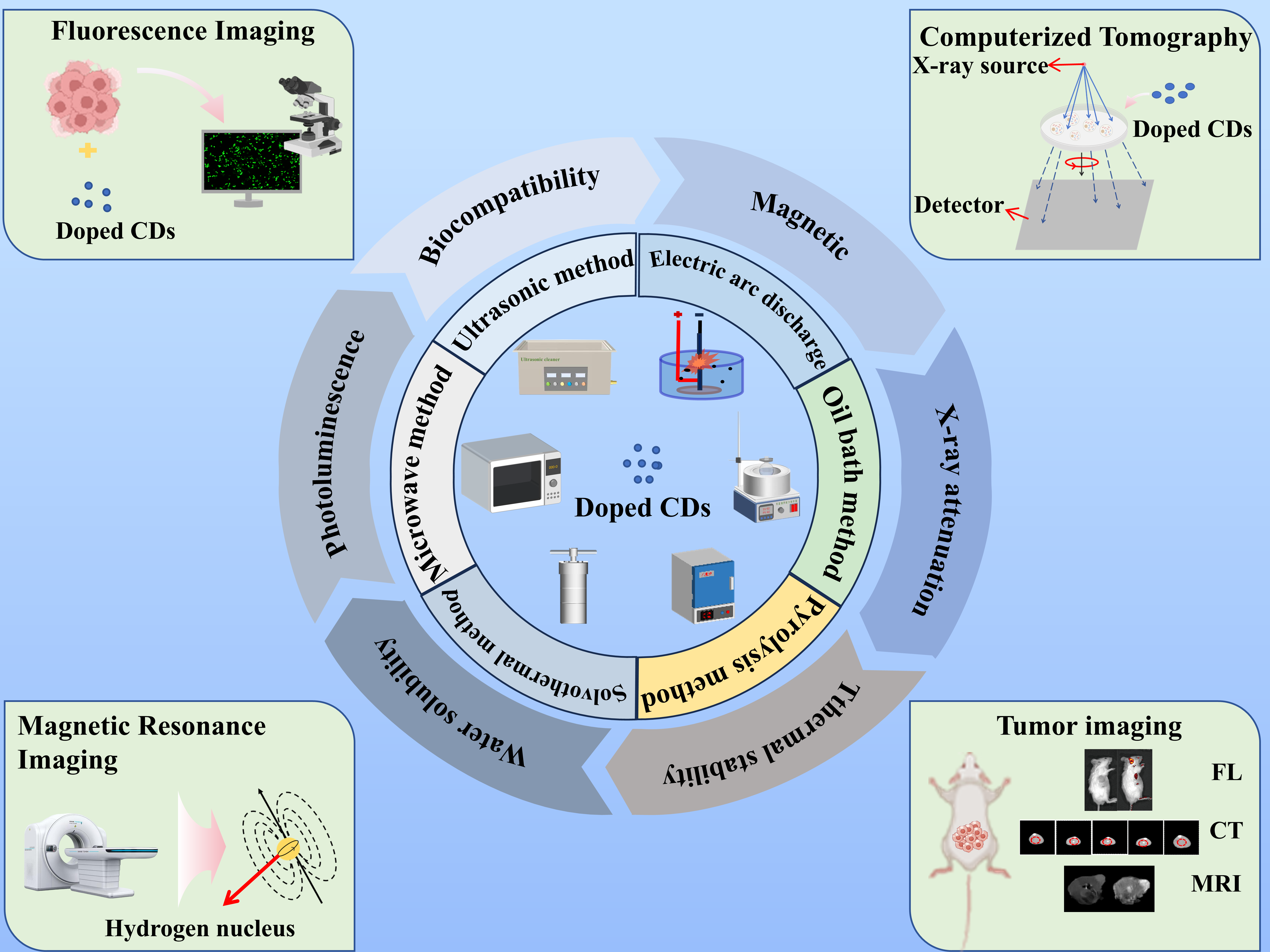
CDs are mainly prepared using two fundamental approaches: the “top-down” approach and the “bottom-up” approach. The former involves the deconstruction of graphitized carbon precursors via physical or chemical means and subsequent size regulation to obtain nanoscale CDs. In contrast, the latter leverages the self-assembly of CDs from small-molecule precursors via pyrolytic carbonization reaction[31-32]. However, for MRI/CT, CDs must be doped with metal ions, nanoparticles, or heteroatoms to achieve magnetic properties and X-ray attenuation capabilities. Therefore, the simple one-step “bottom-up” approach is often more appropriate for preparing CDs for biomedical applications. Notably, common synthesis methods for such CDs include solvothermal, microwave-assisted, and pyrolysis techniques. Most doped CDs for CT applications are prepared via the solvothermal method with water as the reaction medium. Table 1 summarizes the respective advantages and disadvantages of commonly employed synthesis methods, including solvothermal synthesis, microwave synthesis, and pyrolysis synthesis.
表1 碳点不同合成方法的优缺点对比

Table 1 Advantages and disadvantages of different synthesis methods for carbon dots

Synthesis methods Advantages Disadvantages
Solvothermal method Simple handling and easy preparation, High precision dimensional control, Flexible surface functionalization Harsh reaction conditions
Microwave method environmental friendliness, rapid synthesis limitations in product purity and quantum yield
Pyrolysis method strong process stability, high crystallinity of the products Harder surface finishing

2.1 Solvothermal method

The solvothermal method remains the dominant pathway for the fabrication of doped CDs used as multimodal imaging nanoprobes. When water is used as the reaction medium, this method is called the hydrothermal method, which is the most commonly used solvothermal technique. The typical process involves the homogeneous dispersion of a carbon source, a metal salt precursor, and a solvent, which are transferred into a PTFE-lined high-pressure reactor and subjected to high temperature and high pressure for several hours to complete the carbonization process, thereby achieving the controllable synthesis of CDs.
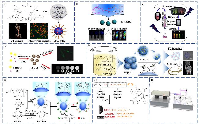
Zhang’s team[33] prepared uniformly dispersed, discrete quasi-spherical iodine-doped CDs (I-CDs) via a simple hydrothermal method, using iodixanol as the iodine source and glycine as the carbon source. Fig.1A illustrates the preparation process of the I-CDs and their application in CT/FL bimodal imaging. Zheng’s team[34] successfully synthesized gadolinium-doped CDs (Gd-CDs) with excellent biocompatibility via a one-step hydrothermal method, using gadopentetate dextran amine (Gd-DTPA) as the gadolinium source and L-arginine as the carbon precursor. Transmission electron microscopy (TEM)-based characterization revealed that the resulting Gd-CDs exhibited a monodisperse spherical morphology and an average particle size of 5.38 nm. Meanwhile, Maghsoudinia’s group[35] synthesized Gd-CDs using citric acid, ethylenediamine, and Gadovist as the starting materials by reacting at 200 ℃ for 4 h under hydrothermal conditions and subsequently utilized folic acid (FA) surface functionalization to obtain Gd-CDs-FA complexes. A chemical activation strategy using carbodiimide (EDC)/N-hydroxysuccinimide (NHS) was further employed to covalently couple bevacizumab (BEV) to the surface of the nanoparticles, resulting in the targeted complex Gd-CDs-FA-BEV. Compared with the original Gd-CDs, which had a fluorescence quantum yield (FLQY) of 49.94%, the modified Gd-CDs-FA-BEV had a FLQY of 83.67%. Although the mechanism underlying the FLQY enhancement of Gd-CDs is not fully understood, it is hypothesized to be closely related to the disorder of carbon ring structures induced by Gd3+ doping[36-37]. Notably, studies have confirmed that the introduction of surface passivation reagents such as ethylenediamine (EDA) into the synthesis system can significantly enhance the FLQY of CDs[38-39]. Moreover, the FLQY has also been found to increase significantly after the addition of FA, a phenomenon attributed to the selective anchoring of amine functional groups by FA molecules on the surface of Gd-CDs. This interaction can optimize excited-state electron transport pathways by modulating the energy level structure of Gd-CDs, thereby enhancing radiative recombination efficiency[40-41]. Huang’s team[42] prepared a new high-FLQY system using a solvent-heating method, employing glutathione (GSH), ethylenediaminetetraacetic acid (EDTA), and ferrous sulfate heptahydrate (FeSO4·7H2O) as precursors to synthesize iron-doped carbon quantum dots (Fe-CQDs) via the solvothermal method. The preparation process of the Fe-CQDs and their in vivo and ex vivo imaging results are shown in Fig.1B. X-ray photoelectron spectroscopy (XPS) confirmed the presence of Fe—O coordination bonds in the prepared material, which are formed by the chelation of Fe2+ with oxygen-containing functional groups on the surface of carbon quantum dots.
图1 不同多模态掺杂CDs的制备途径:(A) 水热法合成I-CDs的合成路径及其在荧光/CT双模成像中的示意图应用[33];(B) 溶剂热法合成Fe-CQDs的路线及其在MRI/FL双模成像中的示意图[42];(C) 采用硅胶柱色谱法分离四种溶剂热法制备的Mn-CDs[43];(D) 微波法制备Gd-CDs的合成路径及其在MRI/FL双模成像中的示意图[44];(E) 简单热解法制备Gd-CDs的合成路径及其在MRI/FL双模成像中的示意图[45];(F) 经低温热解制备并用DTPA对CQDs进行表面修饰的Gd-CDs合成路径示意图[46];(G) 经煅烧制备的Gd-CQDs合成路径及其原子配位固定机制。合成路径及其原子配位固定机制示意图[47];(H) 采用高能脉冲法制备的GC/CS示意图[48]

Fig.1 Preparation paths of different multimodal doped CDs. (A) Synthesis pathway for the preparation of I-CDs by hydrothermal method and its schematic application in FL/CT dual-modal imaging[33]. Copyright 2015, Informa UK Limited; (B) Synthesis pathway for the preparation of Fe-CQDs by solvothermal method and its schematic application in MRI/FL dual-modal imaging[42]. Copyright 2019, Springer Nature; (C) Separation of four Mn-CDs prepared by solvothermal method using silica gel column chromatography[43]. Copyright 2018, Elsevier; (D) Synthesis pathway for the preparation of Gd-CDs by microwave method pathway and schematic diagram of its MRI/FL dual-modal imaging application[44]. Copyright 2014, American Chemical Society; (E) Synthetic pathway of Gd-CDs prepared by simple pyrolysis and schematic diagram of its MRI/FL dual-modal imaging application[45]. Copyright 2017, The Royal Society of Chemistry; (F) Schematic diagram of the synthetic pathway of Gd-CDs prepared by low-temperature pyrolysis followed by the surface functionalization of the CQDs with DTPA[46]. Copyright 2015, Elsevier; (G) Synthetic pathway of Gd-CQDs prepared by calcination and its atomic coordination immobilization mechanism. Schematic diagram of the synthetic pathway and its atomic coordination fixation mechanism[47]. Copyright 2026, Royal Society of Chemistry; (H) Schematic diagram of GC/CS prepared by high-energy pulse method[48]. Copyright 2019, IOP Publishing Ltd.

Beyond Gd3+, Fe2+, and iodine-doped systems, Wang et al.[43] prepared manganese-doped CDs (Mn-CDs) using the solvothermal method. In this study, p-phenylenediamine served as the carbon source, manganese chloride tetrahydrate (MnCl2·4H2O) as the manganese source, and ethanol as the solvent. Fig.1C demonstrates that four manganese-doped configurations were further obtained using silica gelcolumn chromatography (mobile phase: a petroleum ether/ethyl acetate mixed system). Among these systems, CDs-2 showed a longitudinal relaxation rate (r1) of 7.28 mL/(mol·s), which was significantly higher than that of Gd-DTPA, a commonly used clinical contrast agent.
Notably, a systematic comparative study of CDs synthesized in organic and aqueous solvent systems showed that the FLQY of organic-phase-synthesized samples is significantly higher. However, the practical application of the solvothermal synthesis method is limited by its high production cost and the toxicity associated with most organic solvents, making the hydrothermal method more universally applicable for scale-up production[20].

2.2 Microwave method

Microwave synthesis is an efficient technique for CD preparation. This technique is based on the microwave dielectric heating effect. It enables the directed pyrolysis of carbon precursors and the self-assembly of nanostructures via high-frequency oscillations of molecular dipole moments induced by electromagnetic fields, and these oscillations generate localized thermal effects.
Gong’s team[44] successfully prepared Gd-CDs using sucrose, concentrated sulfuric acid (H2SO4), gadolinium trichloride (GdCl3), and diethylene glycol as precursors. The mixture of these components was treated with microwave radiation for 50 s at 750 W. Quantitative analysis via inductively coupled plasma optical emission spectrometry (ICP-OES) showed that gadolinium accounted for 18.2% of the material mass. Moreover, the longitudinal relaxation rate (r1) and transverse relaxation rate (r2) of the Gd-CDs reached 11.356 mL/(mol·s) and 14.026 mL/(mol·s), respectively, indicating the material's strong potential as an MRI nanoprobe. Fig.1D illustrates the microwave synthesis process of the Gd-CDs and the experimental results of MRI/FL bimodal imaging.

2.3 Pyrolysis method

The pyrolysis method for CD preparation is valuable because it drives deoxygenation, dehydrogenation and aromatization of organic precursors via high-temperature thermal cracking (300~800 ℃ typically), forming sp²/sp³ hybridized carbon skeleton structures.
Pan’s team[45] employed gadolinium chloride hexahydrate (GdCl3·6H2O), Gd-DTPA, branched polyethyleneimine (BPEI), and citric acid (CA) as composite precursors to successfully synthesize Gd-encapsulated carbon quantum dots (GCDs) via a low-temperature pyrolysis process. These GCDs exhibited both a high MRI response and strong fluorescence emission characteristics. Fig.1E shows the GCDs prepared via pyrolysis, along with the corresponding cell imaging and MRI imaging data. In comparison, conventional approaches such as hydrothermal synthesis can produce small-sized, highly water-soluble GCDs with excellent MRI response through surface modification. However, these methods suffer from more complex processes and longer reaction times.
Shi’s group[46] used citric acid monohydrate (CA·H2O) as the carbon source and BPEI as a synergistic reactant to produce CDs. Following the synthesis of CDs via a creative low-temperature pyrolysis method, they constructed a CQD-DTPA-Gd composite probe through covalent coupling with cyclo-DTPA dianhydride (cDTPAA) and subsequent ligation with Gd3+. Fig.1F illustrates the synthesis process of the CQD-DTPA-Gd composite probe. Notably, this probe maintained excellent water solubility (dispersion >95%) and cell membrane permeability, and exhibited a significantly enhanced longitudinal relaxation rate (r1), highlighting its promising application in MRI.

2.4 Other methods

Beyond the mainstream synthesis methods, other strategies for CD synthesis have also been explored. Bourlinos’s team[47] prepared Gd-doped carbon quantum dots (Gd-CQDs) via a calcination process using tris(hydroxymethyl)aminomethane (Tris), gadotrienoic acid, and betaine hydrochloride as precursors. Fig.1G illustrates the calcination-mediated preparation process of these Gd-CQDs. Although this approach is simple, the carbonization process significantly reduces the hydrophilicity of the resulting product, which limits its application as a bioprobe. In another study, Chen’s group[49] constructed an innovative cetyltrimethylammonium bromide (CTAB)-templated microporous reactor for the preparation of Gd@C-dots using a domain-limited pyrolysis strategy. This material exhibited a particle size-dependent MRI response, confirming the advantage of particle size controllability. Meanwhile, Zhu et al.[48] prepared gadolinium/carbon composite quantum dots (GC/CS) in a toluene system via high-energy pulsed laser ablation (PLA). This strategy significantly increased the relaxation rates to r1 = 86.5 mL/(mol·s) and r2 = 107.3 mL/(mol·s), providing a novel methodological paradigm for the development of carbon-coated probes (Fig.1H).
Current synthesis technologies for doped CDs used in multimodal imaging have the following characteristics: the solvothermal method is the dominant one, owing to its simple operation, high precision in size control and flexible surface functionalization. However, the high-pressure conditions required for this method pose safety risks that warrant strengthened system-level prevention and control. The microwave-assisted preparation method offers key advantages such as environmental friendliness and rapid synthesis, although it suffers from limitations in product purity and quantum yield. The pyrolysis method is distinguished by its strong process stability and the high crystallinity of the products, but it is constrained by challenges in achieving effective surface hydrophilic modification. Emerging technologies (e.g., combustion, template-assisted synthesis, pulsed laser ablation) are still in the laboratory exploration stage, and the scalability and reproducibility of these processes have not yet been fully realized[50].

3 Properties of doped CDs

Introduction of metal ions, nanoparticles or heteroatoms into CDs can reconfigure their intrinsic electronic structure and modulate their surface defect states. These structural modifications not only realize targeted modulation of the optical properties of CDs but also synergistically enhance their magnetic relaxation efficiency and X-ray attenuation capacity. Studies have shown that metal ions chelated in the carbon skeleton have significantly lower biotoxicity than free metal ions. Such surface functionalization strategies preserve the inherent advantages of CDs while meeting the stringent biocompatibility requirements for biomedical applications. Such optimized CDs exhibit excellent biosafety and hold great potential as multimodal imaging contrast agents, particularly for integrated CT/MRI/FL imaging platforms.

3.1 Optical properties

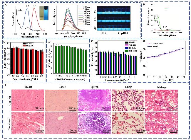
Although doped CDs exhibit favorable photoluminescence, those currently used in biomedical imaging primarily emit in the blue and green spectral regions. This spectral limitation poses a fundamental challenge for deep-tissue imaging: biological tissues strongly absorb and scatter short-wavelength light, leading to shallow penetration depths and thus reducing the imaging resolution and signal-to-noise ratio in deep tissues. In contrast, research into therapeutic applications has increasingly focused on CDs with emission in the first and second near-infrared (NIR-I/II, 1000~1700 nm) windows, owing to their enhanced tissue penetration capability. Consequently, future efforts should prioritize the development of synthetic strategies to tune the emission of doped CDs to cover the NIR region, which meets the critical demand for high-fidelity deep-tissue bioimaging. Therefore, the emission spectra of the doped CDs for biomedical imaging probes discussed in this review are currently limited to the blue-green band. For instance, Li’s team[51] prepared Gd-CDs with a FLQY of 11.7% using citric acid as the carbon source and GdCl3 as the gadolinium source. These Gd-CDs displayed stable blue fluorescence under UV excitation (λex = 365 nm). A systematic investigation of the influence of pH value (adjusted from pH 3 to 10) on the fluorescence performance of this material showed that the fluorescence intensity remained stable over the pH range of 3 to 10, indicating excellent pH stability(Fig.2A). Furthermore, the material demonstrated significant photostability after continuous UV irradiation for 60 min. Jeong et al.[52] used iohexol, a conventional CT contrast agent, as an iodine source to synthesize iodinated CDs (I-CDs), which exhibited favorable fluorescence properties. The team further investigated the influence of internal chemical bonds on the optical performance of the I-CDs. As shown in the ultraviolet visible absorption spectrum in Fig.2B, the I-CDs displayed a dual peak absorption feature: a sharp absorption peak at 226 nm corresponding to the π-π* transition of C $\stackrel{\mathrm{ }\mathrm{ }\mathrm{ }\mathrm{ }}{=}$C bonds in aromatic sp² hybridized domains, and a broad absorption band in the 250~500 nm range attributed to n-π* transitions induced by surface defect states or heteroatoms.
图2 掺杂碳点的光学特性及生物相容性:(A) 从左至右:Gd-CDs在水中的紫外-可见吸收光谱和光致发光光谱,插图为日光下(左)和紫外灯下(右)的照片,激发波长在320~400 nm(每10 nm递增)时记录的Gd-CDs光致发光光谱,Gd-CDs在不同pH值PBS溶液中紫外光下的照片图[51];(B) 乳酸、碘己醇和IDC在365 nm紫外光下的紫外-可见吸收峰[52];(C) Gd-CDs的体外细胞毒性测定[34];(D) 不同浓度Tb-CDs的细胞存活率测定[53];(E) 不同浓度Gd/Yb@CDs对4T1细胞和HeLa细胞增殖的影响(左),以及小鼠经Gd/Yb@CDs治疗30天后是否出现体重波动(右)[55];(F) 静脉注射一定剂量(8.65 mg/kg,以Gd计)的Gd/Yb@CDs 15天前后(脾、肺、心、肝、肾)的器官切片[55]

Fig.2 Optical properties and biocompatibility of doped CDs. (A) From left to right: UV-Vis absorption and PL spectra of Gd-CDs in water, with inset showing daylight (left) and UV (right) photos; photoluminescence spectra under excitation from 320 to 400 nm in 10 nm steps; and digital photos of Gd-CDs in PBS under UV light at different pH levels[51]. Copyright 2020, Future Medicine Ltd.; (B) UV-visible absorption peaks of lactic acid, iohexanol, and IDC under 365 nm UV light[52]. Copyright 2022, American Association for the Advancement of Science; (C) In vitro cytotoxicity assay of Gd-CDs[34]. Copyright 2019, Elsevier; (D) Cell viability assay of Tb-CDs at different concentrations[53]. Copyright 2016, Royal Society of Chemistry; (E) The effects of different concentrations of Gd/Yb@CDs on the proliferation of 4T1 cells and Hela cells (left), with or without body weight fluctuations (right) in mice treated with Gd/Yb@CDs for 30 days[55]. Copyright 2018, American Chemical Society; (F) Organ sections (spleen, lung, heart, liver, kidney) before and after intravenous injection of a certain dose (8.65 mg/kg, calculated as Gd) of Gd/Yb@CDs for 15 days[55]. Copyright 2018, American Chemical Society.

3.2 Biocompatibility

Biocompatibility is an indispensable core criterion in the development of bioimaging probes. For functionalized CDs used in MRI, although the incorporation of paramagnetic metal ions effectively enhances their magnetic responsiveness and imaging performance, the potential biotoxicity caused by ion leaching remains a key concern. Current research efforts are focused on the development of composite carbon quantum dot systems that combine excellent magnetic properties with high biosafety. This review systematically summarizes the current understanding of the biocompatibility of doped CDs systems, focusing on their cytotoxicity, in vivo metabolism, and long-term toxicity.
In the context of exploring CDs as MRI nanoprobes, Pan et al.[45] successfully synthesized Gd-CQDs through an innovative synthesis process, achieving a longitudinal relaxation rate (r1) of 57.42 mL/(mol·s), significantly surpassing the r1 of commercial gadolinium-based contrast agents. Considering the inherently high toxicity of Gd3+, the research team further optimized the synthesis process and successfully reduced the gadolinium content to 1.0% (w/w) without compromising the high relaxation rate. Validation via the dimethylphenol orange colorimetric method showed that Gd3+ forms a stable chelate with the carbon skeleton, which effectively prevents metal ion leaching. MTT cytotoxicity assays demonstrated that the survival rate of HeLa cells remained above 80% even at a Gd-CQD concentration of 1.0 mg/mL (corresponding to a Gd3+ concentration of 0.06 mmol/L). Using a low-temperature one-pot pyrolysis method, the team prepared Gd-CQDs that utilize the carbon-dot framework to effectively anchor gadolinium ions. This structure achieves high relaxivity while mitigating ion leakage, resulting in low cytotoxicity. The findings offer new insights into chelating functional elements within carbon frameworks for advanced bioprobe design. Zheng’s team[34] also synthesized Gd-CDs, which were evaluated in 786-O renal cancer cells and HK-2 tubular epithelial cells using the MTT assay. As shown in Fig.2C, Gd-CDs maintained over 90% cell viability under high-concentration exposure conditions, demonstrating excellent in vitro biocompatibility. Assessment via serum biochemical analysis further confirmed the in vivo metabolic safety of Gd-CQDs. The results revealed no significant differences in renal function markers (blood urea nitrogen and creatinine) between the treated and control groups at any time point. Regarding liver function, a transient slight increase in aspartate aminotransferase and alanine aminotransferase levels was observed 1 day after injection, but these levels returned to normal within 7 days. These findings confirm that the material induces only a mild, reversible effect on the liver, indicative of favorable in vivo metabolic compatibility. Han and colleagues[53] utilized an ethylene glycol-based solvothermal method to fabricate Mn-CDs using EDTA, triethylenetetramine, and MnCl2 as precursors. As shown by the MTT assay, when the Mn-CD concentration was 3 μg/mL, survival rates for HO-8910 ovarian cancer cells and EA.hy926 human umbilical vein fusion cells remained above 85% even after 12 h of co-culture (Fig.2D). Mice administered with 100 mg/kg of Mn-CDs via tail vein injection underwent histopathological evaluation over a three-week observation period. Results revealed no discernible histopathological alterations in major organs, including the heart, liver, spleen, and kidneys, thereby confirming their excellent in vivo compatibility and absence of long-term toxicity.
In investigating CD as CT nanoprobes, Molkenova’s group[54] developed terbium-doped CDs (Tb-CDs) using glucose and terbium chloride hexahydrate. The probes demonstrated a CT contrast of (48.2 ± 3.99) mL/mg(in Hounsfield units(HU)), significantly outperforming commercial iohexanol (6.5 mL/mg(in HU)). The CCK-8 assay confirmed that Tb-CDs do not exhibit cytotoxicity across a concentration range of 0~200 ppm(1 ppm=1×10⁻⁶), and that high-concentration treatments do not induce morphological alterations in cells, exhibiting low cytotoxicity. Despite these favorable preliminary biosafety findings, systematic animal model studies are still required to verify their clinical potential. Meanwhile, Zhao’s team[55] synthesized Gd/Yb@CDs with MRI/CT/FL multimodal imaging capabilities using bis-rare earth elements. Fig.2E shows the results of MTT assays in 4T1 and HeLa cells. After 24 h of incubation with Gd/Yb@CDs at a concentration of 1 mg/mL, no significant impact on cell proliferation was detected. Even when the concentration was increased to 3 mg/mL, cell viability remained above 80%. Additionally, body weight change curves revealed no significant deviations from the control group in the experimental group, further affirming the biosafety of Gd/Yb@CDs. Histopathological analysis of key target organs (spleen, lungs, heart, liver, and kidneys) using an in vivo mouse model revealed that Gd/Yb@CDs were primarily metabolized by the kidneys and exhibited transient renal accumulation. Fig.2F demonstrates that this accumulation did not induce significant nephrotoxicity. The Gd/Yb@CDs have a mean diameter of (5.26 ± 0.93) nm, which is below the renal filtration threshold (6~8 nm) for spherical nanoparticles, thus facilitating efficient renal clearance. Particle size determines the metabolic fate and long-term toxicity of nanomaterials, and smaller hydrodynamic diameters facilitate efficient renal clearance. Therefore, in the design of future probes, precise size regulation should be given equal consideration to imaging performance to achieve an optimal balance between efficacy and biosafety.

3.3 Magnetic properties

Conventional CDs are inherently non-magnetic; however, they can be doped with paramagnetic ions to impart magnetic properties, enabling their application as nanoprobes for MRI. Magnetic doped CDs offer a multifunctional platform for MRI, combining the fluorescence characteristics of CDs with the contrast-enhancing abilities of magnetic materials, which is of great significance for precision medicine. Paramagnetic ions can form stable chelate structures by coordinating with oxygen- or nitrogen-containing functional groups on the surface of CDs. Specifically, the empty orbitals of these ions form coordination bonds with the lone pairs of electrons on functional groups such as carboxyl, hydroxyl, and amino, thereby anchoring them to the CD surface. Additionally, these ions can adsorb small-molecule precursors through electrostatic interactions, inducing their transformation into larger conjugated structural domains, which facilitates the doping process[56]. Magnetic CDs can be categorized into MRI T1 contrast agents and MRI T2 contrast agents. The r2/r1 ratio is a critical parameter for classifying MR contrast agents and assessing their efficiency. MRI contrast agents that exhibit only T1 contrast effects typically have an r2/r1 ratio between 1 and 2, whereas those with predominantly T2 shortening effects exhibit an r2/r1 ratio greater than 10[57].
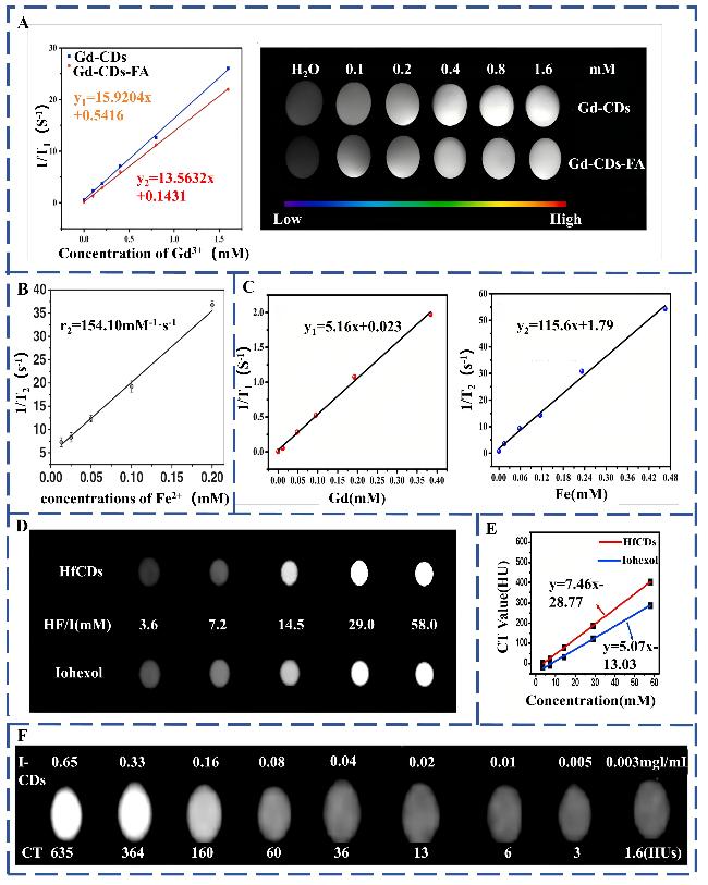
Du et al.[58] synthesized Gd-CDs-FA by first preparing Gd-CDs via a hydrothermal method using citric acid as the carbon source and gadolinium diamine as the gadolinium source. A silane coupling agent (KH-792) was used as an additive, and this was followed by FA modification. The resulting CDs exhibited an r1 value of 13.56 mL/(mol·s). MRI capability was assessed using a 7 T MRI scanner (Biospec 7 T/20 USR, Bruker, Germany). The experimental results, shown in Fig.3A, demonstrate that the T1-weighted signal intensified progressively with increasing Gd3+ concentration. The CDs modified with FA displayed magnetic properties comparable to those of conventional Gd-CDs, indicating their suitability as T1 molecular probes for MRI. The r1 values were likely related to the hydrodynamic radius and specific surface area of the Gd-CDs. Since the r1 value is primarily influenced by the strength of the external magnetic field and the interaction between protons and neighboring molecules, a larger specific surface area enhances the interaction efficiency between Gd3+ and hydrated protons. This geometrical advantage not only facilitates effective contact between the paramagnetic centers and water protons but also reduces the relaxation time (T1), thereby increasing the longitudinal relaxation rate (r1[37].
图3 掺杂碳点的磁性性能:(A) Gd-CDs 和Gd-C不同浓度下Gd³+与1/T1的线性拟合(左),不同浓度Gd³+的Gd-CDs和Gd-CDs-FA的T1加权图像[58];(B) 不同Fe²+浓度的Fe-CDs与1/T2的线性拟合[59];(C) 不同Gd³+浓度的Gd-CQDs@N-Fe3O4纳米颗粒与1/T1的对比(左)及不同Fe²+浓度与1/T2的对比(右)[60];(D) 分别对应不同浓度Hf-CDs和碘海醇的CT加权图像[65];(E) 不同浓度Hf-CDs和碘海醇的CT值线性拟合图[65];(F) CT图像中不同浓度I-CDs的CT图像[67]

Fig.3 Magnetic properties of doped CDs. (A) Linear fitting of Gd-CDs and Gd-C ifferent concentrations of Gd3+ versus 1/T1 (left), and T1-weighted images of Gd-CDs and Gd-CDs-FA with different concentrations of Gd3+[58]. Copyright 2023, Elsevier; (B) Linear fitting of Fe-CDs with different concentrations of Fe2+ versus 1/T2[59]. Copyright Wiley-VCH Verlag; (C) Gd-CQDs@N-Fe3O4 nanoparticles with different concentrations of Gd3+ versus 1/T1 (left) and different concentrations of Fe2+ versus 1/T2 (right)[60]. Copyright 2020, Elsevier; (D) CT-weighted images corresponding to different concentrations of Hf-CDs and Iohexol, respectively[65]. Copyright 2020, Elsevier; (E) Linear fits of CT values of different concentrations of Hf-CDs and Iohexol[65]. Copyright 2020, Elsevier; (F) CT images of different concentrations of I-CDs in the CT images[67]. Copyright 2018, Elsevier.

Liu et al.[59] synthesized Fe3O4-CDs through a green and simple one-pot hydrothermal process using poly-γ-glutamic acid as a precursor. The potential of these CDs as MRI contrast agents was evaluated using a 7 T MRI scanner. T2-weighted MR images exhibited a marked decline in signal intensity with increasing iron concentration. As illustrated in Fig.3B, linear fitting of the image intensity yielded an r2 value of 154.10 mL/(mol·s), confirming the utility of Fe3O4-CDs as MRI T2 contrast agents.
Huang et al.[60] initially synthesized Gd-CDs via a hydrothermal method. Subsequently, through the interaction between carboxyl groups on the Gd-CD surface and the amino groups of amino-functionalized iron oxide (N-Fe3O4), Gd-CDs@N-Fe3O4 nano-composites were obtained via chemical coupling. The changes in 1/T1 and 1/T2 induced by varying concentrations of Gd3+ and Fe2+ were linearly fitted (Fig.3C). The longitudinal relaxation rate (r1) was measured to be 5.16 mL/(mol·s), and the transverse relaxation rate (r2) reached 115.6 mL/(mol·s). This composite exhibited both enhanced T1 and T2 relaxation properties, indicating its potential as a dual-mode MRI contrast agent suitable for both T1-weighted and T2-weighted imaging.

3.4 X-ray attenuation properties

A key principle underlying CT contrast enhancement is the K-edge effect, wherein an element’s X-ray absorption coefficient increases dramatically upon the incident photon energy surpassing the binding energy of its K-shell electrons. The element-specific nature of K-edge energies allows for spectral CT imaging using contrast agents labeled with distinct high-atomic-number elements. This has motivated the exploration of hafnium (Hf), ytterbium (Yb), and bismuth (Bi) as dopants in CDs-based CT contrast agents, given their high atomic numbers and favorable X-ray attenuation properties. Specifically, hafnium (atomic number 72) presents a K-edge at 65.4 keV[61], ytterbium (atomic number 70) at 61 keV[62], and bismuth (atomic number 83) demonstrates an attenuation coefficient of 5.74 cm²/g at 100 keV[63], all of which are well-matched to the X-ray energies employed in clinical CT systems (approximately 100~120 kV(peak))[64]. Su’s team[65] prepared hafnium-doped CDs (Hf-CDs) using a one-pot pyrolysis method. Comparative analyses with the commercial CT contrast agent iodohexanol were performed using concentration-gradient experiments. As shown in Fig.3D and 3E, the CT signal intensities of both Hf-CDs and iohexanol showed a positive correlation with the concentration. Linear fitting revealed that Hf-CDs had a higher slope (7.21 mL/mol(in HU)) than iohexanol (5.07 mL/mol)[66]. Comparative in vivo experiments were conducted in a hormone-induced mouse tumor model, where three-dimensional CT imaging was performed after the intravenous injection of Hf-CDs and iodohexanol, respectively. The Hf-CDs group showed significantly higher CT values at the tumor site as well as improved imaging contrast. This confirmed that Hf-CDs have excellent X-ray attenuation properties and demonstrate immense potential as novel CT nanoprobes. Meanwhile, Molkenova et al.[54] developed multi-element charge transfer co-doped CDs through hydrothermal carbonization and examined their potential as bimodal contrast agents in an Osteosarcoma(OS) model. Experiments in a Balb/C hormone-induced mouse model showed that among the different Hf-doped CDs, P, W, and Hf-CDs-1 exhibited stronger enrichment in tumor tissues and thus provided superior FL/CT dual-mode imaging contrast.
After synthesizing I-CQDs, Su’s team[67] covalently coupled cetuximab to the surface of I-CQDs-NH2 via a modified EDC-NHS reaction[68] to obtain a targeted I-CQDs-C225 complex. With the increase in the I-CQDs concentration, the CT images showed gradual brightening (Fig.3F), verifying the X-ray attenuation ability of I-CQDs-NH2. Moreover, under equivalent iodine concentrations, I-CQDs-C225 provided equal or higher brightness on CT images than iohexanol, which confirmed their enhanced X-ray attenuation capacity and advantages related to imaging sensitivity.

4 Advances in multimodal imaging applications

Clinical studies have shown that iodine-based CT contrast agents significantly increase the risk of contrast-induced nephropathy (CIN) (OR = 2.3~3.8) and are associated with a 1.2%~3.1% incidence of cardiovascular events within 30 days post-administration[69-73]. Similarly, MRI contrast agents based on gadolinium (e.g., gadobutrol) raise biosafety concerns due to their potential toxicity. In this context, carbon-based nanomaterials such as fluorescent CDs offer a promising alternative owing to their quantum confinement effects, tunable surface hydration kinetics, and inherently low cytotoxicity. To overcome the limitations of traditional contrast agents, molecular engineering strategies such as PEG modification and heteroatom or metal ion doping have been employed, and multimodal imaging probes have been constructed. These novel probes offer innovative solutions for precise and safe biomedical imaging[74-75]. Table 2 summarizes the performance parameters of currently clinically used CT and MRI contrast agents, as well as other agents that can serve as multimodal probes.
表2 常用临床造影剂及其他类型纳米探针的性能参数

Table 2 Performance parameters of commonly used clinical contrast agents and other types of nanoprobes

Materials Imaging r1/
(mL/(mol·s))
r2/(mL/(mol·s)) CT values(in HU)/
(mL/mg)
Ref
Iodixanol CT 7.7 76
Iobitridol CT 31.83 54
Iohexol CT 4.233 77
Gd-DTPA MRI 6.27 34
GdAuNP(Type A) MRI 3.403 44.56 78
GdAuNP(Type B) MRI 4.450 56.86 79
GdAuNP(Type C) MRI 5.603 90.29
GdAuNP(Type D) MRI 4.244 49.14
RGD@Fe3O4-Au/Gd MRI 23.5 36.8
Fe3O4-SWCNT@M1 MRI 201.79 80
Cu2-xSe@MnO2 MRI 7.51 81
MnCO3-FP MRI/FL 5.8 49.5 82
ZrMOF@MnO2 MRI/FL 5.97 83
Fe3O4@PGLL MRI/FL 628.6 84
Fe3O4-Ag2S MRI/CT 267.48 3.03 85
HA-FeWO4 MRI/CT 0.64 6.63 5.388 77
Gd-AuNPr MRI/CT 23.1 959.3 86

4.1 Doped CDs for CT/FL imaging

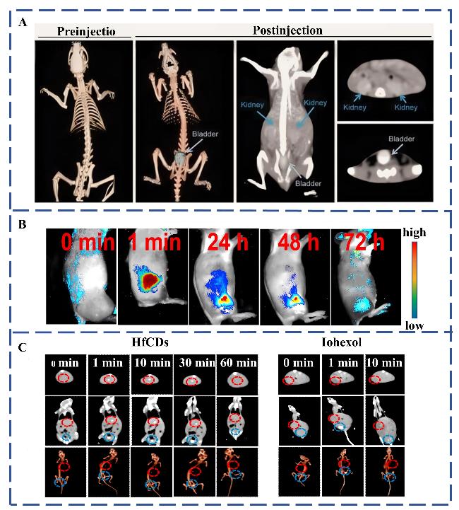
Halogen- and lanthanide-doped CDs have significant applications in medical imaging and diagnostics. The synthesis methods, products, FLQYs, emission wavelengths, and CT values of doped CDs currently used for CT/FL dual-modality imaging are listed in Table 3. Zhang et al.[33] was the first to successfully prepare I-CDs via the hydrothermal method. The X-ray attenuation performance of I-CDs was found to be significantly superior to that of iodixanol, a conventional nonionic dimeric iodine contrast agent. To verify their CT imaging efficacy, the team injected I-CDs into Sprague Dawley rats via the tail vein. Dynamic CT scans (Fig.4A) revealed a gradual increase in the CT value of the kidneys within minutes after the administration of I-CDs, accompanied by enhanced intensity of bladder images. This renal clearance property could effectively circumvent the non-targeted accumulation typically observed with conventional iodine contrast agents, confirming the translational potential of I-CDs as a new generation of kidney-specific CT nanoprobes. Additionally, the research team systematically demonstrated that the photoluminescence intensity of I-CDs peaks when the molar ratio of iodine source to glycine (used as a surface passivator) reaches 1∶1. A pronounced Tyndall effect was observed under red light excitation, further confirming the material’s suitability as a FL probe. Consequently, the I-CDs prepared by this group appeared to be promising nanoprobes for CT/FL dual-modality imaging. However, the aforementioned I-CDs system lacks a targeted design, and its ability to specifically recognize tumor tissues in complex physiological environments has yet to be further explored. Su et al.[67] constructed EGFR-targeted I-CQDs-c225 by functionalizing CDs with cetuximab. These CDs exhibited strong fluorescence and greater sensitivity for CT imaging than iodixanol. Moreover, the team demonstrated that I-CQDs-c225 could specifically localize to cancer cell lysosomes via receptor-mediated endocytosis, offering a novel paradigm for molecular imaging. The studies above indicate that while iodine doping endows CDs with CT imaging capabilities, their X-ray attenuation performance is limited by iodine’s relatively low atomic number. Therefore, as discussed in Section 3.4, researchers have begun to explore high atomic number elements such as hafnium (Hf), ytterbium (Yb), and bismuth (Bi) to achieve superior CT imaging results. Su’s research team[65] also developed a new type of Hf-CDs. In vitro experiments showed that after the co-incubation of these Hf-CDs with HeLa cells, the intracellular cytoplasmic fluorescence intensity gradually increased over time, confirming the efficient cellular uptake of these CDs. In addition, fluorescence monitoring of H22 tumor-bearing mice using the Maestro 500 in vivo optical imaging system revealed elevated fluorescence signal intensity in the tumor region compared to background tissue following the intravenous injection of Hf-CDs (Fig.4B). Meanwhile, CT imaging further demonstrated that the CT value of the tumor’s inner region increased significantly from 114.0 HU (0 min) to 296.0 HU (1 min) and 251.0 HU (30 min) post-injection. At the same time, the bladder signal rose from 109 HU at 0 min to 715 HU at 30 min before gradually decreasing, while the liver signal remained consistently low. No specific enhancement of iohexol signals was observed at the tumor site in the control group, confirming the unique tumor-targeting ability of Hf-CDs (Fig.4C). These in vivo and ex vivo experimental data confirmed that Hf-CDs synergize active targeting with the enhanced permeability and retention (EPR) effect, representing a breakthrough in the design of CT/FL dual-modality imaging probes. The studies described above provide a crucial foundation for the rational design of CT/FL dual-modality imaging probes. However, further investigation is needed to ensure that the elemental doping strategies meet clinical safety standards.
表3 用于CT/FL双模态成像的掺杂碳点的合成方法、原材料及表征

Table 3 Synthetic methods, raw materials and characterization results of doped CDs for CT/FL dual modality imaging

Magnetic CDs Synthesis
methods
Feedstocks FLQY
/%
Emission wavelength
/nm
CT values(in HU)/(mL/mg) Ref
I-CDs Hydrothermal Iodixanol and glycine, 475 33
I-CQDs-C225 Hydrothermal citric acid and iohexol 18 67
IDC Hydrothermal LA and iohexol 52
hf-CD Hydrothermal CA, HfCl4 and thiourea 7.21 65
P, W-CDs solvothermal o-phenylenediamine, Sodium phosphotungstate
Octadecahydrate and NN-dimethylformamide
16.5 54
P, W, Hf-CDs-1 solvothermal o-phenylenediamine, Sodium phosphotungstate
Octadecahydrate and NN-dimethylformamide
27.3 540
P, W, Hf-CDs-2 solvothermal o-phenylenediamine, Sodium phosphotungstate
Octadecahydrate and NN-dimethylformamide
14.2 630
Fe3O4@Au HNPs 122.12 87

LA: Lactobionic acid. CA: Citric acid.

图4 掺杂碳点在CT/FL双模态成像中的应用:(A) 大鼠静脉注射碘标记的碳纳米球(40mg/kg)前后的三维X射线CT成像(左图为注射前,左二图为注射后),大鼠静脉注射碘标记的碳纳米球后的体内CT成像与分析,蓝色箭头和灰色箭头分别代表肾脏和膀胱(右图)[33];(B) 向H22激素小鼠注射铪-CDs后,在肿瘤内注射前(0 min)以及注射后1 min、24 h、48 h和72 h进行的体内荧光成像[65];(C) 静脉注射Hf-CDs后不同时间点获取的主要器官(肿瘤、肝脏和膀胱)的自上而下CT图像,三维重建、冠状面及三维重建图像(左),以及注射碘海醇后冠状面、轴向和三维重建的CT成像效果(右)[65]

Fig.4 Doped CDs for CT/FL imaging. (A) 3-D x-ray CT imaging before and after intravenous injection of I-CDs (40 mg /kg) in rats (left one before injection, left two after injection), In vivo CT imaging and analysis after intravenous injection of I-CDs in rats, Blue arrows and gray arrows represent kidney and bladder (right), respectively[33]. Copyright 2015, Informa UK Limited; (B) In vivo fluorescence imaging by injecting Hf-CDs into H22 hormonal mice before (0 min) and after intratumoral injection for 1 min, 24 h, 48 h, and 72 h[65]. Copyright 2020, Elsevier; (C) Top-down CT images of major organs (tumor, liver, and bladder) acquired at different times after intravenous injection of Hf-CDs, respectively. Three-dimensional reconstruction, coronal as well as three-dimensional reconstruction images (left), and CT imaging effects of coronal, axial and three-dimensional reconstruction after iohexol injection (right)[65]. Copyright 2020, Elsevier.

4.2 Doped CDs for MRI/FL imaging

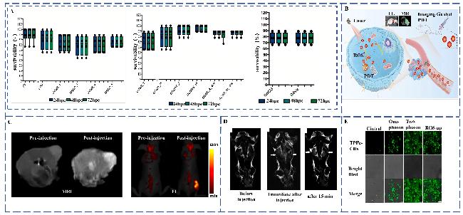
Table 4 summarizes the synthesis methods, products, FLQYs, emission wavelengths, and r1 values and r2 values of doped CDs currently used for MRI/FL dual-modal imaging. Significant progress has been made with regard to the synthesis methods and performance optimization of Gd-CDs in bimodal imaging. High FLQY and low biotoxicity remain the most critical factors when selecting bioimaging contrast agents. In an earlier study, Bourlinos’s team[47] prepared Gd-CDs via pyrolysis, and these Gd-CDs showed excellent T1-weighted imaging contrast (r1 value better than that of gadobutrol) and good cytocompatibility. Additionally, they possessed excitation-related emission properties similar to those of other CDs and thus serve as MRI/FL probes. However, due to limitations in their in vitro model, no further in vivo experiments were carried out. Meanwhile, Wojnicki et al.[95] prepared Gd-CQDs via the hydrothermal method using sucrose and GdCl3·6H2O as precursors and carried out in vivo experiments in zebrafish embryos. Fluorescence spectral analysis confirmed that the Gd-CQDs exhibited strong fluorescence emission, with peak intensity significantly higher than that of both gadobutrol and undoped CQD controls. The longitudinal relaxation rate (r1) of Gd-CQDs was 2.5 times that of gadobutrol after normalization to the gadolinium molar concentration. Subsequently, the team established a toxicological assessment system by incubating zebrafish embryos with varying concentrations of Gd3+ and GdCl3. Fig.5A demonstrate the survival rates of zebrafish embryos incubated with unadulterated CQDs, GdCl3, and gadobutrol containing different Gd3+ doses after different incubation periods. Median survival rates were analyzed using the Kruskal-Wallis test, revealing statistically significant differences (p = 0.0149). Analysis of zebrafish embryo hatchability demonstrated that the Gd-CDs prepared by the team had good in vivo and in vitro biocompatibility and could be used as MRI/FL bimodal probes. However, although the aforementioned Gd-CDs possess favorable magnetic resonance properties, their low fluorescence quantum yield leaves room for optimization as fluorescent probes. The antagonism between magnetic and fluorescence properties poses a key challenge in the development of MRI/FL bimodal probes. In recent years, researchers have successfully developed composite probes that exhibit both excellent magnetic resonance performance and high fluorescence quantum yields by leveraging mechanistic insights. In another study, Wang’s team[84] synthesized Gd-CDs with an FLQY up to 78.5% using a hydrothermal method, and their fluorescence intensities were dually regulated by temperature (180 ℃ peak) and the gadolinium doping concentration (0.8 mmol/L optimum). Experimental characterization further confirmed that the Gd-CDs exhibit excellent magnetic resonance relaxation properties. These properties could be attributed to the synergistic magnetic response mechanism of the unpaired electrons of the 4f orbitals of Gd3+ ions under the action of the carbon substrate ligand field. A recent study aimed at enhancing relaxation rates and applying green synthesis approaches prepared Gd-CDs for MRI/FL dual-modality imaging using the hydrophobic anticancer drug curcumin[90] and a starch/polyethyleneimine system[89]. The prepared Gd-CDs had an r1 = 0.3663 mL/(mol·s) and r2 = 0.1315 mL/(mol·s). Notably, their performance gap when compared to the clinical standard Gd-DOTA (r1 = (4.5±0.3) mL/(mol·s)) mainly stemmed from defects in the gadolinium ligand environment and lattice disorder. Shi et al.[93] achieved a significant improvement in the performance of Gd-DOTA by synthesizing Gd/Ru bimetallic-doped fluorescent CDs. The synergistic enhancement of diagnostic imaging was realized with this material, resulting in improved MRI and FL images performance as well as excellent photodynamic effects. Fig.5B demonstrates the PDT mechanism and experimental results of MRI/FL dual-modality imaging with these probes.
表4 用于MRI/FL双模态成像的掺杂碳点的合成方法、原材料及表征

Table 4 Synthetic methods, raw materials and characterization results of doped CDs for MRI/FL dual-modality imaging

Magnetic CDs Synthesis
methods
Feedstocks FLQY
/%
Emission wavelength /nm r1/
(mL/(mol·s))
r2/
(mL/(mol·s))
Ref
Gd-CQDs calcination Tris base, gadopentetic acid and betaine hydrochloride 445 47
Gd-CQDs Microwave Sucrose, Gd2O3 and DEG 5.4 521 11.356 14.026 44
Gd(III)/CQDs Pyrolysis GdPM 8.9 488 5.5 36
Gd-CQDs Hydrothermal CA, EDA and Gd2O3 43.6 450 6.06 38
Gd-CQDs MSN-templated Gd(NO33 and Gd-DTPA 30.2 420 10 49
Gd-CQDs Hydrothermal CA, DETA and GdCl3 69.89 488 14.33 88
Gd-CQDs Pyrolysis GdCl3·6H2O, Gd-DTPA, CA and BPEI 40 445 57.52 45
Gd-CQDs Hydrothermal L-Arginin and Gd-DTPA 57.75 425 6.27 34
Gd-CQDs@N-
Fe3O4
Hydrothermal CA, EDA, GdCl3 and N-Fe3O4 1.29 500 5.16 116.5 60
Gd-CDs Hydrothermal CA, Gd-DTPA and DETA 78.05 452 7.37 84
Gd-CDs-bev Hydrothermal CA, BEV, Gadovist and EDA 82.5 7.61 35
Gd-CDs-fa-bev Hydrothermal CA, FA, BEV, Gadovist and EDA 83.67 6.03 35
Gd-CQDs Hydrothermal Starch, Gadovist and PEI 13.20 0.2198 0.1315 89
Gd-CDs-FA Hydrothermal FA, CA and EDC 48.20 456 13.56 58
Gd-CQDs Hydrothermal Tumeric, Gadoteric acid and PEI 0.3663 1.8246 90
Gd@CNDs(1.5T) Hydrothermal β-alanine, EDTA and GdCl3 7.92±0.02 8.98±0.01 91
Gd@CNDs(7.0T) Hydrothermal β-alanine, EDTA and GdCl3 10.5±0.3 18.08±0.27 91
Gd-CQDs Hydrothermal Dextran, Gadovist and PEI 0.272 92
Gd/Ru-CDs Microwave CA, GdCl3, Ru2Cl3 and PEI 29.57 637 6.38 93
Gd-CQDs solvothermal OPD, GdCl3 and Gd(NO33 6.3 6.4 38.6 94
Gd-CQDs solvothermal OPD and Gd(NO33·6H2O 7.4 550 94
Gd-CDs Hydrothermal Sucrose and GdCl3·6H2O 0.41 440 95
Mn-CDs solvothermal PDA and MnCl2·4H2O 6.75 555 7.28 43
Mn-CDs Microwave CA, urea and MnCl2 10 525 6.23 96
Mn-CDs Pyrolysis lemon extract, EDA and Mn(OAc)2·4H2O 35.71 463 0.341 2.015 97
Mn-CQDs Hydrothermal Mn, TPP and CA 13 480 98
Mn-CQDs@A/Ce6 Hydrothermal MnCl2·4H2O, Ce6, Waste green tea and FA 12 440 13.888 80.036 99
Mn-CDs-NHF Pyrolysis NHF, MnCl2 17.75 84.46 100
Mn-CDs solvothermal Mn(AcO)2·4H2O and OPD 560 8.8 83.4 101
Mn-CDs solvothermall MnCl2·4H2O and OPD 560 9.7 89
Mn-CDs solvothermal Mn(AcO)2·4H2O and CA 620 4.8 42.2
Mn-CDs solvothermal MnCl2·4H2O and FA 620 6.7 67.1
Mn-CDs Hydrothermal Manganese citrate and urea 23 7.4 102
Mn, S,N-CDs Hydrothermal D-(+)-glucose, EDA, EDTA,f luorescein, mPDA, oPDA and E-MnSO4·H2O 53.9 515 32.3 103
Ho-Mn-CQDs Hydrothermal HoCl3·6H2O, MnCl2 and Cysteine 71.45 477 4.37 21.93 104
BCQD@Mn Hydrothermal citric acid, urea, MnCl2·4H2O and TA 7.24 490 2.43 105
C-Fe3O4 Hydrothermal FeCl3⋅6H2O, FeCl2⋅4H2O and γ-PGA 21.6 154.1 59
Fe-CDs Hydrothermal FeSO4·7H2O, GSH, EDTA 3.8 450 3.92 4.99 42
Fe-CDs Hydrothermal Sucrose, FeCl2,and FeCl3 31 470 118.3 106
Fe-CDs pyrolysis lemon extract, EDA and (NH42Fe(SO42·6H2O 41.72 457 97
Co-CDs pyrolysis lemon extract, EDA and Co(OAc)2·2H2O 75.07 459 97
Ni-CDs pyrolysis lemon extract, EDA and Ni(OAc)2·2H2O 50.84 458
HoBCDs Hydrothermal CA, BPEI and Ho-DTPA 8.2 441 0.1128 107

DEG: Diethylene glycol. GdPM: Gadopentetate monomeglumine. EDA: Ethylenediamine. Gd-DTPA: Diethylene triamine pentaacetic acid. DETA: Diethylenetriamine. BPEI: Branched polyethylenimine. BEV: Bevacizumab. FA: Folic acid. EDTA: Ethylenediaminetetraaceti acid. OPD,oPDA: o-phenylenediamine. PDA: p-phenylenediamine. TPP: Tetraphenylporphyrin. NHF: N-Hydroxyphthalimide. mPDA: m-Phenylenediamine. TA: Tannic acid. γ-PGA: Poly-γ-glutamic acid. GSH: Glutathione. Ho-DTPA: diethylenetriamine pentaacetic acid hydrate holmium(III) dihydrogen salt.

图5 掺杂碳点在MRI/CT/FL多模态成像中的应用:(A) 不同浓度Gd³+条件下,纯CQDs、GdCl3和钆布醇在24 h、48 h和72 h时的存活率[95];(B) Gd/Ru-CDs的MRI/荧光双模态成像以及肿瘤光动力治疗示意图[93];(C) MRI活体成像(左)与荧光活体成像(右)示意图[108];(D) 小鼠注射前、注射后立即及注射15 min后的T2加权活体成像示意图[106];(E) TPFe-CDs与对照组ROSUP在单光子与双光子激发下经照射产生的活性氧(ROS)示意图[109]

Fig.5 Doped CDs for MRI/FL imaging. (A) Survival rates of unadulterated CQDs, GdCl3, and gadobutrol at 24 h, 48 h, and 72 h, respectively, for different concentrations of Gd3+[95]. Copyright 2024, MDPI; (B) Schematic of MRI/FL dual-modality imaging of Gd/Ru-CDs as well as photodynamic treatment of tumors[93]. Copyright 2024, Springer Nature; (C) Schematic of in vivo imaging by MRI (left), and in vivo imaging by FL (right)[108]. Copyright 2021, American Chemical Society; (D) Mouse Schematic of in vivo T2-weighted imaging before, immediately after, and 15 min after injection[106]. Copyright 2019, American Chemical Society; (E) Schematic of ROS production by irradiation under single-photon versus two-photon excitation for TPFe-CDs versus control ROSUP[109]. Copyright 2024, Published by Elsevie.

Following advancements in Gd doping, the potential of manganese-doped CDs (Mn-CDs) in MRI/FL dual-modality imaging and tumor diagnosis and treatment has gradually been recognized. The Mn-CDs reported by Sun et al.[108] combine long-wavelength emission properties with an enhanced MR response, offering a new approach to multifunctional probe design. These Mn-CDs exhibited superior fluorescence performance compared to conventional CDs, as shown in Fig.5C. Notably, the emission wavelength of Mn-CDs was red-shifted from 542 nm (green emission) to 578 nm (orange emission). Simultaneously, they maintained a high longitudinal relaxation value (r1) of 12.69 mL/(mol·s), limiting the leakage of Mn2+ ions into the bloodstream and slowing the relaxation rate. These results confirmed the excellent biocompatibility and effective in vivo bimodal imaging capabilities of the Mn-CDs developed by the team. Ali et al.[102] utilized manganese citrate in the synthesis of magnetofluorescent carbon quantum dots, producing Mn-CDs with high r1 relaxation (7.4 mL/(mol·s)), low cytotoxicity, and FLQY of 23%, demonstrating promising potential for MRI/FL bimodal imaging applications.
Most Mn-CDs function primarily as T1-weighted contrast agents due to their strong longitudinal relaxation effect, serving as complementary fluorescent probes for FL imaging. However, researchers have also made significant progress in exploring Mn-CDs for FL/T2 imaging. Stepanidenko’s team[101] synthesized four Mn-CD systems via a one-pot solvothermal method using OPD, CA, and formamide as carbon sources, complexed with Mn(AcO)2·4H2O and MnCl2·4H2O. Characterization showed that these CDs markedly reduced T1 and T2 relaxation times, with longitudinal (r1) and transverse (r2) relaxation rates outperforming those of commercial manganese dipyridoxal diphosphate-based contrast agents. Among them, the OPD-based CD-1 and CD-2 exhibited significantly enhanced relaxation rates compared to CA/FA-based CD-3 and CD-4/ The r2/r1 ratios ranged from 8.8 to 10, consistent with the properties expected of bimodal T1~T2 contrast agents. Additionally, these Mn-CDs displayed a broad spectral range. Taken together, these features suggested that the Mn-CDs synthesized by the team had strong potential as nanoprobes for FL/T1/T2 imaging. Meanwhile, Irmania’s group[99] applied a green synthesis strategy, using discarded green tea extract as the carbon source and MnCl2·4H2O as the manganese source, to hydrothermally prepare multifunctional nanocomplexes (Mn-CQDs@FA/ Ce6), which were covalently coupled with FA and chlorin e6 (Ce6). The material exhibited excellent photoluminescence, with a FLQY of 12% at an excitation wavelength of 360 nm. Furthermore, Mn-CQDs@FA/Ce6 showed an r2/r1 ratio of 5.77, indicating potential as a T2 contrast agent and promising applications as a PDT agent, enabling integrated diagnosis and treatment.
Beyond manganese-doped systems, transition metal iron-doped CDs also demonstrate remarkable transverse relaxation properties. Das et al.[106] synthesized Fe-CDs using sucrose as the carbon source and FeCl2/FeCl3 as iron sources via a facile hydrothermal method. These Fe-CDs exhibited a transverse relaxation rate (r2) as high as 118.3 mL/(mol·s) and superior fluorescence properties. In vivo MRI experiments in mice confirmed the significant T2 contrast enhancement effect of the CDs, with good biocompatibility and metabolic safety upon gradual intravenous injection. Fig. 5D displays the T2-weighted images at different time points post-injection. Additionally, Zhu et al.[109] reported a transverse relaxation rate (r2) of 10.388 mL/(mol·s) for tumor-targeted Fe-CDs (tFe-CDs), indicating their potential as highly efficient T2-weighted MRI contrast agents. The team further demonstrated, through in vitro experiments in HeLa cells, that tFe-CDs could significantly inhibit cancer cell activity via PDT under laser irradiation. Fig.5E shows that the tFe-CDs prepared by the team exhibited excellent photosensitization under both single-photon and two-photon excitation compared to the conventional commercial reagent ROSUP. This material combined dual-mode imaging with efficient PDT, overcoming the functional limitations of traditional nano-diagnostic agents. It thus holds important prospects for biomedical imaging and therapy integration, offering a novel strategy for advancing precision diagnosis and treatment. Although doping with magnetic ions such as Fe2+ and Gd3+ can significantly enhance the MRI relaxation rate, this improvement often comes at the cost of a sharp decrease in fluorescence quantum yield due to nonradiative energy transfer mediated by the unpaired d/f electrons of these ions. Addressing this magneto-fluorescence antagonism thus represents a key direction for future research.

4.3 Doped CDs for MRI/CT/FL imaging

In recent years, the application of doped CDs has not been limited to MRI/FL or CT/FL bimodal imaging, and growing efforts are being focused on developing MRI/CT/FL trimodal fluorescent probes. Table 5 summarizes the synthesis methods, products, FLQYs, emission wavelengths, CT values, and r1 and r2 values of doped CDs currently used for MRI/CT/FL multimodal imaging. Li’s group[17] successfully constructed manganese and dysprosium co-doped carbon quantum dots (Mn,Dy-CDs) via a hydrothermal method. This material exhibits FL, T1/T2-weighted MRI, and CT imaging capabilities, which endows it with trimodal imaging potential. In vitro experiments revealed that Mn,Dy-CQDs achieved relaxation rates of 7.47 mL/(mol·s) (r1) and 42.686 mL/(mol·s) (r2) for T1 and T2 relaxation, respectively. The X-ray absorption coefficient (per unit logarithmic) of Mn,Dy-CDs was estimated to be 47.344 HU, significantly outperforming clinically used iodinated contrast agents. Photoluminescence spectral analysis showed an optimal emission peak with a 4 nm redshift (from 458 nm to 462 nm) and a moderate increase in emission intensity compared to undoped CQDs. This advancement in doped CDs provides an important theoretical foundation for designing novel multifunctional nanoprobes. Notably, Li et al.[110] developed iodine-functionalized CD-ferroferric oxide complexes (I@CNDs-Fe3O4) to address the osmotic pressure imbalance commonly observed with conventional CT contrast agents. This composite system effectively regulates elemental iodine release kinetics through the nanoconfinement effect. It exhibited a transverse relaxation rate (r2) of 177.4 mL/(mol·s), while its CT value (180 HU/Lg) was nearly seven times higher than previously reported values for C-Fe3O4 quantum dot hybrid nanoparticles (26.1 HU/Lg)[59]. Moreover, I@CNDs-Fe3O4 demonstrated excellent photostability even after 24 h of continuous irradiation. Bismuth-gadolinium co-doped carbon quantum dots (Bi,Gd-CQDs) prepared by Meng’s team[111] featured two distinct emission centers at 518 nm and 602 nm. Their longitudinal relaxation rate (r1 = 4.29 mL/(mol·s)) exceeded that of the clinical agent gadopentetate dimeglumine (r1 = 3.40 mL/(mol·s)), and their CT value reached 164.66 HU/Lg, demonstrating strong multimodal imaging synergy.
表5 用于MRI/CT/FL多模态成像的掺杂碳点的合成方法、原材料及表征

Table 5 Synthetic methods, raw materials and characterization results of doped CDs for MRI/CT/FL multimodal imaging

Doped CDs Synthesis
methods
Feedstocks FLQY
/%
Emission wavelength/nm r1/
(mL/(mol·s))
r2/
(mL/(mol·s))
CT values(in HU)/
(mL/mg)
Ref
I@CNDs-Fe3O4 Hydrothermal FeCl3·6H2O, FeCl2·4H2O and sea vegetable 22.3 177.4 180 110
Gd/Yb-CDs Hydrothermal Na2EDTA, GdCl3, YCl3 and L-arginine 16.82 418 6.65 45.42 55
Mn,Dy-CDs Hydrothermal CA, Na2EDTA, MnCl2·4H2O, DyCl3·6H2O, aminophylline hydrate 3.6 462 7.47 42.686 47.344 17
Bi,Gd-CDs Hydrothermal PDA, GdCl3·6H2O, Na2EDTA, GSH and
Bi(NO33·5H2O
5.11 518/
602
4.29 164.66 111
Mn,S,N-CDs Hydrothermal FeSO4·7H2O, MnSO4·H2O, CuSO4, MgSO4, ZnSO4·7H2O, D-(+)-glucose and EDA 53.9 515 32.3 103
Gd/Yb-CDs Hydrothermal Gd(NO33·6H2O, Yb(NO33·5H2O, CA, urea 14.2 11.16 40.17 112

Na2EDTA: Ethylenediaminetetraacetic acid disodium salt.

Further in-depth studies have shown that doping with rare earth elements can significantly enhance the imaging performance of CDs. Zhao et al.[55] synthesized gadolinium-ytterbium co-doped CDs (Gd/Yb@CDs) via hydrothermal methods. These CDs exhibited an elevated longitudinal relaxation rate of 6.65 mL/(mol·s) and an X-ray attenuation coefficient of 45.42 HU/Lg, representing an improvement of approximately 40% over conventional iodine-based contrast agents (31.83 HU/Lg). Together, these studies indicate that carbon quantum dot-based nanomaterials possess unique advantages for MRI/CT/FL multimodal imaging, with their performance optimization primarily dependent on elemental doping strategies and surface modification techniques.
A persistent challenge in the development of doped CDs for multimodal imaging lies in the paradox between X-ray attenuation and magnetic relaxation. While doping with high atomic number elements such as iodine or tungsten improves CT contrast, their dense electron clouds may interfere with the unpaired electrons of paramagnetic centers, leading to diminished magnetic relaxivity. Striking a delicate balance between these two competing modalities—optimizing CT performance without compromising MRI sensitivity—remains an important direction for future studies.

4.4 The potential of multimodal imaging for clinical applications

Current research on doped CDs for biomedical imaging is primarily centered on their function as multimodal imaging nanoprobes. Concurrently, increasing attention is being directed toward their application in tumor theranostics, aiming to pave the way for clinical translation. Xia et al.[113] synthesized gadolinium/gallium co-doped gallic acid-based CDs (Gd/Ho-CDs) via a microwave-assisted method, yielding a nanoplatform that integrates fluorescence/CT/MRI trimodal imaging capabilities with intrinsic antitumor activity. In a HepG2 tumor-bearing mouse model, Gd/Ho-CDs administration resulted in a marked enhancement of MRI signals within the tumor region, correlating well with fluorescence imaging findings. Furthermore, strong CT signal retention at the tumor site was detectable for up to 24 hours post-injection. The synergistic advantages of these complementary imaging modalities substantially improved the accuracy of image-guided interventions. Additionally, upon laser irradiation, the material effectively ablated tumor cells through photothermal effects, underscoring its theranostic potential.
However, the multimodal imaging capability of doped CDs for targeting specific tumor cells remains to be fully elucidated. The overlapping imaging features of distinct lesions often obscure the interface between normal and specific tumor tissue on conventional imaging, constituting a significant source of diagnostic inaccuracy. Primary renal neuroendocrine tumors (NETs) typify such diagnostic difficulties, and their imaging features often overlap with those of other renal malignancies (e.g., fading renal cell carcinoma and low-fat angiomyolipoma). Given this diagnostic ambiguity, the CT manifestations and MRI signal characteristics (including T1WI and T2WI) of these tumors can be systematically evaluated by integrating multimodal imaging techniques[114]. Therefore, the application of doped CDs for targeted multimodal imaging of tumor tissues warrants further investigation.

5 Conclusion

This review systematically examines recent advances in the application of doped CDs in biomedical imaging, focusing on the development of multimodal imaging nanoprobes. In addition to defining the concepts, structural classifications and synthesis strategies of multimodal imaging probes, this review details the synergistic enhancement effects of metal ion and heteroatom doping on the properties of CDs. The review highlights how these dopants significantly improve the photoluminescence, magnetic responsiveness, and X-ray attenuation coefficients of CDs through functional modifications.
Over the past decade and a half, CDs have achieved multidimensional breakthroughs, including the construction of controlled drug release systems, the design of chemical sensing interfaces, and the development of antimicrobial materials. However, despite the successful fabrication of doped CDs with excellent imaging capabilities, three key challenges remain in their in vivo imaging applications:
(1) Lack of long-term biosafety assessment systems. Currently, doped CDs used in multimodal imaging including MRI typically employ gadolinium, manganese, and iron doping strategies, while doped CDs used in multimodal imaging that includes CT often incorporate iodine and lanthanide rare earth elements. Although CD matrices generally exhibit better biocompatibility and lower systemic toxicity than traditional gadolinium-based (e.g., Gd-DTPA) and iodine-based (e.g., iohexol) contrast agents, their long-term in vivo metabolism and subchronic toxicity continue to pose potential risks. Therefore, there is an urgent need for standardized and durable biosafety evaluation models. Most existing studies rely on in vitro cytotoxicity assays, such as the MTT/MTS and CCK-8 methods, but fewer extend to in vivo animal studies. The latter are predominantly limited to mice and zebrafish embryos, and there is a notable lack of systematic evaluations in primates or large mammals.
(2) Multimodal performance imbalance. This issue manifests primarily due to two core dilemmas: the antagonistic relationship between magnetic and fluorescence performance, and the competitive mechanisms of X-ray attenuation and magnetic relaxation. These contradictions lead to signal interference issues in MRI/FL and MRI/CT bimodal probes, and systems capable of simultaneously optimizing MRI, CT, and FL performances remain exceedingly rare. More critically, the dissociation of functional elements under physiological conditions contributes to performance degradation. Hence, there is a need to focus on the development of multimodal probes that combine stability and high performance.
(3) Targeted issues in clinical translation.Furthermore, as discussed in Section 4.4, current doped CDs probes lack targeted design to meet the demand for precise identification of tumors. This limitation hinders their ability to fundamentally resolve the diagnostic ambiguity arising from overlapping imaging features. Therefore, a critical direction for future research lies in endowing these probes with active recognition capabilities toward specific tumor biomarkers while preserving their inherent multimodal imaging advantages, thereby enabling accurate diagnosis and facilitating clinical translation.
To address the above challenges, future research should focus on the following three aspects. First, in terms of biosafety assessment, researchers can systematically track the metabolic pathways and organ accumulation of doped CDs in non-human primate models via radioisotope labeling and other techniques, and examine their subchronic toxicity under different exposure cycles to fill the gaps in existing long-term safety data. Second, to address the problem of imbalanced multimodal performance, the energy competition between magnetic ions and luminescent centers can be attenuated by regulating the coupling mode of dopant ions, while optimizing the proportion of heavy atom doping to balance CT imaging performance and magnetic relaxation, thereby constructing a stable imaging platform with signals free from mutual interference. Third, to solve the problem of insufficient targeting, high-affinity peptides or antibodies targeting specific tumor markers can be explored and conjugated onto the surface of doped CDs, and their active recognition ability can be validated through in vitro and in vivo studies, thereby overcoming the diagnostic bottleneck caused by lesion overlap in traditional imaging. Overcoming these challenges will further advance the innovative applications of doped CDs in precision medicine and molecular imaging.
[1]
Sun Y P, Zhou B, Lin Y, Wang W, Fernando K A S, Pathak P, Meziani M J, Harruff B A, Wang X, Wang H F, Luo P G, Yang H, Kose M E, Chen B L, Veca L M, Xie S Y. J. Am. Chem. Soc., 2006, 128(24): 7756.

[2]
Wojnicki M, Hessel V. Chem. Eng. J., 2022, 438: 135616.

[3]
Namdari P, Negahdari B, Eatemadi A. Biomed. Pharmacother., 2017, 87: 209.

[4]
Zhao X Y, Liao S, Wang L M, Liu Q, Chen X Q. Talanta, 2019, 201: 1.

[5]
Zhou J, Deng W W, Wang Y, Cao X, Chen J J, Wang Q, Xu W Q, Du P, Yu Q T, Chen J X, Spector M, Yu J N, Xu X M. Acta Biomater., 2016, 42: 209.

[6]
Lim S Y, Shen W, Gao Z Q. Chem. Soc. Rev., 2015, 44(1): 362.

[7]
Jang Y, Shapiro A, Horani F, Kauffmann Y, Lifshitz E. Z. Für Phys. Chem., 2018, 232(9/11): 1443.

[8]
Kou X L, Jiang S C, Park S J, Meng L Y. Dalton Trans., 2020, 49(21): 6915.

[9]
He J, Chen J, Qiu H D. Prog. Chem., 2023, 35(5): 655.

(何静, 陈佳, 邱洪灯. 化学进展, 2023, 35(5): 655.)

[10]
Shabbir H, Wojtaszek K, Rutkowski B, Csapó E, Bednarski M, Adamiec A, Głuch-Lutwin M, Mordyl B, Druciarek J, Kotańska M, Ozga P, Wojnicki M. Molecules, 2022, 27(24): 8728.

[11]
Dang Y Q, Huang R, Feng X Y, Liu G Y, Zhu Y Y, Zhang Y T. Prog. Chem., 2024, 36(4): 575.

(党永强, 黄瑞, 冯香钰, 刘国阳, 朱由余, 张亚婷. 化学进展, 2024, 36(4): 575.)

[12]
Wang J L, Wang Y L, Zheng J X, Yu S P, Yang Y Z, Liu X G. Prog. Chem., 2018, 30(8): 1186.

(王军丽, 王亚玲, 郑静霞, 于世平, 杨永珍, 刘旭光. 化学进展, 2018, 30(8): 1186.)

[13]
Dolai J, Joshi P, Mondal P P, Maity A, Mukherjee B, Jana N R. ACS Appl. Mater. Interfaces, 2024, 16(13): 16003.

[14]
Liu Z C, Li J J, Zhao C, Zhang Z H, Wu P C, Chen J Q, He X, Zhang S J, Tian Y. Adv. Mater., 2025, 37(20): 2410786.

[15]
Zhang Q, Wang X Q, Yuan L L, Yu L N, Shao C Y, Jia H X, Lu S. Anal. Methods, 2024, 16(14): 2063.

[16]
Yu L N, Cao X, Wang X Q, Yuan L L, Chen X, Wang C Y, Shao C Y, Lu S. Anal. Methods, 2025, 17(36): 7178.

[17]
Li C X, Wang Y, Nong H Y, Hu X X, Wu Y, Zhang Y J, Liang C M, Chen K G, Li S L. Nanotechnology, 2022, 33(2): 025101.

[18]
Qin M, Xu M J, Huang D, Wei Y, Meng Y F, Chen W Y. Prog. Chem., 2020, 32(9): 1264.

(秦苗, 徐梦洁, 黄棣, 魏延, 孟延锋, 陈维毅. 化学进展, 2020, 32(9): 1264.)

[19]
Ma T, Deng C Y, Li J, Wang Z Y, Zhou Q, Yu X Q. Prog. Chem., 2025, 37(4): 519.

(马婷, 邓春宇, 李杰, 王周玉, 周倩, 余孝其. 化学进展, 2025, 37(4): 519.)

[20]
Zhong Y M, Chen L, Yu S P, Yang Y Z, Liu X G. ACS Biomater. Sci. Eng., 2023, 9(12): 6548.

[21]
Lin L P, Luo Y X, Tsai P, Wang J J, Chen X. Trac Trends Anal. Chem., 2018, 103: 87.

[22]
Li X C, Fu Y Z, Zhao S J, Xiao J F, Lan M H, Wang B H, Zhang K, Song X Z, Zeng L T. Chem. Eng. J., 2022, 430: 133101.

[23]
Georgakilas V, Perman J A, Tucek J, Zboril R. Chem. Rev., 2015, 115(11): 4744.

[24]
Krok D, Tomal W, Knight A J, Tartakovskii A I, Farr N T H, Kasprzyk W, Ortyl J. Polym. Chem., 2023, 14(38): 4429.

[25]
Szczepankowska J, Khachatryan G, Khachatryan K, Krystyjan M. Int. J. Mol. Sci., 2023, 24(19): 14984.

[26]
Shen C L, Lou Q, Lv C F, Zang J H, Qu S N, Dong L, Shan C X. Adv. Sci., 2019, 6(11): 1802331.

[27]
Li J R, Zhao H G, Zhao X J, Gong X. Nano Lett., 2024, 24(37): 11722.

[28]
Chen X Z, Yan F, Li J R, Gong X. JACS Au, 2025, 5(3): 1350.

[29]
Zhang S W, Shen C L, Li J R, Liao J F, Miao P, Zhao X J, Gong X. Chem. Sci., 2025, 16(31): 14098.

[30]
Li J R, Zhao H G, Zhao X J, Gong X. Adv. Funct. Mater., 2024, 34(44): 2404473.

[31]
Liu M L, Chen B B, Li C M, Huang C Z. Green Chem., 2019, 21(3): 449.

[32]
Wang B Y, Cai H J, Waterhouse G I N, Qu X L, Yang B, Lu S Y. Small Sci., 2022, 2(6): 2200012.

[33]
Du F Y, Zhang M M, Ju H X, Zhang L, Sun M Z, Zhou Z W, Dai Z Y, Zhang L R, Gong A H, Wu C Y. Int. J. Nanomed., 2015: 6943.

[34]
Zheng S H, Yu N N, Han C P, Xie T, Dou B R, Kong Y, Zuo F M, Shi M L, Xu K. Biochem. Biophys. Res. Commun., 2019, 511(2): 207.

[35]
Maghsoudinia F, Tavakoli M B, Samani R K, Motaghi H, Hejazi S H, Mehrgardi M A. J. Drug Deliv. Sci. Technol., 2021, 61: 102288.

[36]
Ren X Y, Liu L H, Li Y, Dai Q, Zhang M, Jing X L. J. Mater. Chem. B, 2014, 2(34): 5541.

[37]
Chen H M, Wang G D, Tang W, Todd T, Zhen Z P, Tsang C, Hekmatyar K, Cowger T, Hubbard R B, Zhang W Z, Stickney J, Shen B Z, Xie J. Adv. Mater., 2014, 26(39): 6761.

[38]
Chen H, Wang L N, Fu H, Wang Z Y, Xie Y J, Zhang Z J, Tang Y. J. Mater. Chem. B, 2016, 4(46): 7472.

[39]
Devi S, Gupta R K, Paul A K, Kumar V, Sachdev A, Gopinath P, Tyagi S. RSC Adv., 2018, 8(57): 32684.

[40]
Saljoughi H, Khakbaz F, Mahani M. Photodiagn. Photodyn. Ther., 2020, 30: 101687.

[41]
Liu H F, Li Z H, Sun Y Q, Geng X, Hu Y L, Meng H M, Ge J, Qu L B. Sci. Rep., 2018, 8: 1086.

[42]
Huang Q, Liu Y, Zheng L L, Wu L P, Zhou Z Y, Chen J F, Chen W, Zhao H W. Microchim. Acta, 2019, 186(8): 492.

[43]
Wang Y R, Wang T R, Chen X, Xu Y, Li H R. Opt. Mater., 2018, 78: 118.

[44]
Gong N Q, Wang H, Li S, Deng Y L, Chen X A, Ye L, Gu W. Langmuir, 2014, 30(36): 10933.

[45]
Pan Y, Yang J, Fang Y N, Zheng J H, Song R, Yi C Q. J. Mater. Chem. B, 2017, 5(1): 92.

[46]
Shi Y P, Pan Y, Zhong J, Yang J, Zheng J H, Cheng J L, Song R, Yi C Q. Carbon, 2015, 93: 742.

[47]
Bourlinos A B, Bakandritsos A, Kouloumpis A, Gournis D, Krysmann M, Giannelis E P, Polakova K, Safarova K, Hola K, Zboril R. J. Mater. Chem., 2012, 22(44): 23327.

[48]
Zhu Z F, Sun H B, Jin J, Wang M L, Zhang Y, Yu D B, Jiang Y. Nanotechnology, 2019, 30(10): 105705.

[49]
Chen H M, Wang G D, Sun X L, Todd T, Zhang F, Xie J, Shen B Z. Adv. Funct. Mater., 2016, 26(22): 3973.

[50]
Wang L P, Ma Y J, Zhou X H, Liu Y, Wu R D. J. Mater. Eng., 2015, 43(5): 101.

[51]
Li Y X, Li B, Wang X C, Meng Y, Bai L, Zheng Y Y. Nanomedicine, 2020, 15(24): 2385.

[52]
Jeong Y, Jin M, Kim K S, Na K. Biomater. Res., 2022, 26: 27.

[53]
Han C P, Xu H T, Wang R, Wang K Y, Dai Y, Liu Q, Guo M X, Li J J, Xu K. J. Mater. Chem. B, 2016, 4(35): 5798.

[54]
Molkenova A, Serik L, Ramazanova A, Zhumanova K, Duisenbayeva B, Zhussupbekova A, Zhussupbekov K, Shvets I V, Kim K S, Han D W, Atabaev T S. RSC Adv., 2023, 13(22): 14974.

[55]
Zhao Y Z, Hao X T, Lu W, Wang R M, Shan X R, Chen Q, Sun G Y, Liu J H. ACS Appl. Nano Mater., 2018, 1(6): 2544.

[56]
Liao H, Wang Z Y, Chen S, Wu H, Ma X J, Tan M Q. RSC Adv., 2015, 5(82): 66575.

[57]
Liang G H, Xiao L F. Biomater. Sci., 2017, 5(10): 2122.

[58]
Du J L, Zhou S Z, Ma Y H, Wei Y Y, Li Q, Huang H, Chen L, Yang Y Z, Yu S P. Colloids Surf. B Biointerfaces, 2024, 234: 113721.

[59]
Liu X, Jiang H, Ye J, Zhao C, Gao S, Wu C, Li C, Li J, Wang X. Adv. Funct. Mater., 2016, 26(47): 8694-8706.

[60]
Huang Y, Li L, Zhang D H, Gan L P, Zhao P, Zhang Y F, Zhang Q, Hua M Q, Jia C M. Magn. Reson. Imag., 2020, 68: 113.

[61]
Marill J, Anesary N M, Zhang P, Vivet S, Borghi E, Levy L, Pottier A. Radiat. Oncol., 2014, 9: 150.

[62]
Xiong L Q, Yang T S, Yang Y, Xu C J, Li F Y. Biomaterials, 2010, 31(27): 7078.

[63]
Li H Y, Sun H Z. Curr. Opin. Chem. Biol., 2012, 161-2: 74.

[64]
Roessler A C, Hupfer M, Kolditz D, Jost G, Pietsch H, Kalender W A. Investig. Radiol., 2016, 51(4): 249.

[65]
Su Y, Liu S, Guan Y Y, Xie Z G, Zheng M, Jing X B. Biomaterials, 2020, 255: 120110.

[66]
Bouzas-Ramos D, Cigales Canga J, Mayo J C, Sainz R M, Ruiz Encinar J, Costa-Fernandez J M. Adv. Funct. Mater., 2019, 29(38): 1903884.

[67]
Su H F, Liao Y, Wu F S, Sun X Z, Liu H J, Wang K, Zhu X J. Colloids Surf. B Biointerfaces, 2018, 170: 194.

[68]
Huang P, Lin J, Wang X S, Wang Z, Zhang C L, He M, Wang K, Chen F, Li Z M, Shen G X, Cui D X, Chen X Y. Adv. Mater., 2012, 24(37): 5104.

[69]
Kistner A, Tamm C, Svensson A M, Beckman M O, Strand F, Sköld M, Nyrén S. BMC Nephrol., 2021, 22: 297.

[70]
Faucon A L, Bobrie G, Clément O. Eur. J. Radiol., 2019, 116: 231.

[71]
Davenport M S, Khalatbari S, Cohan R H, Dillman J R, Myles J D, Ellis J H. Radiology, 2013, 268(3): 719.

[72]
Fu J J, Guo J J, Qin A P, Yu X Y, Zhang Q, Lei X P, Huang Y G, Chen M Y, Li J X, Zhang Y, Liu J P, Dang Y Y, Wu D, Zhao X Y, Lin Z X, Lin Y L, Li S P, Zhang L Y. J. Nanobiotechnol., 2020, 18(1): 110.

[73]
Bolognese L, Falsini G, Schwenke C, Grotti S, Limbruno U, Liistro F, Carrera A, Angioli P, Picchi A, Ducci K, Pierli C. Am. J. Cardiol., 2012, 109(1): 67.

[74]
Wang W, Ni Y R, Xu Z Z. J. Alloys Compd., 2015, 622: 303.

[75]
Zhou J J, Sheng Z H, Han H Y, Zou M Q, Li C X. Mater. Lett., 2012, 66(1): 222.

[76]
Tsai I C, Lee T, Tsai W L, Chen M C, Wu M J, Lee W L, Ting H J. Am. J. Roentgenol., 2008, 190(1): W47.

[77]
Yang C M, Che X L, Zhang Y, Gu D D, Dai G D, Shu J, Yang L. Int. J. Nanomed., 2023, 18: 8023.

[78]
Kisel A, Luo M R, Bailey M D, Ghobrial K, Lukashova L, Lei Y Q, Hitchens T K, Meade T J, Modo M. Biomaterials, 2026, 329: 123947.

[79]
Amraee A, Alamzadeh Z, Irajirad R, Sarikhani A, Ghaznavi H, Ghadiri Harvani H, Mahdavi S R, Shirvalilou S, Khoei S. Cancer Nanotechnol., 2023, 14: 61.

[80]
Zhang Y Y, Liu S W, Li D Y, He C Y, Wang D X, Wei M, Zheng S H, Li J J. Colloids Surf. B Biointerfaces, 2023, 229: 113452.

[81]
Fu D D, Ding X G, Wu J, Li C Y, Wang Q B, Jiang J. Part. Part. Syst. Charact., 2018, 35(7): 1800078.

[82]
Lee K K, Lee J H, Lee S C, Lee C S. Theranostics, 2022, 12(15): 6762.

[83]
Liu Y, Gong C S, Lin L S, Zhou Z J, Liu Y J, Yang Z, Shen Z Y, Yu G C, Wang Z T, Wang S, Ma Y, Fan W P, He L C, Niu G, Dai Y L, Chen X Y. Theranostics, 2019, 9(10): 2791.

[84]
Wang L, Zhou W J, Yang D, Zhe H, Mei S L, Yuan J, Zhang W L, Li H, Fan H, Xie F X, Guo R Q. Anal. Methods, 2021, 13(21): 2442.

[85]
Liu D D, Lai J Y, Wang R, Ye L, Tian Y. ACS Biomater. Sci. Eng., 2019, 5(11): 6196.

[86]
Liu W F, Liu K, Zhao Y, Zhao S, Luo S, Tian Y, Teng Z G, Wang S J, Lu G M. RSC Adv., 2017, 7(26): 15702.

[87]
Linh N T N, Du N H, Dung N T, Tam L T T, Nam P H, Tuyet P T H, Lu L T, Tam L T. Chemistry Open, 2025, 14(10): e202500166.

[88]
Yu C Y, Xuan T T, Chen Y W, Zhao Z J, Liu X X, Lian G H, Li H L. J. Alloys Compd., 2016, 688: 611.

[89]
Molaei M J. Sci. Rep., 2022, 12: 17681.

[90]
Molaei M J. J. Lumin., 2023, 257: 119692.

[91]
Cardo L, Martínez-Parra L, Cesco M, Echeverría-Beistegui B M, Martínez-Moro M, Herrero-Álvarez N, Cabrerizo M B, Carregal-Romero S, Ramos-Cabrer P, Ruiz-Cabello J, Prato M. Small, 2023, 19(31): 2206442.

[92]
Ghahremani H, Molaei M J, Salimi E. Opt. Mater., 2024, 155: 115872.

[93]
Shi Y P, Xia Y N, Zhou M Y, Wang Y F, Bao J F, Zhang Y, Cheng J L. J. Nanobiotechnol., 2024, 22: 88.

[94]
Efimova A A, Badrieva Z F, Brui E A, Miruschenko M D, Aleinik I A, Mitroshin A M, Volina O V, Koroleva A V, Zhizhin E V, Stepanidenko E A, Ushakova E V. J. Opt. Technol., 2024, 91(6): 362.

[95]
Wojnicki M, Wojtaszek K, Tokarski T, Csapó E, Knutelska J, Bednarski M, Skórkowska A, Pomierny-Chamioło L, Kotańska M. Molecules, 2024, 29(17): 4075.

[96]
Ji Z, Ai P H, Shao C, Wang T J, Yan C X, Ye L, Gu W. ACS Biomater. Sci. Eng., 2018, 4(6): 2089.

[97]
Rub Pakkath S A, Chetty S S, Selvarasu P, Vadivel Murugan A, Kumar Y, Periyasamy L, Santhakumar M, Sadras S R, Santhakumar K. ACS Biomater. Sci. Eng., 2018, 4(7): 2582.

[98]
Yue L L, Li H L, Liu Q, Guo D, Chen J, Sun Q, Xu Y Z, Wu F S. Microchim. Acta, 2019, 186(5): 315.

[99]
Irmania N, Dehvari K, Gedda G, Tseng P J, Biomed. Mater. Res. Part B Appl. Biomater., 2020, 108(4): 1616.

[100]
Tiron A, Stan C S, Luta G, Uritu C M, Vacarean-Trandafir I C, Stanciu G D, Coroaba A, Tiron C E. Pharmaceutics, 2021, 13(11): 1982.

[101]
Stepanidenko E A, Vedernikova A A, Badrieva Z F, Brui E A, Ondar S O, Miruschenko M D, Volina O V, Koroleva A V, Zhizhin E V, Ushakova E V. Photonics, 2023, 10(7): 757.

[102]
Ali V, Kefayati H, Shafiee Ardestani M, Pourahmad A. Magn. Reson. Mater. Phys. Biol. Med., 2024, 37(1): 139.

[103]
Kajani A A, Pouresmaeili A, Mehrgardi M A, Javanmard S H. Sci. Rep., 2024, 14: 29111.

[104]
Ghosh T, Nandi S, Girigoswami A, Bhattacharyya S K, Ghosh S K, Mandal M, Ghorai U K, Banerji P, Das N C. ACS Biomater. Sci. Eng., 2024, 10(2): 1112.

[105]
Xu W B, Zhang J, Yang Z J, Zhao M Z, Long H T, Wu Q F, Nian F. J. Mater. Sci. Mater. Med., 2022, 33(2): 16.

[106]
Das B, Girigoswami A, Dutta A, Pal P, Dutta J, Dadhich P, Srivas P K, Dhara S. ACS Biomater. Sci. Eng., 2019, 5(7): 3549.

[107]
Fang Y N, Jia J, Yang J, Zheng J H, Yi C Q. Chin. Chem. Lett., 2018, 29(8): 1277.

[108]
Sun S J, Zhao L N, Wu D, Zhang H X, Lian H C, Zhao X L, Wu A G, Zeng L Y. ACS Appl. Bio Mater., 2021, 4(2): 1969.

[109]
Zhu T T, Cao L, Li X L, Du Y W, Yan H Y, Chang Z M, Dong W F, Li L. Sens. Actuat. B Chem., 2025, 424: 136812.

[110]
Li W P, Zeng G M, Yan J, Liu X L, Jiang X H, Yang J Z, Liu J, Sun D P. Mater. Sci. Eng. C, 2021, 124: 112037.

[111]
Meng Q, Wang Y, Li C X, Hu X X. New J. Chem., 2022, 46(35): 16970.

[112]
Shu G, Jiang T T, Zhang X N, Zhao H X. Chem. Eng. J., 2024, 494: 153086.

[113]
Xia Y N, Li M Q, Zhang R Y, Zhou M Y, Jing L H, Wang W J, Zhang Y, Shi Y P. Adv. Healthc. Mater., 2025, 15(10): e04706.

[114]
Wei B C, Chen Y G, Chen Y Q, Zhang S J. Front. Oncol., 2026, 15: 1597024.

Outlines

/