Home Journals Progress in Chemistry
Progress in Chemistry

Abbreviation (ISO4): Prog Chem      Editor in chief: Jincai ZHAO

About  /  Aim & scope  /  Editorial board  /  Indexed  /  Contact  / 
Review

Self-Enhanced Electrochemiluminescence: From Construction Principles to Advanced Applications in Bioanalytical and Environmental Sensing

  • Fangxin Du ,
  • Gen Liu , *
Expand
  • Key Laboratory of Green and Precise Synthetic Chemistry and Applications, Ministry of Education;School of Chemistry and Chemical Engineering, Huaibei Normal University, Huaibei 235000, China

Received date: 2025-07-11

  Revised date: 2025-09-23

  Online published: 2026-01-07

Supported by

National Natural Science Foundation of China(22304058)

Natural Science Foundation of Anhui Provincial Department of Education(2024AH051690)

Abstract

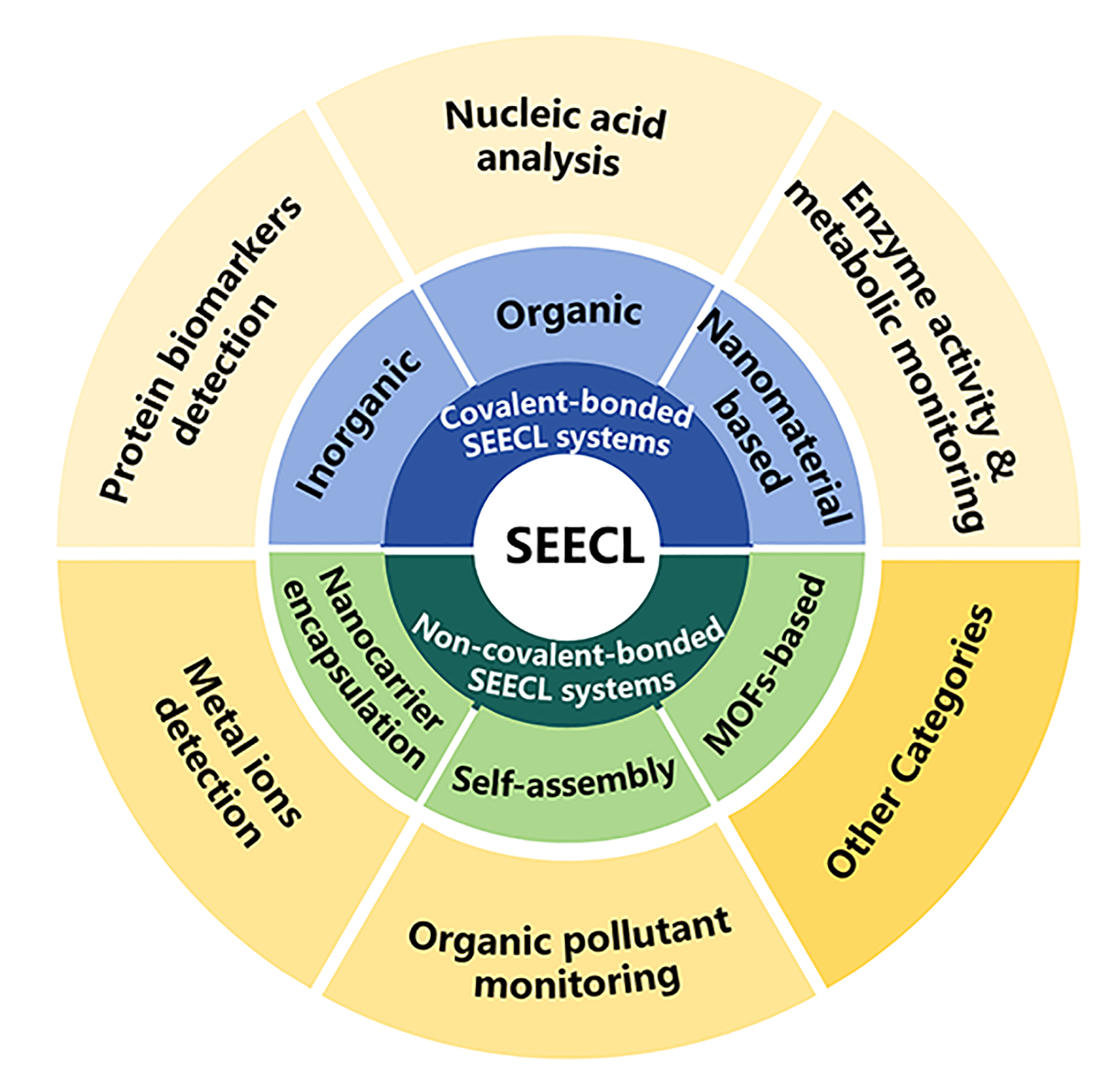
Self-enhanced electrochemiluminescence (SEECL), as an emerging analytical technique, significantly enhances electrochemiluminescence (ECL) efficiency by integrating luminophores and co-reactants into unified nanostructures or molecular frameworks, demonstrating substantial value in the fields of bioanalysis and environmental sensing. Based on the integration mode of luminophores and co-reactants, SEECL structures can be categorized into two types: covalently bonded SEECL and non-covalently bonded SEECL. Covalently bonded SEECL can be further divided into inorganic, organic, and nanoscale covalent bonding SEECL systems, while non-covalently bonded SEECL includes structures such as nanocarrier encapsulation, self-assembly, and metal-organic framework (MOF)-based SEECL. On the basis of summarizing the construction principle of SEECL, this paper summarizes its applications in areas including bioanalysis (protein biomarker detection, nucleic acid analysis, and enzyme activity monitoring), environmental sensing (trace detection of heavy metal ions and organic pollutants), food safety testing, wearable devices, and point-of-care testing (POCT). Additionally, the article addresses unresolved issues such as the stability, biocompatibility of SEECL materials and interference from complex matrices, and prospects its future development directions, providing a reference for subsequent research on SEECL.

Contents

1 Introduction

2 Construction of SEECL systems

2.1 Mechanistic insights into SEECL

2.2 Covalent-bonded SEECL systems

2.3 Non-covalent-bonded SEECL Systems

3 Applications of SEECL

3.1 Bioanalysis

3.2 Environmental sensing

3.3 Other categories

4 Conclusions and prospects
Cite this article

Fangxin Du , Gen Liu . Self-Enhanced Electrochemiluminescence: From Construction Principles to Advanced Applications in Bioanalytical and Environmental Sensing[J]. Progress in Chemistry, 2026 , 38(3) : 601 -614 . DOI: 10.7536/PC20250713

1 Introduction

Electrochemiluminescence (ECL) is a process wherein species generated at an electrode surface undergo electron transfer reactions to form excited states, which release energy in the form of light upon returning to the ground state[1-3]. Compared with other optical detection techniques, ECL eliminates external light sources, simplifying instrumentation and avoiding background interference from ambient light. Additionally, ECL offers superior controllability in excitation, reaction timing, and spatial localization due to its electrochemical activation. In 1929, Harvey[4] first observed anodic and cathodic luminescence in alkaline luminol solutions. However, technical limitations restricted widespread attention to ECL until the 1960s, when advancements in electronics and optoelectronic detection technologies catalyzed its exploration across biosensor, environmental monitoring, and food safety analysis[5-11].
However, traditional ECL systems rely on intermolecular electron transfer between emissive species and exogenously added co-reactants, which suffer from significant energy loss and low electron transfer efficiency, thereby limiting their sensitivity. To overcome these challenges, self-enhanced ECL (SEECL) strategies have been developed by integrating luminophores and co-reactants within unified nanostructures or molecular frameworks. The development of SEECL traces back to the late 20th century when Liang et al. labeled ampicillin with bipyridine ruthenium, demonstrating that hydrolysis-induced intramolecular electron transfer significantly enhanced light emission[12]. Subsequent studies elucidated the mechanisms of SEECL in ruthenium terpyridine complexes and cationic iridium(III) complexes[13-14], indicating this design paradigm shortened electron transfer pathways, minimized energy dissipation, and achieved remarkable improvements in luminous efficiency and system stability. After that, advances in materials science and nanotechnology have dramatically propelled the development of SEECL. The integration of nanomaterials, which combines emissive materials and co-reactants through covalent or non-covalent interactions, significantly promotes electron transfer and energy delivery within the system, thereby enhancing the ECL signal. In this paper, SEECL systems are divided into two primary categories: covalent-bonded SEECL and non-covalent-bonded SEECL, according to the integration mode of luminophores and co-reactants. Its applications in biological analysis (protein biomarker detection, nucleic acid analysis and enzyme activity monitoring), environmental sensing (trace detection of heavy metal ions and organic pollutants) and other fields (food safety detection, wearable devices, and point-of-care testing) are summarized. Additionally, the unresolved challenges in SEECL development and its prospective directions are outlined.

2 Construction principles of SEECL systems

The core principle of SEECL involves transforming the interaction between the luminophore and co-reactant from the traditional “intermolecular” mode to an “intramolecular” or “nanostructure-confined” mode. By shortening the electron transfer distance and reducing energy loss, this approach achieves self-enhanced signals. Based on the integration modes of the luminophore and co-reactant, SEECL can be divided into covalent-bonded SEECL and non-covalent-bonded SEECL.

2.1 Mechanistic insights into SEECL

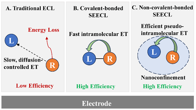
In traditional ECL systems, luminophore radicals (e.g., Ru(bpy)32+) and co-reactant radicals (e.g., TPrA•) rely on Brownian motion for collision. The diffusion-limited rate constant of this process (kdiff ∼106~107 L/(mol·s)) is far slower than the excited-state lifetime (~600 ns) of typical ECL luminophores[1]. This mismatch leads to radical deactivation via solvent quenching before effective collision, resulting in significant energy waste (Fig.1A). To address this, the SEECL strategy spatially “locks” the luminophore and co-reactant together, transforming the slow intermolecular electron transfer into highly efficient intramolecular or nanoconfined electron transfer.
图1 (A)传统ECL、(B)共价键合SEECL和(C)非共价键合SEECL体系中发光体(L)与共反应剂(R)之间的电子传递路径

Fig.1 (A) Electron transfer (ET) pathways between luminophore (L) and co-reactant (R) in traditional ECL, (B) covalent-bonded SEECL, and (C) non-covalent-bonded SEECL systems.

For covalent-bonded SEECL systems, direct chemical bonding between the luminophore and co-reactant constructs the shortest electron transfer pathway, enabling intramolecular electron transfer with rates (kIET∼1010 L/(mol·s)) significantly faster than diffusion control[14], thereby improving the generation efficiency of excited states (Fig. 1B). For non-covalent-bonded SEECL systems, the luminophore and co-reactant are confined within a nanoscale space despite the absence of chemical bonds. This creates a “localized high-concentration” microenvironment that markedly increases the frequency of effective collisions[15], overcoming the bottleneck of macroscopic diffusion and achieving “pseudo-intramolecular” efficient electron transfer (Fig. 1C).

2.2 Covalent-bonded SEECL systems

The most general approach for constructing SEECL is to conjugate the luminophore and co-reactant within the same nanostructure or molecule covalently. Various types of ECL luminophores, including metal complexes, organic compounds, and nanomaterials, can all form SEECL systems through covalent conjugation with co-reactants.

2.2.1 Inorganic covalent-bonded SEECL systems

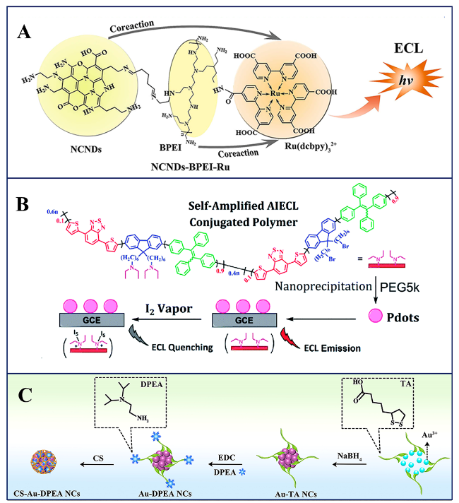
Tris(bipyridine)ruthenium(II) (Ru(bpy)32+) and its derivatives, due to their excellent electrochemical and optical properties, have been extensively studied and applied in SEECL systems. By introducing functional groups such as carboxyl and amino groups onto the ligands, Ru(II) complexes can form SEECL luminophores by covalent conjugation with amino acid or alkyl amine, such as L-lysine[16], L-cysteine[17], tris(2-aminoethyl)amine (TAEA)[18], tris(3-aminopropyl)amine (TAPA)[19]NN-diisopropylethylenediamine (DPEA)[20], polyethylenimine (PEI)[21], and polyamidoamine (PAMAM)[22]. Within this category, PEI has emerged as the most widely adopted linker, primarily attributed to its amine-enriched structure and anionic polyelectrolyte characteristics, and a variety of nanocomposites based on Ru(II) complexes/PEI systems have been developed for ECL biosensing applications, such as AuNR@Ru-LA/PEI[23], Ru-PEI/Au@ZIF-8[24], Ru-PEI@PCN-333(Al)[25], and MXenes-BPQDs@Ru-PEI[26]. In Addition, amine-rich nitrogen-doped carbon nanodots (NCNDs), functioning as both ECL label carriers and co-reactants, have been applied to form covalent NCND-Ru(bpy)32+ hybrid, which exhibits enhanced ECL emission through intramolecular electron transfer[27]. Furthermore, Hu et al.[28] attempted to covalently link NCNDs with Ru(dcbpy)3Cl2 using branched PEI (BPEI), synthesizing a dual co-reactant self-enhanced nanocomposite (NCNDs-BPEI-Ru) which generated an ultra-strong initial ECL signal on glassy carbon electrode (Fig.2A).
图2 (A)无机[28]、(B)有机[36]和(C)基于纳米材料的[38]共价键合SEECL体系

Fig.2 (A) Inorganic covalent-bonded SEECL system[28], Copyright 2022, Elsevier. (B) Organic covalent-bonded SEECL system[36], Copyright 2021, Royal Society of Chemistry. (C) nanomaterial-based covalent-bonded SEECL system[38], Copyright 2019, Elsevier

2.2.2 Organic covalent-bonded SEECL systems

In SEECL systems where organic molecules serve as the luminophore, the most extensively studied and applied systems remain the luminol or luminol derivative/PEI systems. In such systems, nanomaterials applied to load the luminophore and PEI typically exhibit the capability to catalyze H2O2 for reactive oxygen species (ROS) generation, such as CuMn-CeO2-PEI-luminol[29], PTC-PEI-luminol[30], and Ni-TCPP(Fe)-PEI-luminol[31], achieving dual enhancement effects from both intermolecular and intramolecular co-reactants. Except for PEI, cysteine[32], glutathione[33], and Schiff bases[34] have also been reported to successfully construct SEECL luminophores by covalent conjugation with N-aminobutyl-N-ethylisoluminol (ABEI). Additionally, aggregation-induced ECL (AIECL), as an emerging strategy, has opened up new avenues for the development of SEECL. Li et al.[35] coordinated 1,1,2,2-tetra (4-carboxylbiphenyl) ethylene (H4TCBPE), which served as a metal-organic framework (MOF) ligand with Zr(IV), followed by covalent conjugation with PEI to form a unique SEECL complex (Zr-TCBPE-PEI). Meanwhile, Wang et al. developed a conjugated polymer modified with a tertiary amine as a co-reactive group, which exhibited both AIECL and SEECL behaviors, enabling applications in the detection of iodine vapor (Fig.2B[36] and doxorubicin[37].

2.2.3 Nanomaterial-based covalent-bonded SEECL systems

Amine compounds are commonly used as co-reactants for nanomaterials to achieve SEECL. As shown in Fig. 2C, Yang et al.[38] covalently conjugated the co-reactant DPEA to gold nanoclusters (Au NCs), significantly enhancing the ECL efficiency of Au NCs at low potentials. Cao et al.[39] synthesized SEECL CsPbBr3 perovskite NCs by using oleylamine as both a co-reactant and stabilizer through the ligand-assisted reprecipitation method, which exhibited an ECL efficiency of up to 57.08% in aqueous solution (using the Ru(bpy)32+-TPrA system as the reference). Similarly, CdTe@ZnS quantum dots (QDs) combined with polyaniline (GDY@PANI/CdTe@ZnS)[40], 3D graphitic carbon combined with nitride-3-(dibutylamino)propylamine (3D g-C3N4-NV-DBAPA)[41] can also form SEECL through covalent conjugation, and be applied to high-sensitivity biosensing.

2.3 Non-covalent-bonded SEECL systems

In recent years, researchers have increasingly attempted to integrate luminophores and co-reactants into a unified nanostructure using non-covalent approaches, such as nanocarrier encapsulation, self-assembly, and MOF-based methods. These flexible and efficient strategies have significantly enriched the SEECL systems.

2.3.1 Nanocarrier encapsulation SEECL systems

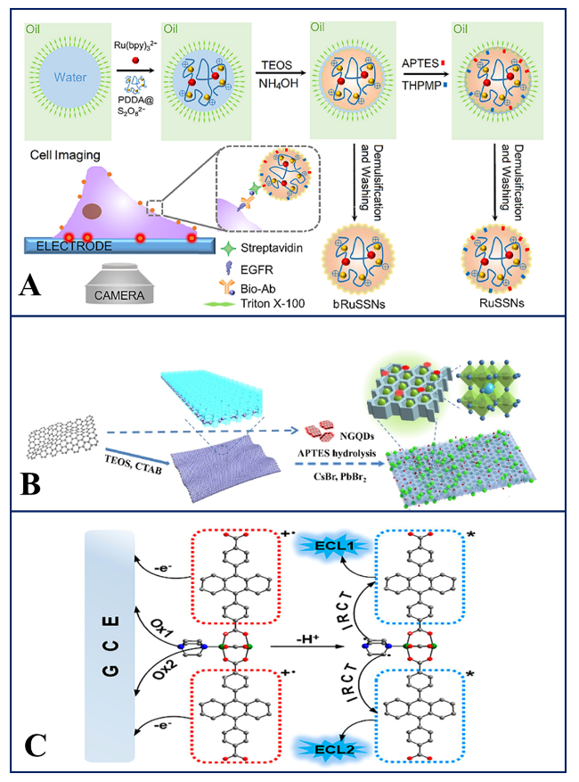
SiO2 nanoparticles (NPs) are widely used in the construction of SEECL as encapsulation materials due to the controllable porous structure, high specific surface area, and biocompatibility. The abundant silanol groups (―SiOH) on SiO2 NPs enable grafting functional groups such as amino (―NH2), thiol (―SH), and carboxyl (―COOH) through silane coupling agents, facilitating covalent immobilization or electrostatic adsorption of luminophores and co-reactants. A variety of SiO2 NPs encapsulated SEECL emitters have been reported, such as DEAMTES@RuSiO2[42], Ru@SiO2-CNQDs[43], Ru@SiO2-BPQDs[44], and Ru-TEPA@m-SiO2[45]. As shown in Fig. 3A, positively charged Ru(bpy)32+ and negatively charged peroxydisulfate were co-doped into SiO2 NPs via a process involving PDDA@S2O82- formation, encapsulation, and SiO2 NPs synthesis, achieving a 2.9-fold stronger ECL emission with excellent uniformity, monodispersity, and biocompatibility[46]. Besides the typical combination of SiO2 NPs and Ru complexes, Li et al.[47] encapsulated CsPbBr3 QDs and co-reactants into a SiO2 matrix, which was in situ generated by the hydrolysis of tetramethyl orthosilicate. The synthesized CPB-CoR@SiO2 nanocomposite exhibited a 10.2-fold enhanced ECL efficiency compared to the standard Ru(bpy)32+/TPrA system.
图3 (A) 纳米封装型[46]、(B) 自组装型 [53]和(C) 基于MOF的[58]SEECL体系

Fig.3 (A) Nanocarrier encapsulation SEECL system[46], Copyright 2023, American Chemical Society. (B) Self-assembly SEECL system[53], Copyright 2022, Elsevier. (C) MOFs-based SEECL system[58], Copyright 2021, American Chemical Society

2.3.2 Self-assembly SEECL systems

The common feature of self-assembled SEECL lies in its reliance on various nanomaterials (such as Si NPs, graphene QDs, etc.), utilizing non-covalent forces including electrostatic adsorption, nanoconfinement host-guest interactions, and coordination-driven assembly to bring luminophores and co-reactants into close proximity[48-55]. These systems differ in terms of luminophore and co-reactant selection as well as assembly strategies. Luminophores encompass perovskite nanocrystals (PNCs), ruthenium complexes, etc., while co-reactants include amines, peroxides, NGQDs, and PEI. Assembly strategies range from simple electrostatic adsorption (NH2-Ru@SiO2-NGQDs)[52] to complex architectures such as three-layered nanoparticles (with Ru(II)-doped SnS2 as the middle layer, PAMAM as the inner layer, and AuNPs as the outer layer)[49] or two-dimensional mesoporous SiO2-confined CsPbBr3 and NGQDs[53]. These differences in approaches influence the ECL enhancement mechanisms, with some focusing on shortening charge transfer paths and others emphasizing amplifying local co-reactant concentrations. As illustrated in Fig.3B, Wei et al.[53] constructed a self-enhanced quaternary complex by sequentially loading NGQDs and CsPbBr3 PNCs onto graphene-supported two-dimensional mesoporous SiO2 nanosheets (2D mSiO2-G). The confined space provided by mesoporous SiO2 enabled tight contact between NGQDs (co-reactant) and PNCs (luminophore) via spatial confinement, enhancing stability and accelerating charge transfer. Their synergistic effect achieved a highly efficient self-enhanced ECL.

2.3.3 MOFs-based SEECL systems

The MOF structure provides a confined environment. Specifically, by introducing luminophores and co-reactants as ligands into the MOF matrix, intramolecular or intra-network charge transfer can be facilitated, thereby enhancing ECL. The most common ligand combinations include those of Ru complexes/oxalic acid[56-57] and anthracene-based ligands/amines[58-59]. As Fig. 3C shows, Zhu et al.[58] developed a MOF-based SEECL system by integrating 9,10-di(p-carboxyphenyl)anthracene (DPA) and 1,4-diazabicyclo[2.2.2]octane (D-H2) as dual ligands on a single metal node, where their sequential oxidation generated cation radicals (DPA+) and neutral radicals (D-H), respectively, enabling radical recombination to produce excited DPA* for ECL emission without external co-reactants.
Some MOF-based SEECL systems utilize resonance energy transfer (RET) or localized surface plasmon resonance (LSPR) effects to further modulate ECL. For example, Mei et al.[60] incorporated the luminophore 4,4'-(anthracene-9,10-diyl) dibenzoic acid and co-reactant D-H2 within a Zn-MOF framework, and utilized the LSPR effect of AuNPs triggered by Zn-MOF emission to co-enhance the near-electromagnetic field, creating a feedback loop that potentiates signal amplification for sensitive detection. Yang et al. [56] integrated the co-reactants C2O42- and Ru(bpy)32+ to form the Ru-Zn-MOF luminophore as the ECL donor, and applied polydopamine-modified ZnO nanoflowers as ECL acceptors to regulate RET by quenching the Ru-Zn-MOF ECL signal, achieving ultrasensitive neuron-specific enolase (NSE) detection.

3 Applications of SEECL

3.1 Bioanalysis

3.1.1 Protein biomarker detection

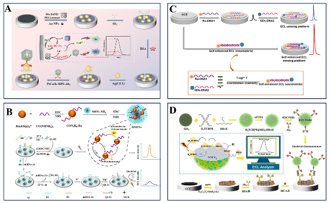
Through material innovation and signal amplification strategies, SEECL has significantly improved the detection sensitivity of disease biomarker detection. For example, Ru-based complexes combined with PAMAM dendrimers and palladium nanowires achieved ultrasensitive detection of carcinoembryonic antigen (CEA) with a limit of detection (LOD) of 0.3 pg/mL[61], while manganese-based PEI-immobilized luminol, combined with the ECL-RET strategy, further reduced the LOD of CEA to 10 fg/mL (Fig.4A[62]. Similarly, in cardiovascular disease biomarker detection, Ru-PAMAM/AuNPs complex integrating GOD-FA-PtCu3-Ab2 bioconjugate with dual quenching effect, also enabled the LOD of cardiac troponin I (cTnI), reducing to a femtogram-level (12 fg/mL)[22]. Except for cancer and cardiovascular diseases, SEECL has been extensively applied in the detection of biomarkers for diabetic nephropathy[19], inflammation[63-64] and Alzheimer’s disease (AD)[65]. For example, Xie et al.[66] prepared a luminescent material (PFO-SDS-PEI) with both SEECL and fluorescence (FL) properties for FL/ECL dual-mode detection of Tau protein (a biomarker of AD), achieving LODs of 549.16 ag/mL and 5.45 ag/mL for FL and ECL, respectively. Additionally, the detection of the SARS-CoV-2 nucleocapsid protein[67] further expanded the application of ECL in infectious disease diagnosis. These advancements highlighted the critical role of SEECL in early-stage disease diagnosis.
图4 用于检测(A) 蛋白标志物[62]、(B) 核酸[68]、(C) 金属离子[84]和(D) 藻类毒素[91]SEECL传感器

Fig.4 SEECL sensors for detecting (A) protein markers [62], Copyright 2023, American Chemical Society. (B) Nucleic acids[68], Copyright 2023, Elsevier. (C) Metal ions[84], Copyright 2021, American Chemical Society. (D) Algal toxins[91], Copyright 2024, Elsevier

3.1.2 Nucleic acid analysis

SEECL shows its unique advantages in nucleic acid analysis, and realizes the ultrasensitive and highly specific detection of nucleic acid biomarkers[68-71] and pathogen nucleic acids[28-30]. Among these, systems with Ru(II) complexes as the core luminophore are particularly prominent. For example, a SEECL biosensor integrating Ru(dcbpy)32+-CON4H6 in mesoporous SiNPs was constructed via covalent linkage of DNA strands (Q1-H2) to detect microRNA-21 through strand displacement reaction and G-quadruplex formation, advancing ultra-sensitive microRNA analysis for breast cancer diagnosis (Fig.4B[68]. Another typical strategy involves DNA nanotube-mediated quenching and subsequent 3D DNA nanomachine-driven signal recovery, in which NCNDs-BPEI-Ru nanocomposite was developed as an ECL signal probe for ultrasensitive detection of Mycobacterium tuberculosis (MTB) DNA fragment, with a low LOD of 1.4 amol/L[28]. Through precise regulation of luminophore-nucleic acid interactions and signal amplification via nanomaterials/enzymatic reactions, these works have advanced the in-depth application of ECL in precise nucleic acid analysis.

3.1.3 Enzyme activity and metabolic monitoring

Enzyme activity and metabolic monitoring are critical directions in disease diagnosis, drug screening, and environmental toxicity assessment. SEECL, leveraging its high sensitivity, low background noise, and tunable signal amplification, has emerged as a key tool in this field[72-81]. For example, Xu et al.[80] developed a ratiometric thrombin (TB) biosensor based on PEDOT-Au@Luminol modified electrode, in which a bifunctional peptide (with antifouling and TB recognizable sequences) was applied to capture SEECL probe (PAMAM-CuInZnS/ZnS QDs), achieving a LOD of 1.82 fmol/L. Cao et al.[23] integrated CsPbBr3 QDs with urease on an electrode surface and dynamically monitored live cell metabolism by utilizing urease-catalyzed urea hydrolysis to modulate the ECL signal of the QDs, achieving a LOD of 8.8×10⁻¹¹ mol/L for urea.

3.2 Environmental detection

3.2.1 Metal ions detection

SEECL enables ultrasensitive detection of various heavy metal ions such as Pb2+[82], Co2+[83], Hg2+[52,84], I-[7,36],and UO22+[85]. For example, Deng et al.[82] developed Ru-PAMAM-HIFAuNPs composite materials, which achieved a LOD of 4.0×10⁻¹⁴ mol/L for Pb2+ through an intermolecular/intramolecular dual co-reactant enhancement mechanism, enabling trace lead pollution monitoring. Li et al.[83] synthesized two Ru(II) complexes featuring Schiff base cavities, which enhance ECL through phenolic hydroxyl group oxidation and imino radical resonance mechanisms. They further applied a Co2+-bonded salen cavity for sensitive ECL sensing of Co2+, achieving a LOD of 21 nmol/L. As Fig. 4C shows, through Hg2+-mediated T-Hg2+-T duplex assembly strategy, a SEECL sensing platform was developed to detect Hg2+ with a low LOD of 3.3 fmol/L, using tris(4,4'-dicarboxylic acid-2,2'-bipyridyl)ruthenium(II) dichloride-modified DNA1 (Ru-DNA1) and g-C3N4 QDs-linked DNA2 (QDs-DNA2) as materials[84]. These studies demonstrate the potential of SEECL to break through the LOD in metal ion detection through material innovation and strategy optimization, providing critical tools for environmental toxicological assessment.

3.2.2 Organic pollutant monitoring

Given the severe threats posed by organic pollutants to ecological environments and human health, SEECL has emerged as a pivotal tool for ultrasensitive detection of organic pollutants such as pesticides[86-88], antibiotics[34,89], hormones[90], and toxins[91]. For pesticide detection, PEI/RuSi-MWCNTs[86], NH2-RuDS@NGQDs[88], and PAN@Ru@PEI@Nafion[87] were synthesized to construct SEECL sensors for detecting profenofos, carbaryl, and α-naphthol (hydrolysates of carbaryl), respectively. It is worth noting that the co-reactant and luminophore in PAN@Ru@PEI@Nafion were not connected via covalent or non-covalent interactions; they were fabricated into nanofibers through electrospinning instead. In addition to Ru(II) complexes, the ABEI-anchored Schiff base Fe complex[34] and water-soluble Ir-PEI composite[89] were respectively applied for the detection of neomycin and tetracycline, with detection limits of 0.21 pmol/L and 6.14 nmol/L. Moreover, an ECL immunosensor for microcystin-LR was proposed based on an AIECL-assisted self-enhancement strategy recently, which used ternary H4TCBPE@SiO2-DBAE nanoparticles as probes and Ti3C2Tx/MoS2/Au hybrid as the immunosensing substrate, achieving a LOD of 31 fg/mL (Fig. 4D[91].

3.3 Other categories

3.3.1 Food safety monitoring

SEECL is of great significance in food safety monitoring due to its high sensitivity and rapidity, and has become a powerful tool for trace detection of hazardous substances such as mycotoxins[92-94], bacteria[41,95] (e.g., Staphylococcus aureusBurkholderia pseudomallei), and algal toxins[55]. Key advancements stem from material innovations (e.g., helical carbon nanotubes, 3D g-C3N4 porous frameworks, Au@PEI-ABEI@Pt hydrogels, perovskite composites, and N,S-doped graphene QDs) and strategy optimizations (e.g., dual co-reactants, molecular imprinting, MOF enhancement, dual-quenching mechanisms, and LSPR amplification). For example, Fang et al.[32] developed a helical carbon nanotube-based aptasensor using an ABEI/GSH SEECL system and octahedral anatase mesocrystals carrier, combined with horseradish peroxidase (HRP)-catalyzed ROS amplification, achieving a LOD of 1 fg/mL for Zearalenone (ZEN) in corn and hazelnut samples. Additionally, an Au@PEI-ABEI@Pt hydrogel-based sensor, featuring a large specific surface area and porous structure to promote electron transfer, integrated with CRISPR/Cas12a signal amplification, enabled ultrasensitive detection of Burkholderia pseudomallei at 5 CFU/mL in complex samples[95]. For Ochratoxin A (OTA) detection, an “on-off-on” aptasensor based on AuNPs-PEI-MWCNTs electrodes and Ru@SiO2-BPQDs luminophores was proposed. Utilizing dual electron transfer pathways and Fc quenching/release mechanisms, it achieved a low LOD of 0.03 ng/mL, demonstrating high selectivity in mycotoxin analysis[44].

3.3.2 Portable biosensors and wearable devices

Innovative applications of SEECL technology have been extended to portable biosensors and wearable devices, enabling on-site, real-time detection of both physiological and pathological targets. For wearable applications, a flexible ECL platform was first developed for sweat analysis, integrating high-luminescent nanospheres on gold nanotube networks coated with elastic molecularly imprinted polymers, enabling stable ECL signals and mechanical compliance during deformation, and successfully detecting lactate and urea in sweat with high fidelity[96]. In point-of-care testing scenarios, a dry chemistry-based bipolar ECL immunoassay device was designed for AD detection, utilizing a self-enhanced Ru(II)-poly-L-lysine complex and lateral flow fiber chip to eliminate co-reactant addition and rinsing, completing AD7c-NTP detection in 6 minutes with a low LOD of 0.15 pg/mL[97]. For infectious disease diagnostics, a SEECL array chip was constructed for SARS-CoV-2 N-protein detection, combining Ru/PEI@SiO2 nanomaterials with a single-electrode system and 3D-printed portable smartphone-integrated housing, achieving a record-low LOD of 0.47 pg/mL and detecting viral antigens as low as 1.92 pg/mL in patient serum[98]. These advancements demonstrate that SEECL has great potential in developing portable, wearable, and high-performance biosensors for diverse healthcare and diagnostic needs.
In summary, SEECL integrates luminophores and co-reactants into a single nanostructure or molecular framework through covalent or non-covalent interactions. By leveraging diverse nanomaterials and signal amplification strategies, SEECL has found wide application in biological analysis, environmental monitoring, food safety, and other fields. As illustrated in Table 1, we systematically summarize its construction methods and application scopes.
表1 SEECL体系构造方法及应用范围的对比分析

Table 1 Comparative analysis of the SEECL system from construction method to application scope

Construction method SEECL system Target Linear range LOD Ref
Covalent-bonded Ru-Amp@CNTs-PEI-AuNCs TB 1.0 fmol/L~1.0 pmol/L 0.33 fmol/L 72
Covalent-bonded PTCA-PEI-Ru(II)/CNTs Apurinic/apyrimidinic endonuclease 1 1 fg/mL~1 pg/mL 0.3 fg/mL 73
Covalent-bonded PTCA-PEI-Ru(II) TB 1.0×10⁻¹⁴~1.0×10⁻¹⁰ mol/L <fmol/L level 75
Covalent-bonded CS/Ru-PEI@ZIF-8/PtNPs Telomerase activity 50~106 HeLa cells 11 cells 76
Covalent-bonded GO-PEI-Ru-AuNPs CEA 0.10 pg/mL~80 ng/mL 0.045 pg/mL 99
Covalent-bonded Ru-PEI@PCN-333(Al) MOF Caspase-3 - 0.017 pg/mL 25
Covalent-bonded Ru-PEI/Au@ZIF-8 SARS-CoV-2 RNA 1 fmol/L~100 pmol/L 0.67 fmol/L 24
Covalent-bonded PEI/RuSi-MWCNTs Profenofos 1×10⁻²~1×10³ ng/mL 1.482×10⁻³ ng/mL 86
Covalent-bonded SiO2-PEI NPs-Ru Spermine 10~100 nmol/L 12.2 nmol/L 100
Covalent-bonded AuNR@Ru-LA/PEI Urea 1.0×10⁻¹⁰~1.0×10⁻⁴ mol/L 8.8×10⁻¹¹ mol/L 23
Covalent-bonded PEI@Ru-Hf-MOL Mucin 1 (MUC1) 1 fg/mL~10 ng/mL 0.48 fg/mL 101
Covalent-bonded DPs/Ru-PAMAM-HIFAuNPs Pb2+ 1.0×10⁻¹³~1.0×10⁻⁷ mol/L 4.0×10⁻¹⁴ mol/L 82
Covalent-bonded PdNWs-PAMAM-Ru CEA 0.001~80 ng/mL 0.3 pg/mL 61
Covalent-bonded Ru-PAMAM/AuNPs cTnI 0.1 pg/mL~0.2 ng/mL 12 fg/mL 22
Covalent-bonded TAEA-PTCA@GO/Ru MicroRNA 10 amol/L~1.0 pmol/L 3.3 amol/L 18
Covalent-bonded G4-Ru-DPEA N‑Acetyl-β-D-glucosaminidase 0.1 pg/mL~1 ng/mL 0.028 pg/mL 20
Covalent-bonded MSNs-Ru-CON4H6 MicroRNA-21 0.1 fmol/L~1 nmol/L 0.03 fmol/L 68
Covalent-bonded Zn-Ru-EDA PCT 1.00×10⁻⁶~10 ng/mL 0.47 fg/mL 102
Covalent-bonded Ru-Schiff base Co2+ 0.9~6.3 μmol/L 21 nmol/L 83
Covalent-bonded PtNPs/Ru-L-Lys/Mn-ZnONRs CA15-3 0.05~120 U/mL 0.014 U/mL 16
Covalent-bonded Ru-L-Lys-Zr-MOL MUC1 1 fg/mL~100 pg/mL 0.72 fg/mL 103
Covalent-bonded AuNRs/L-Cys@Ru(dcbpy)32+ cTnI 0.25 pg/mL~0.1 ng/mL 0.083 pg/mL 17
Covalent-bonded Ru(II)-L-Cys cTnI 0.001~100 ng/mL 0.4416 pg/mL 104
Covalent-bonded AuNPs-Ru-Arg@NH2-Ti3C2-MXene CEA 0.01~150 ng/mL 1.5 pg/mL 105
Covalent-bonded Ru-N-SiNPs Paclitaxel 1~200 nmol/L 0.3 nmol/L 74
Covalent-bonded Ru-BCDs-PEI MicroRNA-133a 500 fmol/L~1 nmol/L 60 fmol/L 21
Covalent-bonded MXenes-BPQDs@Ru-PEI Exosomes 1.1×10²~1.1×10⁷ particle/μL - 26
Covalent-bonded BCN NSs-Ru TK1 mRNA 100 amol/L~100 nmol/L 32.3 amol/L 106
Covalent-bonded NCNDs-BPEI-Ru MTB DNA 50 amol/L~1 nmol/L 1.4 amol/L 28
Covalent-bonded Ru-CoO@N-C (MOF) SARS-CoV-2 8 fg/mL~4 ng/mL 1.6 fg/mL 67
Covalent-bonded Ir-PEI Ttetracycline (TET) 5 ng/mL~5 mg/mL 2.73 ng/mL 89
Covalent-bonded TPrA@Ir-SiO2 AD biomarker (P-tau181) 0.1 pg/mL~0.1 μg/mL 68.58 fg/mL 65
Covalent-bonded CuMn-CeO2-PEI-luminol DNA of Group B Streptococci 0.1 fmol/L~1 nmol/L 63 amol/L 29
Covalent-bonded PTC-PEI-luminol Helicobacter pylori DNA 10 fmol/L~10 nmol/L 2.4 fmol/L 30
Covalent-bonded Ni-TCPP (Fe)-PEI-luminol h-FABP 100 fg/mL~100 ng/mL 44.5 fg/mL 31
Covalent-bonded PEI-luminol hydrogels BCR/ABL fusion gene 10.0 fmol/L~10.0 nmol/L 3.75 fmol/L 71
Covalent-bonded Au@PEI-ABEI@Pt Burkholderia pseudomallei - 5 CFU/mL 95
Covalent-bonded ABEI-PEI-Au-Pd-NFC Procalcitonin (PCT) 10 fg/mL~100 ng/mL 3.46 fg/mL 63
Covalent-bonded ABEI-PEI-Au@AgNCs β2-Microglobulin 0.01 pg/mL~200 ng/mL 3.3 fg/mL 107
Covalent-bonded PdIr-L-Cys-ABEI Laminin 1 pg/mL~120 ng/mL 0.27 pg/mL 108
Covalent-bonded ABEI-Cys/Au-Pd-Pt/MoS2 Cystatin C 1.0 fg/mL~5.0 ng/mL 0.35 fg/mL 33
Covalent-bonded OAM-GSH-ABEI ZEN 1.0×10⁻⁴~10 ng/mL 33 fg/mL 32
Covalent-bonded Fe-PDL-ABEI Neomycin 0.3 pmol/L~0.1 nmol/L0.1 nmol/L~1 μmol/L 0.21 pmol/L 34
Covalent-bonded Conjugated polymer & tertiary amine, AIECL I2 - 0.13 ppt 36
Covalent-bonded Conjugated polymer & tertiary amine, AIECL Doxorubicin (DOX) - 0.9% (mDOX/mpolymer 37
Covalent-bonded H4TCBPE@SiO2-DBAE Microcystin-LR 50 fg/mL~10 ng/mL 31 fg/mL 91
Covalent-bonded CS-Au-DPEA NCs MUC1 1 fg/mL~1 ng/mL 0.54 fg/mL 38
Covalent-bonded GDY@PANI/CdTe@ZnS α-synuclein (α-syn) 0.2 fmol/L~8 nmol/L 0.02 fmol/L 40
Covalent-bonded 3D g-C3N4-NV-DBAPA Staphylococcus aureus - 10.3 amol/L 41
Nanocarrier encapsulation Ru-HPNSs MUC1 1.0 fg/mL~100 pg/mL 0.31 fg/mL 109
Nanocarrier encapsulation Ru-CNQDs@SiO2 Hg2+ 0.1 nmol/L~10 μmol/L 33 pmol/L 43
Nanocarrier encapsulation Ru@SiO2-BPQDs OTA 0.1~320 ng/mL 0.03 ng/mL 44
Nanocarrier encapsulation PDDA@S2O82-@RuSSNs Membrane
protein
- - 46
Self-assembly (H-Mn3O4-PEI-PtNPs-Ru)n-Eu3+ Cyclin A 2 0.001~100 ng/mL 0.3 pg/mL 48
Self-assembly Au-PAMAM-Ru(II)-SnS2 NPs TB 5.0×10⁻¹⁵~1.0×10⁻⁹ mol/L 2.6×10⁻¹⁵ mol/L 49
Self-assembly Ru-SiO2@PEI NSE 1.0×10⁻¹¹~1.0×10⁻⁵ mg/mL 1.0×10⁻¹¹ mg/mL 110
Self-assembly NCDs@PEI-rGO/RuNSs Dopamine 0.01~50 μmol/L 10 nmol/L 50
Self-assembly PdNPs/PEI-GO-QDs Diclofenac 0.001~1000 ng/mL 0.3 pg/mL 111
Self-assembly Nss-TiO2-RM@Ru@BiNBs Sialic acid 3.5×10⁻⁵~350 ng/mL 1.17×10⁻⁵
ng/mL
77
Self-assembly NGQDs-NH2-Ru@SiO2 ZEN 10 fg/mL~10 ng/mL 1 fg/mL 92
Self-assembly PEI-CdS/Au@SiO2@RuDS/PANI Creatinine 0.05 nmol/L~5 μmol/L 0.02 nmol/L 51
Self-assembly PeQDs-NCDs@HZIF-8 T4 polynucleotide kinase 1.0×10⁻⁵~5.0×10⁻² U/mL 6.2 × 10⁻⁶ U/mL 78
Self-assembly NH2-Ru@SiO2-NGQDs Hg2+ 5.0×10⁻¹¹~1.0×10⁻⁶ mol/L 3.0 × 10⁻¹¹ mol/L 52
Self-assembly Ru@MOF@NCND-Ru@SmS2 QDs CA19-9 0.0005~200 U/mL 0.00013 U/mL 112
Self-assembly PNCs-NGQDs@mSiO2-G OTA 10-5~1.0 ng/mL 0.2 pg/mL 53
Self-assembly Zr-MOF@PEI@AuAg MicroRNA-144
MicroRNA-155
1.0×10⁻¹⁶~1.0×10⁻¹⁰ mol/L 19 amol/L
26 amol/L
69
Self-assembly AuNCs-DEAET Carboxylesterase 1×10⁻⁶~1×10² U/L 9.1×10⁻⁷ U/L 54
Self-assembly (Hf) MOL-Ru-PEI-Pd CEA 0.1 pg/mL~100 ng/mL 20 fg/mL 113
Self-assembly RuSiNPs@N,S-GQDs Okadaic acid 0.003~40 ng/mL 0.001 ng/mL 55
Self-assembly ZnNi-MOF/PEI-L012 CA15-3 0.0005~50 U/mL 5.75×10⁻⁵ U/mL 114
MOF-based DPA/D-H2@Zn-MOF - - - 58
MOF-based DPA/DEAEA@Zn-MOF MicroRNA-21 100.0 amol/L ~10.0 pmol/L 61.7 amol/L 59
MOF-based Ru/H2C2O4@Zn-MOF NSE 10 fg/mL~100 ng/mL 3.3 fg/mL 56
MOF-based Ru/H2C2O4@Zn-MOF Squamous cell carcinoma antigen 1 fg/mL~100 ng/mL 0.26 fg/mL 57
MOF-based DPA/D-H2@Zn-MOF Deoxynivalenol 1 ×10⁻⁴~100 ng/mL 0.036 pg/mL 60

4 Conclusions and prospects

By integrating luminophores and co-reactants into a single nanostructure or molecule through covalent or non-covalent interactions, SEECL has overcome the bottleneck of low intermolecular electron transfer efficiency in traditional ECL systems. Meanwhile, combined with diverse nanomaterials and signal modulation strategies, SEECL has been widely applied in bioanalytical and environmental monitoring fields, successfully achieving ultrasensitive detection. Furthermore, the applications in food safety, wearable devices, and point-of-care testing demonstrate its cross-disciplinary adaptability. Despite significant advancements, SEECL still faces the following challenges and opportunities:
(1)In-depth mechanistic elucidation: Further clarification is needed on the structure-property relationships of charge transfer dynamics and energy transfer efficiency within complex nanostructures, particularly the dynamic processes under multi-component synergistic interactions.
(2)Material innovation and stability: The development of novel luminous systems with high luminous efficiency, long-term stability, and biocompatibility is required, such as QDs-MOF hybrid materials or nanostructures functionalized with single-atom catalytic sites.
(3)Multimodal sensing integration: Exploring integration strategies between SEECL and complementary techniques (e.g., FL, LSPR) to enable multi-dimensional signal output and the construction of intelligent sensing platforms.
(4)Expansion of practical applications: Addressing interference from complex matrices (e.g., blood, soil) by developing specific recognition elements and signal separation algorithms to promote the practical implementation of SEECL in clinical diagnostics and environmental emergency monitoring.
(5)Intelligence and miniaturization: Combining microfluidic chips or miniature/microelectrode arrays with artificial intelligence (AI) algorithms to develop portable SEECL sensing devices, facilitating on-site rapid detection and big-data-driven early disease warning.
Future research should prioritize interdisciplinary integration, leveraging collaborative innovation in nanotechnology, synthetic biology, and data science to advance SEECL toward higher sensitivity, stronger anti-interference capability, and broader applicability. This will ultimately enable its large-scale deployment in precision medicine, green environmental governance, and other fields.
[1]
Richter M M. Chem. Rev., 2004, 104(6): 3003.

[2]
Hu L Z, Xu G B. Chem. Soc. Rev., 2010, 39(8): 3275.

[3]
Yang Y K, Wang X M, Fang G Z, Yun Y G, Guo T, Wang S. Prog. Chem., 2016, 28(9): 1351.

(杨钰昆, 王小敏, 方国臻, 云雅光, 郭婷, 王硕. 化学进展, 2016, 28(9): 1351.)

[4]
Harvey N. J. Phys. Chem., 1929, 33(10): 1456.

[5]
Hu X, Guo Q, Liu Y, Sun Q, Meng T, Zhang R. Prog. Chem., 2017, 29(2/3): 300.

[6]
Gong M, Wang X Y, Wang X N. Prog. Chem., 2019, 31(6): 894.

(宫苗, 王晓英, 王晓宁. 化学进展, 2019, 31(6): 894.)

[7]
Cao X, Wang Z, Liu B, Li X, Wu S, Jiang J, Feng J, Ju H, Wang N. Anal. Chem., 2024, 96(44): 17824.

[8]
Lin Y, Tan X C, Wu Y Y, Wei F C, Wu J W, Ou P P. Prog. Chem., 2022, 34(4): 898.

(林瑜, 谭学才, 吴叶宇, 韦富存, 吴佳雯, 欧盼盼. 化学进展, 2022, 34(4): 898.)

[9]
Wang Z Y, Li C Q, Pei Y, Li M X, Liu Y L, Xu J J, Hua D B. Anal. Chem., 2024, 96(36): 14604.

[10]
Wang H J, Sun D, Wang R Y, Zhang H. Prog. Chem., 2024, 36(12): 1972.

(王慧静, 孙蝶, 王睿颖, 张卉. 化学进展, 2024, 36(12): 1972.)

[11]
Du F X, Chen Y Q, Meng C D, Lou B H, Zhang W, Xu G B. Curr. Opin. Electrochem., 2021, 28: 100725.

[12]
Liang P, Dong L W, Martin M T. J. Am. Chem. Soc., 1996, 118(38): 9198.

[13]
Sun S G, Yang Y, Liu F Y, Fan J L, Peng X J, Kehr J, Sun L C. Dalton Trans., 2009(38): 7969.

[14]
Swanick K N, Ladouceur S, Zysman-Colman E, Ding Z F. Angew. Chem. Int. Ed., 2012, 51(44): 11079.

[15]
Kato K, Fa S X, Ohtani S, Shi T H, Brouwer A M, Ogoshi T. Chem. Soc. Rev., 2022, 51(9): 3648.

[16]
Zhang L, He Y, Wang H, Yuan Y, Yuan R, Chai Y. Biosens. Bioelectron., 2015, 74: 924.

[17]
Zhou Y, Zhuo Y, Liao N, Chai Y Q, Yuan R. Talanta, 2014, 129: 219.

[18]
Chen A Y, Gui G F, Zhuo Y, Chai Y Q, Xiang Y, Yuan R. Anal. Chem., 2015, 87(12): 6328.

[19]
Wang H J, Yuan Y L, Zhuo Y, Chai Y Q, Yuan R. Anal. Chem., 2016, 88(4): 2258.

[20]
Wang H J, Yuan Y L, Zhuo Y, Chai Y Q, Yuan R. Anal. Chem., 2016, 88(11): 5797.

[21]
Ye J, Liu G Y, Yan M X, Zhu Q J, Zhu L P, Huang J S, Yang X R. Anal. Chem., 2019, 91(20): 13237.

[22]
Zhang L, Xiong C Y, Wang H J, Yuan R, Chai Y Q. Sens. Actuat. B Chem., 2017, 241: 765.

[23]
Feng D, Xiao M, Yang P. Anal. Chem., 2023, 95(2): 766.

[24]
Yang F, Wang W, Zhang M, Tao W X, Wang Y W, Shi J M, Ding Y D, Xie M H, Zhang S, Fan Z Q, Zhang K. Environ. Sci. Nano, 2022, 9(9): 3417.

[25]
Luo W W, Chu H Y, Wu X Z, Ma P Y, Wu Q, Song D Q. Talanta, 2022, 239: 123083.

[26]
Fang D D, Zhao D D, Zhang S P, Huang Y T, Dai H, Lin Y Y. Sens. Actuat. B Chem., 2020, 305: 127544.

[27]
Carrara S, Arcudi F, Prato M, De Cola L. Angew. Chem. Int. Ed., 2017, 56(17): 4757.

[28]
Hu J C, Zhang Y, Chai Y Q, Yuan R. Sens. Actuat. B Chem., 2022, 354: 131252.

[29]
Ling J J, Zhao M, Chen F J, Zhou X Y, Li X S, Ding S J, Tang H. Biosens. Bioelectron., 2019, 127: 167.

[30]
Tang H J, Chen W X, Li D D, Duan X L, Ding S J, Zhao M, Zhang J. Sens. Actuat. B Chem., 2019, 293: 304.

[31]
Gan X F, Han D B, Wang J M, Liu P, Li X R, Zheng Q Y, Yan Y R. Biosens. Bioelectron., 2021, 171: 112735.

[32]
Fang D D, Zeng B S, Zhang S P, Dai H, Lin Y Y. Microchim. Acta, 2020, 187(5): 303.

[33]
Li Y J, Wang Y H, Bai L J, Lv H Y, Huang W, Liu S C, Ding S J, Zhao M. Anal. Chim. Acta, 2020, 1125: 86.

[34]
Zong L P, Chen X Z, Zhu D R, Li X J, Li F, Cosnier S, Zhang X-J, Marks R S, Shan D. ACS Sens., 2022, 7(10): 3085.

[35]
Li J S, Jia H Y, Ren X, Li Y Y, Liu L, Feng R Q, Ma H M, Wei Q. Small, 2022, 18(13): 2106567.

[36]
Wang Z Y, Xu M Y, Zhang N, Pan J B, Wu X Q, Liu P, Xu J J, Hua D B. J. Mater. Chem. A, 2021, 9(21): 12584.

[37]
Wang Z Y, Liu P, Pei Y, Li Y L, Wang C. J. Drug Deliv. Sci. Technol., 2023, 80: 104137.

[38]
Yang F, Zhong X, Jiang X Y, Zhuo Y, Yuan R, Wei S P. Biosens. Bioelectron., 2019, 130: 262.

[39]
Cao Y, Zhu W L, Li L L, Zhang Z Y, Chen Z X, Lin Y H, Zhu J J. Nanoscale, 2020, 12(13): 7321.

[40]
Bahari D, Babamiri B, Salimi A, Nikkhah M, Hallaj R, Mohtasham H, Allahdadi S, Mousavi S M M. Electroanalysis, 2024, 36(7): e202300395.

[41]
Wang Y X, Jiang X Y, Cao W W, Wang H J, Yuan R. Talanta, 2024, 275: 126144.

[42]
Ning Z Q, Yang E L, Zheng Y J, Chen M Y, Wu G Q, Zhang Y J, Shen Y F. Anal. Chem., 2021, 93(25): 8971.

[43]
Li L B, Zhao W L, Zhang J Y, Luo L J, Liu X H, Li X, You T Y, Zhao C J. J. Colloid Interface Sci., 2022, 608: 1151.

[44]
Sang M S, Meng X Y, Zhang Y H, Li Z Y, Zhou Q L, Jing X Z, Sun X, Zhao W P. Anal. Bioanal. Chem., 2023, 415(23): 5833.

[45]
Gao L H, Huang Y T, Ren H Z, Chen S S, Zhang S P, Dai H. J. Electroanal. Chem., 2023, 947: 117767.

[46]
Yu S Q, Hu X F, Pan J B, Lei J P, Ju H X. Anal. Chem., 2023, 95(45): 16593.

[47]
Li L, Zhang Z, Chen Y, Xu Q, Zhang J R, Chen Z, Chen Y, Zhu J J. Adv. Funct. Mater., 2019, 29(32): 1902533.

[48]
Wang H J, Yuan Y L, Chai Y Q, Yuan R. Small, 2015, 11(30): 3703.

[49]
Zhao M, Huang D, Zhuo Y, Chai Y Q, Yuan R. Electrochim. Acta, 2016, 212: 734.

[50]
Li L B, Liu D, Mao H P, You T Y. Biosens. Bioelectron., 2017, 89: 489.

[51]
Cao N, Zhao F Q, Zeng B Z. Sens. Actuat. B Chem., 2020, 306: 127591.

[52]
Li L B, Chen B N, Luo L J, Liu X H, Bi X Y, You T Y. Talanta, 2021, 222: 121579.

[53]
Wei J L, Chen L M, Cai X Y, Lai W Q, Chen X, Cai Z X. Biosens. Bioelectron., 2022, 216: 114664.

[54]
Guo W Z, Xia M Y, Peng D, Zhao Y Y, Nie Y M, Zhou Y M. Anal. Chem., 2024, 96(6): 2369.

[55]
Li S L, Peng J W, Lin X Y, Chen J, Wu Y F, Chen Q S, Chen X M. Microchim. Acta, 2024, 191(12): 737.

[56]
Yang J, Qin D M, Wang N, Wu Y S, Fang K J, Deng B Y. Analyst, 2023, 148(18): 4539.

[57]
Luo Z, Li J, Hu S L, Zheng L L, Xu L X, Deng B Y. Microchem. J., 2025, 209: 112693.

[58]
Zhu D, Zhang Y, Bao S S, Wang N N, Yu S Q, Luo R G, Ma J, Ju H X, Lei J P. J. Am. Chem. Soc., 2021, 143(8): 3049.

[59]
Wang X Y, Wang X, Hu C Y, Guo W, Wu X J, Chen G X, Dai W J, Zhen S J, Huang C Z, Li Y F. Biosens. Bioelectron., 2022, 213: 114443.

[60]
Mei X N, Yao S, Xu L H, Shi Y T, Yang H M, Zhou S, Zhang J, Yan M, Yu J H, Zhang Y. Sens. Actuat. B Chem., 2025, 431: 137432.

[61]
Wang H, Yuan Y, Chai Y, Yuan R. Biosens. Bioelectron., 2015, 68: 72.

[62]
Lai W J, Li J J, Jiang M Z, Li P L, Wang M, Ma C Y, Zhao C L, Qi Y, Hong C L. Anal. Chem., 2023, 95(18): 7109.

[63]
Song X Z, Yu S Q, Zhao L, Guo Y J, Ren X, Ma H M, Wang S F, Luo C N, Li Y Y, Wei Q. Anal. Chem., 2022, 94(26): 9363.

[64]
Wang C, Zhang N, Wei D, Feng R, Fan D W, Hu L H, Wei Q, Ju H X. Biosens. Bioelectron., 2019, 142: 111521.

[65]
Dai C J, Xu Y Y, Ke L B, Zhu M J, Deng R X, Wang X D, Zhou Y Y. ACS Sens., 2025, 10(2): 1083.

[66]
Xie J P, Yang G M, Yuan R, Chen S H. Biosens. Bioelectron., 2025, 271: 117055.

[67]
Mohtasham H, Bahari D, Keihan A H, Salimi A, Mehrebani R T, Rahimi-Nasrabadi M. Talanta, 2024, 277: 126252.

[68]
Sun W J, Zhang N, Ren X, Wu D, Jia Y, Wei Q, Ju H X. Biosens. Bioelectron., 2023, 242: 115750.

[69]
Yin T Y, Wu D, Du H T, Jie G F. Biosens. Bioelectron., 2022, 217: 114699.

[70]
Xiao S Y, Wang X Y, Yang C P, Jiang Y J, Zhen S J, Huang C Z, Li Y F. Anal. Chem., 2022, 94(2): 1178.

[71]
Duan Y, Song Y, Fan N K, Yao Y Z, Deng S X, Ding S J, Shen B, Yin Q F. Biosens. Bioelectron., 2022, 197: 113784.

[72]
Gui G F, Zhuo Y, Chai Y Q, Xiang Y, Yuan R. Anal. Chem., 2014, 86(12): 5873.

[73]
Zhuo Y, Liao N, Chai Y Q, Gui G F, Zhao M, Han J, Xiang Y, Yuan R. Anal. Chem., 2014, 86(2): 1053.

[74]
Liang W B, Zhuo Y, Xiong C Y, Zheng Y N, Chai Y Q, Yuan R. Anal. Chem., 2015, 87(24): 12363.

[75]
Zhao M, Liao N, Zhuo Y, Chai Y Q, Wang J P, Yuan R. Anal. Chem., 2015, 87(15): 7602.

[76]
Xiong C Y, Liang W B, Zheng Y N, Zhuo Y, Chai Y Q, Yuan R. Anal. Chem., 2017, 89(5): 3222.

[77]
Fang D D, Zhang S P, Dai H, Hong Z S, Lin Y Y. Electrochim. Acta, 2019, 326: 134956.

[78]
Cao Y, Zhou Y, Lin Y H, Zhu J J. Anal. Chem., 2021, 93(3): 1818.

[79]
Yuan P X, Song S S, Zhan J L, Chen C, Wang A J, Feng J J. Anal. Chem., 2023, 95(50): 18572.

[80]
Xu Y, Wang Z B, Ding C F, Luo X L. Sens. Actuat. B Chem., 2020, 322: 128613.

[81]
Jia Y, Du Y, Ru Z Z, Fan D W, Yang L, Ren X, Wei Q. Anal. Chem., 2023, 95(16): 6725.

[82]
Deng W, Hong L R, Zhao M, Zhuo Y, Gao M. Analyst, 2015, 140(12): 4206.

[83]
Li P P, Jin Z Y, Zhao M L, Xu Y X, Guo Y, Xiao D. Dalton Trans., 2015, 44(5): 2208.

[84]
Li L B, Zhang J Y, Zhao W L, Liu X H, Luo L J, Bi X Y, Cheng L, You T Y. ACS Appl. Nano Mater., 2021, 4(2): 1009.

[85]
Wang Z Y, Gao H, Liu P, Wu X Q, Li Q, Xu J J, Hua D B. Chin. Chem. Lett., 2022, 33(7): 3456.

[86]
He Z Y, Wang H F, Liu W Z, Sun J S, Huang J C, Han J, Li B X, Xu R, Zhang Y H, Hua J, Guo Y M, Lu F Y, Shi C. Heliyon, 2024, 10(3): e25167.

[87]
Li L B, Zhou L M, Liu X H, You T Y. J. Mater. Chem. B, 2020, 8(16): 3590.

[88]
Luo L J, Liu X H, Bi X Y, Li L B, You T Y. Sens. Actuat. A Phys., 2020, 312: 112167.

[89]
Zhou Y Y, Dong J H, Mao Z W, Zhao Y B, Li L Z. Anal. Chim. Acta, 2023, 1278: 341694.

[90]
Liu X H, Li L B, Luo L J, Bi X Y, Yan H, Li X, You T Y. J. Colloid Interface Sci., 2021, 586: 103.

[91]
Du Y, Jiang S, Yuan H M, Cui L, Zhang C Y. Sens. Actuat. B Chem., 2024, 401: 134906.

[92]
Luo L J, Ma S, Li L B, Liu X H, Zhang J Y, Li X, Liu D, You T Y. Food Chem., 2019, 292: 98.

[93]
Xia M K, Yang X, Jiao T H, Oyama M, Chen Q S, Chen X M. Food Chem., 2022, 381: 132276.

[94]
Luo L J, Liu X H, Bi X Y, Li L B, You T Y. Biosens. Bioelectron., 2023, 222: 114991.

[95]
Wang Y X, Shen B, Luo N N, Li C, Wu H P, Wang Y S, Tian S, Li X M, Liu R, Li X M, Chen J M, Cheng W, Ding S J, Chen R, Xiao M F, Xia Q F. Chem. Eng. J., 2024, 486: 150279.

[96]
Chen M M, Cheng S B, Ji K L, Gao J W, Liu Y L, Wen W, Zhang X H, Wang S F, Huang W H. Chem. Sci., 2019, 10(25): 6295.

[97]
Liang Y, Xue K F, Shi Y Y, Zhan T T, Lai W, Zhang C S. Anal. Chem., 2023, 95(6): 3434.

[98]
Nie W, Zhang R X, Hu C, Jin T C, Wei X, Cui H. Biosens. Bioelectron., 2023, 240: 115662.

[99]
Yuan Y L, Zhang L, Wang H J, Chai Y Q, Yuan R. Anal. Chim. Acta, 2018, 1001: 112.

[100]
Sornambigai M, Roselin Pavithra A S, Hansda S, Senthil Kumar S. Analyst, 2024, 149(13): 3555.

[101]
Hu G B, Xiong C Y, Liang W B, Yang Y, Yao L Y, Huang W, Luo W, Yuan R, Xiao D R. Biosens. Bioelectron., 2019, 135: 95.

[102]
Bu S C, Song L, Ding Y L, Yang Y Q, Liang Y F, Chai Y Q, Zhang P, Fu Y Z, Yuan R. Anal. Chem., 2024, 96(26): 10809.

[103]
Yang Y, Hu G B, Liang W B, Yao L Y, Huang W, Yuan R, Xiao D R. Nanoscale, 2019, 11(20): 10056.

[104]
Zhan T T, Su Y, Lai W, Chen Z Y, Zhang C S. Biosens. Bioelectron., 2022, 214: 114494.

[105]
Luo W W, Ye Z X, Ma P Y, Wu Q, Song D Q. Analyst, 2022, 147(9): 1986.

[106]
Hu J C, Zhang Y, Chai Y Q, Yuan R. Anal. Chem., 2022, 94(32): 11345.

[107]
Yang H Y, Wang H J, Xiong C Y, Chai Y Q, Yuan R. ACS Appl. Mater. Interfaces, 2017, 9(41): 36239.

[108]
Jiang X Y, Wang H J, Wang H J, Zhuo Y, Yuan R, Chai Y Q. Nanoscale, 2016, 8(15): 8017.

[109]
Chen A Y, Zhao M, Zhuo Y, Chai Y Q, Yuan R. Anal. Chem., 2017, 89(17): 9232.

[110]
Zhou L M, Huang J S, Yu B, You T Y. Sci. Rep., 2016, 6: 22234.

[111]
Chen W L, Zhu Q, Tang Q H, Zhao K, Deng A P, Li J G. Sens. Actuat. B Chem., 2018, 268: 411.

[112]
Bahari D, Babamiri B, Moradi K, Salimi A, Hallaj R. Biosens. Bioelectron., 2022, 195: 113657.

[113]
Ren X, Xie Z X, Wang H, Wang L J, Gao Z F, Ma H M, Zhang N, Fan D W, Wei Q, Ju H X. Anal. Chim. Acta, 2024, 1287: 342091.

[114]
Cui C, Liu N X, Nian Q R, Xia Y T, Zhao L C, Hu L Q, Bai H J, Jiang D C. Talanta, 2025, 283: 127120.

Outlines

/