Home Journals Progress in Chemistry
Progress in Chemistry

Abbreviation (ISO4): Prog Chem      Editor in chief: Jincai ZHAO

About  /  Aim & scope  /  Editorial board  /  Indexed  /  Contact  / 
Review

Nanomaterials-Mediated Autophagy-Based Cancer Treatment

  • Jin Weitao ,
  • Yang Ting ,
  • Jia Jimei ,
  • Zhou Xiaofei , *
Expand
  • Hebei Agriculture University,Baoding 071000, China
*Corresponding author e-mail:

Received date: 2023-03-30

  Revised date: 2023-05-21

  Online published: 2023-09-11

Abstract

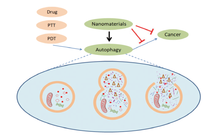
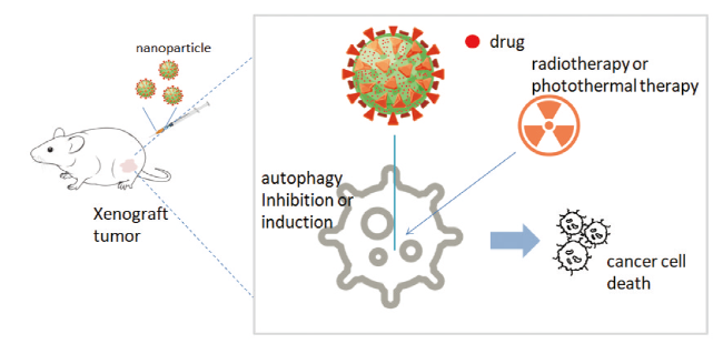
With the rapid development of nanotechnology, nanomaterials have been widely used in the field of cancer treatment. It is well known that autophagy, as a process that maintains cellular homeostasis, plays a dual role in promoting survival and death in cancer development. The level of autophagy in cancer cells is significantly higher than that in normal cells, resulting in various treatment strategies being ineffective. Synergistic treatment of cancer by perturbing autophagy has become a viable option, but traditional autophagy perturbing agents such as chloroquine may lead to certain other side effects. In addition, it has been demonstrated that nanomaterials can be used as a novel autophagy perturber, but the mechanism by which nanomaterials interfere with autophagy needs to be more deeply understood. This review provides an overview of the dual relationship between cancer and autophagy, and highlights the mechanisms by which various nanomaterials induce cancer cell death or apoptosis by perturbing autophagy, or enhance the sensitivity of cancer cells to conventional cancer therapy by perturbing autophagy, and the mechanisms by which they modulate autophagy.

Contents

1 Introduction

2 Cancer and autophagy

2.1 Autophagy inhibits the occurrence of cancer

2.2 Autophagy promotes cancer development

3 Effect of nanomaterials on autophagy

4 Nanomaterials treat cancer by perturbing autophagy

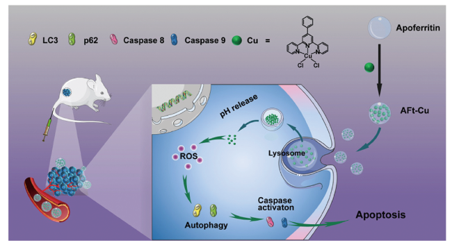
4.1 Metallic nanomaterials

4.2 Oxide nanomaterials

4.3 Carbon based nanomaterials

4.4 Other Nanomaterials

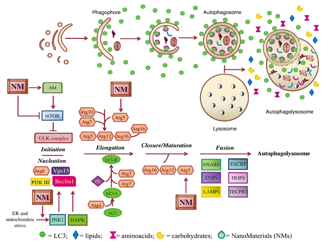
5 Mechanisms of autophagy perturbed by nanomaterials

5.1 Oxidation stress

5.2 Perturbation of autophagy-related signaling pathways

5.3 Lysosomal dysfunction

6 Conclusion

Cite this article

Jin Weitao , Yang Ting , Jia Jimei , Zhou Xiaofei . Nanomaterials-Mediated Autophagy-Based Cancer Treatment[J]. Progress in Chemistry, 2023 , 35(11) : 1655 -1673 . DOI: 10.7536/PC230330

1 Introduction

Cancer is one of the leading causes of death in most countries, particularly before the age of 70[1]. Cancer treatment, as a major public issue, has been widely concerned. Traditional cancer treatments include chemotherapy, radiotherapy, immunotherapy, targeted therapy, and hormone therapy[2][3][4][5,6][7]. However, these traditional treatments have inevitable disadvantages, such as the toxicity of chemotherapeutic drugs to normal organs, which is considered to be the main side effect of chemotherapy[8]. This defect may cause various unknown diseases. Therefore, nanomedicine has significant advantages over these traditional treatments, such as nanocarriers can prolong drug release time, targeted delivery, reduce side effects, and control drug release[9]. With the development of nanotechnology, a variety of nanoparticles are now used in clinical therapy[10].
Autophagy is an evolutionarily conserved catabolic cellular process[11]. Autophagy can be divided into three categories: macroautophagy, microautophagy, and chaperone-mediated autophagic[12][13][14]. The process of autophagy is mainly the aggregation of autophagy-related proteins and lipids to engulf damaged organelles or misfolded proteins to form autophagosomes, which are transported to lysosomes to fuse to form autophagic lysosomes, and autophagic lysosomes degrade to release nutrients[15]. Autophagy is closely related to cancer. The cytoprotective mechanisms of autophagy have been extensively studied to overcome resistance to cancer therapies, including radiotherapy, targeted therapy, immunotherapy, and chemotherapy[16]. Notably, autophagy plays a dual role in cancer development[17]. Therefore, rational regulation of autophagy can assist cancer therapy, thereby improving the therapeutic effect. The most common strategies for regulating autophagy to synergistically treat cancer are the addition of autophagy inhibitors (chloroquine, hydroxychloroquine, etc.) or autophagic inducers (rapamycin, etc.)[18,19]. On the one hand, within tumors with elevated levels of autophagy, tumor therapeutics and autophagy inhibitors are used synergistically, which inhibits tumor survival under metabolic and chemotherapeutic stress. On the other hand, excessive stimulation of autophagy by autophagy inducers in combination with cytotoxic drugs, especially in apoptosis-deficient cells, can lead to autophagic death of tumor cells[20]. However, their pharmacological activity can cause some side effects, such as chloroquine and hydroxychloroquine, which can cause retinopathy, so this method of adding autophagy disruptors has been greatly limited in clinic[21][22]. Therefore, it is urgent to find safer ways to interfere with autophagy level or autophagy flux to treat cancer.
Nanomaterials have been reported to have the ability to regulate autophagy[23]. Studies have shown that many nanomaterials can affect the autophagy of cancer cells to varying degrees, and at the same time, cooperate with chemotherapy, radiotherapy or immunotherapy to treat cancer[20,24]. Or directly induce autophagic death, apoptosis, or iron death of cancer cells[25,26]. Due to the different physical and chemical properties of nanomaterials, the mechanisms of autophagy regulation by various nanoparticles are quite different, mainly including oxidative stress, autophagy-related signaling pathways and lysosomal dysfunction[27~30]. In this review, the dual effects of autophagy and cancer are introduced, focusing on the strategies of various nanomaterials to treat cancer synergistically or directly by affecting autophagy, as well as the mechanisms by which nanomaterials regulate autophagic cells, and the development direction of the combination of nanomaterials and autophagic in cancer therapy is analyzed.

2 Cancer and autophagy

Physiologically, autophagy provides nutrients for cell growth and reduces the environmental stress of cells by removing organelles and proteins damaged during stress and growth. As the major cellular catabolic pathway, the autophagy system plays an important homeostatic function, and defects or deficiencies in this degradation system have been implicated in the cellular pathogenesis of many human diseases, ranging from neurodegenerative disorders to cancer[31]. Autophagy plays important physiological and pathological roles in all mammalian cells. Autophagy in cancer progression falls into two main categories: (1) The transition of healthy cells to metastatic and therapy-insensitive tumors may involve a temporary (but not stable) loss of autophagic capacity. The mechanism of restoration of the efficient autophagic response after malignant transformation remains to be elucidated. (2) In specific settings, carcinogenesis and cancer progression may depend on the permanent loss or gain of autophagic capacity[32]. In addition, it can also increase the resistance of tumors to external stimuli, such as starvation relief and resistance to cancer treatment. This is determined by nutrient availability, microenvironmental stress, pathogenic conditions, and the presence of the immune system[33]. The effect of autophagy on cancer may also be related to the type of autophagy, the level of autophage, and environmental factors. For example, oxidative stress, activation of DNA damage response, and genomic instability resulting from loss of autophagy, a known cause of cancer initiation and progression, have been identified. In addition, the expression of p62 caused by autophagy deficiency promotes tumor growth, but it is not clear how p62 promotes tumor growth[34]. However, intact autophagy functions as a tumor suppressor mechanism by limiting damage and mutation to the genome and inhibiting tumor initiation[33]. Marsh et al. Found that in breast cancer models, inhibition of autophagy can effectively reduce the growth of primary tumors, but promote the spread of tumors to the lungs[35]. There is a mechanism for autophagy to promote or inhibit cancer. On the one hand, autophagy promotes cancer by (1) increasing the resistance of cancer cells to endogenous conditions that often cause cell death; (2) reduce that sensitivity of cancer cell to treatment-induced cell death; (3) maintaining the state of cancer cells that have entered a dormant or senescent state as a result of treatment; (4) Ensure the maintenance of the tumor stem cell room. On the other hand, autophagy inhibits cancer by (1) maintaining genetic/genomic stability; (2) disposal of potentially mutagenic endogenous reactive oxygen species (ROS), (3) maintenance of normal bioenergetic function, (4) degradation of oncogenic protein; (5) endogenous antiviral and antibacterial effects of cells; (6) activating oncogene-induced senescence (OIS) and oncogene-induced cell death (OICD); (7) maintaining a normal stem cell compartment; (8) multi-pronged anti-inflammatory effect; (9) plays a key role in the elicitation and execution of anticancer immune surveillance[32]. With regard to the regulation of autophagy, various strategies have been developed and have shown potential in cancer prevention and treatment, and nanoparticles can enhance the sensitivity of tumors to chemotherapy, radiation, or photothermal therapy by regulating autophagy, or by inducing excessive autophagia of cancer cells, leading to autophagic death of cancer cells, etc. (Fig. 1).
图1 纳米材料通过扰动自噬治疗癌症示意图

Fig.1 Schematic diagram of nanomaterials for cancer treatment by perturbing autophagy

2.1 Autophagy inhibits carcinogenesis

Autophagy was originally thought to be a tumor suppressor mechanism. Beclin 1 is a haploinsufficient tumor suppressor gene, and the expression of Beclin 1 is down-regulated in cancer tissues of several patients with hepatocellular carcinoma, suggesting that autophagy may inhibit tumorigenesis[33]. The inhibitory function of autophagy on tumors may be attributed to its ability to remove cellular protein aggregates and damaged organelles. DNA damage, accumulation of protein aggregates and mitochondrial damage, increased peroxisome number and endoplasmic reticulum (ER) content, increased apoptosis, inflammation and fibrosis in tumor tissue can promote tumorigenesis[36]. However, human cells can alleviate these phenomena through autophagy, thus inhibiting the occurrence of tumors. Fang et al. Found that GAS8-AS1 inhibited the growth of ovarian cancer by activating Beclinl-mediated autophagy[37]. Ostendorf et al. Found that the use of rapamycin or gene induction of autophagy in breast cancer cells significantly inhibited the metastasis of cancer cells[38]. In addition to this, cancer cells with uncontrolled autophagy can also undergo cell death, also known as cell death-type II, possibly due to excessive degradation of cellular components and organelles required for cell homeostasis[39]. It has been found that mesoporous silica nanoparticles can inhibit AKT-mTOR-p70S6K signaling pathway to cause pro-death autophagy, so as to achieve the purpose of breast cancer treatment[40]. In addition to direct cell death-type II through excessive autophagy, other cell death, such as apoptosis and cell iron death, can also be triggered by autophagy. Wen et al. Synthesized water-soluble ultrasmall iron oxide nanoparticles[30]. The iron oxide nanoparticles can activate autophagy by inducing Beclin 1/ATG 5 overexpression in glioblastoma cells, and then promote the increase of iron death markers in cells, including iron ion concentration, intracellular reactive oxygen species and lipid reactive oxygen species, and finally lead to iron death in glioblastoma. In summary, intact autophagy inhibits tumorigenesis by limiting genomic damage and mutation, and thus plays a cancer-suppressive role[33].

2.2 Autophagy promotes cancer development

In some tumors, cancer cells enhance their adaptability to the harsh living environment of hypoxia or nutrient deficiency through autophagy[41]. Autophagy can protect cancer cells from apoptosis and promote their multidrug resistance to chemotherapy. Recent studies have shown that chemotherapeutic agent-triggered autophagy may promote cancer cell resistance to certain chemotherapeutic agents[42]. Autophagy inhibition may enhance the sensitivity of cancer cells to chemotherapeutic agents[43]. It was found that doxorubicin could inhibit the growth of U2OS and Saos-2 osteosarcoma cells in a dose-dependent manner, and Western blotting experiments showed that the expression of LC3-Ⅱ was increased and the expression of p62 protein was decreased in osteosarcoma cells treated with doxorubicin, indicating that doxoruicin-induced autophagy. After using autophagy inhibitor 3-MA to inhibit autophagy, it was found that the inhibitory effect of doxorubicin on cancer cells was significantly improved. The results demonstrate that osteosarcoma cells resist doxorubicin-induced apoptosis through autophagy, and inhibition of autophagy can significantly increase the therapeutic level[44]. Cancer treatment is still facing great challenges due to tumor recurrence and metastasis caused by acquired resistance of cancer cells to treatment, so a large number of studies have focused on increasing the toxicity of drugs to tumors by inhibiting protective autophagy. It has been reported that the tumor inhibition effect of combined treatment with hydroxychloroquine-loaded mesoporous silica nanosystem is about 70% higher than that of radiotherapy alone[45]. In addition, recent studies have shown that induction of autophagy can also increase the sensitivity of cancer cells to chemotherapeutic drugs[46]. In conclusion, enhancing cancer therapeutic efficacy by regulating autophagy has become an effective cancer treatment strategy.

3 Effect of nanomaterials on autophagy

Autophagy plays a key role in protecting cells from foreign substances and maintaining cell stability. It has been reported that various nanoparticles can affect autophagy of cells. Nanomaterial-mediated autophagy has dual effects on humans[47]. On the one hand, nanomaterials have been used in all aspects of people's lives, and the perturbation of autophagy caused by the exposure of nanomaterials may also be a potential mechanism for nanotoxicity, resulting in adverse effects[48]. On the other hand, proper regulation of autophagy by nanoparticles under certain conditions can promote the treatment of some autophagy-related diseases, such as cancer, neurological diseases and inflammation[49~51]. A wide variety of nanomaterials have been used for biomedical applications such as molecular imaging and intracellular drug delivery. Nanoparticles have unique properties such as higher tissue permeability, large surface area/volume ratio, targeted drug delivery, and relatively low cost[52]. When traditional chemotherapy and radiotherapy are considered to be ineffective, nanomedicine has become one of the new treatment strategies.
Nanoparticles have been recognized as a novel regulator of autophagy[53]. Compared with traditional small molecule autophagy modulators, the significant feature of nanomaterials in regulating autophagy is the aggregation of nanomaterials in autophagosomes and autophagic lysosomes[54]. Because most of these nanomaterials are inorganic and not easily digested by lysosomes, they can affect autophagic flux to a certain extent, which can not be accomplished by small molecules[55]. In addition, the side effects of small molecule autophagy modulators greatly limit their use in the clinic[56]. Therefore, nanomaterials can be used as an alternative therapeutic option to regulate autophagy. The differences between nanoparticles and small molecule autophagy modulators can be divided into three categories. First of all, the difference of inducing autophagy is that small molecules are mostly autophagy regulatory proteins specifically targeted to cells[57]. Nanoparticles can not only interact with cell surface and intracellular proteins, but also damage organelles, such as mitochondria and endoplasmic reticulum. Second, the mechanism in blocking autophagic flux is different: the small molecule acts as a base to affect lysosomal pH, thereby blocking autophagic flux[58]. Nanoparticles mostly accumulate in lysosomes and are not easily digested, causing lysosomal damage (including lysosomal alkalinization and lysosomal membrane permeabilization). Finally, for inhibiting autophagy: small molecules can target autophagy-related proteins to inhibit autophagy. Nanoparticles have rarely been reported to inhibit the upstream phase of autophagy. A variety of nanoparticles have been found to regulate autophagy, as shown in Table 1, including silica nanoparticles, iron oxide nanoparticles, gold nanoparticles, silver nanoparticles, zinc oxide nanoparticles, carbon-based nanoparticles, and the like. It is important to note that this list presents only a small subset of the literature reports. Nevertheless, the following conclusions can be drawn: on the one hand, nanoparticles can induce pro-survival autophagy in cancer cells, such as copper-palladium alloy nanoparticles, which can induce pro-survival autophagy without destroying lysosomal function by enhancing the ability of mitochondria to produce ROS[59][59]; On the other hand, certain nanoparticles can induce pro-death autophagy in cancer cells[60].
表1 纳米材料通过调控自噬治疗癌症

Table 1 Nanomaterials treat cancer by regulating autophagy

NMs Size (nm) shape or
dispersity
Coating Drug or therapies Model cells Mechanism Autophagy Effect ref
Au NPs 20 sphere PEG immunotherapy Hepa1-6 cells;
RAW 264.7 cells
Lysosome alkalization; membrane permeabilization Inhibition Increased sensitivity 79
Au NPs 30~60 Peanut Bare SKOV-3 cell ROS upregulation Inhibition Apoptosis 78
Ag NPs 66.92 sphere Bare PC-3 cell lysosome injury; cell hypoxia Inhibition Cell death 93
Fe3O4-Au NPs 15~25 sphere DOX HepG2 cells Enhancing autophagosome formation Induction Reduce drug resistance 80
Ag NPs 59 sphere Bare HT-29 cells JNK activation and
eIF2α phosphorylation
Induction Apoptosis 97
Fe@Au
NPs
core-shell structure OECM1 cell Mitochondria damage Inhibition Cell death 73
Ag NPs 26.5 sphere PVP HeLa cell PtdIns3K-dependent Induction Ehances the anticancer activity 90
Fe3O4 NPs 36 sphere PEG PTX U251 cell ROS upregulation Induction Reduce drug resistance 98
Ag NPs 13 sphere A549 cell ROS upregulation Induction Apoptosis 83
Ag NPs 8 sphere protein Cisplatin OS cell; HCC cell MAPK pathways Induction Reduce drug resistance 89
Fe2O3 NPs 10 sphere DMSA SK-Hep-1 cell ROS upregulation;
MAPK pathways
Induction Cell death 99
CuO NPs 10 sphere MCF7 cell ROS dependent Induction Growth inhibition 100
ZnO NPs 21 Sorafenib Huh 7 cell Promoting p53 Gene Induction Apoptosis 101
ZnO NPs 63 MCF7 cell Inhibition Apoptosis 102
SiO2 NPs 86 sphere HCT-116 cells ER stress Induction Cell survival 103
Ag NPs 15.38 sphere PVP Radiotherapy U251 cells ROS upregulation Induction Increased sensitivity 88
IONPs 37 PEG U251 cells Beclin 1/ATG 5 pathways Induction Ferroptosis 30
Gd2O3 NPs Cisplatin HeLa cells Inhibition Reduce drug resistance 104
SiO2 NPs 125 sphere Propranolol HemSCs cells ER stress Induction Cell death 105
ZnO NPs 20 SKOV3 cells ROS upregulation Induction Apoptosis 106
SiO2 NPs 198 sphere PDA; PEG DOX MCF7 cells AKT-mTOR-p70S6K pathway Induction Cell death 40
ND 191 Hypoxia HeLa cells; MCF7 cells Inhibition Apoptosis 107
ZnO NPs Cisplatin SGC7901 cells;BGC823 cells Inhibition Reduce drug resistance 108
GO 450 sheet DMSO Cisplatin Skov-3 cells Induction Cell death 109
DWCNTs Tube DHD/K12/Trb cell line Intracellular acidification Induction Cell death 110
Se NPs 70 Amorphous solid Astragalus Polysaccharides MCF7 cells ROS upregulation and Mitochondria damage Inhibition Apoptosis 111
BPQDs 140 Monodisperse Platelet membrane Hederagenin MCF7 cells; RAW 264.7 cells ROS upregulation and Mitochondria damage Induction Apoptosis 112
Co3O4 NPs 200 sphere Photothermal therapy U-87 MG cells Llysosomal function damage Blockage of autophagic flux Cell death 113
Considering the complex physicochemical properties of nanomaterials, there are no established rules to predict whether nanomaterials induce pro-survival or pro-death autophagy. In addition, part of the nanoparticles may inhibit the autophagic activity of cells or block autophagic flux[61]. The physical and chemical properties of nanoparticles, such as size, chemical composition, surface chemical modification and shape, can affect their autophagy regulation ability. For example, different carbon nanotubes have different autophagy regulation effects[62,63]. Autophagy can be induced by tetrapod copper-palladium alloy nanoparticles, but not by spherical copper-palladium alloy nanoparticles of the same composition[59]. Different nanoparticles have different mechanisms of autophagy regulation due to their different physical and chemical properties, for example, nanoparticles can induce autophagy through PI13K-Akt-mTOR signaling pathway, or affect autophagic cells by stimulating oxidative stress[64][65].
In conclusion, it has been confirmed that many nanoparticles can affect the level of autophagy of cells, and at the same time, due to the different physical and chemical properties of nanoparticles, the regulation of autophagy of cells is also different. Rational use of the effect of nanoparticles on autophagy can maximize the therapeutic effect of cancer, such as the use of nanoparticle-induced pro-death autophagy, or the use of nanoparticles to regulate autophage to improve the sensitivity of cancer cells to chemotherapeutic drugs. In addition, Huang et al. Showed a reliable universal mechanism for nanomaterials to regulate autophagy, that is, nanoparticles induce autophagy in a dispersion-dependent manner, demonstrating that regulating the dispersion of nanoparticles may be a new way to regulate autophagia[66]. But the understanding of the mechanism by which nanoparticles regulate autophagy is still very lacking.

4 Nanomaterials treat cancer by perturbing autophagy

4.1 Metal nanomaterials

With the continuous exploration of inorganic nanomaterials, gold nanoparticles have become an important concern in the field of nanomedicine, and more and more studies have revealed its potential prospects. The unique physicochemical properties, biocompatibility, and highly developed chemistry of gold nanoparticles (AuNPs) have facilitated breakthroughs in the field of cancer research, mainly focused on applications in cancer diagnosis and therapy[67]. It is well known that cellular uptake is closely related to the shape of nanoparticles. However, the shapes of gold nanoparticles are very rich, and they have been found to be: gold nanotubes, gold nanorods, gold nanoclusters, gold nanostars, gold nanocages, gold nanoshells and gold nanospheres[68][69][70][71][72][73][74]. These rich shape structures offer promising prospects for nanomedicines. Mitochondrial dysfunction and elevated reactive oxygen species have been proposed as potential mechanisms of autophagy induced by gold nanoparticles[75]. The thioredoxin (Trx) system has been reported to play an important role in maintaining cellular balance and regulating cell death. Inhibition of the Trx system causes an increase in reactive oxygen species in the cell, which disrupts cellular homeostasis[76]. Gold nanoparticles can act as a thioredoxin reductase inhibitor to destroy the Trx system and increase the level of reactive oxygen species, eventually leading to autophagy and apoptosis[77]. In addition, gold nanoparticles can also induce autophagy through other pathways. Piktel et al. Reported that the autophagy level of ovarian cancer cells treated with a peanut-shaped gold nanoparticle was significantly increased. The mechanism study found that gold nanoparticles could interfere with the level of reactive oxygen species by disrupting the balance between glutathione (GSH) and glutathione oxidized form (GSSG)[78]. At the same time, gold nano-peanut induced the expression of NADPH subunit 4 (NOX4) in ovarian cancer cells, which promoted oxidative stress. In addition, the gold nano-peanut also destroyed the mitochondrial function of the cell. Excessive oxidative stress activates the JNK signaling pathway in cells, which induces apoptosis and autophagy in ovarian cancer cells. Gold nanoparticles can not only directly promote cell death by perturbing autophagy, but also play an anti-drug resistance role by interfering with autophagy. tumor-associated macrophages (TAMs) are one of the major components of the tumor microenvironment and an important factor in tumor progression and drug resistance. Zhang et al. Reported that gold nanoparticles could induce lysosomal alkalinization and membrane permeabilization in TAM, thereby blocking autophagic flux for the purpose of inhibiting tumor-associated macrophage M2 polarization[79]. Wang et al. Prepared magnetic gold fluorescent polymer nanoparticles based on Fe3O4-Au nanoparticles[80]. It promotes autophagy by inducing the formation of autophagosomes, significantly reducing doxorubicin resistance in cancer cells. Gold nanoparticles have been extensively studied in the past 20 years due to their unique advantages in the biomedical field, especially in the fields of therapy and imaging. However, there are still many challenges for gold nanoparticles, mainly including the ambiguity of cytotoxicity and biological processes, as well as the difficulty of commercialization of gold nanoparticles[81].
Silver nanoparticles have received much attention in mechanosensitive chemotherapy, immunotherapy, drug delivery mechanisms, and nanomedicine[82]. In recent years, cancer therapy based on silver nanoparticles has been widely studied. Que et al. Found that 13 nm silver nanoparticles can effectively induce apoptosis of lung cancer cells[83]. Liu et al. Developed a nanocomposite based on silver nanoparticles that can effectively deliver drugs and tumor targeting[84]. Although the exact mechanism of silver nanoparticles against cancer cells has not been fully elucidated, it is well known that their toxicity is dependent on the generation of reactive oxygen species (ROS)[85]. Table 1 shows many examples of silver nanoparticles promoting apoptosis by inducing the production of reactive oxygen species, which in turn can induce autophagy by promoting the production of reactive oxygen species[86]. This is because the silver nanoparticles ingested by cells are mainly accumulated in lysosomes, which can release a large number of silver ions and promote the production of reactive oxygen species after decomposition, and then the reactive oxygen species inhibit the AKT-mTOR signaling pathway and activate autophagy[87]. Mitochondria are the main site of reactive oxygen species (ROS) production, so high production of ROS may cause mitochondrial damage in cells[88]. This may be a pathway by which silver nanoparticles induce apoptosis by promoting reactive oxygen species production. Fageria et al. Biosynthesized a protein-coated silver nanoparticle, which was found to exert cytotoxic effects by increasing the level of reactive oxygen species and inducing apoptosis[89]. Interestingly, inhibition of autophagy can increase the production of reactive oxygen species and promote cell death. At the same time, silver nanoparticles also sensitized cancer cells resistant to cisplatin. Lin et al. Reproved this finding, inhibiting autophagy by chemical inhibitors or by knocking out the autophagy-essential gene ATG5 significantly enhanced the cytotoxic effect of silver nanoparticles in cancer cells[90]. Yuan et al. Used the combination of silver nanoparticles and cisplatin to treat human cervical cancer cells, and the results showed that silver nanoparticles and cisplatin had a synergistic effect, and the combination of silver nanoparticles and Cisplatin significantly improved the apoptosis and autophagy of HeLa cells[91]. In addition to chemotherapy, reactive oxygen species production induced by silver nanoparticles increases the sensitivity of cancer cells to radiation[88]. It has been reported that silver nanoparticles induce autophagy while blocking autophagic flux through lysosomal damage, such as alkalinization or lysosomal membrane permeabilization[92]. Chen et al. Demonstrated that silver nanoparticles induced autophagy by activating AMPK/mTOR signaling pathway through cell hypoxia and energy deficiency, and at the same time, silver nanoparticles caused lysosomal damage, including the decrease of lysosomal number and lysosomal protease activity, thus blocking autophagy flux[93].
Compared with gold and silver nanoparticles, copper nanoparticles have attracted much attention due to their availability, low cost, and high similarity to noble metals. Copper or copper-based nanomaterials have been successfully used in the field of cancer therapy because of their good photothermal conversion properties[94]. It has been proved that the addition of copper to palladium nanoparticles can increase autophagy induction. It was found that copper palladium nanoparticles could promote the production of reactive oxygen species in cells, especially in mitochondria. It was simultaneously demonstrated that copper-palladium nanoparticle-induced autophagy was pro-survival, as inhibition of autophagy enhanced TNP-1-induced cancer cell killing[59]. Liu et al. Found that cysteamine copper nanoparticles can be used as a new radiosensitizer, and found that cysteamine copper nanoparticles can induce autophagy and apoptosis of colorectal cancer cells.It is speculated that autophagy and apoptosis may be related to the significantly increased production of reactive oxygen species by cysteamine-copper nanoparticles, but the relationship between reactive oxygen species and autophagy and apoptosis is not clear[95]. Xiong et al. Prepared apoferritin-coated copper (II) complex nanoparticles (AFt-Cu)[96]. Nanoparticles were found to increase ROS production in cancer cells in a concentration-dependent manner, and ROS production was found to be due to nanoparticle activation of the Caspase8/9 pathway. Subsequently, cancer cells were co-treated with nanoparticles using apoptosis inhibitor (Z-VAD), Caspase8 inhibitor, and autophagy inhibitor, and the results demonstrated that both autophagy and apoptosis significantly promoted the death of cancer cells (Fig. 2). There are relatively few studies on the effects of copper or copper-based nanoparticles on autophagy in cancer, and a more detailed understanding of the mechanisms by which copper or copper-based nanoparticles affect autophagy is needed.
图2 脱铁蛋白包裹铜多吡啶复合物的纳米颗粒诱导多重耐药的结直肠癌细胞自噬依赖性凋亡示意图[97]

Fig.2 Schematic diagram of autophagy-dependent apoptosis of multi-drug resistant colorectal cancer cells induced by nanoparticles of apoferritin -coated copper polypyridine complex[97]. Copyright © 2022, American Chemical Society

To sum up, silver nanoparticles can disturb autophagy in tumor cells, and then cause apoptosis of cancer cells. The use of silver nanoparticles for cancer therapy holds broad promise. However, due to the limitation of space, there are many literatures that use the autophagy effect of nanomaterials to promote tumor therapy, which are not included in this article, and we apologize to the authors.

4.2 Oxide nanomaterials

Mesoporous silica nanoparticles (MSNs), as a controlled release platform for small molecule drugs, have become one of the research hotspots in the field of nano-drug delivery since the 1990s. As drug carriers, they have outstanding characteristics such as good biocompatibility, good chemical stability, high porosity, various surface chemical modifications, and low cost[114]. Recently, the US Food and Drug Administration (FDA) approved only small-sized (< 10 nm) silica nanoparticles for use as imaging AIDS as investigational new probes in a clinical trial[115]. This provides a basis for the future development of silica nanoparticles as drug carriers. Silica nanomaterials can induce autophagy perturbation in a variety of cells, including inducing autophagy through cytoskeleton disruption, oxidative stress, Endoplasmic reticulum (ER) stress, and mitochondrial damage pathways, or blocking autophagic flux by damaging lysosomes[116]. It has been shown that reactive oxygen species (ROS) is a major inducer of autophagy, and oxidative stress caused by excessive production of ROS can activate autophagy[117]. Wu et al. Found that mesoporous silica nanoparticles promoted the increase of autophagy level in cells by inducing the generation of reactive oxygen species in hemangioma stem cells[118]. Because autophagy plays an important role in degrading misfolded proteins and relieving cellular ER stress, and the production of ROS can lead to ER stress, propranolol (PRN), which can block the degradation of autophagosomes, was added.Finally, the accumulation of autophagosomes mediated by PRN-loaded mesoporous silica nanoparticles further induces ER stress, and then promotes apoptosis by down-regulating Bcl-2 expression to achieve the purpose of treating hemangioma. In addition, mesoporous silica nanoparticles can also interfere with the level of autophagy by perturbing autophagy-related signaling pathways. Duo et al. Reported a mesoporous silica nanoparticle coated with polydopamine (PDA) and polyethylene glycol (PEG). The presence of PDA enables the drug delivery system to sensitively control drug release, while the combination of PEG enhances the stability and biocompatibility in vivo, making the nanomedicine system safer and more efficient[40]. Studies have shown that this mesoporous silica nanosystem can inhibit AKT-mTOR-p70S6K signaling pathway and cause stronger pro-death autophagy. Silica can not only induce pro-death autophagy in cancer cells to promote cancer therapy, but also cause protective autophagy to prevent anti-cancer drugs from acting. It has been reported that silica nanoparticles significantly increased the expression of LC3-Ⅰ and LC3-Ⅱ in pancreatic cancer cells and promoted the cytoprotective autophagy ability of pancreatic cancer cells[119]. The level of autophagy induced by silica nanoparticles in cells may be directly related to the time of silica treatment[103]. The results suggest that the perturbation of autophagy caused by mesoporous silica nanoparticles can be used for cancer therapy.
The use of iron oxide nanoparticles for targeted delivery of chemotherapeutic agents, magnetic resonance imaging (MRI), and transfection has become extremely important in recent in vitro and in vivo studies. At the same time, iron oxide nanoparticles have the advantages of hereditary magnetism, non-toxicity, sensitivity to external stimuli and biodegradability[120]. These advantages demonstrate the broad development prospects of iron oxide in cancer therapy. It has been found that iron oxide nanoparticles can react with reactive oxygen species in vivo[121]. Because autophagy is highly correlated with the production of reactive oxygen species, iron oxide nanoparticles may affect autophagy by perturbing cellular reactive oxygen species levels. Xie et al. Found that alkylated iron oxide nanoparticles can induce a large amount of iron accumulation in hepatoma cells, and then produce excessive reactive oxygen species in hepatoma cells, thus activating autophagy and causing serious damage to autophagy flux[99]. To understand the mechanism of cell death caused by nanoparticles, the expression level of p-p38 in cells was examined. The results demonstrate that autophagic cell death induced by alkylated iron oxide nanoparticles through promoting overproduction of reactive oxygen species is dependent on the function of the P38 mitogen-activated protein kinase (MAPK) signaling pathway. In recent years, iron death, a cell death mode closely related to iron oxide, has appeared in the public eye. Iron death is an iron-dependent programmed cell death characterized by high intracellular iron concentration and reactive oxygen species[30]. It is known that reactive oxygen species are also closely related to autophagy, so the synergistic treatment of autophagy and iron death has become an emerging therapeutic strategy for cancer. Based on this new idea, Wen et al. Found that ultra-small iron oxide nanoparticles induced iron death of glioblastoma cells through Beclin-1/ATG 5-dependent autophagy pathway[30]. Iron oxide nanoparticles can induce the expression of Beclin-1 protein in cancer cells, and then increase the level of autophagy[61]. The increase of autophagy can promote the release of iron ions from iron oxide nanoparticles in lysosomes, and autophagy also participates in lipid peroxidation, resulting in excessive production of intracellular reactive oxygen species and cell iron death[98]. Huang et al. Analyzed the mRNA expression levels of autophagy-related factors (ATG3, ATG5, MAP1ALC3a, MAP1ALC3b, MAP2ALC3c), cell death-associated proteins (BAK1, BID) and some iron death regulators in human ovarian cancer hepatocytes (HuOCSCs) treated with superparamagnetic iron oxide nanoparticles by qPCR.The results showed that superparamagnetic iron oxide nanoparticles could induce oxidative stress, reduce the autophagic activity of ovarian cancer stem cells, activate iron death, and inhibit the proliferation, invasion, drug resistance and tumorigenicity of HuOCSCs[26]. In general, iron oxide nanoparticle-based iron death therapy is expected to be the most promising approach for future cancer treatment.
The FDA has approved zinc oxide as a safe metal oxide[122]. Zinc oxide (ZnO) nanoparticles are widely used in cancer therapy and diagnosis due to their excellent physicochemical properties. It has been reported that zinc oxide nanoparticles can specifically penetrate and aggregate in tumor cells[123]. The generation of toxic reactive oxygen species in the presence of zinc oxide nanoparticles, combined with the enhanced penetration and retention (EPR) effect noted in cancerous tissues, demonstrates their high potential as anticancer agents[124]. Abdel et al. Found that zinc oxide nanoparticles can promote the excessive production of reactive oxygen species in cells, and further cause lipid peroxidation, thereby inducing apoptosis[125]. However, the specific mechanism of apoptosis induced by zinc oxide nanoparticles is not yet clear. Because the main production site of reactive oxygen species is mitochondria, it is speculated that the mechanism of apoptosis induced by zinc oxide nanoparticles may be related to mitochondria[126]. Du et al. Found that the increase of reactive oxygen species may be induced by zinc ions released from zinc oxide nanoparticles, which may cause DNA damage and further up-regulate the expression of p53 protein[127]. At the same time, it was observed that the expression of pro-apoptotic BAX was significantly up-regulated in cancer cells treated with zinc oxide nanoparticles, while the expression of anti-apoptotic Bcl-2 was significantly decreased. Because mitochondria are the main site of reactive oxygen species (ROS) production, ROS production induced by zinc oxide nanoparticles can easily cause mitochondrial damage. Proapoptotic Bcl-2 family proteins can induce permeabilization of the outer mitochondrial membrane and promote mitochondrial membrane potential loss[128]. Mitochondrial damage can further promote apoptosis. It is known that autophagy is an important method to maintain cell homeostasis, so the increase of reactive oxygen species, mitochondrial damage and the expression of apoptosis-related proteins caused by zinc oxide nanoparticles may lead to changes in autophagy[129]. Many studies have shown that zinc oxide nanoparticles induce apoptosis accompanied by the disturbance of autophagy. Bai et al. Showed that zinc oxide nanoparticles could induce significant cytotoxicity, apoptosis and autophagy in human ovarian cells through the production of reactive oxygen species and oxidative stress[106]. Multiple stress-induced signal transduction pathways can induce autophagy and apoptosis in the same cell, such as p53, BH3-only proteins, kinases (AKT, DAPK, and JNK)[130]. Yang et al. Found that zinc oxide nanoparticles could induce the increase of autophagy level in hepatoma cells, and promote the expression of apoptotic protein Caspase-3 and apoptotic gene p53[101]. Zinc oxide nanoparticles can also block the autophagy pathway in some cancer cells, thereby inhibiting the level of autophagy. The regulation of autophagy is of great significance to improve the sensitivity of tumors to chemotherapeutic drugs[131]. For cisplatin-resistant gastric cancer cells, Miao et al. Found that zinc oxide nanoparticles can not only inhibit the proliferation, migration and invasion of gastric cancer cells, but also inhibit autophagy caused by cisplatin, thereby reducing the resistance of gastric cancer cells to cisplatin[108].
Rare earth materials include 17 different elements with similar chemical properties. It has been reported that lanthanide oxide nanoparticles and upconversion nanomaterials can induce autophagy, and their induction activity is similar to that of trehalose, a known autophagy inducer. At the same time, it was found that the nanocrystals formed by the combination of lanthanide oxide nanoparticles and upconversion nanomaterials could be used for cancer therapy after being modified by short peptides with arginine-glycine-aspartic acid motifs[132]. Cerium is one of the lanthanide rare earth elements, and cerium oxide nanoparticles have been widely used in antioxidant therapy, eye protection, neuroprotection, and cancer therapy[116]. Low doses of cerium oxide nanoparticles have been reported to cause mitochondrial damage, overexpression of apoptosis-inducing factor, and induction of autophagy[133]. This provides a research basis for rare earth oxide nanoparticles to treat cancer by perturbing autophagy. Gadolinium, another lanthanide rare earth element, has been the most studied in this field. It has been reported that gadolinium oxide nanoparticles are a new type of autophagy inducer, and its autophagy regulation mechanism may be related to intracellular oxidative stress and mitochondrial dysfunction[134]. Gadolinium oxide nanoparticles can not only directly induce death-promoting autophagy in breast cancer cells, but also increase the sensitivity of lung cancer cells to radiation through autophagy-mediated death[135,136]. Gadolinium oxide nanoparticles can also reduce the level of autophagy, and Zhang et al. Found that gadolinium oxide nanoparticles can block the late stage of autophagic flux and increase autophagosome accumulation in cells in a dose-dependent manner by observing the number of autophagosomes in cancer cells after gadolinium oxide nanoparticle treatment[104]. At the same time, studies have demonstrated that gadolinium oxide nanoparticles can reduce the sensitivity of cancer cells to chemotherapeutic drugs, which is achieved by autophagy inhibition. Previous studies have found that neodymium oxide nanoparticles induced autophagy in non-small cell lung cancer NCI-H460 cells, accompanied by cell cycle arrest in S phase, mild disruption of mitochondrial membrane potential and inhibition of proteasome activity[137]. In addition, certain rare earth oxide nanoparticles can cause autophagic flux defects in cells through lysosomal dysfunction, such as La2O3, Gd2O3, Sm2O3, and Yb2 O 3 [ 116 ]. As the research on the regulation of autophagy by rare earth nanomaterials is relatively mature, it is hopeful to develop a more ideal strategy for the treatment of cancer by regulating autophagy by rare earth nanomaterials.
There are many other oxide nanoparticles used in the diagnosis and treatment of cancer, such as alumina (Al2O3) nanoparticles, titanium dioxide (TiO2) nanoparticles, manganese oxide (MnO) nanocrystals, zirconia (ZrO2) nanoparticles and copper oxide (CuO) nanoparticles have been successfully used in the diagnosis and treatment of cancer[138]. The molecular mechanism of cytotoxicity caused by alumina nanoparticles may be mediated by autophagy[139]. Autophagy induced by alumina nanoparticles also has potential applications in the biomedical field. In tumors, α-Al2O3 nanoparticles can be phagocytosed by dendritic cells and delivered to CD8+T cells via the autophagosome-associated cross-presentation pathway, thereby significantly enhancing antitumor immune response and antitumor efficacy[140]. Titanium dioxide is investigated as an alternative for the treatment of cancer diseases under different activation therapies. The combination of titanium dioxide nanoparticles with various therapies for cancer treatment has significantly enhanced the ability to kill cancer cells by inducing the production of reactive oxygen species[141]. In addition, titanium dioxide nanoparticles can also affect autophagy by interfering with the expression of endoplasmic reticulum and p53, thus exerting cytotoxic effects[142]. Azimee et al. Found that titanium dioxide nanoparticles can promote the production of reactive oxygen species and impair lysosomal function, and then block the autophagic flux of gastric cancer cells[143]. Therefore, low concentrations of titanium dioxide nanoparticles can also enhance the toxicity of 5-fluorouracil to gastric cancer cells by blocking autophagy. Manganese oxide nanocrystals can promote the production of autophagosomes in cancer cells, thereby reducing the resistance of cancer cells to doxorubicin[144]. Zirconia nanoparticles can induce HeLa cell death through ROS mediated mitochondrial apoptosis and autophagy[145]. Copper oxide nanoparticles have been shown to be a potential radiosensitizer by inducing excessive autophagy[146]. However, the mechanism by which these nanoparticles promote cancer therapy based on perturbed autophagy is less studied. There is an urgent need to seek more ideal nanoparticles and to understand in more detail the mechanisms by which they perturb autophagy.

4.3 Carbon-based nanomaterials

Carbon-based nanomaterials mainly refer to fullerene and its derivatives (fullerenols), carbon nanotubes (CNTs), carbon dots (CDs), graphene oxide (GO), and nanodiamonds (NDs)[147]. These nanomaterials have gained much attention in the fields of biomedicine, nanomedicine, energy and environmental applications[148]. Due to the inherent hydrophobic nature of carbon nanomaterials, they can be loaded with drugs through hydrophobic interactions or π-π stacking and can be used as an effective drug delivery platform[149]. Carbon-based nanomaterials can increase the sensitivity of tumors to chemotherapeutic drugs by enhancing cancer cell apoptosis and inhibiting cancer cell proliferation[150]. In addition, carbon-based nanomaterials can also cause autophagy perturbation in a variety of cells and improve the efficacy of cancer therapy.
Fullerenes can produce reactive oxygen species (ROS) through the triplet excited state when irradiated by visible or ultraviolet light, which makes them superior in photodynamic therapy (PDT)[147]. In addition, fullerenes have the ability to accumulate in tumors, based on enhanced permeability and retention effects, and are highly permeable to enter tumor cells through blood vessels[151]. Fullerene C60 crystals promote autophagy in osteosarcoma cells by inducing the production of reactive oxygen species and induce cell death. Meanwhile, blocking autophagic flux by autophagy inhibitors or other pathways can significantly enhance the cytotoxicity of C60 crystals[152]. Fullerene C60 nanoparticles have been shown to induce autophagy and sensitize cancer cells for chemotherapeutic killing. Wei et al. Studied the mechanism by which C60 nanoparticles induced autophagy to enhance the sensitivity of drug-resistant cells to chemotherapeutic drugs, and found that C60 could induce autophagy by promoting the production of reactive oxygen species, thereby reducing the drug resistance of cells[153]. Interestingly, autophagy induced by autophagy inducers does not increase the toxicity of chemotherapeutic drugs, so it is speculated that C60 can induce autophagia without being degraded in lysosomes and thus block autophagic flux. Yang et al. Found that fullerenol C60 nanoparticle treatment of cells led to the accumulation of autophagosomes in cells, and the study found that fullerenol disrupted autophagic flux by disrupting lysosomal functions, including lysosomal membrane permeabilization, lysosomal alkalinization, and decreased Capthesin B activity[154].
Carbon nanotubes have excellent optical properties, thermal conductivity, electrical conductivity, easy functionalization and high drug loading, so they can be used in cancer therapy and diagnosis in a multi-functional way[155,156]. Phan et al. Prepared polyampholyte-grafted single-walled carbon nanotubes loaded with doxorubicin for tumor therapy, and the results showed that carbon nanotubes significantly improved the therapeutic effect of doxorubicin[157]. It has been confirmed that carbon nanotubes can induce excessive autophagy in cancer cells and cause irreversible cell damage, eventually leading to cell death[158]. Carbon nanotubes can not only promote the expression of autophagy genes through endoplasmic reticulum stress, but also inhibit autophagy by inducing the reduction of oxidative stress between cells[159][160]. Fiorito et al. Confirmed that multi-walled carbon nanotubes induced autophagy in tumor cells and promoted the production of reactive oxygen species to block G2 phase cells to inhibit cancer cell proliferation[110].
Because of its unique structure, carbon dots show many advantages, such as surface modification, long-term penetration and metabolism in vivo, unique blood-brain barrier permeability, specific cell targeting and so on[161]. This makes it promising in the field of cancer therapy. At the same time, carbon dots are photosensitizers, which can be used in photodynamic therapy. Murali et al. Prepared carbon quantum dots encapsulated with hematoporphyrin (HP) photosensitizer. Under deep red light irradiation, carbon quantum dots can promote the production of reactive oxygen species in cells, thus promoting the death of human breast cancer cells[162]. In hepatocytes, carbon dots promote apoptosis by inducing overproduction of reactive oxygen species, and cells reduce the cytotoxicity of carbon dots through protective autophagy[163]. Carbon dots can induce not only protective autophagy but also pro-death autophagy. Tian et al. Used pentacyclic triterpenoids (PT) as raw materials to prepare carbon dots, which can specifically target the mitochondria of tumor cells and cause mitochondrial damage[164]. Cell death is then induced by three pathways. Significant changes in the content of oxidative stress markers malondialdehyde (MDA), glutathione, and reactive oxygen species were observed in the carbon dot-treated tumor cells, which induced iron death of the cells. Reactive oxygen species induced Caspase-3-dependent apoptosis by activating the JNK signaling pathway. In addition, carbon dots also induce autophagic cell death of tumor cells by causing mitochondrial damage. This therapeutic strategy based on precise targeting of carbon dots to mitochondria and promoting cell death through multiple pathways provides a broader idea for future cancer treatment.
Among many nanomaterials, two-dimensional graphene oxide is widely used in biomedical fields, including drug delivery, photothermal and photodynamic therapy, due to its ultra-high specific surface area and dispersion[165][166][167]. One of the most promising is that graphene oxide can regulate the tumor microenvironment and the sensitivity of tumors to therapeutic drugs[168]. The sensitivity of tumors to therapeutic drugs is closely related to autophagy, so graphene oxide may regulate the sensitivity of cancer cells to drugs by interfering with autophagy[169]. Lin et al. Found that graphene oxide can increase the sensitivity of colorectal cancer cells and ovarian cancer cells to cisplatin by inducing autophagy[109]. The mechanism of autophagy induction is to promote the initiation and progression of autophagy, which is not related to the late stage of autophagic (such as autophagosome formation and autolysosome formation). However, GO had no significant effect on the autophagy level of lung cancer cell A549. In addition to the indirect treatment of cancer by sensitization, graphene oxide can also directly induce cell death through autophagy. Kr Krłtowski et al. Found that reduced graphene oxide can induce apoptosis and autophagy in tumor cells, which may be caused by decreased mitochondrial membrane potential, dysregulated mitochondrial protein expression, and activation of Caspase-9 and Caspase-3[170]. Xiao et al. Reported that graphene oxide could activate autophagy by inducing ER stress in nasopharyngeal carcinoma cells, thereby inhibiting the proliferation of cancer cells[171]. In conclusion, the strategy of graphene oxide to treat cancer based on the mechanism of autophagy perturbation has greatly promoted the treatment of tumors.
In recent years, nanodiamond has become a widely used nanocarrier because of its large specific surface area, strong affinity with biomolecules, low toxicity and good biocompatibility[172]. It has been reported that nanodiamond is a safe and effective autophagy inhibitor[173]. Because autophagy is the main way for hypoxic cells to obtain energy, nanodiamond inhibition of autophagy can be combined with apoptosis of cancer cells in hypoxic environment, and has no obvious toxicity to normal cells[107]. Nanodiamond can reduce the resistance of tumor cells to chemotherapeutic drugs and photothermal therapy by inhibiting autophagy, thereby enhancing the combined therapeutic effect of chemotherapy and photothermal therapy in vitro and in vivo[174]. Nanodiamond can also enhance the therapeutic effect of cancer by inducing autophagy. Li et al. Designed a drug complex based on polyglycerol-functionalized doxorubicin-loaded nanodiamond, and found that nanodiamond could stimulate the immunogenicity of glioblastoma by inducing autophagy, and ultimately promote the immunotherapy of tumors[175].
All in all, these five carbon-based nanomaterials have shown good autophagy regulation and have been fully utilized in many cancer therapies. However, studies on the physicochemical properties of carbon-based nanomaterials and the mechanism of autophagy perturbation are extremely limited. In order to make better use of carbon-based nanomaterials to treat tumors, it is necessary to understand the mechanism of autophagy regulated by carbon-based nanomaterials in more detail, and to clarify the relationship between physical and chemical properties and autophagy.

4.4 Other nanomaterials

In addition to the nanoparticles introduced above, which have been used in the biomedical field of cancer therapy, there are some very promising nanoparticles that have been used in cancer therapy, such as selenium nanoparticles and black phosphorus nanomaterials. In recent years, a large number of studies have proved that these nanoparticles can be used for cancer treatment, but the reports on their anti-cancer mechanism are slightly insufficient.
Selenium nanoparticles have the advantages of low toxicity, good degradability, and high antitumor, antimicrobial, and antiviral activities[176]. Selenium nanoparticles can be widely used as immunomodulators to enhance anti-tumor immune response[177]. In addition, selenium nanoparticles can also increase the sensitivity of cancer cells to radiotherapy by promoting the production of reactive oxygen species and inducing apoptosis in cancer cells[178,179]. Altered intracellular levels of reactive oxygen species often cause autophagy. Selenium nanoparticles have been reported as a novel autophagy inhibitor and autophagy inducer[111,180]. In human keratinocytes, selenium nanoparticles can activate autophagy by promoting oxidative stress, thereby inducing apoptosis[181]. Selenium nanoparticles not only exert toxic effects through autophagy, but also can be used in the field of cancer therapy. Autophagy plays an important role in the treatment of cancer with selenium nanoparticles[182]. Mi et al. Synthesized silymarin-functionalized selenium nanoparticles[183]. It was found that the nanoparticles could significantly inhibit the expression of PI3K/AKT/mTOR pathway in AGS cells, and induce the expression of Bax/Bcl-2, cytochrome C and the cleavage of Caspase protein, and ultimately induce autophagy and apoptosis of gastric cancer cells.
Phosphorus is one of the essential elements for human biological activities. Therefore, black phosphorus nanomaterials are almost non-toxic, have excellent physicochemical properties, such as good biocompatibility and biodegradability, excellent optical properties, and high drug loading, and have been successfully applied to cancer therapy[184]. Black phosphorus nanomaterials are mainly divided into black phosphorus nanosheets and black phosphorus quantum dots according to their spatial dimensions, and both nanomaterials can be prepared by a variety of methods[185,186]. Studies have shown that black phosphorus nanomaterials have high cell uptake and can induce apoptosis and autophagic death of cancer cells, but have no effect on normal cells[187]. Zhou et al. Found that compared with the traditional chemotherapy drug doxorubicin, black phosphorus nanosheets can selectively induce G2/M phase arrest, apoptosis and autophagy-mediated programmed cell death of cancer cells[188]. In addition, Shang et al. Constructed a black phosphorus quantum dot nanoplatform[189]. It was found that the nanoplatform could significantly reduce the mitochondrial membrane potential and increase the production of reactive oxygen species, thereby inducing apoptosis and autophagy in cancer cells.
In conclusion, many nanomaterials can be used to treat cancer by perturbing autophagy, but the clinical application of nanomaterials still faces many obstacles, as well as the difficulty or high cost of some nanomaterials. Therefore, it is necessary to find more ideal nanomaterials and to clarify the mechanism of their direct or synergistic treatment of cancer.

5 Mechanism of nanomaterials perturbing autophagy

As a novel regulator of autophagy, nanomaterials can affect autophagy through multiple pathways. Nanoparticles have different autophagy regulation mechanisms due to their different physicochemical properties[190]. Because the study of nanomaterials perturbing autophagy is still in its infancy, our understanding of how nanomaterials affect autophagy is very limited. In this section, we discuss only a few possible perturbation mechanisms. The mechanisms by which nanomaterials interfere with autophagy can be divided into three main categories: oxidative stress, perturbation of autophagy-related signaling pathways, and lysosomal dysfunction[190,191].

5.1 Oxidative stress

Oxidative stress is considered to be one of the main causes of cytotoxicity caused by nanoparticles, and plays an important role in the regulation of autophagy by nanoparticles[192]. Reactive oxygen species (ROS) are a class of chemically reactive molecules containing oxygen. They are natural by-products of normal oxygen metabolism and play a key role in cellular homeostasis. The sources of reactive oxygen species mainly include mitochondria, endoplasmic reticulum, peroxisomes, and NADPH oxidase complex[193]. Wang et al. Used chitosan nanoparticles to explore the effect of nanoparticles on autophagy of tumor cells[194]. The results showed that the autophagy mediated by chitosan nanoparticles was related to reactive oxygen species, because the addition of NAC, a scavenger of reactive oxygen species, significantly attenuated the autophagy induced by nanoparticles. However, the mechanism of reactive oxygen species production induced by nanoparticles is not clear. Mitochondria are known to play an important role in the generation of intracellular ROS. Nanomaterials can increase or decrease the production of reactive oxygen species by interfering with mitochondrial function, thereby affecting autophagy[73]. Khan et al. Found that mitochondria in myocardial cells of rats treated with silver nanoparticles were damaged, resulting in a large accumulation of reactive oxygen species in the cells[195]. It was also found that silver nanoparticles induced autophagy, which was closely related to the cytotoxicity of silver nanoparticles. Shang et al. Explored the anti-tumor activity of zirconia nanoparticles, and found that the level of reactive oxygen species in HeLa cells treated with zirconia nanoparticles was significantly increased, and mitochondrial apoptosis and autophagy were induced[145]. Oxidative stress is also associated with nanoparticle-mediated iron death[196]. In conclusion, oxidative stress is the most common mechanism by which nanoparticles regulate autophagy, and the rational use of oxidative stress is beneficial for cancer therapy.

5.2 Perturbation of autophagy-related signaling pathways

In mammals, starvation-induced autophagy is regulated by approximately 20 autophagy-related proteins[33]. As shown in Fig. 3, nanomaterials can affect autophagy-related signaling pathways through multiple pathways at different stages of the autophagy process, such as activating JNK1 and DAPK signaling pathways through ER and mitochondrial stress, or directly affecting mTOR-dependent signaling pathways, as well as altering the expression of autophagy-related genes (e.g. Atg5)[43]. The mammalian target of rapamycin (mTOR) pathway plays a key role in various cellular functions and is one of the major regulatory pathways of autophagy[197]. Nanoparticles can inhibit autophagy by up-regulating the transduction of mTOR signaling, or down-regulate mTOR signaling to induce autophagy[20]. It has been reported that a variety of nanomaterials can induce mTOR-dependent autophagy, such as tungsten disulfide (WS2) nanosheets induced mTOR-dependent autophagy in human cells[198]. In addition, Roy et al. Found that zinc oxide nanoparticles induced autophagy and apoptosis in macrophages[199]. Phosphorylated Akt, PI3K and mTOR were significantly decreased in cells, which proved that zinc oxide nanoparticles induced autophagy by inhibiting PI3K-Akt-mTOR signaling pathway. Li et al. Found that silver nanoparticles increased the level of calcium ion (Ca2+) in cells, which led to the activation of calmodulin-dependent protein kinase β (CaMKKβ) and adenosine 5 '-monophosphate-activated protein kinase (AMPK), thereby down-regulating the level of mammalian target of rapamycin (mTOR) and up-regulating the level of Beclin-1, thus activating autophagy in SH-SY5Y cells[200]. In addition, Zhao et al. Prepared polydopamine-coated gold-silver nanoparticles and investigated the toxic mechanism of the nanoparticles on human bladder cancer cells (T24 cells)[201]. The results showed that the nanoparticles triggered autophagy in T24 cells under laser irradiation, and the expression of phosphorylated Akt and ERK in cells was significantly reduced, while the nanoparticles promoted the production of reactive oxygen species. In conclusion, polydopamine-coated gold-silver nanoparticles induced autophagy in T24 cells through reactive oxygen species and Akt-ERK signaling pathways. In addition, nanoparticles can also regulate autophagy by activating a variety of signaling pathways, including MAPK signaling pathway, Toll signaling pathway and HIF-α signaling pathway[191].
图3 纳米材料影响自噬相关信号通路示意图[44]

Fig.3 Schematic diagram of signal pathways related to the influence of nanomaterials on autophagy[44]. Copyright © 2014, American Chemical Society

Nanoparticles affect autophagy by interfering with Beclin-1 expression[202]. Guo et al. Reported that zinc oxide nanoparticles increased the expression of Atg5, Atg12, and Beclin1 in cells, enhanced the formation of autophagosomes, and promoted the expression of LC3 protein, thereby activating the autophagy signaling pathway[203]. Transcription factor EB (TFEB) is a major regulator of autophagy, and Lin et al. Found that silver nanoparticles induced the dephosphorylation of TFEB at serine 142 and serine 211[204]. Nuclear translocation of TFEB results in enhanced expression of genes essential for autophagy, thereby inducing protective autophagy. Although there are many proteins related to autophagy regulation, there are still some methods to study how nanoparticles affect autophagy by regulating autophagy-related proteins. Ruan et al. Developed a new algorithm for computational prediction of master autophagy-regulating kinases (cMAKs) for integration and analysis of multi-omics data[205]. Using cMAK, 21 protein kinases were predicted to be involved in the regulation of autophagy activation induced by biennial silica nanoparticles. Subsequently, siRNAs corresponding to the predicted kinases were created by a small interfering RNA (siRNA) library and transfected into cells, and finally, it was determined that the silica nanoparticles induced autophagy by activating CDK4 (cyclin-dependent kinase 4) and CDK7 (cyclin-dependent kinase 7). In conclusion, due to the different physical and chemical properties and biological functions of nanoparticles, the mechanisms of autophagy regulation are also diverse, so the specific regulatory mechanisms need to be further studied.

5.3 Lysosomal dysfunction

It is well known that autophagy is a lysosome-based degradation pathway. During autophagy, the role of lysosomes is to combine with autophagosomes to form autophagic lysosomes and degrade autophagosomes, thereby releasing nutrients[15]. Therefore, lysosomal dysfunction is often accompanied by a blockade of autophagic flux[206]. Lysosomal dysfunction is mainly divided into lysosomal alkalinization and lysosomal membrane permeabilization (LMP)[154]. It has been found that many nanoparticles enter cells through the endocytosis pathway and accumulate in lysosomes, but they are not digested by lysosomes. Therefore, nanoparticles can greatly affect lysosomal function, thus interfering with autophagic flux[207]. One of them is that the presence of nanoparticles increases the pH of lysosomes and thus affects lysosomal function[181,208]. The optimal pH for most lysosomal enzymes is 4.5, which is determined by the large amount of acid phosphatase within the lysosome[209]. The survey found that lysosomal acidity and the ability to block autophagic flux were reflected in a decrease in acid phosphatase activity[21]. Wang et al. Found that low molecular weight polyethylenimine in the nanoassembly can significantly reduce the activity of acid phosphatase, indicating that low molecular weight polyethylenimine can alkalize lysosomes, thereby blocking the combination of lysosomes and autophagosomes, and ultimately blocking autophagic flux to reverse the drug resistance of A549/T cells to a certain extent[210]. Cathepsins (lysosomal cysteine and aspartic proteinases) are generated by the cleavage of lysosomes to form active mature forms from inactive membrane-bound proverniers, and they contribute to autophagolysosomal degradation. Silica nanoparticles were found to significantly reduce aspartic protease activity and reduce acid phosphatase activity in human endothelial cells. It is manifested by a significant increase in LMP and disrupts the acidic environment of lysosomes, eventually blocking autophagic flux[211]. Ma et al. Studied the mechanism of autophagosome accumulation in cells caused by gold nanoparticles[212]. The results showed that the accumulation of autophagosomes caused by gold nanoparticles was positively correlated with the particle size, which was due to the accumulation of 50 nm gold nanoparticles around lysosomes, and the increase of lysosomal pH and lysosomal enlargement. The impairment of lysosomal function leads to the accumulation of autophagosomes in cells. Because there are a large number of hydrolytic enzymes in lysosomes, if the lysosomal contents are released into the cytoplasm, it may lead to the inactivation of some organelles, and eventually lead to autophagy and cell death[213]. So the lysosomal membrane is essential for cell survival. It has been demonstrated that silver nanoparticles prevent the merging of lysosomes and autophagosomes by reducing the level of lysosome-associated membrane proteins, changing the acidic microenvironment of lysosomes and promoting lysosomal membrane permeabilization, which leads to the impairment of autophagic flux and subsequent neuroinflammation, thus aggravating the cytotoxicity of silver nanoparticles[214].

6 Sum up

Cancer has become one of the leading causes of human death. However, traditional cancer treatments have been unable to meet people's needs, which is related to their side effects and poor therapeutic effect. Due to their excellent physicochemical properties, nanocarriers can accurately deliver drugs to the tumor site and control drug release. Therefore, nano-drug delivery has gradually become one of the main strategies in the field of cancer treatment. Autophagy is the process of maintaining cellular homeostasis. In tumor cells, autophagy plays a dual role, which can not only inhibit tumorigenesis, but also promote tumor growth by releasing energy to alleviate the energy deficiency of cancer cells. It has been proposed to improve the therapeutic effect of cancer by regulating autophagy, but traditional autophagy modulators, such as chloroquine and rapamycin, have different degrees of harm to the human body. At the same time, it has been found that many nanomaterials exert their cytotoxic effects based on autophagy, which proves that nanomaterials can be used as a regulator of autophagy. Therefore, regulating autophagy by nanomaterials to cooperate with cancer therapy or directly inducing death-promoting autophagy in cancer cells has become a feasible cancer therapy. It has been reported that many nanoparticles can perturb autophagy to synergistically treat cancer, such as silica nanoparticles, iron oxide nanoparticles, gold nanoparticles, silver nanoparticles, zinc oxide nanoparticles and carbon-based nanoparticles. However, due to the different physical and chemical properties of nanomaterials, the mechanisms of autophagy regulation are also different. It has been found that nanoparticles can affect the autophagy level of cells by regulating oxidative stress, autophagy-related signaling pathways, and lysosomal dysfunction. However, the mechanism of many nanoparticles on autophagy is still unclear. Iron death, as a new form of cell death, is closely related to autophagy. Therefore, the use of nanoparticles to induce iron death is also an alternative cancer treatment strategy. However, due to the limited research on iron death, there is still a need to understand the induction mechanism of iron death in more detail and apply it to the field of cancer treatment. In addition, some nanomaterials also have adverse effects on normal cells, and their clinical application is very limited. Large-scale production of nanomaterials is also very difficult, such as gold nanoparticles. In the following research, it is necessary to find more safe and efficient nanomaterials for cancer therapy. Moreover, the mechanism of the effect of nanomaterials on autophagy still needs further study.
[1]
Sung H, Ferlay J, Siegel R L, Laversanne M, Soerjomataram I, Jemal A, Bray F. CA A Cancer J. Clin., 2021, 71(3): 209.

[2]
Lee L Y, Cazier J B, Angelis V, Arnold R, Bisht V, Campton N A, Chackathayil J, Cheng V W, Curley H M, Fittall M W, Freeman-Mills L, Gennatas S, Goel A, Hartley S, Hughes D J, Kerr D, Lee A J, Lee R J, McGrath S E, Middleton C P, Murugaesu N, Newsom-Davis T, Okines A F, Olsson-Brown A C, Palles C, Pan Y, Pettengell R, Powles T, Protheroe E A, Purshouse K, Sharma-Oates A, Sivakumar S, Smith A J, Starkey T, Turnbull C D, Várnai C, Yousaf N, Kerr R, Middleton G. Lancet, 2020, 395(10241): 1919.

[3]
Kerkmeijer L G W, Groen V H, Pos F J, Haustermans K, Monninkhof E M, Smeenk R J, Kunze-Busch M, de Boer J C J, van der Voort van Zijp J, van Vulpen M, Draulans C, van den Bergh L, Isebaert S, van der Heide U A. J. Clin. Oncol., 2021, 39(7): 787.

[4]
Esfahani K, Roudaia L, Buhlaiga N, Del Rincon S V, Papneja N, Miller W H. Curr. Oncol., 2020, 27(12): 87.

[5]
Korde L A, Somerfield M R, Carey L A, Crews J R, Denduluri N, Hwang E S, Khan S A, Loibl S, Morris E A, Perez A, Regan M M, Spears P A, Sudheendra P K, Symmans W F, Yung R L, Harvey B E, Hershman D L. J. Clin. Oncol., 2021, 39(13): 1485.

[6]
Liu S J, Ren J, ten Dijke P. Signal Transduct. Target. Ther., 2021, 6: 8.

[7]
Mori K, Mostafaei H, Sari Motlagh R, Pradere B, Quhal F, Laukhtina E, Schuettfort V M, Kramer G, Abufaraj M, Karakiewicz P I, Kimura T, Egawa S, Shariat S F. BJU Int., 2022, 129(4): 423.

[8]
Oun R, Moussa Y E, Wheate N J. Dalton Trans., 2018, 47(19): 6645.

[9]
Qiu Z Y, Yu Z H, Xu T, Wang L Y, Meng N X, Jin H W, Xu B Z. Cells, 2022, 11(23): 3761.

[10]
Pallares R M, Mottaghy F M, Schulz V, Kiessling F, Lammers T. J. Nucl. Med., 2022, 63(12): 1802.

[11]
Chen T, Tu S Y, Ding L, Jin M L, Chen H C, Zhou H B. J. Biomed. Sci., 2023, 30(1): 5.

[12]
Klionsky D J, Emr S D. Science, 2000, 290(5497): 1717.

[13]
Li W W, Li J, Bao J K. Cell. Mol. Life Sci., 2012, 69(7): 1125.

[14]
Liu Y, Tan L, Tan M S. Mol. Cell. Biochem., 2023, 478(10): 2173.

[15]
Mizushima N, Levine B, Cuervo A M, Klionsky D J. Nature, 2008, 451(7182): 1069.

[16]
Lee M J, Park J S, Bin Jo S, Joe Y A. Biomol. Ther., 2023, 31(1): 1.

[17]
Choi K S. Exp. Mol. Med., 2012, 44(2): 109.

[18]
Liu N, Luo T T, Zhang J, Han L N, Duan W Q, Lu W X, Qiu H R, Lin Y, Wu Y M, Zhang H, Yang F F, Ge D. Curr. Med. Chem., 2023, 30(40): 4605.

[19]
Takeuchi H, Kondo Y, Fujiwara K, Kanzawa T, Aoki H, Mills G B, Kondo S. Cancer Res., 2005, 65(8): 3336.

[20]
Negi S, Chaudhuri A, Kumar D N, Dehari D, Singh S, Agrawal A K. Drug Deliv. Transl. Res., 2022, 12(11): 2589.

[21]
Li N, Han S C, Ma B H, Huang X, Xu L S, Cao J, Sun Y. Nanoscale Adv., 2021, 3(6): 1656.

[22]
Jorge A, Ung C, Young L H, Melles R B, Choi H K. Nat. Rev. Rheumatol., 2018, 14(12): 693.

[23]
Tavakol S, Ashrafizadeh M, Deng S, Azarian M, Abdoli A, Motavaf M, Poormoghadam D, Khanbabaei H, Afshar E G, Mandegary A, Pardakhty A, Yap C T, Mohammadinejad R, Kumar A P. Biomolecules, 2019, 9(10): 530.

[24]
Guan Y H, Wang N, Deng Z W, Chen X G, Liu Y. Biomaterials, 2022, 282: 121434.

[25]
Ghaznavi H, Shirvaliloo M, Zarebkohan A, Shams Z, Radnia F, Bahmanpour Z, Sargazi S, Saravani R, Shirvalilou S, Shahraki O, Shahraki S, Nazarlou Z, Sheervalilou R. Mol. Pharmacol., 2021, 100(2): 119.

[26]
Huang Y Y, Lin J J, Xiong Y, Chen J, Du X L, Liu Q, Liu T. J. Biomed. Nanotechnol., 2020, 16(11): 1612.

[27]
Wang F J, Salvati A, Boya P. Open Biol., 2018, 8(4): 170271.

[28]
Khan M I, Mohammad A, Patil G, Naqvi S A H, Chauhan L K S, Ahmad I. Biomaterials, 2012, 33(5): 1477.

[29]
Jia L, Hao S L, Yang W X. Nanomedicine, 2020, 15(14): 1419.

[30]
Wen J, Chen H, Ren Z, Zhang P, Chen J, Jiang S. Nano Convergence, 2021, 8(1): 10.

[31]
Popp L, Segatori L. ACS Omega, 2019, 4(1): 573.

[32]
Galluzzi L, Pietrocola F, Bravo-San Pedro J M, Amaravadi R K, Baehrecke E H, Cecconi F, Codogno P, Debnath J, Gewirtz D A, Karantza V, Kimmelman A, Kumar S, Levine B, Maiuri M C, Martin S J, Penninger J, Piacentini M, Rubinsztein D C, Simon H U, Simonsen A, Thorburn A M, Velasco G, Ryan K M, Kroemer G. EMBO J., 2015, 34(7): 856.

[33]
Li X, He S, Ma B. Mol. Cancer, 2020, 19(1): 12.

[34]
White E. J. Clin. Invest., 2015, 125(1): 42.

[35]
Marsh T, Debnath J. Autophagy, 2020, 16(6): 1164.

[36]
Chao X J, Qian H, Wang S G, Fulte S, Ding W X. Clin. Mol. Hepatol., 2020, 26(4): 606.

[37]
Fang Y J, Jiang P, Zhai H, Dong J S. Oncotargets Ther., 2020, 13: 10431.

[38]
Ostendorf B N, Tavazoie S F. Dev. Cell, 2020, 52(5): 542.

[39]
Cordani M, Somoza Á. Cell. Mol. Life Sci., 2019, 76(7): 1215.

[40]
Duo Y H, Li Y, Chen C K, Liu B Y, Wang X Y, Zeng X W, Chen H B. RSC Adv., 2017, 7(63): 39641.

[41]
Arif A, Khawar M B, Mehmood R, Abbasi M H, Sheikh N. Asian Biomed., 2022, 16(3): 111.

[42]
Li Y J, Lei Y H, Yao N, Wang C R, Hu N, Ye W C, Zhang D M, Chen Z S. Chin. J. Cancer, 2017, 36(1): 52.

[43]
Panzarini E, Dini L. Mol. Pharmaceutics, 2014, 11(8): 2527.

[44]
Zhao D X, Yuan H P, Yi F, Meng C Y, Zhu Q S. Mol. Med. Rep., 2014, 9(5): 1975.

[45]
Li Y, Cho M H, Lee S S, Lee D E, Cheong H, Choi Y. J. Control. Release, 2020, 325: 100.

[46]
Luo M, Han L. Autophagy, 2023, 19(8): 2393.

[47]
Zhou X F, Jin W T, Sun H N, Li C J, Jia J B. Sci. Total Environ., 2022, 823: 153629.

[48]
Xiao J T, Tu B J, Zhou X, Jiang X J, Xu G, Zhang J, Qin X, Sumayyah G, Fan J C, Wang B, Chen C Z, Zou Z. J. Nanobiotechnology, 2021, 19(1): 162.

[49]
Long X H, Yan J Q, Zhang Z R, Chang J, He B, Sun Y, Liang Y. NPG Asia Mater., 2022, 14: 71.

[50]
Nouri Z, Sajadimajd S, Hoseinzadeh L, Bahrami G, Arkan E, Moradi S, Abdi F, Farzaei M H. J. Food Biochem., 2022, 46(12): e14408.

[51]
Yang M, Duan J, Huang P, Sun Z. Toxicol. Lett., 2019, 314S: S198.

[52]
Xiong Q, Liu A, Ren Q, Xue Y, Yu X, Ying Y, Gao H, Tan H, Zhang Z, Li W, Zeng S, Xu C. Cell Death Dis., 2020, 11(5): 366.

[53]
Zabirnyk O, Yezhelyev M, Seleverstov O. Autophagy, 2007, 3(3): 278.

[54]
Zhou H L, Gong X Q, Lin H Y, Chen H M, Huang D T, Li D, Shan H, Gao J H. J. Mater. Chem. B, 2018, 6(48): 8127.

[55]
Zhang Y J, Zhang L, Gao J H, Wen L P. Acc. Chem. Res., 2019, 52(11): 3164.

[56]
Kotcherlakota R, Rahaman S T, Patra C R. Curr. Top. Med. Chem., 2018, 18(30): 2599.

[57]
Limpert A S, Lambert L J, Bakas N A, Bata N, Brun S N, Shaw R J, Cosford N D P. Trends Pharmacol. Sci., 2018, 39(12): 1021.

[58]
Gorshkov K, Chen C Z, Bostwick R, Rasmussen L, Tran B N, Cheng Y S, Xu M, Pradhan M, Henderson M, Zhu W, Oh E, Susumu K, Wolak M, Shamim K, Huang W W, Hu X, Shen M, Klumpp-Thomas C, Itkin Z, Shinn P, Carlos de la Torre J, Simeonov A, Michael S G, Hall M D, Lo D C, Zheng W. ACS Infect. Dis., 2021, 7(6): 1389.

[59]
Zhang Y J, Sha R, Zhang L, Zhang W B, Jin P P, Xu W G, Ding J X, Lin J, Qian J, Yao G Y, Zhang R, Luo F C, Zeng J, Cao J, Wen L P. Nat. Commun., 2018, 9: 4236.

[60]
He G P, Ma Y L, Zhu Y, Yong L, Liu X, Wang P, Liang C, Yang C L, Zhao Z G, Hai B, Pan X Y, Liu Z J, Liu X G, Mao C B. Adv. Healthcare Mater., 2018, 7(17): 1800332.

[61]
Ren X Q, Chen Y T, Peng H B, Fang X L, Zhang X L, Chen Q Y, Wang X F, Yang W L, Sha X Y. ACS Appl. Mater. Interfaces, 2018, 10(33): 27701.

[62]
Wu L, Zhang Y, Zhang C K, Cui X H, Zhai S M, Liu Y, Li C L, Zhu H, Qu G B, Jiang G B, Yan B. ACS Nano, 2014, 8(3): 2087.

[63]
Solhjoo A, Sobhani Z, Sufali A, Rezaei Z, Khabnadideh S, Sakhteman A. Colloids Surf. B Biointerfaces, 2021, 205: 111823.

[64]
Yu Y B, Duan J C, Yu Y, Li Y, Sun Z W. J. Biomed. Nanotechnol., 2017, 13(5): 485.

[65]
Gautam A, Rakshit M, Nguyen K T, Kathawala M H, Nguyen L T H, Tay C Y, Wong E, Ng K W. NanoImpact, 2019, 15: 100177.

[66]
Huang D T, Zhou H L, Gao J H. Sci. Rep., 2015, 5: 14361.

[67]
Chatterjee S, Liang F. Anti Cancer Agents Med. Chem., 2022, 22(13): 2354.

[68]
Ye S J, Marston G, McLaughlan J R, Sigle D O, Ingram N, Freear S, Baumberg J J, Bushby R J, Markham A F, Critchley K, Coletta P L, Evans S D. Adv. Funct. Mater., 2015, 25(14): 2117.

[69]
Taylor M L Jr. Wilson R E, Amrhein K D, Huang X. Bioengineering-Basel, 2022, 9(5): 200.

[70]
Gorbachevskii M V, Stavitskaya A V, Novikov A A, Fakhrullin R F, Rozhina E V, Naumenko E A, Vinokurov V A. Appl. Clay Sci., 2021, 207: 106106.

[71]
Odion R, Liu Y, Vo-Dinh T. IEEE J. Sel. Top. Quantum Electron., 2021, 27(5): 4800109.

[72]
Zhang M, Chen Y Z, Li D D, He Z D, Wang H R, Wu A H, Fei W D, Zheng C H, Liu E G, Huang Y Z. ACS Appl. Mater. Interfaces, 2022, 14(34): 38550.

[73]
Wu Y N, Yang L X, Shi X Y, Li I C, Biazik J M, Ratinac K R, Chen D H, Thordarson P, Shieh D B, Braet F. Biomaterials, 2011, 32(20): 4565.

[74]
Nasseri B, Turk M, Kosemehmetoglu K, Kaya M, Pişkin E, Rabiee N, Webster T J. Int. J. Nanomed., 2020, 15: 2903.

[75]
Huang K B, Wang F Y, Tang X M, Feng H W, Chen Z F, Liu Y C, Liu Y N, Liang H. J. Med. Chem., 2018, 61(8): 3478.

[76]
Lu J, Holmgren A. Free. Radic. Biol. Med., 2014, 66: 75.

[77]
Lin Y X, Gao Y J, Wang Y, Qiao Z Y, Fan G, Qiao S L, Zhang R X, Wang L, Wang H. Mol. Pharmaceutics, 2015, 12(8): 2869.

[78]
Piktel E, Ościłowska I, Suprewicz Ł, Depciuch J, Marcińczyk N, Chabielska E, Wolak P, Wollny T, Janion M, Parlinska-Wojtan M, Bucki R. Int. J. Nanomed., 2021, 16: 1993.

[79]
Zhang S Y, Xie F Y, Li K C, Zhang H, Yin Y, Yu Y, Lu G Z, Zhang S H, Wei Y, Xu K, Wu Y, Jin H, Xiao L, Bao L L, Xu C, Li Y L, Lu Y, Gao J. Acta Pharm. Sin. B, 2022, 12(7): 3124.

[80]
Wang G N, Qian K, Mei X F. Nanoscale, 2018, 10(22): 10467.

[81]
Sharifi M, Attar F, Saboury A A, Akhtari K, Hooshmand N, Hasan A, El-Sayed M A, Falahati M. J. Control. Release, 2019, 311/312: 170.

[82]
Raja G, Jang Y K, Suh J S, Kim H S, Ahn S H, Kim T J. Cancers, 2020, 12(3): 664.

[83]
Que Y M, Fan X Q, Lin X J, Jiang X L, Hu P P, Tong X Y, Tan Q Y. RSC Adv., 2019, 9(37): 21134.

[84]
Liu E G, Zhang M, Cui H, Gong J B, Huang Y Z, Wang J X, Cui Y N, Dong W B, Sun L, He H N, Yang V C. Acta Pharm. Sin. B, 2018, 8(6): 956.

[85]
Miranda R R, Sampaio I, Zucolotto V. Colloids Surf. B Biointerfaces, 2022, 210: 112254.

[86]
Zhu L Y, Guo D W, Sun L L, Huang Z H, Zhang X Y, Ma W J, Wu J, Xiao L, Zhao Y, Gu N. Nanoscale, 2017, 9(17): 5489.

[87]
Li H Y, Chen J Q, Fan H Z, Cai R, Gao X S, Meng D J, Ji Y L, Chen C Y, Wang L M, Wu X C. Nanoscale, 2020, 12(11): 6429.

[88]
Wu H, Lin J, Liu P D, Huang Z H, Zhao P, Jin H Z, Ma J, Wen L P, Gu N. Biomaterials, 2016, 101: 1.

[89]
Fageria L, Pareek V, Dilip R V, Bhargava A, Pasha S S, Laskar I R, Saini H, Dash S, Chowdhury R, Panwar J. ACS Omega, 2017, 2(4): 1489.

[90]
Lin J, Huang Z H, Wu H, Zhou W, Jin P P, Wei P F, Zhang Y J, Zheng F, Zhang J Q, Xu J, Hu Y, Wang Y H, Li Y J, Gu N, Wen L P. Autophagy, 2014, 10(11): 2006.

[91]
Yuan Y G, Gurunathan S. Int. J. Nanomed., 2017, 12: 6537.

[92]
Mao B H, Tsai J C, Chen C W, Yan S J, Wang Y J. Nanotoxicology, 2016, 10(8): 1021.

[93]
Chen Y, Yang T, Chen S Q, Qi S Y, Zhang Z H, Xu Y. J. Biochem. Mol. Toxicol., 2020, 34(5): e22474.

[94]
Xu N, Yang Y F, Chen L, Lin J. ACS Omega, 2020, 5(18): 10415.

[95]
Liu Z P, Xiong L, Ouyang G, Ma L, Sahi S, Wang K P, Lin L W, Huang H, Miao X Y, Chen W, Wen Y. Sci. Rep., 2017, 7: 9290.

[96]
Xiong K, Zhou Y, Karges J, Du K J, Shen J C, Lin M W, Wei F M, Kou J F, Chen Y, Ji L N, Chao H. ACS Appl. Mater. Interfaces, 2021, 13(33): 38959.

[97]
Bao J, Jiang Z Y, Ding W L, Cao Y P, Yang L, Liu J B. Nanotechnol. Rev., 2022, 11(1): 1911.

[98]
Chen H, Wen J. Eur. J. Pharmacol., 2022, 921: 174860.

[99]
Xie Y X, Jiang J N, Tang Q Y, Zou H B, Zhao X, Liu H M, Ma D, Cai C L, Zhou Y, Chen X J, Pu J, Liu P F. Adv. Sci., 2020, 7(16): 2070091.

[100]
Laha D, Pramanik A, Maity J, Mukherjee A, Pramanik P, Laskar A, Karmakar P. Biochim. Biophys. Acta BBA Gen. Subj., 2014, 1840(1): 1.

[101]
Yang R, Wu R, Mei J, Hu F R, Lei C J. Eur. Rev. Med. Pharmacol. Sci., 2021, 25(3): 1557.

[102]
Farasat M, Niazvand F, Khorsandi L. Biologia, 2020, 75(1): 161.

[103]
Wei F J, Wang Y M, Luo Z W, Li Y, Duan Y X. Sci. Rep., 2017, 7: 42591.

[104]
Zhang T Y, Zhong C, Xie Z X. Curr. Nanosci., 2021, 16(6): 945.

[105]
Wu H W, Wang X, Liang H, Zheng J W, Huang S Y, Zhang D S. Acta Biomater., 2020, 107: 272.

[106]
Bai D P, Zhang X F, Zhang G L, Huang Y F, Gurunathan S. Int. J. Nanomed., 2017, 12: 6521.

[107]
Chen N, Han Y P, Luo Y, Zhou Y F, Hu X J, Yu Y, Xie X D, Yin M, Sun J L, Zhong W Y, Zhao Y, Song H Y, Fan C H. Mater. Horiz., 2018, 5(6): 1204.

[108]
Miao Y H, Mao L P, Cai X J, Mo X Y, Zhu Q Q, Yang F T, Wang M H. World J. Gastroenterol., 2021, 27(25): 3851.

[109]
Lin K C, Lin M W, Hsu M N, Guan Y C, Chao Y C, Tuan H Y, Chiang C S, Hu Y C. Theranostics, 2018, 8(9): 2477.

[110]
Fiorito S, Flahaut E, Groppi F, Sabbioni E, Bakalis E, Zerbetto F, Serafino A, Rapino S, Paolucci F, Andreola F, Moroni N, Pittaluga E, Zonfrillo M, Valenti G, Mastrofrancesco A. Carbon, 2014, 78: 589.

[111]
Duan Z Y, Liang M D, Yang C C, Yan C Q, Wang L W, Song J Q, Han L L, Fan Y Z, Li W, Liang T G, Li Q S. J. Trace Elem. Med. Biol., 2022, 73: 127006.

[112]
Shang Y H, Wang Q H, Wu B, Zhao Q Q, Li J, Huang X Y, Chen W S, Gui R. ACS Appl. Mater. Interfaces, 2019, 11(31): 28254.

[113]
Huang X Q, Cai H H, Zhou H B, Li T, Jin H, Evans C E, Cai J Y, Pi J. Acta Biomater., 2021, 121: 605.

[114]
Xu D J, Song X X, Zhou J H, Ouyang X L, Li J P, Deng D W. Colloids Surf. B Biointerfaces, 2021, 197: 111452.

[115]
Möller K, Bein T. Chem. Mater., 2019, 31(12): 4364.

[116]
Guo L L, He N Y, Zhao Y X, Liu T H, Deng Y. Theranostics, 2020, 10(7): 3206.

[117]
Li Q L, Hu H J, Jiang L Z, Zou Y, Duan J C, Sun Z W. Toxicol. Res., 2016, 5(4): 1216.

[118]
Wu H W, Wang X, Liang H, Zheng J W, Huang S Y, Zhang D S. Acta Biomater., 2020, 107: 272.

[119]
Liu L W, Zhou H L, Chen W D, Yang X M, Wu S N, Shi W H, Lin Y, Chen L L, Xu H H. Sci. Adv. Mater., 2022, 14(5): 904.

[120]
Naz S, Shamoon M, Wang R, Zhang L, Zhou J, Chen J H. Int. J. Mol. Sci., 2019, 20(4): 965.

[121]
Ayyanaar S, Kesavan M P, Balachandran C, Rasala S, Rameshkumar P, Aoki S, Rajesh J, Webster T J, Rajagopal G. Nanomed. Nanotechnol. Biol. Med., 2020, 24: 102134.

[122]
George B P, Rajendran N K, Houreld N N, Abrahamse H. Molecules, 2022, 27(20): 6862.

[123]
Ibraheem S, Ali Kadhim A, Ali Kadhim K, Kadhim I A, Jabir M. Int. J. Biomater., 2022, 2022: 1.

[124]
Taylor Z, Marucho M. Nanomaterials, 2020, 10(2): 269.

[125]
Ibrahim Abdel Aziz I, Riyad A A, Hussian A A, Mazen G M, Kannaiyan M. Bioinorg. Chem. Appl., 2022, 2022: 1.

[126]
Zhang X M, Song Y, Zhu X Y, Wang W J, Fan X L, El-Aziz T M A. Int. J. Biochem. Cell Biol., 2023, 157: 106374.

[127]
Du Y Q, Zhang J, Yan S Y, Tao Z H, Wang C C, Huang M Z, Zhang X W. IET Nanobiotechnol., 2019, 13(5): 536.

[128]
PatrÓn-Romero L, Alfredo Luque-Morales P, Loera-Castañeda V, Lares-Asseff I,Leal-Ávila M Á, Alvelais-Palacios J A, Plasencia-LÓpez I, Almanza-Reyes H. Crystals, 2022, 12(8): 1089.

[129]
Wen Z H, Kuo H M, Shih P C, Hsu L C, Chuang J M J, Chen N F, Sun H W, Liu H T, Sung C S, Chen W F. Biomed. Pharmacother., 2023, 160: 114359.

[130]
Mariño G, Niso-Santano M, Baehrecke E H, Kroemer G. Nat. Rev. Mol. Cell Biol., 2014, 15(2): 81.

[131]
Hu Y, Zhang H R, Dong L, Xu M R, Zhang L, Ding W P, Zhang J Q, Lin J, Zhang Y J, Qiu B S, Wei P F, Wen L P. Nanoscale, 2019, 11(24): 11789.

[132]
Zhang Y J, Zheng F, Yang T L, Zhou W, Liu Y, Man N, Zhang L, Jin N, Dou Q Q, Zhang Y, Li Z Q, Wen L P. Nat. Mater., 2012, 11(9): 817.

[133]
Hussain S, Al-Nsour F, Rice A B, Marshburn J, Yingling B, Ji Z X, Zink J I, Walker N J, Garantziotis S. ACS Nano, 2012, 6(7): 5820.

[134]
Akhtar M J, Ahamed M, Alhadlaq H. Nanomaterials, 2020, 10(9): 1675.

[135]
Li F, Li Z, Jin X, Liu Y, Li P, Shen Z, Wu A, Zheng X, Chen W, Li Q. Nanoscale Res. Lett., 2019, 14(1): 328.

[136]
Akhtar M J, Ahamed M, Alhadlaq H, Alrokayan S. Curr. Drug Metab., 2019, 20(11): 907.

[137]
Chen Y, Yang L S, Feng C, Wen L P. Biochem. Biophys. Res. Commun., 2005, 337(1): 52.

[138]
Gao Y, Hu L L, Liu Y, Xu X Y, Wu C. Biomed Res. Int., 2019, 2019: 2928507.

[139]
Fan R, Chen J, Gao X C, Zhang Q L. Food Chem. Toxicol., 2021, 157: 112587.

[140]
Zhao J J, Pan N, Huang F, Aldarouish M, Wen Z F, Gao R, Zhang Y Y, Hu H M, Shen Y F, Wang L X. Bioconjugate Chem., 2018, 29(3): 786.

[141]
PÉrez-Larios A, Rodríguez-Barajas N, Anaya-Esparza L M, Villagrán-de la Mora Z, Alberto Sánchez-Burgos J. Anti Cancer Agents Med. Chem., 2022, 22(12): 2241.

[142]
Alinovi R, Goldoni M, Pinelli S, Ravanetti F, Galetti M, Pelosi G, De Palma G, Apostoli P, Cacchioli A, Mutti A, Mozzoni P. Toxicol. Vitro, 2017, 42: 76.

[143]
Azimee S, Rahmati M, Fahimi H, Moosavi M A. Life Sci., 2020, 248: 117466.

[144]
Lu Y, Zhang L, Li J, Su Y D, Liu Y, Xu Y J, Dong L, Gao H L, Lin J, Man N, Wei P F, Xu W P, Yu S H, Wen L P. Adv. Funct. Mater., 2013, 23(12): 1534.

[145]
Shang Y H, Wang Q H, Li J, Liu H T, Zhao Q Q, Huang X Y, Dong H, Chen W S, Gui R, Nie X M. Front. Chem., 2021, 9: 522708.

[146]
Jiang Y W, Gao G, Jia H R, Zhang X D, Zhao J, Ma N N, Liu J B, Liu P D, Wu F G. ACS Biomater. Sci. Eng., 2019, 5(3): 1569.

[147]
Thomas D T, Baby A, Raman V, Balakrishnan S P. ChemistrySelect, 2022, 7(36): e202202455.

[148]
Sajjadi M, Nasrollahzadeh M, Jaleh B, Soufi G J, Iravani S. J. Drug Target., 2021, 29(7): 716.

[149]
Saleem J, Wang L, Chen C. Adv. Healthcare Mater., 2018, 7(20): 1800525.

[150]
Erdmann K, Ringel J, Hampel S, Rieger C, Huebner D, Wirth M P, Fuessel S. Nanotechnology, 2014, 25(40): 405102.

[151]
Xu B, Yuan L, Hu Y, Xu Z, Qin J, Cheng X. Front. Pharmacol., 2021, 11: 598155.

[152]
Xu J, Wang H S, Hu Y, Zhang Y S, Wen L P, Yin F, Wang Z Y, Zhang Y C, Li S Y, Miao Y Y, Lin B H, Zuo D Q, Wang G Y, Mao M, Zhang T, Ding J X, Hua Y Q, Cai Z D. Adv. Sci., 2019, 6(8): 1970051.

[153]
Wei P F, Zhang L, Lu Y, Man N, Wen L P. Nanotechnology, 2010, 21(49): 495101.

[154]
Yang L Y, Hua S Y, Fan J P, Zhou Z Q, Wang G C, Jiang F L, Xie Z X, Xiao Q, Liu Y. ACS Appl. Bio Mater., 2020, 3(2): 977.

[155]
Tang L, Xiao Q Q, Mei Y J, He S, Zhang Z Y, Wang R T, Wang W. J. Nanobiotechnology, 2021, 19(1): 423.

[156]
Thiruvengadam M, Rajakumar G, Swetha V, Ansari M, Alghamdi S, Almehmadi M, Halawi M, Kungumadevi L, Raja V, Sabura Sarbudeen S, Madhavan S, Rebezov M, Ali Shariati M, Sviderskiy A, Bogonosov K. Micromachines, 2021, 12(12): 1502.

[157]
Phan Q T, Patil M P, Tu T T K, Le C M Q, Kim G D, Lim K T. Polymer, 2020, 193: 122340.

[158]
Wang Z J, Tao J H, Chen J N, Liu Q. Lett. Drug Des. Discov., 2020, 17(4): 366.

[159]
Yang Q Y, Wang M L, Sun Y B, Peng S M, Ding Y H, Cao Y. Chin. Chemical Lett., 2019, 30(6): 1224.

[160]
Zhang H J, Chen F F, Li Y, Shan X D, Yin L, Hao X J, Zhong Y C. Environ. Toxicol., 2021, 36(2): 238.

[161]
Shen C L, Liu H R, Lou Q, Wang F, Liu K K, Dong L, Shan C X. Theranostics, 2022, 12(6): 2860.

[162]
Murali G, Kwon B, Kang H, Modigunta J K R, Park S, Lee S, Lee H, Park Y H, Kim J, Park S Y, Kim Y J, In I. ACS Appl. Nano Mater., 2022, 5(3): 4376.

[163]
Zhang T, Qu J, Yao Y, Zhang Y, Ma Y, Wu D M, Cao Y N, Yang M R, Zhang Y J, Tang M, Pu Y P. Chemosphere, 2020, 251: 126440.

[164]
Tian L Y, Ji H X, Wang W Z, Han X Y, Zhang X Y, Li X, Guo L P, Huang L Q, Gao W Y. Bioorg. Chem., 2023, 130: 106259.

[165]
Alemi F, Zarezadeh R, Sadigh A R, Hamishehkar H, Rahimi M, Majidinia M, Asemi Z, Ebrahimi-Kalan A, Yousefi B, Rashtchizadeh N. J. Drug Deliv. Sci. Technol., 2020, 60: 101974.

[166]
Işiklan N, Hussien N A, Türk M. Colloids Surf. A Physicochem. Eng. Aspects, 2023, 656: 130322.

[167]
Guo S, Song Z M, Ji D K, Reina G, Fauny J D, Nishina Y, MÉnard-Moyon C, Bianco A. Pharmaceutics, 2022, 14(7): 1365.

[168]
Zhu J Q, Xu M, Gao M, Zhang Z H, Xu Y, Xia T, Liu S J. ACS Nano, 2017, 11(3): 2637.

[169]
Taheriazam A, Abad G G Y, Hajimazdarany S, Imani M H, Ziaolhagh S, Zandieh M A, Delaram Bayanzadeh S, Mirzaei S, Hamblin M R, Entezari M, Aref A R, Zarrabi A, Ertas Y N, Ren J, Rajabi R, Deldar Abad Paskeh M, Hashemi M, Hushmandi K. J. Control. Release, 2023, 354: 503.

[170]
Krętowski R, Cechowska-Pasko M. Int. J. Mol. Sci., 2022, 23(16): 9285.

[171]
Xiao H, Yang X, Luo L, Ning Z. Int. J. Clin. Exp. Path., 2018, 11(12): 5801.

[172]
Perevedentseva E, Lin Y C, Cheng C L. Expert Opin. Drug Deliv., 2021, 18(3): 369.

[173]
Cui Z F, Zhang Y, Xia K, Yan Q L, Kong H T, Zhang J C, Zuo X L, Shi J Y, Wang L H, Zhu Y, Fan C H. Nat. Commun., 2018, 9: 4347.

[174]
Cui X Y, Liang Z Y, Lu J Q, Wang X, Jia F, Hu Q, Xiao X Q, Deng X W, Wu Y, Sheng W. Nanoscale, 2021, 13(31): 13375.

[175]
Li T F, Xu Y H, Li K, Wang C, Liu X, Yue Y, Chen Z, Yuan S J, Wen Y, Zhang Q, Han M, Komatsu N, Zhao L, Chen X. Acta Biomater., 2019, 86: 381.

[176]
Lin W, Zhang J, Xu J, Pi J. Front. Pharmacol., 2021, 12(1): 682284.

[177]
Hu Y, Liu T, Li J X, Mai F Y, Li J W, Chen Y, Jing Y Y, Dong X, Lin L, He J Y, Xu Y, Shan C L, Hao J L, Yin Z N, Chen T F, Wu Y Z. Biomaterials, 2019, 222: 119397.

[178]
Tian J, Wei X, Zhang W, Xu A. Front. Bioeng. Biotechnol., 2020, 8: 598997.

[179]
Yu B, Liu T, Du Y X, Luo Z D, Zheng W J, Chen T F. Colloids Surf. B Biointerfaces, 2016, 139: 180.

[180]
Domínguez-Álvarez E, Rácz B, Marć M A, Nasim M J, SzemerÉdi N, Viktorová J, Jacob C, Spengler G. Drug Resist. Updat., 2022, 63: 100844.

[181]
Kirwale S, Pooladanda V, Thatikonda S, Murugappan S, Khurana A, Godugu C. Nanomedicine, 2019, 14(15): 1991.

[182]
Huang G N, Liu Z M, He L Z, Luk K H, Cheung S T, Wong K H, Chen T F. Biomater. Sci., 2018, 6(9): 2508.

[183]
Mi X J, Choi H S, Perumalsamy H, Shanmugam R, Thangavelu L, Balusamy S R, Kim Y J. Phytomedicine, 2022, 99: 154014.

[184]
Liu H, Mei Y J, Zhao Q Q, Zhang A N, Tang L, Gao H B, Wang W. Pharmaceutics, 2021, 13(9): 1344.

[185]
Guan L, Xing B R, Niu X Y, Wang D, Yu Y, Zhang S C, Yan X Y, Wang Y W, Sha J. Chem. Commun., 2018, 54(6): 595.

[186]
Guo T, Wu Y, Lin Y, Xu X, Lian H, Huang G M, Liu J Z, Wu X P, Yang H H. Small, 2018, 14(4): 1702815.

[187]
Geng S Y, Pan T, Zhou W H, Cui H D, Wu L, Li Z B, Chu P K, Yu X F. Theranostics, 2020, 10(11): 4720.

[188]
Zhou W H, Pan T, Cui H D, Zhao Z, Chu P K, Yu X F. Angew. Chem. Int. Ed., 2019, 58(3): 769.

[189]
Shang Y H, Wang Q H, Wu B, Zhao Q Q, Li J, Huang X Y, Chen W S, Gui R. ACS Appl. Mater. Interfaces, 2019, 11(31): 28254.

[190]
Wei M, Le W D. Autophagy: Biology and Diseases. Singapore: Springer Singapore, 2019, 273.

[191]
Li Y, Ju D. Adv. Exp. Med. Bio., 2018, 1048: 71.

[192]
Stern S T, Adiseshaiah P P, Crist R M. Part. Fibre Toxicol., 2012, 9(1): 20.

[193]
Di Meo S, Iossa S, Venditti P. J. Endocrinol., 2017, 233(1): R15.

[194]
Wang H, Yu X W, Su C, Shi Y J, Zhao L. Artif. Cells Nanomed. Biotechnol., 2018, 46(1): 293.

[195]
Khan A A, Alanazi A M, Alsaif N, Al-Anazi M, Sayed A Y A, Bhat M A. Saudi. J. Biol. Sci., 2021, 28 (5): 2762.

[196]
Wang H Y, Cheng Y, Mao C, Liu S, Xiao D S, Huang J, Tao Y G. Mol. Ther., 2021, 29(7): 2185.

[197]
McAuliffe P F, Meric-Bernstam F, Mills G B, Gonzalez-Angulo A M. Clin. Breast Cancer, 2010, 10: S59.

[198]
Zhou X F, Yan B. Nanoscale, 2019, 11(22): 10684.

[199]
Roy R, Singh S K, Chauhan L K S, Das M, Tripathi A, Dwivedi P D. Toxicol. Lett., 2014, 227(1): 29.

[200]
Li L, Li L, Zhou X J, Yu Y, Li Z Q, Zuo D Y, Wu Y L. Nanotoxicology, 2019, 13(3): 369.

[201]
Zhao X M, Qi T Y, Kong C F, Hao M, Wang Y Q, Li J, Liu B C, Gao Y Y, Jiang J L. Int. J. Nanomed., 2018, 13: 6413.

[202]
Mondal S, Giri A, Zhang Y J, Kumar Pal S, Zhou W, Wen L P. J. Biomed. Mater. Res., 2017, 105(5): 1299.

[203]
Guo D D, Wang Z, Guo L J, Yin X W, Li Z H, Zhou M X, Li T L, Chen C, Bi H S. Eur. J. Pharmacol., 2021, 907: 174294.

[204]
Lin J, Liu Y M, Wu H, Huang Z H, Ma J F, Guo C, Gao F, Jin P P, Wei P F, Zhang Y J, Liu L, Zhang R, Qiu L X, Gu N, Wen L P. Small, 2018, 14(13): 1703711.

[205]
Ruan C, Wang C W, Gong X Q, Zhang Y, Deng W K, Zhou J Q, Huang D T, Wang Z N, Zhang Q, Guo A Y, Lu J H, Gao J H, Peng D, Xue Y. Autophagy, 2021, 17(6): 1426.

[206]
Zhang L, Chen X R, Wu J Z, Ding S P, Wang X, Lei Q F, Fang W J. RSC Adv., 2018, 8(8): 4130.

[207]
Rathore B, Sunwoo K, Jangili P, Kim J, Kim J H, Huang M N, Xiong J, Sharma A, Yang Z G, Qu J L, Kim J S. Biomaterials, 2019, 211: 25.

[208]
Manshian B B, Pokhrel S, Mädler L, Soenen S J. J. Nanobiotechnology, 2018, 16(1): 85.

[209]
Trombetta E S, Ebersold M, Garrett W, Pypaert M, Mellman I. Science, 2003, 299(5611): 1400.

[210]
Wang C D, Li Z P, Xu P, Xu L S, Han S C, Sun Y. J. Nanobiotechnology, 2022, 20(1): 476.

[211]
Abulikemu A, Zhao X Y, Qi Y, Liu Y F, Wang J, Zhou W, Duan H W, Li Y B, Sun Z W, Guo C X. Environ. Pollut., 2022, 304: 119202.

[212]
Ma X W, Wu Y Y, Jin S B, Tian Y, Zhang X N, Zhao Y L, Yu L, Liang X J. ACS Nano, 2011, 5(11): 8629.

[213]
Boya P. Antioxid. Redox Signal., 2012, 17(5): 766.

[214]
Shang M T, Niu S Y, Chang X R, Li J Y, Zhang W L, Guo M H, Wu T S, Zhang T, Tang M, Xue Y Y. Food Chem. Toxicol., 2022, 170: 113469.

Outlines

/